Are self-ligating brackets related to less formation of
Streptococcus mutans
colonies? A systematic review
Leonard Euler Andrade Gomes do Nascimento1, Margareth Maria Gomes de Souza2, Angela Rita Pontes Azevedo3, Lucianne Cople Maia4
How to cite this article: Nascimento LEAG, Souza MMG, Azevedo ARP, Maia LC. Are self-ligating brackets related to less formation of Streptococcus mutans colonies? A systematic review. Dental Press J Orthod. 2014 Jan-Feb;19(1):60-8. doi: http://dx.doi.org/10.1590/2176-9451.19.1.060-068.oar
Submitted: August 14, 2012 - Revised and accepted: February 01, 2013
Contact address: Lucianne Cople Maia
Disciplina de Odontopediatria da Faculdade de Odontologia da Universidade Federal do Rio de Janeiro – Caixa Postal 68066 – Universitária – CCS
CEP: 21.941-971 – Rio de Janeiro/RJ – Brazil – E-mail: [email protected] 1 Visiting professor, Federal University of Piauí (UFPI).
2 Full professor, Department of Pediatric Dentistry and Orthodontics, Federal University of Rio de Janeiro (UFRJ).
3 Doctorate student in Orthodontics, UFRJ.
4 Full Professor, Department of Pediatric Dentistry and Orthodontics, UFRJ.
» The authors report no commercial, proprietary or financial interest in the products or companies described in this article.
Objective:To verify, by means of a systematic review, whether the design of brackets (conventional or self-ligating) influ-ences adhesion and formation of Streptococcus mutans colonies. Methods: Search strategy: four databases (Cochrane Central Register of Controlled Trials, Ovid ALL EMB Reviews, PubMed and BIREME) were selected to search relevant articles cov-ering the period from January 1965 to December 2012. Selection Criteria: in first consensus by reading the title and abstract. The full text was obtained from publications that met the inclusion criteria. Data collection and analysis: Two reviewers inde-pendently extracted data using the keywords: conventional, self-ligating, biofilm, Streptococcus mutans, and systematic review; and independently evaluated the quality of the studies. In case of divergence, the technique of consensus was adopted. Results: The search strategy resulted in 1,401 articles. The classification of scientific relevance revealed the high quality of the 6 eligible articles of which outcomes were not unanimous in reporting not only the influence of the design of the brackets (conventional or self-ligating) over adhesion and formation of colonies of Streptococcus mutans, but also that other factors such as the quality of the bracket type, the level of individual oral hygiene, bonding and age may have greater influence. Statistical analysis was not feasible because of the heterogeneous methodological design. Conclusions: Within the limitations of this study, it was concluded that there is no evidence for a possible influence of the design of the brackets (conventional or self-ligating) over colony formation and adhesion of Streptococcus mutans.
Keywords:Biofilms. Orthodontic brackets. Streptococcus mutans. Review.
Objetivo:verificar, por meio de uma revisão sistemática, se o design dos braquetes (convencionais ou autoligáveis) apresenta influência na aderência e formação de colônias de Streptococcus mutans. Métodos: quatro bases de dados (Cochrane Central Register of Controlled Trials; Ovid ALL EMB Reviews; PubMed e BIREME) foram selecionadas para a busca por artigos relevantes, do período de janeiro de 1965 a dezembro de 2012. Os critérios de seleção foram inicialmente aplicados aos títulos e abstracts e o texto integral foi obtido de publicações que cumprira os critérios de inclusão. Dois revisores, de forma independente, extraíram os dados utilizando as palavras-chave “convencionais”, “autoligados”, “biofilme”, “Streptococcus mutans” e “revisão sistemática” e avaliaram a qualidade metodológica dos es-tudos incluídos. No caso de divergência, foi adotada a técnica do consenso. Resultados: a estratégia de busca resultou em 1.401 artigos. A classificação da relevância científica revelou alta qualidade dos 6 artigos elegíveis, cujos desfechos não foram unânimes em relatar a influência do design dos braquetes (convencionais ou autoligáveis) sobre a aderência e a formação de colônias de Streptococcus mutans, e que outros fatores como características dos tipos de braquetes, o nível de higiene bucal individual, colagem e idade dos indivíduos, podem ter maior influência. O tratamento estatístico foi inviável por causa do desenho metodológico heterogêneo. Conclusões: dentro das limitações do presente estudo, concluiu-se que não há evidência de uma possível influência do design dos braquetes (convencionais ou autoligáveis) sobre a aderência e a formação de colônias de Streptococcus mutans.
INTRODUCTION
Increased oral microbiota of Streptococcus mutans and Lactobacillus is associated with the onset of tooth demin-eralization and periodontal disease, especially in orth-odontic patients who present greater risk of colonization
by these microrganisms.1-4 It seems that the main factor
behind the increase in the accumulation of dental plaque and inlammatory response is the appearance of new locations of retention around the components of ixed
orthodontic appliance.5 The devices used in
orthodon-tic appliances (bands, wires, ligatures or brackets) can promote changes in the oral environment, such as pH,
amount of Streptococcus mutans, bioilm6-9 and enamel
de-calciication.10-16 The clinical characteristics and the
phys-ical properties of the bracket types are very diferent,17
and, thus, can directly inluence the amount of bioilm
adhesion and, consequently, gingivitis.5,18-22 The
charac-teristics of both the surface of the teeth and the gingiva inluence the spontaneous formation of plaque, not only
in quantity, but also in quality.18,23-30 Saliva composition
and secretion rate also inluence plaque formation.27
Conventional brackets (C) are associated with the use of either elastomeric or stainless steel ligature to keep the
orthodontic wire inside the slot.8 In Orthodontics, the
term self-ligating (SL) refers to orthodontic brackets that have their own mechanism for opening and closing the slot, and do not require any metal or elastomeric
liga-ture as a method for wire ligation.31,32 All these methods
have advantages and disadvantages, but in relation to
bio-ilm retention, the literature8,33 suggests that it is greater
with elastomeric ligatures. Orthodontic treatment with C brackets usually presents some periodontal changes as side efects caused by diiculty in periodontal hygiene and also by greater accumulation and qualitative
altera-tion of plaque.3,5,6,8,19,20 Thus, in order to improve the
de-iciency of conventional brackets systems, SL were devel-oped so as to, according to the manufacturers and some
studies,8,34-38 allow better hygiene. They claim that SL
brackets are less susceptible to bacterial colonization due to their shape and absence of elastomeric and metal
liga-tures.33 It is questionable, however, if the adhesion of
mi-croorganisms and the development of bioilm is reduced by the removal of ligatures of conventional brackets and with the use of the opening and closing mechanism of SL systems. Even with the changes in modern bracket types, the problem of plaque accumulation around the brackets is still persistent in daily orthodontic practice.37,39
Over the years, many publications6-11,33,34,38-41 have
reported diferent results concerning microorganism adhesion and bioilm development for C and SL brack-ets. Bioilm adhesion on brackets is measured by difer-ent systems, which hinders the evaluation of scidifer-entiic quality. Therefore, it was proposed to verify, through a systematic review, whether bracket design (convention-al or self-ligating) inluences adhesion and formation of Streptococcus mutans colonies. Additionally, the method-ological soundness of the studies included in the review was assessed in terms of quality.
MATERIAL AND METHODS
Search strategy
The strategy of this review was based on the Na-tional Health Service Center for Reviews and
Dissemi-nation.42 Four databases (Cochrane Central Register of
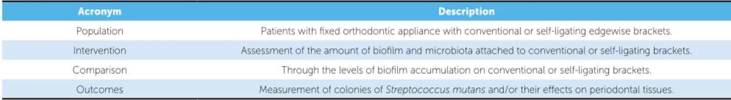
Controlled Trials; Ovid ALL EMB Reviews, PubMed and Bireme) were selected to ind relevant articles pub-lished between January 1965 and December 2012. The search used the keywords “conventional” and/or “self-ligating” crossed with combinations of the terms bioilm and / or Streptococcus mutans and / or systematic review. Two reviewers separately sought additional relevant publications, which may not have been in the searched databases, by manually searching for papers in libraries and contacting authors. There were no language restric-tions. As a irst step, the reviewers selected the articles by reading titles and abstracts. Full texts were obtained from publications that met the inclusion criteria. Ater the articles were selected, their scientiic relevance was independently assessed by the reviewers, and in case of divergence, the technique of consensus was adopted. This review used the PICO (Population Intervention
Comparator Outcomes) strategy43 to develop both the
research and the bibliography (Table 1).
Inclusion and exclusion criteria
studies, treatment plans that included extractions of premolars as well as studies that included patients younger than 11 years of age, with periodontal prob-lems, who were users of antibiotics and oral antiseptic solutions, alcoholics and smokers. Articles mention-ing patients who used mechanical and anchormention-ing de-vices, as well as Hyrax, were also excluded.
Assessment of the scientific relevance of the eligible studies
The following data were collected from each one of the papers selected: author/year of publication, jour-nal, study design, age, teeth involved, bracket type and brand, ligature type, objective and method of analysis, follow-up, statistical analysis and outcome. A quality
as-sessment44 was performed on each article, according to
the following ten criteria:
1) Study design (randomized clinical trials [RCT], prospective [P] or controlled clinical trials [CCT]) = 2 points.
2) Adequate study description = 1 point. 3) Adequate sample size = 1 point.
4) Adequate sample selection description = 1 point. 5) Drop outs description = 1 point.
6) Adequate description of bioilm measurement method = 0.5 point.
7) Blind study = 0.5 point. 8) Adequate statistics = 1 point.
9) Confounding factors considered = 1 point; and 10) Clinical signiicance = 1 point.
The ten criteria speciied above were used to identify the scientiic relevance of the methodological quality of the reviewed papers. The rating was “low” when the points given were less than or equal to 4, “medium” from 5 to 8 points and “high” for 9 or 10 points.
RESULTS
Search strategy outcomes
The search strategy resulted in 1,401 articles, out of which 195 were repeated references. The exclusion cri-teria used by both independent reviewers excluded 1,194 articles, which were not considered as relevant to the review, thus, totalizing twelve potentially relevant
ar-ticles.33,45-55 They were chosen for retrieval and evaluation
of the full text, for which a summarized data extraction sheet was used (Table 2). Out of the twelve full-text ar-ticles that were retrieved, 6 were excluded because: one
article45 presented premolar extractions in its sample,
three47,49,51 were in vitro studies, and two50,53 did not pro-vide a direct comparison between C and SL brackets systems. This resulted in six articles33,46,48,52,54,55 that were suitable for the inal analysis as they evaluated periodontal and clinical variables originating from bacterial adhesion in patients with C and SL brackets (Fig 1).
Assessment of the scientific relevance of the eligible studies
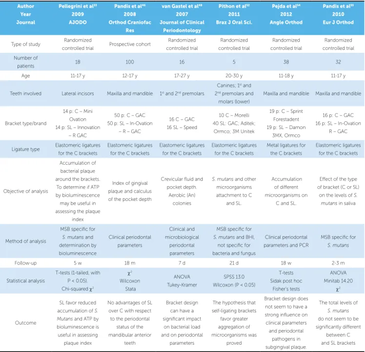
The six articles33,46,48,52,54,55 included in this review (Ta-ble 3) met the inclusion criteria, although with diferences among their methods of study, sampling, analysis and fol-low-up. All the eligible studies33,46,48,52,54,55 compared both systems: conventional and self-ligating edgewise brackets.
Pandis46 also made reference to gingival plaque and
calcu-lus index, whereas the article by van Gastel48 examined
the amount of gingival luid and anaerobic and aerobic
colonies. Another study carried out by Pandis54 collected
saliva 2-3 months ater orthodontic appliances had been bonded. Mitis salivarius culture medium (MS), speciic for Streptococcus mutans, was used to count the colony
form-ing units (CFU). Pithon52 collected the plaque samples
directly from SL and C brackets of diferent brands, and 3
Acronym Description
Population Patients with ixed orthodontic appliance with conventional or self-ligating edgewise brackets.
Intervention Assessment of the amount of bioilm and microbiota attached to conventional or self-ligating brackets.
Comparison Through the levels of bioilm accumulation on conventional or self-ligating brackets.
Outcomes Measurement of colonies of Streptococcus mutans and/or their efects on periodontal tissues.
When evaluating the scientiic relevance of the six
eligible articles,33,46,48,52,54,55 we found that the
descrip-tion of the sample selecdescrip-tion was appropriate, however, the number of drop outs was declared in studies by Pel-legrini,33 Pandis,46 van Gastel48 and Pejda54. All
stud-ies33,48,52,54 provided the approval of the Institutional
Re-view Board, except for the articles by Pandis,46,55 who
asked for the consent of patients / parents before starting the study, only. Considering the confounding factors, similar oral routine and hygiene instructions were given to the subjects taking part in these six studies.33,46,48,52,54,55 weeks ater bonding, the CFU was carried out in the
fol-lowing culture medium: MS, speciic for S. mutans, and BHI (Brain Heart Infusion), not speciic for bacteria and
fungi. In this study,52 CFU was visually performed ater
24, 48 and 72 hours of incubation. Pejda et al54 collected
the plaque samples of subgingival sulcus ater 18 weeks of treatment, counting 5 periodontal pathogens by PCR,
while Pellegrini et al33 collected the samples from tooth
surfaces surrounding the brackets ater 5 weeks of bond-ing, and the CFU was analyzed by MS and biolumines-cence of ATP (adenosine triphosphate).
Table 2 - Search data, search strategies and number of results for each database.
Figure 1 - Review flowchart.
Database Search strategies Results Selected papers
Cochrane C.R.C. Trials conventional OR self-ligating 160 2
Ovid ALL EMB Reviews
exp Orthodontic Appliances / OR edgewise.mp. AND exp Orthodontic Appliance Design/ OR exp
Orthodontic Brackets/ OR self-ligating.mp. OR exp Orthodontic Appliances/ AND bioilm.mp. OR exp Dental Bioilm Index/ AND streptococcus mutans.mp. OR exp Streptococcus mutans/
53 4
PubMed (NLM) conventional AND self-ligating, OR bioilm OR Streptococcus mutans 788 5
Bireme conventional OR self-ligating 400 1
TOTAL 1,401 12
Trials
160 papers Cochrane C.R.C
n = 1,401 Search production including titles +/- abstracts
Ineligible publications after screening all titles and abstracts
n = 1,194
Studies excluded after detailed access to the full text. Exclusion criteria included: sample
with premolars extraction,45 in vitro studies,47,49,51
use of one bracket system only.50,53
12 papers to access the full text
Studies included in the review
n = 6
195 repeated references
Inclusion
Eligibility
Identiica
tion
Scr
eening
Ovid ALL EMB PubMed (NLM)
53 papers 788 papers 400 papers
Bireme
asked whether they had already received any kind of orthodontic treatment with ixed appliances, since this can have consequences for the smoothness of the tooth enamel and for microbial adhesion at the beginning of bioilm formation.5,20,21 All six studies33,46,48,52,54,55 used appropriate statistical methods. The examiner’s
calibra-tion level was reported in one single study,54 and only
two papers54,55 identiied the sample calculation.
Smok-ing or medical conditions were clearly identiied in
In the papers,46,54 full alignment of the mandibular arch
was necessary to eliminate crowding as a confounding factor, but the clinical variables were assessed by the same periodontist. The examiner in the study carried
out by Pandis46 was not blinded, which could have
in-luenced the outcome of the research, making the
re-sults biased. The study conducted by Pithon52 did not
describe whether it had a blinded examiner, however, as a confounding factor, randomized participants were
Table 3 - Summarized data of the six studies included in the review.
p = patients; y = years; m = months; w = weeks; d = days; h = hours; C = conventional brackets; SL = self-ligating brackets; S. = Streptococcus; SEM = scanning electron microscopy; ATP = adenosine triphosphate; MSB = Mitis Salivarius agar; BHI = brain heart infusion; PCR = polymerase chain reaction.
Author Year
Journal
Pellegrini et al33 2009
AJODO
Pandis et al46 2008
Orthod Craniofac Res
van Gastel et al48 2007
Journal of Clinical Periodontology
Pithon et al52 2011
Braz J Oral Sci.
Pejda et al54 2012
Angle Orthod
Pandis et al55 2010
Eur J Orthod
Type of study Randomized
controlled trial Prospective cohort
Randomized controlled trial Randomized controlled trial Randomized controlled trial Randomized controlled trial Number of
patients 18 100 16 5 38 32
Age 11-17 y 12-17 y 17-27 y 20-30 y 11-18 y 11-17 y
Teeth involved Lateral incisors Maxilla and mandible 1st and 2nd premolars
Canines; 1st and
2nd premolars and
molars (lower)
Maxilla and mandible Maxilla and mandible
Bracket type/brand
14 p: C – Mini
Ovation 14 p: SL – Innovation
– R GAC
50 p: C – GAC 50 p: SL – In-Ovation
– R – GAC
16 C – GAC 16 SL – Speed
10 C – Morelli
40 SL: GAC; Aditek; Ormco; 3M Unitek
19 p: C – Sprint
Forestadent 19 p: SL – Damon
3MX, Ormco
16 p: C – GAC
16 p: SL – In-Ovation R – GAC
Ligature type Elastomeric ligatures for the C brackets
Elastomeric ligatures for the C brackets
Elastomeric ligatures for the C brackets
Elastomeric ligatures for the C brackets
Metal ligatures for the C brackets
Elastomeric ligatures for the C brackets
Objective of analysis
Accumulation of bacterial plaque
around the brackets. To determine if ATP
by bioluminescence may be useful in
assessing the plaque index
Index of gingival
plaque and calculus of the pocket depth
Crevicular luid and pocket depth.
Aerobic (An) colonies
S. mutans and other microorganisms
attachment to C and SL.
Accumulation of diferent
microorganisms on C and SL.
Efect of the type of bracket (C or SL)
on the levels of S. mutans in saliva
Method of analysis
MSB speciic for
S. mutans and determination by bioluminescence Clinical periodontal parameters Clinical and microbiological periodontal parameters
MSB speciic for
S. mutans and BHI, not speciic for
bacteria and fungus
Clinical periodontal parameters and PCR
MSB speciic for
S. mutans
Follow-up 5 w 18 m 7 d 21 d 18 w 2-3 m
Statistical analysis
T-tests (1-tailed, with
P < 0.05). Chi-squared c2
c2 Wilcoxon Stata ANOVA Tukey-Kramer SPSS 13.0 Wilcoxon (P < 0.05) T-tests Sidak post hoc
Fisher’s tests
ANOVA Minitab 14.20
c2
Outcome
SL favor reduced accumulation of S. Mutans and ATP by bioluminescence is
useful in assessing plaque index
No advantages of SL over C with respect
to the periodontal status of the
mandibular anterior teeth
Bracket design can have a
signiicant impact on bacterial load
and on periodontal parameters
The hypothesis that self-ligating brackets
favor greater aggregation of
microorganisms was proved
Bracket design does
not seem to have a strong inluence on
clinical parameters and periodontal
pathogens in subgingival plaque.
The total levels of
S. mutans
do not seem to be signiicantly diferent
studies by van Gastel,48 Pejda54 and Pandis.55 As for the
other studies,33,46,52 these conditions were declared only
ater the authors were requested to do so. The inal score of the scientiic relevance, in accordance with the Jadad scale,44 was 10.0 for Pellegrini33 and Pejda54, 9.5 for van
Gastel48 and Pandis55, and 9.0 for Pandis46 and Pithon52
(Table 4), which revealed high-quality researches and methodological soundness.
Assessment of the eligible studies outcomes
Among the selected studies, four46,48,54,55 had their
outcomes consistent in reporting that (a) SL brackets have no advantages over C in periodontal condition
of anterior mandibular teeth;46 (b) the design of the
brackets can have significant impact on bacterial load
and periodontal parameters;48 and (c) in subgingival
plaque and saliva, there seems to be no significant differences in the total levels of S. Mutans and
peri-odontal pathogens between C and SL.54,55 However, a
study52 confirmed the hypothesis that SL brackets
fa-vor the accumulation of micro-organisms, while
an-other study33 reported that SL brackets promote lower
retention of S. mutans when compared to C (Table 3).
The outcomes of the eligible studies33,46,48,52,54,55 were
not unanimous in reporting that there is evidence of a possible influence of bracket design (convention-al or self-ligating) over adhesion and formation of Streptococcus mutans colonies.
DISCUSSION
A systematic review can conirm the quality of a re-search as well as the methodological soundness of works selected from the literature. Additionally, it can present them for consideration of the clinical and scientiic com-munities. Evidence-based practice requires the con-struction of a research question and a literature review.
Conventionally, to attach the wire to the brackets, three methods are used: metal ligature, elastomeric ligatures, and the open-close devices of SL brackets. All these methods have advantages and disadvantages, but with regard to the accumulation of biofilm, the
literature8,33 suggests that elastomeric ligatures
fa-vor the retention of biofilm in comparison with the other two methods of ligatures. The question pre-pared for this review aimed to verify whether bracket design (conventional or self-ligating) influences the formation of Streptococcus mutans colonies. Micro-organisms exhibit significant adherence to brackets because there are favorable ecological niches in the porous (rough and irregular surfaces of these
brack-ets).39,47,49,51,56 Thus, the characteristics of the bracket
surface can be considered as harboring favorable sites for the adhesion of biofilm.
Search strategy outcomes
This research was highly sensitive, addressing evi-dence of minimum bias. The study carried out by
Pellegrini et al33 2009
AJODO
Pandis et al46 2008
Orthod Craniofac Res
van Gastel et al48 2007
Journal of Clinical Periodontology
Pithon et al52 2011
Braz J Oral Sci.
Pejda et al54 2012
Angle Orthod
Pandis et al55 2010
Eur J Orthod
Type of study 2.0 2.0 2.0 2.0 2.0 2.0
Study description 1.0 1.0 1.0 1.0 1.0 1.0
Sample size 1.0 1.0 1.0 0.5 1.0 1.0
Sample selection description 1.0 1.0 1.0 1.0 1.0 1.0
Drop out description 1.0 0.5 0.5 1.0 1.0 0.5
Measurement method 0.5 0.5 0.5 0.5 0.5 0.5
Blind study 0.5 --- 0.5 --- 0.5 0.5
Statistics 1.0 1.0 1.0 1.0 1.0 1.0
Confounding factors 1.0 1.0 1.0 1.0 1.0 1.0
Clinical signiicance 1.0 1.0 1.0 1.0 1.0 1.0
Scale score (Jadad44) 10.0 9.0 9.5 9.0 10.0 9.5
Quality standard assessed high high high high high high
Jordan and LeBlanc50 was excluded due to: (a) hav-ing assessed one bracket system only, (b) havhav-ing a not blinded examiner and (c) presenting unspecified statistical analyses. The in vitro studies that were
ex-cluded47,49,51 did not have the inherent
characteris-tics which contribute to the development of intra-oral biofilm, and may provide bias results for clinical
periodontal conditions.22 The differences observed
between the results of some papers33,46,48-50,52 may be
related to factors that include: variations in the shape, material and size between SL and C brackets, the individual level of oral hygiene, salivary flow, treat-ment variables, types of ligatures, bonding
proce-dures and age of the individuals involved.24,45,49,51,55
Thus, bracket type itself would not be a deciding fac-tor for biofilm development, but its composition and material type should be included as factors behind
Streptococcus mutans colonies formation.56
Assessment of the scientific relevance of the eligible studies
The statistical analysis of our results was not fea-sible, given that the methodological designs of the eli-gible articles were heterogeneous. However, the sci-entiic relevance assessment revealed high-quality re-searches and methodological soundness of all six
stud-ies,33,46,48,52,54,55 as shown in their inal scores, according
to the Jadad scale.44
Although SL brackets do not require ligatures, their opening and closing mechanism may provide sites for biofilm adhesion similarly to conventional
brackets.46 This mechanism of SL brackets is not
re-newed, as it occurs with elastomeric modules in con-ventional brackets. Moreover, plaque calcification in SL leads to a malfunction of the opening and closing mechanisms. Thus, the theoretical advantages of self-ligating over conventional brackets can be eliminated,
as confirmed by other studies.46,52 When using
con-ventional brackets, neither the elastomeric rings nor the metal ligatures seem to affect the distribution of bacterial morphotypes in brackets or on the enamel
surface.3 Aged elastomeric surfaces can apparently
favor plaque retention in comparison with polished stainless steel ligatures, but there are no differences between periodontal conditions of patients treated
with these two types of ligatures.8,57 Nevertheless,
some studies41,58 report that brackets with elastomeric
rings favor damage to gingival conditions, with sig-nificant accumulation of biofilm, while the metal ligature had lower retention of biofilm in comparison
with other brackets. Some reports59,60 affirm that C
brackets are directly related to the retention of
bio-film, however, the study conducted by Pithon et al52
suggests that cross-infection caused by replacement of elastomeric rings is controllable with the use of C brackets, because this type of brackets favors lower formation of S. Mutans colonies, which agrees with
the study by van Gastel et al48 that showed no
differ-ence between C and SL in gingival bleeding.
Assessment of the retrieved studies outcomes
The increase in oral microbiota attachment of Strep-tococcus mutans and Lactobacillus is associated with the
use of orthodontic appliances,6,8,9,33,45 with both C or
SL brackets. This increase leads to higher cariogenic plaque, pH low enough to change the clinical
peri-odontal parameters46,48,54 and increased risk of enamel
demineralization.6,47
Some eligible studies52,54 evaluated not only the
presence of S. mutans, but also of other microorgan-isms related to periodontal disease in patients with C
or SL brackets. The study conducted by Pejda et al54
found 23.8 times more chance of inding Aggregatibacter actinomycetemcomitans (AA) in subgingival plaque of pa-tients with C brackets, but the increase in AA does not represent a risk factor for local periodontitis, as studies
by Paolantonio et al61,62 conirm. The diferences found
between the results of the study by Pithon et al52 and
the other studies assessed33,46,48,54,55 may have been due
to methodological diferences in some of these
stud-ies46,48,54,55 in which the CFU were counted from
ma-terial collected from saliva; Pellegrini et al33 collected
it from tooth surfaces surrounding the bracket; and,
in the study by Pithon,52 it was directly collected from
the surface of brackets (winglets, slot and cervical re-gion). That was the reason why this latest study should have found statistically signiicant diferences that re-veal greater accumulation of bioilm in SL brackets.
Clinical implications
Some studies8,33-39 report that SL brackets are less
the correct orientation and cooperation of patients24,55 than by simply choosing one system of brackets instead of
another. The outcomes of the eligible studies33,46,48,52,54,55
were not unanimous in reporting a possible inluence of bracket design (conventional or self-ligating) over the adhesion and formation of Streptococcus mutans colonies.
The decision of orthodontists on prescribing the use of SL instead of C in their clinical routine, aiming at improving hygiene / plaque accumulation, cannot yet
be applied due to lack of scientiic evidence.46,48,52,54,55
Ater this review, we presume that there is not enough evidence to support the use of ixed appliances with SL brackets in place of systems with C or vice versa,
which agrees with the study by Fleming et al.63
Based on the limitations of some works,64,66
fur-ther studies on ofur-ther types of brackets, for example, esthetic self-ligating ones, must be performed to vi-sualize the periodontal complications arising from diferent shapes, sizes and material types of brackets, and with that, guide the development of new systems of brackets design in order to reduce the formation of Streptococcus mutans colonies.
CONCLUSIONS
Within the limitations of this study, it was conclud-ed that there is no evidence for a possible inluence of bracket design (conventional or self-ligating) over colo-ny formation and adhesion of Streptococcus mutans.
1. O’Reilly MM, Featherstone JDB. Demineralization and remineralization around orthodontic appliances: an in vivo study. Am J Orthod Dentofacial Orthop. 1987;92(1):33-40.
2. Øgaard B, Rølla G, Arends J. Orthodontic appliances and enamel demineralization. Part 1. Lesion development. Am J Orthod Dentofacial Orthop. 1988;94(1):68-73.
3. Gwinnett JA, Ceen F. Plaque distribution on bonded brackets. Am J Orthod. 1979;75(6):667-77.
4. Mizrahi E. Surface distribution of enamel opacities following orthodontic treatment. Am J Orthod Dentofacial Orthop. 1983;84(4):323-31. 5. Alexander SA. Efects of orthodontic attachments on the gingival
health of permanent second molars. Am J Orthod Dentofacial Orthop. 1991;100(4):337-40.
6. Balenseifen JW, Madonia JV. Study of dental plaque in orthodontic patients. J Dent Res. 1970;49(2):320-4.
7. Menzaghi N, Saletta M, Garattini G, Brambilla E, Strohmenger L. Changes in the yeast oral lora in patients in orthodontic treatment. Prev Assist Dent. 1991;17(4):26-30.
8. Forsberg CM, Brattström V, Malmberg E, Nord CE. Ligature wires and elastomeric rings: two methods of ligation, and their association with microbial colonization of Streptococcus mutans and lactobacilli. Eur J Orthod. 1991;13(5):416-20.
9. Rosenbloom RG, Tinanof N. Salivary Streptococcus mutans levels in patients before, during, and after orthodontic treatment. Am J Orthod Dentofacial Orthop. 1991;100(1):35-7.
10. Saemundsson SR, Bergmann H, Magnusdottir MO, Holbrook WP. Dental caries and Streptococcus mutans in a rural child population in Iceland. Scand J Dent Res. 1992;100(5):299-303.
REFERENCES
11. Sansone C, Van Houte J, Joshipura K, Kent R, Margolis HC. The association of mutans streptococci and non-mutans streptococci capable of acidogenesis at a low pH with dental caries on enamel and root surfaces. J Dent Res. 1993;72(2):508-16.
12. Mattousch TJ, van der Veen MH, Zentner A. Caries lesions after orthodontic treatment followed by quantitative light-induced luorescence: a 2-year follow-up. Eur J Orthod. 2007;29(3):294-8.
13. Mizrahi E. Enamel demineralization following orthodontic treatment. Am J Orthod. 1982;82(1):62-7.
14. Benson PE, Parkin N, Millett DT, Dyer FE, Vine S, Shah A. Fluorides for the prevention of white spots on teeth during ixed brace treatment. Cochrane Database Syst Rev 2004;(3):CD003809.
15. Gorelick L, Geiger AM, Gwinnett AJ. Incidence of white spot formation after bonding and banding. Am J Orthod. 1982;81(2):93-8.
16. Øgaard B. Prevalence of white spot lesions in 19-year-olds: a study on untreated and orthodontically treated persons 5 years after treatment. Am J Orthod Dentofacial Orthop. 1989;96(5):423-7.
17. Anhoury P, Nathanson D, Hughes CV, Socransky S, Feres M, Chou LL. Microbial proile on metallic and ceramic bracket materials. Angle Orthod. 2002;72(4):338-43.
18. Loe H, Theilade E, Jensen SB. Experimental Gingivitis in Man. J Periodontol. 1965;36:177-87.
19. Zachrisson S, Zachrisson BU. Gingival condition associated with orthodontic treatment. Angle Orthod. 1972;42(1):26-34.
22. van Pelt AW, Weerkamp AH, Uyen MH, Busscher HJ, de Jong HP, Arends J. Adhesion of Streptococcus sanguis CH3 to polymers with diferent surface free energies. Appl Environ Microbiol. 1985;49(5):1270-5.
23. Socransky SS, Hafajee AD, Smith C, Dibart S. Relation of counts of microbial species to clinical status at the sampled site. J Clin Periodontol. 1991;18(10):766-75.
24. Quirynen M, Dekeyser C, van Steenberghe D. The inluence of gingival inlammation, tooth type, and timing on the rate of plaque formation. J Periodontol. 1991;62(3):219-22.
25. Quirynen M, Dekeyser C, van Steenberghe D. Discriminating power of ive plaque indices. J Periodontol. 1991;62(2):100-5.
26. Ramberg P, Axelsson P, Lindhe J. Plaque formation at healthy and inlamed gingival sites in young individuals. J Clin Periodontol. 1995;22(1):85-8. 27. Rowshani B, Timmerman MF, Van der Velden U. Plaque development in
relation to the periodontal condition and bacterial load of the saliva. J Clin Periodontol. 2004;31(3):214-8.
28. Quirynen M, Marechal M, Busscher H, el-Abiad M, Arends J, Van Steenberghe D. The inluence of surface characteristics on the early bacterial colonization of intra-oral hard surfaces. J Clin Dent. 1988;1 Suppl A:A14-9.
29. Bollen CM, Quirynen M. [Specimen collection in dental plaque and oral microbiology]. Rev Belge Med Dent. 1994;49(2):44-51.
30. Satou J, Fukunaga A, Satou N, Shintani H, Okuda K. Streptococcal adherence on various restorative materials. J Oral Rehabil. 2004;31(3):278-85. 31. Berger J. The engaging concept of self-ligation. Ont Dent. 1999;76(3):26-33. 32. Cacciafesta V, Sfondrini MF, Ricciardi A, Scribante A, Klersy C, Auricchio F.
Evaluation of friction of stainless steel and esthetic self-ligating bráquetes in various bracket-archwire combinations. Am J Orthod Dentofacial Orthop. 2003;124(4):395-402.
33. Pellegrini P, Sauerwein R, Finlayson T, McLeod J, Covell DA Jr, Maier T, et al. Plaque retention by self-ligation vs elastomeric orthodontic brackets: quantitative comparison or oral bacteria detection with adenosine triphosphate-driven bioluminescence. Am J Orthod Dentofacial Orthop. 2009;135(4):426.e1-9.
34. Damon DH. The rationale, evolution and clinical application of the self-ligating bracket. Clin Orthod Res. 1998;1(1):52-61.
35. Shivapuja PK, Berger J. A comparative study of conventional ligation and self-ligation bracket systems. Am J Orthod Dentofacial Orthop. 1994;106(5):472-80.
36. Paduano S, Cioi I, Iodice G, Rapuano A, Silva R. Time eiciency of self-ligating vs conventional brackets in orthodontics: efect of appliances and ligating systems. Prog Orthod. 2008;9(2):74-80.
37. Yu YL, Qian YF. The clinical implication of self-ligating brackets. Shanghai Kou Qiang Yi Xue. 2007;16(4):431-5.
38. Fernandes C, Almeida R. self-ligating appliances: evolution or revolution? Aust Orthod. 2008;24(1):97-103.
39. Türkkahraman H, Sayin MO, Bozkurt FY, Yetkin Z, Kaya S, Onal S. Archwire ligation techniques, microbial colonization, and periodontal status in orthodontically treated patients. Angle Orthod. 2005;75(2):231-6. 40. Polson AM, Subtelny JD, Meitner SW, Polson AP, Sommers EW, Iker HP.
Long-term periodontal status after orthodontic treatment. Am J Orthod Dentofacial Orthop. 1988;93(1):51-8.
41. Garcez, AS, Suzuki SS, Ribeiro MS, Mada EY, Freitas AZ, Suzuki H. Bioilm retention by 3 methods of ligation on orthodontic brackets: a microbiologic and optical coherence tomography analysis. Am J Orthod Dentofacial Orthop. 2011;140(4):e93-8.
42. National Health Service (NHS) Centre for reviews and dissemination. Undertaking systematic reviews of research on efectiveness [report]. York (UK): York Publishing Services; 2001. Available from: www.york.ac.uk/inst/ crd/crdrep.htm.
43. European Food Safety Authority. Application of systematic review methodology to food and feed safety assessments to support decision making. EFSA J. 2010;8(6):1637. Available from: http://www.efsa.europa.eu 44. Jadad AR, Moore RA, Carrol D, Reynolds DJ, Gavaghan DJ, McQuay HJ.
Assessing the quality of reports of randomized clinical trials: is blinding necessary? Controlled Clin Trials 1996;17(1):1-12.
45. Sukontapatipark W, el-Agroudi MA, Selliseth NJ, Thunold K, Selvig KA. Bacterial colonization associated with ixed orthodontic appliances. A
scanning electron microscopy study. Eur J Orthod. 2001;23(5):475-84. 46. Pandis N, Vlachopoulos K, Polychronopoulou A, Madianos P, Eliades T. Periodontal condition of the mandibular anterior dentition in patients with conventional and self-ligating brackets. Orthod Craniofac Res. 2008;11(4):211-5.
47. Faltermeier A, Burgers R, Rosentritt M. Bacterial adhesion of Streptococcus mutans to esthetic bracket materials. Am J Orthod Dentofacial Orthop. 2008;133(4 Suppl):S99-103.
48. van Gastel J, Quirynen M, Teughels W, Coucke W, Carels C. Inluence of bracket design on microbial and periodontal parameters in vivo. J Clin Periodontol 2007;34(5):423-31.
49. Brusca MI, Chara O, Sterin-Borda L, Rosa AC. Inluence of diferent orthodontic brackets on Adherence of microorganisms in vitro. Angle Orthod. 2007;77(2):331-6.
50. Jordan C, LeBlanc DJ. Inluences of orthodontic appliances on oral populations of mutans streptococci. Oral Microbiol Immunol. 2002;17(2):65-71.
51. Fournier A, Payant L, Bouclin R. Adherence of Streptococcus mutans to orthodontic bráquetes. Am J Orthod Dentofacial Orthop. 1998;114(4):414-7. 52. Pithon MM, Santos RL, Nascimento LE, Ayres AO, Alviano D, Bolognese AM. Do self-ligating brackets favor greater bacterial aggregation? Braz J Oral Sci. 2011;10(3):208-12.
53. Ristic M, Vlahovic Svabic M, Sasic M, Zelic O. Clinical and microbiological efects of ixed orthodontic appliances on periodontal tissues in adolescents. Orthod Craniofac Res. 2007;10(4):187-95.
54. Pejda S, Varga ML, Milosevic SA, Mestrovic S, Slaj M, Repic d, Bosnjak A. Clinical and microbiological parameters in patients with self-ligating and conventional brackets during early phase of orthodontic treatment. Angle Orthod. 2013;83(1):133-9.
55. Pandis N, Papaioannou W, Kontou E, Nakou M, Makou M, Eliades T. Salivary Streptococcus mutans levels in patients with conventional and self-ligating brackets. Eur J Orthod. 2010;32(1):94-9.
56. Carneiro RC. Estudo da microbiota do bioilme supragengival de pacientes em tratamento ortodôntico com diferentes tipos de braquetes [tese]. Belo Horizonte (MG): Pontifícia Universidade Católica; 2008.
57. Gameiro GH, Nouer DF, Cenci MS, Cury JA. Enamel demineralization with two forms of archwire ligation investigated using an in situ caries model: a pilot study. Eur J Orthod. 2009;31(5):542-6.
58. Souza RA, Borges de Araújo Magnani MB, Nouer DF, Oliveira da Silva C, Klein MI, Sallum EA, et al. Periodontal and microbiologic evaluation of 2 methods of archwire ligation: Ligature wires and elastomeric rings. Am J Orthod Dentofacial Orthop. 2008;134(4):506-12.
59. Eliades T, Eliades G, Brantley WA. Microbial attachment on orthodontic appliances: I. Wettability and early pellicle formation on bracket materials. Am J Orthod Dentofacial Orthop. 1995;108(4):351-60.
60. Batoni G, Pardini M, Ota F, Guica MR, Gabriele M, Campa M, Senesi S. Efect of removable orthodontic appliances on oral colonization by mutans streptococci in children. Eur J Oral Sci. 2001;109(6):388-92.
61. Paolantonio M, Festa F, di Placido G, D’Attilio M, Catamo G, Piccolomini R. Site-speciic subgingival colonization by Actinobacillus actinomycetemcomitans in orthodontic patients. Am J Orthod Dentofacial Orthop. 1999;115(4):423-8.
62. Paolantonio M, Pedrazzoli V, di Murro C, di Placido G, Picciani C, Catamo G, et al. Clinical signiicance of Actinobacillus actinomycetemcomitans in young individuals during orthodontic treatment. A 3-year longitudinal study. J Clin Periodontol. 1997;24(9 Pt 1):610-7
63. Fleming PS, Johal A. Self-ligating brackets in orthodontics. Angle Orthod. 2010;80:575-584.
64. Maza JL, Elguezabal N, Prado C, Ellacuria J, Soler I, Ponton J. Candida albicans adherence to resin–composite restorative dental material: inluence of whole human saliva. Oral Surg Oral Med Oral Pathol Oral Radiol Endod. 2002;94(5):589-92.
65. Addy M, Shaw WC, Hansford P, Hopkins M. The efect of orthodontic appliances on the distribution of Candida and plaque in adolescents. Br J Orthod. 1982;9(3):158-63.