Departments of 1Infectology, 3Neurology and 5Neurosurgery, Institute of Infectious Disease Emilio Ribas, Sao Paulo SP, Brazil; 2Center for Clinical Decisions Sciences, Department of Public Health, Erasmus University Medical Center, Rotterdam, The Netherlands; 4Clinical Research Unit in Human Retrovirus, University of Campinas, Sao Paulo SP, Brazil.
Received 27 January 2004, received in final form 6 April 2004. Accepted 28 May 2004.
Dr. José E. Vidal - Rua Capote Valente 668/78 - 05409-002 São Paulo SP - Brasil. E-mail: [email protected]
CEREBRAL TUBERCULOMAS IN AIDS PATIENTS
A forgotten diagnosis?
José E. Vidal
1, Adrián V. Hernández
2, Augusto C. Penalva de Oliveira
3,4,
Alexandre Leite de Souza
1, Geraldine Madalosso
1, Paula R. Marques da Silva
1, R. Dauar
4ABSTRACT - The human immunodeficiency virus (HIV) infection epidemics increased the prevalence, mul-ti-drug resistance and disseminated forms of tuberculosis. The central nervous system (CNS) tuberculosis has high mortality and morbidity, and it is usually divided into diffuse (meningitis) and localized (tubercu-loma and abscess) forms. We report three cases of cerebral tubercu(tubercu-lomas in AIDS patients: one with defin-itive diagnosis, confirmed with histopathology, and two with probable diagnosis, based on clinical infor-mation, radiological images, Mycobaterium tuberculosis isolation out of the CNS and adequate response to antituberculous treatment. Further, we discuss diagnostic, therapeutic and prognostic issues of tuber-culomas, with emphasis in the distinction from cerebral tuberculous abscesses. Despite of their infrequent presentation, tuberculomas should be considered in the differential diagnosis of cerebral expansive lesions in patients with AIDS.
KEY WORDS: tuberculoma, tuberculosis, central nervous system, acquired immunodeficiency syndrome.
Tuberculomas cerebrais em pacientes com aids: um diagnóstico esquecido?
RESUMO - A epidemia da infecção pelo vírus da imunodeficiência humana (HIV) aumentou a prevalência, multiresistência e formas disseminadas da tuberculose. O acometimento neurológico da tuberculose apre-senta elevada morbidade e mortalidade, classificando-se em formas difusas (meningite) e localizadas (tuberculoma e abscesso). Relatamos três casos de tuberculomas cerebrais em pacientes com AIDS: um deles com diagnóstico definitivo, confirmado com histopatologia e dois com diagnóstico provável, baseado em informação clínica, radiológica, isolamento de Mycobaterium tuberculosis fora do sistema nervoso central e adequada resposta ao tratamento tuberculostático. Discutimos também aspectos diagnósticos, terapêu-ticos e prognósterapêu-ticos dos tuberculomas, enfatizando suas diferenças com os abscesos tuberculosos cerebrais. Apesar de serem relatados de forma infreqüente, os tuberculomas devem sempre ser considerados no diag-nóstico diferencial das lesões expansivas cerebrais em pacientes com AIDS.
PALAVRAS-CHAVE: tuberculoma, tuberculose, sistema nervoso central, sindrome da imunodeficiência adquirida.
Tuberculosis (TB) has become more frequent
since the emergence of human
immunodeficien-cy virus (HIV) infection epidemics. Nowadays, there
are important challenges, which complicate the
ma-nagement of HIV-TB co-infected patients. Among
them, it is emphasized the increase of
disseminat-ed and extra-pulmonary TB forms, the multi-drug
resistance and the increase in mortality
1.
The central nervous system (CNS) TB is found in
5-10% of patients with pulmonary TB
2. These
cas-es corrcas-espond to either latent infection reactivations
or disseminated infections. Tuberculous
meningi-tis is the most common clinical form and it is five
times more frequent in patients with HIV infection
than in patients without it
3. The focal forms of CNS
tuberculosis are infrequently described and they are
divided into tuberculoma and abscess
4.
794 Arq Neuropsiquiatr 2004;62(3-B)
CASES
Case 1 -A 36-year-old Brazilian man was admitted in December 2002 with a seven-days history of tiredness, headache, nausea and vomiting. He had diagnosed HIV and cerebral toxoplasmosis in October 2002. The patient irregularly used zidovudine, lamivudine, efavirenz, sulfa-diazine, pyrimethamine, and folinic acid. On physical exa-mination, he showed a decrease in consciousness level and no focal neurological signs. Altered laboratory tests included hemoglobin 9.2 g/dl, leucocytes 1100/mm3,
platelets 95000/mm3and sodium 128 mmol/l. A chest
X-ray showed a scary lesion in upper right lung. Toxoplasma gondii serology was positive and CD4+ lymphocytes
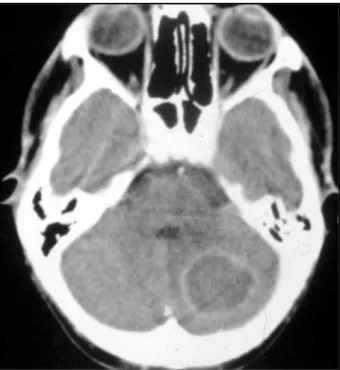
count was 7 cells/mm3. A cerebral computed
tomogra-phy (CT) scan showed a solitary lesion in the left cere-bellum, with contrast enhancement and mass effect (Fig 1). The cerebrospinal fluid examination (CSF) was normal. Empirical treatment for toxoplasmic encephali-tis with sulfadiazine 6 g, pyrimethamine 25 mg and fo-linic acid 15 mg daily was initiated. Three weeks later, a new CT scan showed increase in size of the cerebellar lesion and surgery was indicated. The histological exam-ination showed presence of few acid-fast bacilli (AFB), necrotic tissue and absence of microabscesses. Investi-gation for other causes, including bacteria and fungi, were negative. Thus, treatment with isoniazid 400 mg, rifampin 600 mg, pyrazinamide 2 g, and prednisone 50 mg daily was initiated. Four weeks later, the patient evi-denced meaningful clinical and imaging improvement and he was discharged to complete nine months of an-tituberculous treatment.
Case 2 -A 31-year-old Brazilian woman was admit-ted in August 2002 because of weight loss, tiredness, fe-ver, headache and productive cough for four weeks and a convulsive episode one week before. She had been diagnosed as being infected with HIV infection since 1985 and she had Pneumocystis carinii pneumonia in 1999 and pulmonary TB in 2001. The patient irregularly used zi-dovudine, lamivudine, efavirenz and trimethoprim-sul-famethoxazole. On physical examination, she had fever, diffuse non-painful adenomegalia and no neurological signs. Laboratory tests showed hemoglobin 8.6 g/dl, leucocytes 2.900/mm3, alkaline phosphatase 460 UI/l,
aspartate aminotransferase 83 UI/l, alanine aminotransfe-rase 62 UI/l and lactose dehydrogenase 657 UI/l. A chest X-ray showed bilateral pleural thickness and enlarged mediastinal area. Her tuberculin skin test, using purified protein derivative (PPD), and T. gondii serology were neg-ative. The CD4+lymphocytes count was 31 cells/mm3
and the HIV viral load was 234000 copies/ml. An abdom-inal ultrasonography evidenced hepatosplenomegalia, intra-abdominal adenomegalia and ascitic fluid. A cere-bral CT scan showed multiple nodular and ring-enhanc-ing lesions (Fig 2A), and one lesion with central calcifica-tion and ring-enhancement. The CSF was normal and a cervical node biopsy yielded abundant AFB with caseous necrosis. Isoniazid 400 mg, rifampin 600 mg, pyrazina-mide 2 g, ethambutol 1.2 g, sulfadiazine 6 g, pyrime-thamine 25 mg, and folinic acid 15 mg daily were initi-ated. Three weeks later, the patient did not have any clinical improvement and the cerebral lesions remained unchanged. The culture of AFB yielded M. tuberculosis
resistant to rifampin. Streptomycin 1 g was added to iso-niazid, pyrazinamide and ethambutol, and the toxoplas-mosis treatment was stopped. One month later, the pa-tient was asymptomatic and she was discharged to
com-Fig 1. Contrast-enhanced computed tomography scan of case 1, showing a large unique ring-enhancing lesion in the left cere-bellum, sourrounded by discrete edema.
plete nine months of treatment. Two months after, a CT scan showed moderate cortical and subcortical atrophy without focal lesions (Fig 2B).
Case 3 -A 30-year-old Brazilian woman was admit-ted in August 2002 with a three-weeks history of fever, productive cough, headache, abdominal pain, vomiting and diarrhea. She had diagnosed HIV infection in June 2001 and she had a history of disseminated TB (pul-monary and nodal) in September 2001, with abandon-ment of antituberculous treatabandon-ment at the third month. On physical examination, the patient had important weight loss, moderate dehydration, abdominal disten-tion, hepatosplenomegalia, ataxia and psychomotor slowness. Altered laboratory tests were hemoglobin 11.2 g/dl, leucocytes 3100/mm3, alkaline phosphatase 514
UI/l, aspartate aminotransferase 57 UI/l and alanine ami-notransferase 53 UI/l, and lactose dehydrogenase 711 UI/l. A chest X-ray showed multiple micronodular lesions in both lungs compatible with miliary tuberculosis. The PPD and T. gondii serology were negative, the CD4+
lymphocytes count was 35 cells/mm3and the HIV viral
load was 54800 copies/ml. The direct examination of spu-tum for AFB was positive. A CT scan showed two cere-bellar lesions, one of them multiloculated (Fig 3A). The CSF examination revealed a protein of 150 mg/L with both normal glucose level and white cell count. No AFB were seen on Ziehl-Neelsen staining and culture was neg-ative. Treatment with isoniazid 400 mg, rifampin 600 mg, pyrazinamide 2 g, and ethambutol 1.2 g daily was start-ed. One month later, the patient evidenced important clinical and imaging improvement and she was dischar-ged to complete nine months of treatment. The culture of sputum yielded M. tuberculosis sensible to isoniazid, rifampin and pyrazinamide. Three months later, the pa-tient was asymptomatic and had a normal cerebral CT scan (Fig 3B).
DISCUSSION
The introduction of highly active antiretroviral
therapy (HAART) improved significantly the survival
and quality of life of AIDS patients in developed
countries and in Brazil
5. TB prevalence has also
decreased as a consequence of HAART. However,
TB is still a public health problem in Brazil and
repre-sents the most frequent AIDS-defining disease
5.
There is limited information about focal forms
of cerebral TB in AIDS patients and recent reviews
over CNS opportunistic infections did not include
TB
6,7. This is possible due to the fact that the
num-ber of CNS TB cases in developed countries is very
limited, as demonstrated in a detailed
neuropatho-logical study
8. In the AIDS era, Lesprit et al.
9found
only 21 cases of cerebral TB (including
tuberculo-mas and abscesses), and Vidal et al.
10found nine
cases of tuberculous abscess. The lack of reports
of CNS TB in developing countries, where the TB
prevalence is high, can be due to underdiagnosis
and underregister of cases and, indeed, to the lack
of publications.
Tuberculomas can develop through four
patho-physiological mechanisms. First, invasion of
bacil-li from the CSF (TB meningitis). Second, as a
conse-quence of a disseminated TB. Third, paradoxical
reaction in patients with antituberculous
treat-ment, with or without antiretrovirals. Finally, local
reactivation of latent foci
2,11. Cerebral TB
infec-tion may produce local areas of cerebritis with
formation of tuberculomas. It is unknown why an
abscess is produced in some cases and in another
it is a tuberculoma. Farrar
12considers that if the
quantity of bacilli is high enough or the
immuni-ty is depressed, a focal cerebritis may progress to
an abscess. On the other hand, the most of authors
consider that the abscesses result from the
liquefac-tion of tuberculomas
13.
The focal forms of CNS TB show different
anato-mopathological characteristics, which were defined
by Whitener
14. Tuberculomas have a central region
with caseous necrosis, a capsule of collagen and
giant multinuclear, epitheliod and mononuclear
cells. On the other hand, abscess require to fulfill
the following criteria: (1) macroscopic evidence
of pus, (2) inflammatory reaction in the abscess wall,
which consists of granulate vascular tissue and
a-cute and chronic inflammatory cells, and (3)
de-monstration of AFB in the purulent material or in
the abscess wall, or positive culture of
M.
tubercu-losis.
Definitive diagnosis of cerebral tuberculoma
needs histopathologic evidence in the cerebral
sue obtained by biopsy, trepanation or necropsy.
Probable diagnosis of tuberculomas requires
epi-demiological, clinical and laboratorial
informa-tion and also an adequate response to
antitubercu-lous treatment
2.
The clinical presentation of tuberculomas and
tuberculous abscesses is similar to the main CNS
ex-pansive lesions in AIDS patients
9,10. However, the
tuberculous abscesses usually have a more
accelera-ted clinical course. Therefore, it is important to
per-form a detailed anamnesis and physical
examina-tion to find exposure informaexamina-tion, previous TB
di-sease and treatment as well as symptoms and signs
out of the CNS.
Tomographic findings of the focal forms of CNS
TB are unspecific and less clear than the findings of
the magnetic resonance (MRI)
4. However, it seems
that there is an adequate correlation between
his-topathological and tomographic findings of
tuber-culomas. The radiological presentation depends
on whether the granuloma is noncaseating,
caseat-ing with a solid center, or caseatcaseat-ing with a liquid
center
4. The noncaseating granulomas are
round-ed or oval, multiple, isodense or lightly hypodense
lesions, with nodular contrast enhancement. These
are the classical lesions described for
tuberculo-mas and they were present in the patient 2. The
caseating granulomas with solid center and
caseat-ing granulomas with liquid center are isodense or
hypodense lesions with annular contrast
enhance-ment. The degree of surrounding edema is variable
and is thought to be inversely proportional to the
maturity of the lesion
4. Usually, the caseating
gran-ulomas with solid center show thicker walls. On the
other hand, the caseating granulomas with liquid
center may be undistinguishable from the
bacter-ial or TB abscesses. This fact was evident in patients
1 and 3. However, the TB abscesses might differ from
tuberculomas because they show a more rapid
course, usually have larger size (often > 3cm),
thin-ner walls, and mostly unique and multiloculated
lesions
2,4,15. Moreover, there are tuberculomas which
are solitary and large (patient 1), multiloculated
(patient 3) and with an important mass effect
9.
The “target sign”, defined as a central nest of
calci-fication or a central contrast enhancement
sur-rounded by a ring of enhancement, is
characteris-tic of tuberculomas
16,17. However, recent studies
have suggested that only the “target sign” with
cen-tral calcification is pathognomonic of tuberculoma
4.
Unfortunately, the “target sign” is an infrequent
finding
4,15.
796 Arq Neuropsiquiatr 2004;62(3-B)
In contrast to the cerebral TB abscesses, which
require surgical and pharmacological treatment
10,12,14,
tuberculomas generally respond quite well with
only pharmacological treatment
16,17. Nevertheless,
like was verified in some of our patients, the
differen-tiation between this two diagnoses is difficult and
complicates the surgical decision.
We conclude that tuberculomas should be
consi-dered in the differential diagnosis of CNS
expansi-ve lesions in patients with AIDS. A careful
individ-ual evaluation, including clinical (anamnesis,
phys-ical examination), laboratorial (negative
T. gondii
serology, evidence of TB out of the CNS) and
imag-ing information (previous or active images in chest
X-ray, “target sign” in cerebral CT) could increase
the diagnostic probability of tuberculoma. In
con-trast to TB abscesses, the conservative treatment of
tuberculomas usually determines a good outcome.
REFERENCES
1. Sepkowitz KA, Raffalli J, Riley L, Kiehn TE, Armstrong D. Tuberculosis in the AIDS era. Clin Microbiol Rev 1995;8:180-199.
2. Zuger A, Franklin DL. Tuberculosis. In Scheld RJ, Whitley RJ, Durack DT (eds). Infections of the central nervous system. Philadelphia: Lippincott-Raven, 1997:417-443.
3. Berenguer J, Moreno S, Laguna F, et al. Tuberculosis meningitis in patients infected with the human immunodeficiency virus. N Engl J Med 1992;326:668-672.
4. Bernaerts A, Vanhoenacker FM, Parizel PM, et al. Tuberculosis of the central nervous system: overview of neuroradiological findings. Eur Radiol 2003;13:1876-1890.
5. Marins JR, Jamal LF, Chen SY, et al. Dramatic improvement in survival among adult Brazilian AIDS patients. AIDS 2003;17:1675-1682. 6. Mamidi A, DeSimone JA, Pomerantz RJ. Central nervous system
infec-tions in individuals with HIV-1 infection. J Neurovirol 2002;8:158-167. 7. Skiest DJ. Focal neurological disease in patients with acquired
immun-odeficiency syndrome. Clin Infect Dis 2002;34:103-115.
8. Gray F, Chretien F, Vallat-Decouvelaere AV, Scaravelli F. The changing pattern of HIV neuropathology in the HAART era. J Neuropathol Exp Neurol 2003;5:429-440.
9. Lesprit P, Zagdanski A-M, de la Blanchardiere A, et al. Cerebral tuber-culosis in patients with the acquired immunodeficiency syndrome (AIDS). Medicine (Baltimore) 1997;76:423-431.
10. Vidal JE, Cimerman S, Silva PR, Sztajnbok J, Coelho JF, Lins DL. Tuberculous brain abscess in a patient with AIDS: case report and lit-erature review. Rev Inst Med Trop Sao Paulo 2003;45:111-114. 11. Shelburne SA 3rd, Hamill RJ, Rodriguez-Barradas MC, et al. Immune
reconstitution inflammatory syndrome: emergence of a unique syndrome during highly active antiretroviral therapy. Medicine (Baltimore) 2002;81:213-227.
12. Farrar DJ, Flanigan TP, Gordon NM, Gold RL, Rich JD. Tuberculous brain abscess in a patient with HIV infection: case report and review. Am J Med 1997;102:297-301.
13. Dastur HM. A tuberculoma review with some personal expiriences: I. Brain. Neurol India 1972;20:111-126.
14. Whitener DR. Tuberculous brain abscess: report of a case and review of the literature. Arch Neurol 1978;35:148-155.
15. Whiteman ML. Neuroimaging of central nervous system tuberculosis in HIV-infected patients. Neuroimaging Clin N Am 1997;7:199-213. 16. Harder E, Al-Kawi MZ, Carney P. Intracranial tuberculoma:
conserva-tive management. Am J Med 1983;74:570-576.