Hemodynamic and respiratory support using
venoarterial extracorporeal membrane oxygenation
(ECMO) in a polytrauma patient
Uso de suporte hemodinâmico e respiratório por meio de
oxigenação extracorpórea por membrana (ECMO) venoarterial
em um paciente politraumatizado
INTRODUCTION
he use of extracorporeal membrane oxygenation (ECMO) as respiratory support has been widely acknowledged as a rescue technique for refractory hypoxemia in H1N1-infected patients.(1) Recently, our institution adopted
the use of ECMO in the intensive care unit (ICU) for selected refractory cardiopulmonary dysfunction cases.
In the venoarterial modality, venous blood is oxygenated and pumped back into the arterial system, providing total/nearly total cardiorespiratory support. his method is mostly used in patients who are diicult to wean from cardiopulmonary bypass following acute myocardial infarction or in cases of refractory cardiac arrest.(2)
Few investigators have reported the use of ECMO for simultaneous post-traumatic cardiac and pulmonary dysfunctions.(3,4) In this article, we report
the case of a 48-year-old male patient with cardiogenic shock and hypoxemia due to cardiac and pulmonary contusions, who was successfully supported by venoarterial ECMO until cardiorespiratory recovery.
CASE REPORT
A 48-year-old male patient, with no previous comorbidities, was brought to the emergency service of Hospital das Clínicas after a serious traic accident (automobile versus motorcycle). he patient was a motorcycle rider who was not wearing a helmet. At the accident site, he had an oxygen saturation of
Estevão Bassi1, Luciano César
Pontes Azevedo1,2,3, Eduardo Leite
Vieira Costa2,3,4, Alexandre Toledo
Maciel1,2,3, Edzangela Vasconcelos1,4,
César Biselli Ferreira5, Luiz Marcelo
Sá Malbouisson5, Marcelo Park1,2,3
1. Intensive Care Unit, Discipline of Emergency Medicine, Hospital das Clínicas da Faculdade de Medicina da Universidade de São Paulo – USP – São Paulo (SP), Brazil.
2. Research and Education Institute, Hospital Sírio-Libanês – São Paulo (SP), Brazil.
3. Intensive Care Unit, Hospital Sírio-Libanês – São Paulo (SP), Brazil. 4. Respiratory Intensive Care Unit, Discipline of Pulmonology, Hospital das Clínicas da Faculdade de Medicina da Universidade de São Paulo – USP – São Paulo (SP), Brazil.
5. Intensive Care Unit, Discipline of General Surgery and Trauma, Hospital das Clínicas da Faculdade de Medicina da Universidade de São Paulo – USP – São Paulo (SP), Brazil.
ABSTRACT
here are few reports in the literature regarding the use of venoarterial extracorporeal membrane oxygenation (ECMO) for double-dysfunction from both heart and lung contusions in polytrauma patients. his article reports a 48-year-old patient admitted after a traic accident. He rapidly progressed to shock with low cardiac output due to myocardial contusion and refractory hypoxemia due to
pulmonary contusion, an unstable chest wall and bilateral pneumothorax. ECMO was an efective rescue procedure in this dramatic situation and was successfully discontinued on the fourth day after the trauma. he patient also developed an extensive brain infarction and eventually died on the seventh day after admission.
Keywords: Oxygenation; Shock,
cardiogenic; Acute lung injury; Craniocerebral trauma; Case reports
Study conducted at the Intensive Care Unit of the Emergency Medicine Service of Hospital das Clínicas da Faculdade de Medicina da Universidade de São Paulo – USP – São Paulo (SP), Brazil.
Conlicts of interest: he ECMO membranes were donated by Maquet Cardiopulmonary.
Submitted on June 10, 2011 Accepted on August 18, 2011
Corresponding author:
Marcelo Park
Rua Enéas Carvalho de Aguiar, 255 Disciplina de Emergências – 5º andar Zip Code: 05403-000 - São Paulo (SP), Brazil.
90% (while breathing room air), an unstable chest wall, a heart rate of 130 beats per minute, an arterial blood pressure of 80/40 mmHg, and a Glasgow coma scale (GCS) rating of 12. During transport to the hospital, orotracheal intubation, right hemithorax relief puncture and volume expansion with 1,000 mL of saline solution were performed.
Upon admission to the emergency room, the patient was lying on a rigid bed with cervical collar in place. He had a level 3 GCS and miotic pupils and was mechanically ventilated. He had reduced breath sounds and severe chest subcutaneous emphysema and was hypotensive. Focused assessment with sonography for trauma (FAST)
was negative. Chest tubes were placed bilaterally, and 350 mL of blood had drained from the right side. he patient was persistently hypotensive, despite volume expansion with crystalloid solutions and administration of vasopressor drugs.
Computed tomography (CT) imaging of the head, chest, abdomen and pelvis showed a small left frontal contusion (without an indication for surgical treatment) with mild lateral ventricular asymmetry, suggesting probable right brain edema; multiple costal fractures; extensive pneumothorax and pneumopericardium; bilateral pulmonary contusions; vertebral spinous process fractures; fractures of the lumbar transverse
Figure 1 - A) Admission chest radiograph showing bilateral pneumothorax and extensive right lung consolidation, in addition to several costal fractures. B) Initial chest computed tomography showing pneumothorax and extensive bilateral consolidation, compatible with polytrauma and pulmonary contusion. C) Chest radiograph upon withdrawal of extracorporeal support showing improvement in the pulmonary contusion and pneumothorax.
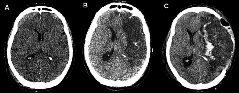
Figure 2 - A) Head computed tomography showing the initially mild asymmetric lateral ventricles, suggesting brain edema likely
from ischemia. B) Head computed tomography on the 5th day from admission, already without extracorporeal support, showing
processes; and a fracture of the left ilium extending to the pubis and acetabulum (chest and head CT shown in igures 1 and 2).
After admission to the ICU, diiculty with adequate ventilation persisted due to the patient’s extensive pulmonary contusions, even after efective bilateral lung drainage. Additionally, the patient required increasing doses of noradrenalin and dobutamine due to persistent signs of low cardiac output (diaphoresis, coldness and slow capillary illing). Bedside echocardiography was performed, and the subcostal window showed an extremely dilated (diastolic diameter 6 cm) and hypokinetic left ventricle, with an estimated ejection fraction of 0.08 (Teicholz). he esophageal Doppler measured a cardiac index of 0.8 L/m2.
About 18 hours after the trauma, despite the administration of 4 mcg/kg/min noradrenalin and 20 mcg/kg/min dobutamine, the patient’s hemodynamics progressively worsened, with a mean blood pressure of 50 mmHg, profuse sweating and delayed peripheral perfusion. he patient was then placed under assisted pressure controlled mechanical ventilation with an inspired oxygen fraction (FiO2) of 1.0, a positive end-expiratory pressure (PEEP) of 10 cmH2O, an inspiratory
pressure of 25 cmH2O (15 cmH2O driving pressure), an inspiratory time of 0.75 seconds and a respiratory rate of 30. Using these parameters, arterial blood gas showed a PaO2 of 56 mmHg, an oxygen saturation of 84% and 3.1 mEq/L (28 mg/dL) lactate. Subsequent tests showed progressive worsening of the physiological parameters, with a central venous saturation of 57% (see Baseline column in Table 1).
Given the imminent risk of death from cardiogenic shock and refractory hypoxemia, our institution’s ECMO team chose to start venoarterial ECMO support as a rescue procedure. Using the Seldinger technique, 22 Fr draining cannulas were inserted into the right common femoral vein. A return cannula was placed in the right femoral artery with an 8F catheter for distal perfusion of the right lower limb. A centrifuge magnetic pump with a polymethylpentene oxygenation membrane (Rotalow/Jostra Quadrox, Maquet Cardiopulmonary AG, Hirrlinger, Germany) was used. he blood low was initially at 4,500 mL/min with a 6,000 mL/min gas low (pure oxygen Sweeper).
he ECMO team of Hospital das Clínicas de São Paulo and Hospital Sirio-Libanês consists of nurses, physicians and physiotherapists. he entire shift team
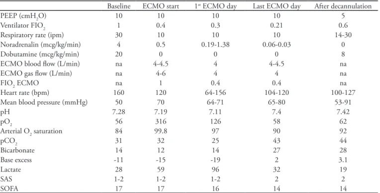
Table 1 – Clinical progression including hemodynamic, respiratory, neurological (Sedation-Agitation Scale) and organ dysfunction (Sequential Organ Failure Assessment score – SOFA) parameters
Baseline ECMO start 1st ECMO day Last ECMO day After decannulation
PEEP (cmH2O) 10 10 10 10 5
Ventilator FIO2 1 0.4 0.3 0.21 0.6
Respiratory rate (ipm) 30 10 10 10 14-30
Noradrenalin (mcg/kg/min) 4 0.5 0.19-1.38 0.06-0.03 0
Dobutamine (mcg/kg/min) 20 0 0 0 8
ECMO blood low (L/min) na 4-4.5 4 4-4.5 na
ECMO gas low (L/min) na 4-6 4 4 na
FIO2 ECMO na 1 0.4 0.4 na
Heart rate (bpm) 160 120 64-156 104-120 100-127 Mean blood pressure (mmHg) 50 70 64-71 65-80 53-91
pH 7.28 7.19 7.11 7.4 7.42
pO2 56 316 126 58 62
Arterial O2 saturation 84 99.8 97 90 92
pCO2 31 32 25 43 44
Bicarbonate 14 12 14 27 28
Base excess -11 -15 -19 2 3.1
Lactate 28 59 96 32 19
SAS 1-2 1-2 1-2 2 2
SOFA 17 17 16 14 14
ECMO – extracorporeal membrane oxygenation; PEEP – positive end-expiratory pressure; FiO2 – inspired oxygen fraction; SAS - Sedation-Agitation Scale; SOFA - Sequential Organ Failure Assessment score.
rather than one speciic person was responsible for managing the device.
Progressive hemodynamic and respiratory improvement occurred about 8 hours after ECMO was started. his allowed us to wean the patient from dobutamine and taper the noradrenaline dose to 0.5 mcg/kg/min. a mean blood pressure of 70 mmHg was maintained. he absence of a pressure curve and a pulse pressure led us to infer that the entire blood low was mediated by the ECMO. Minimal mechanical ventilation parameters were maintained, with a PEEP of 10 cmH2O, an inspiratory pressure of 20 cmH2 O and a FiO2 of 0.3 (controlled pressure mode).(5)
Blood gas analysis showed that the patient’s hypoxia had been corrected; however, he continued to have metabolic acidosis and signiicant hyperlactatemia. he ECMO parameters were adjusted according to the perfusion and oxygenation indices (Table 1).
During the ICU stay, this patient was given analgesia with continuous fentanyl (0.25 – 0.5 mcg/kg/minute). He continued to be obtunded (GCS 5T; Sedation Agitation Scale (SAS) 1-2). On the second day following admission, bedside cranial ultrasonography showed optic sheath widening (6 mm) and midline shift; however, due to the patient’s critical clinical status, no new CT scans were possible during ECMO. Because the nature of the intracranial event could not be precisely established, we chose to maintain analgesia with fentanyl while monitoring the consciousness level until imaging could be performed. Pain was assessed based on behavioral and physiological reactions.
Because of heavy bleeding from the chest tube (1.5 L during the irst day), which required multiple transfusions, and the above described neurological conditions, an anticoagulant was not given during the ECMO.
Despite the presence of acute renal failure (requiring hemodialysis), low platelet counts and signs of extremity ischemia (worse in the right leg where the arterial return cannula was located), cardiac and pulmonary functions progressively improved. On the 4th day of support, a
pulse pressure curve was detected by invasive blood pressure monitoring, and the echocardiogram-estimated left ventricle ejection fraction was 0.3. he pulmonary condition also improved, as assessed by chest x-ray. Dobutamine inotropic support was restarted, and the patient was successfully decannulated (Table 1).
he day after decannulation, the patient had anisocoria and a decreased consciousness level (GCS 3T, SAS1). Repeat head CTs showed a left hemisphere infarction (Figure 2). Left frontotemporal decompressive craniectomy with duraplasty was performed. Postoperative
imaging showed signiicant post-decompression bleeding (Figure 2) with neurological deterioration. he next day, clinical examinations were compatible with brain death, which could not be conirmed due to intraoperative use of thionembutal. About 24 hours later, somatic death was diagnosed.
DISCUSSION
he use of extracorporeal support for severe hypoxemia in children is supported by relatively strong clinical evidence.(2) Little evidence has supported the use of this
technique in adult patients.(5) Recently, however, interest
in the use of ECMO in adults has intensiied, partly due to technological advances (such as biocompatible and durable membranes) and especially due to the large number of refractory hypoxemia cases that occurred during the H1N1 inluenza epidemics.(1)
Extracorporeal support was a key component of the successful Australian treatment regimen for H1N1-related refractory hypoxemia.(1) A recent randomized trial showed
a possible beneit from extracorporeal support in patients with severe acute respiratory failure secondary to acute lung injury/adult acute respiratory distress syndrome. In this trial, ECMO was used to prevent lung injury caused by mechanical ventilation using low ventilation volumes and pressures. However, this study has been criticized because ECMO was only used in 68 of the 90 randomized patients, and there was a relatively high rate of death among patients during transfer to ECMO-specialized sites.(5)
he lack of well-designed clinical trials of ECMO in adult patients prevents us from drawing clear conclusions about the utility of the procedure in adult critical care patients, as highlighted in a recent systematic review.(6)
herefore, the current status of ECMO is that of a rescue measure for failed traditional therapeutics. Because of this, a multidisciplinary team was established for using ECMO in selected refractory hypoxemia cases. With similar indications to those proposed by CESAR,(5)
this team aims to ofer an alternative for patients in whom usual hypoxemia management measures (e.g., alveolar recruitment maneuvers, nitric oxide and high-frequency ventilation) are inefective and/or harmful (e.g., barotrauma or high airway pressure needed for maintaining acceptable ventilation).
Signiicant hypoxemia is a common complication of pulmonary contusion, and the use of extracorporeal oxygenation in some of these patients has been reported.(7)
reports in several important ways. his patient had refractory hypoxemia from several injury mechanisms (pulmonary contusion, bilateral extensive pneumothorax and an unstable chest wall) and had a PaO2/FiO2 ratio of 56. Measures frequently used in this context would be inappropriate or even harmful. Alveolar recruitment maneuvers could worsen the bilateral air istulae, and prone positioning is contraindicated due to the extreme hemodynamic instability.
Additionally, the patient had shock that was refractory to volume expansion, vasopressors and inotropic drugs. Echocardiography revealed clear cardiogenic shock, likely due to myocardial contusion. Considering the imminent risk of death and that there were no other therapeutic options, we chose to use venoarterial ECMO with total cardiorespiratory support to simultaneously support the patient’s respiratory and hemodynamic functions. Signiicant improvement was quickly achieved (Table 1), allowing the progressive recovery of cardiac and pulmonary functions, and we were able to discontinue extracorporeal support after 4 days.
During this time, clinical (consciousness level) and imagery signs (transcranial ultrasound and widened optical sheath) were indicative of intracranial hypertension. However, the patient’s complete dependency on hemodynamic and respiratory support prevented him from undergoing a head CT. Unfortunately, the patient died from his intracranial injury, which could not be assessed and treated in a timely fashion.
Few reports discuss full cardiopulmonary support with venoarterial ECMO in trauma patients. Perchinsky et al. reported 50% survival in a series of 6 patients using this procedure as a rescue measure for severe polytrauma patients deteriorating in spite of the conventional therapy.(3) More recently, Masiakos et al. reported the
successful management of a patient with pulmonary and myocardial contusions in addition to right ventricular papillary muscle rupture with signiicant tricuspid regurgitation, who presented with signiicant hypoxemia, hemodynamic instability and diicult-to-manage ventricular arrhythmias.(4)
In our report, we successfully used extracorporeal support as a rescue measure for cardiorespiratory dysfunction that would otherwise have been rapidly fatal. ECMO was efective as a bridging strategy, allowing
decannulation on the 4th day after the trauma. he patient
died from head trauma, which was not related to nor treated with extracorporeal support. Unlike the report by Masiakos et al.,(4) no additional ICU professionals (e.g.,
a perfusionist) were necessary during the extracorporeal support; only our institutional ECMO team was involved.
In summary, this article reports a polytrauma patient with refractory hypoxemia due to pulmonary contusion and refractory cardiogenic shock due to cardiac contusion. Venoarterial ECMO was successfully used as a bridging strategy for cardiac and pulmonary recovery, and the extracorporeal support was discontinued on the 4th day
after trauma. his extracorporeal support method can be lifesaving in selected patients. However, additional studies are necessary to evaluate how this promising technology may best be used clinically.
Participants of the Hospital das Clínicas de São Paulo and Hospital Sírio-Libanês ECMO team:
Luciano Cesar Pontes Azevedo, Marcelo Park, André Luiz de Oliveira Martins, Eduardo Leite Vieira Costa, Guilherme Paula Pinto Schettino, Marcelo Brito Passos Amato, Carlos Roberto Ribeiro Carvalho, Mauro Tucci, Alexandre Toledo Maciel, Fernanda Maria Queiroz Silva, Leandro Utino Taniguchi, Edzângela Vasconcelos, Raquel de Nardi, Cláudio Machtans, Michele Nardi and Adriana Sayuri Hirota.
RESUMO
Existem poucos relatos na literatura sobre o uso de oxige-nação extracorpórea por membrana venoarterial por dupla dis-função decorrente de contusão cardíaca e pulmonar no paciente politraumatizado. Relatamos o caso de um paciente de 48 anos, vítima de acidente de motocicleta e automóvel, que evoluiu ra-pidamente com choque refratário com baixo débito cardíaco por contusão miocárdica e hipoxemia refratária decorrente de contusão pulmonar, tórax instável e pneumotórax bilateral. O suporte extracorpóreo foi uma medida efetiva de resgate para esse caso dramático, e o seu uso pôde ser interrompido com su-cesso no 4º dia após o trauma. O paciente evoluiu com extenso infarto cerebral, morrendo no 7º dia de internação.
Descritores: Oxigenação; Choque cardiogênico; Lesão
REFERENCES
1. Australia and New Zealand Extracorporeal Membrane Oxygenation (ANZ ECMO) Inluenza Investigators, Davies A, Jones D, Bailey M, Beca J, Bellomo R, Blackwell N, et al. Extracorporeal Membrane Oxygenation for 2009 Inluenza A(H1N1) Acute Respiratory Distress Syndrome. JAMA. 2009;302(17):1888-95.
2. Sidebotham D, McGeorge A, McGuinness S, Edwards M, Willcox T, Beca J. Extracorporeal membrane oxygenation for treating severe cardiac and respiratory disease in adults: Part 1--overview of extracorporeal membrane oxygenation. J Cardiothorac Vasc Anesth. 2009;23(6):886-9.
3. Perchinsky MJ, Long WB, Hill JG, Parsons JA, Bennett JB. Extracorporeal cardiopulmonary life support with heparin-bonded circuitry in the resuscitation of massively injured trauma patients. Am J Surg. 1995;169(5):488-91.
4. Masiakos PT, Hirsch EF, Millham FH. Management
of severe combined pulmonary and myocardial contusion with extracorporeal membrane oxygenation. J Trauma. 2003;54(5):1012-5.
5. Peek GJ, Mugford M, Tiruvoipati R, Wilson A, Allen E, halanany MM, Hibbert CL, Truesdale A, Clemens F, Cooper N, Firmin RK, Elbourne D; CESAR trial collaboration. Eicacy and economic assessment of conventional ventilatory support versus extracorporeal membrane oxygenation for severe adult respiratory failure (CESAR): a multicentre randomised controlled trial. Lancet. 2009;374(9698):1351-63. Erratum in Lancet. 2009;374(9698):1330.
6. Mitchell MD, Mikkelsen ME, Umscheid CA, Lee I, Fuchs BD, Halpern SD. A systematic review to inform institutional decisions about the use of extracorporeal membrane oxygenation during the H1N1 inluenza pandemic. Crit Care Med. 2010;38(6):1398-404.