Rev. Assoc. Med. Bras. vol.62 número4
Texto
Imagem
Documentos relacionados
In this case, the uncommon localization of the ischemic event not detected in cranioencephalic magnetic resonance imaging, the lack of focal deficits and changes in other
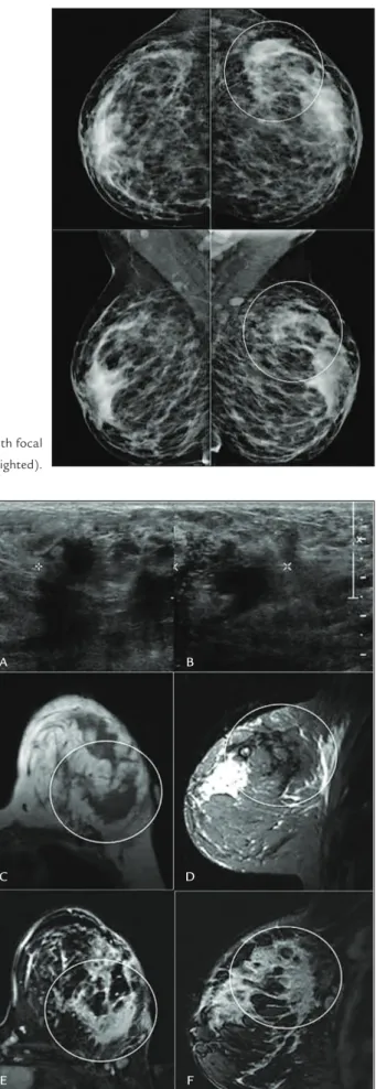
The authors report the case of a 45-year-old patient and describe the findings at structural magnetic resonance imaging and at advanced sequences, correlating them
Technological developments in imaging, such as elastography, magnetic resonance imaging-guided biopsy, magnetic resonance imaging-ultrasonography fusion, can revolutionize the
Esaote) were examined and a round lesion was observed on the left cerebral hemisphere, with ill-defined margins, apparent midline shift, moderate peritumoral
We present the case of a 53 year old man that fulfilled the diagnostic criteria for HAM/TSP but had at the magnetic resonance imaging (MRI) of the spinal cord evidences
Utility of preoper- ative magnetic resonance imaging mielography for identifying dural defects in patients with spinal extradural arachnoid cysts: case report.. Hatashita S, Kondo
Proton magnetic resonance spectroscopic imaging and magnetic resonance imaging volumetry in the lateralization of temporal lobe epilepsy: a series of 100 patients. Connelly A,
We report the case of a 34-year-old male who presented with complete features of Kjellin’s syndrome, with typical retinal findings observed on multimodal imaging (spectral
