NASAL AERATION AND RESPIRATORY MUSCLE
STRENGTH IN MOUTH BREATHERS’ CHILDREN
Aeração nasal e força muscular respiratória
em crianças respiradoras orais
Renata Andrade da Cunha (1), Daniele Andrade da Cunha (1),
Luciana Ângelo Bezerra (1), Ana Carolina Cardoso de Melo (1),
Décio Medeiros Peixoto (2), Tetsuo Tashiro (3), Hilton Justino da Silva (1)
(1) Universidade Federal de Pernambuco-UFPE, Recife,
Per-nambuco, Brasil.
(2) Departamento Materno-Infantil da Universidade Federal de
Pernambuco-UFPE, Recife, Pernambuco, Brasil.
(3) Departamento de Educação Física da Universidade
Fede-ral de Pernambuco-UFPE, Recife, Pernambuco, Brasil. Source of aid: Study conducted at Clinical Hospital of Federal University of Pernambuco - HC/UFPE - Recife (PE), Brazil, master scholarship granted by the Higher Education Personnel Improvement Coordination - CAPES. Financial support from the
National Counsel of Technological and Scientiic Development
(CNPq) - Support for Research Projects/ Universal 14/2011 - Track A - up to R$ 20.000,00, process: 475641/2011-6.
Conlict of interest: non-existent
Due to lack of air low passage of the stimulus
through the nasal duct, do not happen the pressures
and strains that ensure the correction of the maxillary sinuses. The nostrils become cracks narrow nasal with a volume and elasticity reduced by the disuse, present pale nasal mucosa, poor iltration and air
heating during breathing2.
With decreasing nasal air passage, the air will
reach the lungs through mechanically shorter and
easier way. Thus, the child makes less efort to breathe, aggravating the whole mechanical venti
-lation with the commitment of the lungs, abnormal respiratory rate, subject to the expansion and
contraction of the lungs and alveolar ventilation.
Thus, the diaphragm action will be reduced, as
compromise their length-tension relationship, incapacitating him to produce adequate peak
tension, leading to relaxation and requiring less respiratory muscle strength, which develops weakness with retraction muscular3,4.
Changes can also happen with the abdominal muscles, which associated to constant intake of air,
INTRODUCTION
For eiciently nasal breathing occurs, there needs
to be a condition of air passage through the nostrils. When there is impossibility of breathing through the
nasal route, that breath will occur predominantly at
the mouth, being called oral breathing1.
ABSTRACT
Purpose: to observe whether there is a relationship between respiratory muscle strength and degree
of nasal aeration in Mouth Breathing children. Methods: this is an observational and a comparative
cross-sectional study. 32 Mouth Breathing children with allergic rhinitis (21 boys and 11 girls) and 30 nasal breathing without allergic rhinitis (09 boys and 21 girls) participated, 7-12 years, subjected to evaluation for nasal aeration with Altmann mirror and to evaluation of respiratory muscle strength with digital manovacuometer (MVD®30). Results: there was no correlation between nasal aeration
and respiratory muscle strength in each subgroup. There was diference comparing values of maximal expiratory pressure between mouth breathers boys and girls (p=0,0064), and between nasal breathers boys and girls (p=0,0030). There was also diference maximal inspiratory pressure between mouth breathers boys and girls (p=0,0324), and between nasal breathers boys and girls (p=0,0210).
Conclusion: it was not possible to conirm that there is a relationship between the degree of nasal
aeration and respiratory muscle strength in Mouth Breathing.
angle to the height of the anterior nasal volunteer
spine who was seated and with your head straight, supported column on the chair and feet lat on the loor. After two quiet exhalation, nasal aeration was measured by checking with black marker blurry area
on the mirror itself. The total time of this procedure
ranged from ive to ten minutes.
Then this labeling was transferred to a sheet of
Altmann Mirror Reference Book. Each sheet of the
reference block was scanned by means of an HP
Photosmart printer series D110, the data having been subsequently measured through 1.46r Image
J software (http://imagej.nih.gov/ij), yielding the extent cm².
After the nasal aeration, a physical therapist conducted the evaluation of respiratory muscle
strength, through the maximum inspiratory and expiratory pressures (PImax and PEmax). We used a portable digital manovacueometer (MVD®
300-Globalmed-Brazil), graduated in cmH2O, with the
examination mode Of-Line, which has measurement
resolution of 1 cmH20 and 480 cmH20 full scale,
coupled with a mouth scientiic with a 2 mm hole in order to provide exhaust air and thus prevent the
increase in the oral cavity pressure generated by of
unwanted oral wall muscles contraction, minimizing the cheek efect and thereby avoiding inter -ference in the results, according to some authors
recommendations5.
By convention and to standardize measures,
children were sitting, with their spine supported in
the back of the chair, upper members supported
on the thighs and feet lat on the loor6. The PImax
and PEmax measurements were recorded during maximum efort against nasal tract occluded by a
nose clip, preventing air leakage through the nose, and generated the air outlet on the mouth in the
inhalation and exhalation, following the previus
study criteria6,7.
During the PEmax and PImax evaluation, it was
requested that the child undertake a deep inspiration
or expiration until reaching the total lung capacity (CPT) or expiratory volume reserve (VR), respec
-tively, and then exhale or inhale vigorously through the mouth piece that the children arrested with their
lips to prevent air leakage around the same. The peak
expiratory and inspiratory force was sustained for at least one second, with a minimum interval of one second between each peak. The children performed three to ive attempts to obtain the pressures being considered the most valuable, both for PEmax as PImax, measured in cmH2O7. Sometimes, in case of signs of fatigue manifestation, the assessment test
of PImax and PEmax was stopped and restarted.
The total time of this procedure ranged from 15 to 30 minutes.
leads the oral breathing child to present a laccid and protruding abdomen, resulting in muscle weakness both inspiratory, as expiratory4.
Whereas, due to the change in breathing mode, orofacial changes occurs and respiration
mechanism too, this study aims to observe whether there is a relationship between respiratory muscle
strength and nasal aeration area in mouth breathing children.
METHODS
The research project was iled, evaluated and
approved by the Ethics in Human Beings Research Committee of the Federal University Health Sciences Center of Pernambuco (CEP/CCS/UFPE)
under the registration number 492/11 and CAAE
0484.0.172.000 -11. There is an observational study
and cross-comparison between two groups, carried
out from October 2012 to April 2013.
According to the inclusion criteria, 32 children participated in oral breathing secondary to allergic
rhinitis conirmed in medical records and that
breathing through the mouth into the time of the
survey and 30 nasal breathing children without allergic rhinitis, of both genders, between 7 and 12 years. The volunteers were in attendance at the
Allergy and the Pediatric ambulatories of Clinical Hospital of the Federal University of Pernambuco (HC/UFPE).
Were adopted as exclusion criteria for both groups: children with diiculty in understanding
simple orders, evaluated by means of spontaneous conversation, or neurological changes; genetic and
endocrine disorders that interfere in the growth and
development; cardiovascular changes and people
with severe heart disease; deviated nasal septum;
cleft lip, cleft lip and palate; in use of braces;
reporting respiratory infectious disease of the lower airway such as asthma; physical therapy and/or
speech therapy prior or ongoing intervention.
All oicials who were accompanying children at the time of evaluation were interviewed and informed
of the research through the Informed Consent Free
Term (TCLE).
The interview consisted of maternal and child
socioeconomic data, family housing, smell
condi-tions and aspects of the child’s sleep, followed by
information on the medical history of the child.
Measurement of nasal aeration area was
conducted by the Altmann milimetrically nasal mirror
graded from Altmann (Pro-Fono®), for a speech
therapist, who was standing and ahead of the child
using disposable gloves. After the air conditioning
RESULTS
It was evaluated 62 children: 32 (51.61%) for the
mouth breathers group, distributed in 21 children
(65.63%) for the male subgroup and 11 (34.37%) for females, and 30 children (48.39%) for the nose breathers group, distributed in 21 (70%) for the subgroup of girls and 09 (30%) for boys. The average age was 8.7 ± 1.4 years for mouth breathing group and 9.0 ± 1.3 for nose breathers group, with no diference between groups (p=0.3207).
Regarding the distribution of the sample according to housing conditions, family income, maternal variables and breastfeeding participants
included, there were diferences between the groups only for the variable family income (p=0.0437)
(Table 1).
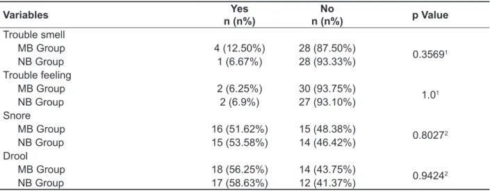
No diference was observed between the two
groups regarding the sample distribution according to variables related to smell, taste, snoring and drooling (Table 2).
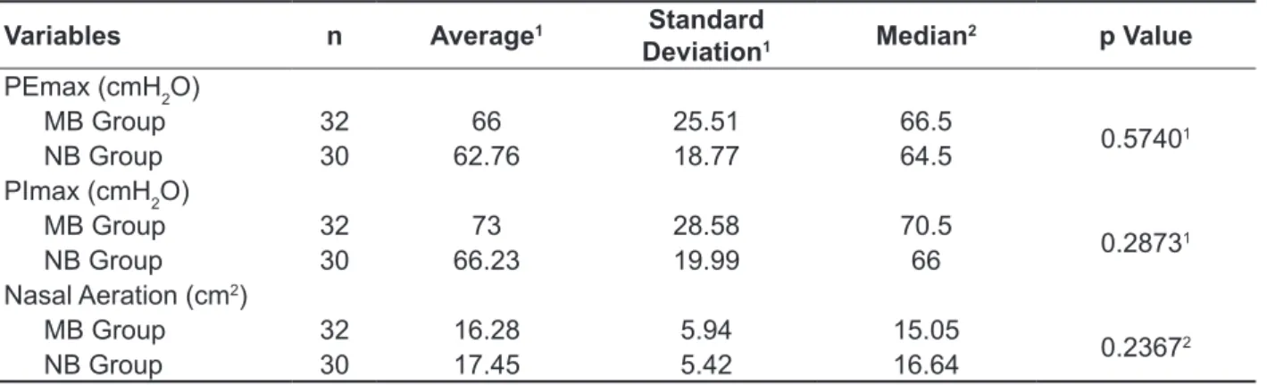
The average values were expressed, standard deviation and median for PEmax, PImax and nasal aeration between the two groups, according to the
applied test (Table 3).
It is noticed that there was no correlation between
nasal aeration and respiratory muscle strength
(PEmax and PImax) within each subgroup (Table 4). However, when comparing the values of the PEmax and PImax between boys and girls mouth breathers, signiicant diferences (p=0.0064 and p=0.0324, respectively) (Table 5).
The same was true for nose breathers group, where p=0.0030 for PEmax and p=0.0210 for PImax
(Table 5). The sample size estimate and statistical result
analysis were performed through BioEstat software,
5.3 version, performing previously the Shapiro-Wilk
normality test which considered the sample with normal distribution. Then sample size estimate were
performed to linear correlation test, considering a
90% of power test and an alpha level of 0.05 in the
ratio of 1:2, estimating a minimum sample size for
the mouth breathers group: ten children were female and 185 were male and, for the nose breathers
group: 20 females and 370 males. From the results
of the pilot study, which tended to get close corre
-lation between the values of the nasal aeration area and the values of maximal respiratory pressures for females, it was decided to separate by gender.
In results analyzing, it was used the chi-square test of Pearson or Fisher’s exact test, when
necessary, for the analysis of categorical variables. According to the result of normality test, Pearson
correlation test or Spearman’s correlation test was used to assess the correlations between the values of respiratory pressures (PEmax and PImax) and
nose through the air outlet (nasal aeration) in both groups.
For comparison between groups, the Mann-Whitney test (Wilcoxon Rank-Sum Test) was
applied to p <0.05 (non-parametric data), consid-ering analyze the data based on the median or the Student t test for p <0.05 (parametric data), based
on the mean and standard deviation. Diferences were considered signiicant when p <0.05 for all
calculations.
This study hypothesis was that there is a relationship between the nasal aeration area and respiratory muscle strength (PEmax and PImax) in
Table 1 – Distribution of the sample according to housing conditions, family income, maternal variables and breastfeeding participants
Variables MB Group
n (n%)
NB Group
n (n%) p Value
Piped water at home
Yes 29 (90.62%) 30 (100%)
0.23851
No 3 (9.38%) 0 (0%)
Flush toilet at home
Yes 28 (87.5%) 27 (90%)
1.0001
No 4 (12.5%) 3 (10%)
Home light
Yes 32 (100%) 30 (100%)
1.0001
No 0 (0%) 0 (0%)
Family monthly income (MW)
≤1 MW 22 (68.75%) 13 (43.33%) 0.04372
>1 MW 10 (31.25%) 17 (56.67%)
Breastfeeding
< 4 months* 20 (66.66%) 13 (46.42%)
0.11992
≥ 4 months * 10 (33.34%) 15 (53.58%)
1Fisher’s Exact Test; 2Chi-Square Test; p<0,05 (statistically signiicant values)
Legend: n=number of children; n%=number of children in percentage; NB=nose breathers; MB=mouth breathers MW=minimum wage; *02 participants were excluded because they were unable to answer
Table 2 – Distribution according to variables related to smell, taste, snoring and drooling
Variables Yes
n (n%)
No
n (n%) p Value
Trouble smell
0.35691
MB Group 4 (12.50%) 28 (87.50%)
NB Group 1 (6.67%) 28 (93.33%)
Trouble feeling
1.01
MB Group 2 (6.25%) 30 (93.75%)
NB Group 2 (6.9%) 27 (93.10%)
Snore
0.80272
MB Group 16 (51.62%) 15 (48.38%)
NB Group 15 (53.58%) 14 (46.42%)
Drool
0.94242
MB Group 18 (56.25%) 14 (43.75%)
NB Group 17 (58.63%) 12 (41.37%)
1Fisher’s Exact Test; 2Chi-Square Test; p<0,05 (statistically signiicant values)
Table 3 – Measures of the maximum respiratory pressures and the area of nasal aeration in mouth breathers and nose breathers children
Variables n Average1 Standard
Deviation1 Median
2 p Value
PEmax (cmH2O)
MB Group 32 66 25.51 66.5
0.57401
NB Group 30 62.76 18.77 64.5
PImax (cmH2O)
MB Group 32 73 28.58 70.5
0.28731
NB Group 30 66.23 19.99 66
Nasal Aeration (cm2)
MB Group 32 16.28 5.94 15.05
0.23672
NB Group 30 17.45 5.42 16.64
1Student t Test; 2The Mann-Whitney Test; p<0,05 (statistically signiicant values)
Legend: n=number of children; NB=nose breathers; MB=mouth breathers; PEmax=maximum expiratory pressure; PImax=maximum
inspiratory pressure
Table 4 – Correlation between the area of nasal aeration and the maximum respiratory muscle strength (PEmax and PImax) in mouth breathers and nose breathers children
Variables
Girls Boys
MB Group (n=11)
NB Group (n=21)
MB Group (n=21)
NB Group (n=9)
PEmax X Nasal Aeration p=0.31061 p=0.42322 p=0.39422 p=0.30841
rs=0.3371 r=-0.1845 r=0.1961 rs=0.3833 PImax X Nasal Aeration p=0.3071 p=0.21772 p=0.18212 p=0.54391
rs=0.1636 r=0.2807 r=0.3028 rs=-0.2343
1Pearson’s Correlation Test; 2Spearman’s Correlation Test; p<0,05 (statistically signiicant values)
Legend: p=p value; r=Pearson’s correlation coeicient; rs=Spearman’s correlation coeicient; n=number of children; NB=nose brea
-thers; MB=mouth brea-thers; PEmax = maximum expiratory pressure; PImax=maximum inspiratory pressure
Table 5 – Comparison between the area of nasal aeration and maximum respiratory pressures (PEmax and PImax) in mouth breathers and nose breathers children
Variables MB Group NB Group
p Value p Value
PEmax (girls) and PEmax (boys) 0.00641 0.00302
PImax (girls) and PImax (boys) 0.03241 0.02101
Nasal Aeration (girls) and Nasal Aeration (boys) 0.59222 0.66722
1Student t Test; 2The Mann-Whitney Test; p<0,05 (statistically signiicant values)
Legend: PEmax=maximum expiratory pressure; PImax=maximum inspiratory pressure
DISCUSSION
There are several factors that can lead to mouth breathing, and allergic rhinitis is possibly the most
common cause of chronic airway obstruction, afecting 15-20% of population8. In this study, we
found a signiicantly higher number of male children with allergic rhinitis and mouth breathing.
This inding can be explained because the boys
have a higher prevalence of allergic rhinitis, the
main entity associated with mouth breathing, and a lower airway caliber9. However, according to other
studies performed8,10 there is no direct relationship
from the mouth breathing, caused by allergic rhinitis, for males.
Family income is cited as an important
because the airlow is dynamic. In addition, you have no control over the voluntary expiration of the individual and can be seen an expiration with more or less efort, even with corrections in these cases.
The combination of the aforementioned factors
coupled with the lack of precise functional diagnosis
of mouth breathing may have been decisive for the
values of the nasal aeration areas have not shown diferences between the groups, although the mouth breathing group median (15.05 cm²) was slightly lower than that of nasal breathing. Although the values of the mean and median do not difer much from each other, we could not compare the results of
the areas of the nasal aeration of the current study
with those of another study16, or with others in the literature, because they do not compare average
with median due statistical test applied.
Although mirror using along with the clinical history and physical examination are considered the
gold standard in Orofacial Motricity area of speech therapy, there are no studies that point predicted
values of nasal aeration to normal subjects. The dii -culty in standardizing protocols for characterization of mouth breath sample in this research is a factor that may also have interfered directly on the data
obtained. Diagnosis of mouth breathing was deined
in the medical records, often building on nasal
obstruction, this factor alone that not only deines
the situation of mouth breathing, given that the signs and symptoms may not have been considered.
Regarding the inspiratory and expiratory maximum pressure (PImax and PEmax), the PImax, generated from maximum expiratory eforts,
measures the strength of the inspiratory muscles
(diaphragm and external intercostal), while PEmax, generated from maximum inspiratory eforts, measures the strength of the expiratory muscles (abdominal and internal intercostal). The PImax and PEmax indirectly indicate the respiratory muscle
strength21.
To measure these pressures and thus quantify the strength of respiratory muscles, it used a digital
manovacuometer that provides accurate result with
evaluation 1 in 1 cmH2O, record the peak pressure
in the display, allowing the values remain stored on
the device, besides having 2 mm hole minimizes
the cheek efect. The digital is recommended rather than the analog, because the latter makes it diicult
to record the peak pressure, presents scale ranges
from 4 cmH2O, easily descalibration and does not
have the hole 2 mm22.
In this study, most girls had lower values for inspi
-ratory and expi-ratory pressures compared with boys in both groups. Similar results were observed in
previous study10. This is because females have vital
capacity (air volume that can expel from the lungs
found in this study is that a good portion of respon-dents from mouth breathers group have monthly
incomes below the minimum wage compared to the nasal breathers group and were diferences between groups, corroborating the indings of other
authors12 also they found lower averages for mouth
breathers group.
Given that the low-income population is evident
as higher risk because it involves demographic and economic factors, undoubtedly, the prevalence of
respiratory diseases in children would be reduced if
they had better residence conditions11. The latter data contradicts some points of the results of this study, since all mouth breathing children lived in homes
with light and most of them had running water and lushing toilets at home. So the more unfavorable
for the socioeconomic situation, the prevalence of
respiratory diseases tend to be bigger11.
In addition, it was found, in the present study
and on a study of 200713, that many of the children
weaned before four months of life. The correct breathing pattern may be impaired by weaning before six months of life, because it compromises
the proper oral motor development and the lack of
physiological sucking the breast allows the instal -lation of malocclusion, motor-oral amendment and mouth breathing4,14.
Regarding the smell and taste, a literature
review found that the mouth breathing results in
the decrease in smell and taste, there is a limitation
in the operation of these sense15. However, these
indings are not consistent with the results of this study, since most mouth breathing children showed no trouble smell, as well as nose breathers group.
A large number of children in both groups reported no trouble feeling like some foods. Three other
authors16 observed that a small number of
volun-teers had no complaints regarding taste.
Studies show that snoring and drooling relationship with mouth breathing can cause respi -ratory and sleep problems, caused by constant
mouth opening, because the space of rinopharynx
(throat region) is reduced due to nasal obstruction
or allergic state4,17. In this study, despite snoring
and drooling data have been approximated, but no diference between the two groups the mouth
breathers had a higher frequency of these aspects also been observed in previus studies8,13,16,18.
The use of Altmann millimetred mirror was nominated for this study due to its wide clinical appli
-cation, playback facility and manipulation16,19, and
does not cause discomfort to paciente20,21. However,
the subjectivity of this equipment to obtain the nasal aeration is criticized in literature, questioning the
test sensitivity. The mirror does not allow the estab
In addition, this age due to the abdominal muscles immaturity, the compliance of this region is
larger, allowing the abdomen to expand more easily
and predominance during respiration (inspiration
and expiration), facilitating the lung expansion24
which justify an result without diference between groups for the PEmax.
Thus, the younger the mouth breathing children, less orofacial and lung function changes
they present, suggesting that with growth, these
changes can accentuate18. Even though there is
evidence that mouth breathing children can behave as the nasal breathing because of possible muscle compensation arising from the mouth breathing, it is believed that there may be a direct relationship
between the values of the nasal aeration area and the values of maximal respiratory pressures.
Much of children that chronically breathe through
the mouth shows a decrease in nasal aeration, reduces the efort to inhale and exhale because the air comes quickly to the lungs without these organs to expand and retract properly and decrease the
diaphragm and abdominal action. With these conse-quences of mouth breathing, there is a commitment in respiratory muscle strength, reducing the
maximum respiratory pressures. Also, it is believed
that these relationships can be better understood
with the most comprehensive studies of mouth
breathing functional diagnostics and more accurate equipment such as acoustic rhinometry.
The Pathophysiology of the stomatognathic system research group of this institution has invested
in knowledge about this product and suggests it to
better assess nasal geometry, to conduct an nasal area investigation more precise than the Altmann mirror. The rhinometry is a quantitative method that
allows mapping of the nasal anatomy, measuring its volume in diferent points27. This method can
improve the aerodynamic upper airway charac
-teristics deinition of mouth breathing children, contributing to the study of the relationship between
the area and nasal volume and respiratory muscle strength.
In addition, the evaluation of nasal function can
help the child to observe how much air low from
the nose and to encourage use it more breathing as
well as the early assessment of the strength of the breathing muscles help in the lungs use awareness for expansion and lung retraction properly.
It is thinking of the existence of these relation
-ships that we suggests a regular monitoring in mouth breathing children with assessments of nasal and lung function, in order to observe whether there is
any change in the long-term respiratory component.
after maximum deep breath), decreased maximal expiratory low and a lower surface pulmonary difusion and airways diameter dereased23.
The main results observed were that mouth breathing children behaved as nasal breathing when
respiratory pressure variables and nasal aeration
were correlated. The fact of not having found corre
-lation between these variables can suggest that the
transition from nasal breathing for mouth breathing induces changes in respiratory muscle structure,
developing compensation strategies to live with the consequences of mouth breathing without
the appearance of noticeable changes, favoring breathing24,25.
As regards the muscles involved with nasal
breathing (nasolabial lifters - dilates the nostrils),
a study conducted an experiment in Wistar rats, which were induced mouth breathing and these muscles showed a relative decrease in fatigable 2b iber type. This means that the nasolabial lifters were more resistant to fatigue as they adapted to the new condition of mouth breathing, allowing the
maintenance of functional position of these involved
muscles in this function without altering the muscle activity and consequently without altering the nasal function (nasal inspiration and exhalation)25.
Another explanation for the lack of relationship would be that the children may have ordered the
accessory muscles of inspiration
(sternocleido-mastoid and trapezius) along with the major muscles of inspiration (diaphragm and external intercostal)
during manovacuometry26, even with the body
stabilization control by the physiotherapist during evaluations.
This accessory activation may be imperceptible to the eye of the professional, but a study in mouth breathing children, 8-12 years revealed that these
same accessory muscles showed increased muscle
activity, perceived by electromyography. Because
of airway obstruction, a stronger diaphragm
contraction happens, preceded by muscular action
inspiratory accessory, shown by increased activity
of the sternocleidomastoid muscle during nasal
inspiration of children with mouth breathing26. Other
authors found no signiicant changes among groups
of mouth and nasal breathing in relation to the
composition of the muscle ibers of the diaphragm, ie, this muscle was equivalent behavior in mouth
and nasal breathers25.
It is noteworthy that children up to the age of ten
are in the process of alveolar multiplication and rib
and the maximal respiratory pressures values in
mouth breathing children.
ACKNOWLEDGEMENTS
The Hospital of the Federal University of
Pernambuco which enabled this research and the National Counsel of Technological and Scientiic
Development-CNPq (Universal Notice MCT/CNPq 14/2011 - Track A - Process: 475641 / 2011-6) for
inancial support .
CONCLUSION
In this study, low family income tends to inluence
the development of mouth breathing.
For the values of the nasal aeration area, the
group of mouth breathers had lower medians, but with no diference between groups.
For the PEmax and PImax values, the boys showed higher values than girls in both groups, with a diference.
However, it was not possible to conirm the direct relationship between the nasal aeration area values
REFERENCES
1. Andrada e Silva MA, Marchesan IQ, Ferreira LP,
Schmidt R, Ramires RR. Postura, tônus e mobilidade de lábios e língua de crianças respiradoras orais. Rev CEFAC. 2012;14(5):853-60.
2. Okuro RT. Efeitos da respiração bucal e da projeção anterior da cabeça na força muscular
respiratória e na capacidade de exercício em
crianças e adolescentes [dissertação]. Campinas (SP): Faculdade de Ciências Médicas da Faculdade de Campinas-UNICAMP; 2012.
3. Aragão W. Aragao’s Function Regulator, the estomatognathic system and postural changes in
children. J clin pediatr dent. 1991;15(4):226-31.
4. Felcar JM, Bueno IR, Massan ACS, Torezan RP, Cardoso JR. Prevalência de respiradores bucais em crianças de idade escolar. Ciênc saúde colet. 2010;15(2):437-44.
5. Oliveira LC, Campos TF, Borja RO, Claves
GSS, Delgado RN, Mendes REF et al. Pressões
respiratórias máximas de pico e sustentada
em crianças. Rev Bras Saúde Matern Infant. 2012;12(4):357-64.
6. Brasileiro-Santos MS, Lima AMJ, Hunka MBS,
Neves TS, Andrade MA, Santos AC. Atividade mioelétrica dos músculos respiratórios em crianças
asmáticas durante manobra inspiratória máxima.
Rev Bras Saúde Matern Infant. 2012;12(3):251-7.
7. Black LF, Hyatt RE. Maximal respiratory pressures: normal values and relationship to age and sex. Am Rev Respir Dis. 1969;99(5):696-702.
8. Popoaski C, Marcelino TF, Sakae TM, Schmitz
LM, Correa LHL. Avaliação da qualidade de
vida em pacientes respiradores orais. Int Arch Otorhinolaryngol. 2012;16(1):74-81.
9. Rappai M, Collop N, Kemp S, deShazo R. The nose and sleep-disordered breathing: what we know and what we do not know. Chest. 2003;124(6):2309-23.
RESUMO
Objetivo: observar se existe relação entre força muscular respiratória e área da aeração nasal em
crianças respiradoras orais. Métodos: trata-se de um estudo do tipo observacional, transversal
com-parativo entre dois grupos. Participaram 32 crianças com Respiração Oral secundária à rinite alérgica
(21 meninos e 11 meninas) e 30 respiradoras nasais sem rinite alérgica (09 meninos e 21 meninas),
7 a 12 anos, submetidas à avaliação da aeração nasal com o espelho de Altmann e à avaliação da
força muscular respiratória com o manovacuômetro digital (MVD®30). Resultados: não houve cor-relação entre aeração nasal e força muscular respiratória em cada subgrupo. Houve diferença
com-parando-se valores das pressões expiratórias máximas entre meninos e meninas respiradores orais (p=0,0064) e entre meninos e meninas respiradores nasais (p=0,0030). Também houve diferença das pressões inspiratórias máximas entre meninos e meninas respiradores orais (p=0,0324) e entre meninos e meninas respiradores nasais (p=0,0210). Conclusão: não foi possível conirmar a relação
entre a área de aeração nasal e a força muscular respiratória nos respiradores orais.
19. Altmann EBC. Espelho nasal milimetrado; 1994 [Acesso em 30 mar 2011]. Disponível em: http://www.profono.com.br/produtos_descricao. asp?lang=pt_BR&codigo_produto=21.
20. Cunha DA, Silva HJ, Moraes KJR, Cunha RA, Régis RMFL, Silva EGF et al. Aeração nasal em crianças asmáticas. Rev CEFAC. 2011;13(5):783-9.
21. Ribeiro SNS, Fontes MJF, Duarte MA. Avaliação da força muscular respiratória e da função pulmonar
por meio de exercício em crianças e adolescentes
com asma: ensaio clínico controlado. Pediatria.
2010;32(2):98-105.
22. Montemezzo D, Velloso M, Britto RR, Parreira VF. Pressões respiratórias máximas: equipamentos e procedimentos usados por isioterapeutas
brasileiros. Fisioter Pesqui. 2010;17(2):147-52.
23. Harms CA. Does gender afect pulmonary function and exercise capacity? Respir Physiol
Neurobiol. 2006;151(2-3):124-31.
24. Brant TCS, Parreira VF, Mancini MC, Becker
HMG, Reis AFC, Britto RR. Breathing pattern and thoracoabdominal motion in mouth-breathing
children. Rev Bras Fisioter. 2008;12(6):495-501. 25. Gelhaye M, Martrette JM, Legrand-Frossi C, Trabalon C. Myosin heavy chain expression and
muscle adaptation to chronic oral breathing in rat. Respir Physiol Neurobiol. 2006;154:443-52.
26. Ribeiro EC, Marchiori SC, Silva AMT. Electromyographic analysis of trapezius and sternocleidomastoideus muscles during nasal and oral inspiration in nasal- and mouth-breathing
children. J Electromyog Kinesiol. 2002;2:305-16.
27. Melo ACC, Gomes, AOC, Cavalcanti AS, Silva HJ. Acoustic rhinometry in mouth breathing patients:
a systematic review. Braz J Otorhinolaryngol.
2015;81(2):212-8. 10. Okuro RT, Morcillo AM, Ribeiro MAGO, Sakano
E, Conti PBM, Ribeiro JD. Respiração bucal e anteriorização da cabeça: efeitos na biomecânica
respiratória e na capacidade de exercício em crianças. J Bras Pneumol. 2011;37(4):471-9. 11. Menezes VA, Leal RB, Moura MM, Granville-Garcia AF. Inluência de fatores socioeconômicos e demográicos no padrão de respiração: um estudo
piloto. Braz J Otorhinolaryngol. 2007;73(6):826-34.
12. Silvério KCA, Ferreira APS, Johanns CM, Wolf A,
Furkin AM, Marques JM. Relação de escolaridade,
faixa etária e proissão de mães com a oferta de chupeta e mamadeira a seus ilhos. Rev CEFAC.
2012;14(4):610-5.
13. Cunha DA, Silva GAP, Motta MEFA, Lima CR,
Silva HJ. A respiração oral em crianças e suas repercussões no estado nutricional. Rev CEFAC.
2007;9(1):47-54.
14. Neu AP, Silva AMT, Mezzomo CL,
Busanello-Stella AR, Moraes AB. Relação entre o tempo e o tipo de amamentação e as funções do sistema estomatognático. Rev CEFAC. 2013;15(2):420-6. 15. Paz FR, Pinto MMA, Silva HJ. A diminuição do olfato como consequência da respiração oral. J Soc Bras Fonoaudiol. 2003;4(14):56-8.
16. Melo FMG, Cunha DA, Silva HJ. Avaliação da aeração nasal pré e pós a realização de manobras de massagem e limpeza nasal. Rev CEFAC.
2007;9(3):375-82.
17. Izu SC, Itamoto CH, Pradella-Hallinan M, Pizarro
GU, Tuik S, Pignatari S, Fujita RR. Obstructive
sleep apnea syndrome (OSAS) in mouth breathing children. Braz J Otorhinolaryngol. 2010;76(5):552-6. 18. Imbaud T, Wandalsen G, Nascimento Filho E, Wandalsen NF, Mallozi MC, Solé D. Respiração bucal em pacientes com rinite alérgica: fatores associados e complicações. Rev Bras Alerg
Imunopatol. 2006;29(4):183-7.
Received on: March 17, 2015
Accepted on: April 29, 2015
Mailing address:
Renata Andrade da Cunha
Avenida Prof. Moraes Rego, 123, Cidade Universitária
Recife – PE – Brasil
CEP: 50670-901