Coronary computed tomography angiography
with 320-row detector and using the AIDR-3D:
initial experience
Angiotomografia computadorizada de coronárias com tomógrafo com 320 fileiras
de detectores e utilizando o AIDR-3D: experiência inicial
Roberto Sasdelli Neto1, Cesar Higa Nomura1, Ana Carolina Sandoval Macedo1, Danilo Perussi Bianco1,
Fernando Uliana Kay1, Gilberto Szarf1, Gustavo Borges da Silva Teles1, Hamilton Shoji1,
Pedro Vieira Santana Netto1, Rodrigo Bastos Duarte Passos1, Rodrigo Caruso Chate1,
Walther Yoshiharu Ishikawa1, João Paulo Bacellar Costa Lima1, Marcelo Assis Rocha1, Vinícius Neves Marcos1,
Bruna Bonaventura Failla2, Marcelo Buarque de Gusmão Funari1
1 Hospital Israelita Albert Einstein, São Paulo, SP, Brazil.
2 Universidade Metodista de São Paulo, São Bernardo do Campo, SP, Brazil.
Corresponding author: Roberto Sasdelli Neto – Avenida Albert Einstein, 627/701, 4th floor, building D – Morumbi – Zip code: 05652-900 – São Paulo, SP, Brazil – Phone: (55 11) 2151-2487 – E-mail: [email protected]
Received on: Nov 1, 2012 – Accepted on: July 5, 2013
ABSTRACT
Coronary computed tomography angiography (coronary CTA) is a powerful non-invasive imaging method to evaluate coronary artery disease. Nowadays, coronary CTA estimated effective radiation dose can be dramatically reduced using state-of-the-art scanners, such as 320-row detector CT (320-CT), without changing coronary CTA diagnostic accuracy. To optimize and further reduce the radiation dose, new iterative reconstruction algorithms were released recently by several CT manufacturers, and now they are used routinely in coronary CTA. This paper presents our first experience using coronary CTA with 320-CT and the Adaptive Iterative Dose Reduction 3D (AIDR-3D). In addition, we describe the current indications for coronary CTA in our practice as well as the acquisition standard protocols and protocols related to CT application for radiation dose reduction. In conclusion, coronary CTA radiation dose can be dramatically reduced following the “as low as reasonable achievable” principle by combination of exam indication and well-documented technics for radiation dose reduction, such as beta blockers, low-kV, and also the newest iterative dose reduction software as AIDR-3D.
Keywords: Coronary angiography; Coronary artery disease; Multidetector computed tomography; Radiation, ionizing; Exposure control to radiation; Image processing, computed-assisted; Myocardial ischemia; Diagnostic imaging; Cardiac-gated imaging techniques; Cardiac imaging techniques
RESUMO
Descritores: Angiografia coronária; Doença da artéria coronariana; Tomografia computadorizada multidetectores; Radiação ionizante; Controle da exposição a radiação; Processamento de imagem assistida por computador; Isquemia miocárdica; Diagnóstico por imagem; Técnicas de imagem de sincronização cardíaca; Técnicas de imagem cardíaca
INTRODUCTION
Coronary computed tomography angiography (CTA) examination’s role was established on the last American College of Cardiology/American Heart Association (ACC/AHA) guidelines as a non-invasive imaging method to evaluate coronary artery disease
and some cardiovascular diseases(1). Nowadays,
coronary CTA estimated effective radiation dose can be dramatically reduced by using state-of-the-art scanners, such as dual-source CT (DSCT) and
320-row detector CT (320-CT)(2), without changing
coronary CTA diagnostic accuracy(3). To optimize
and further reduce the radiation dose, new iterative reconstructions algorithms were recently released by
several scanners manufacturers(4), and now they are
routinely used in coronary CTA. This paper presents our first experience in using coronary CTA with
320-CT and the Adaptive Iterative Dose Reduction 3D (AIDR-3D). In addition, it describes the coronary CTA indications as well as the acquisition protocols related to this new CT application for radiation dose reduction.
THE CORONARY CTA INDICATIONS
Each coronary CTA indication demands a proper CTA scan protocol, which might increase the radiation dose. For example, protocols designed to evaluate coronary artery bypass graft surgery and “triple-rule-out coronary CTA” often require higher doses than standard coronary
CTA(1).
The majority of patients referred to our institution for coronary CTA have prior equivocal cardiovascular exams, such as the treadmill stress test and the single-photon emission computed tomography (SPECT), as main exam indication.
Other indications for coronary CTA related to current appropriate criteria score (ACCF/SCCT/ACR/ A H A / A S E / A S N C / N A S C I / S C A I / S C M R 2 0 1 0 Appropriate Use Criteria for Cardiac Computed
Tomography) are displayed on chart 1(1).
Chart 1. Indications for coronary computed tomographic angiography
Indication Clinical features Score*
Non-acute symptomatic patients Interpretable electrocardiogram AND able to exercise A-7**
Uninterpretable electrocardiogram OR unable to exercise A-8
Acute (urgent presentation) symptomatic patients Normal electrocardiogram and cardiac biomarkers A-7
Non-diagnostic electrocardiogram OR equivocal cardiac biomarkers A-7
Acute chest pain of uncertain cause U-6***
Use of computed tomographic angiography in the setting of prior test results – prior electrocardiogram exercise testing
Normal electrocardiogram exercise test and continued symptoms A-7
Prior electrocardiogram exercise testing and Duke Treadmill Score - intermediate risk findings A-7
Discordant electrocardiogram exercise and imaging results A-8
Evaluation of computed tomographic calcium score >100 in symptomatic or between 100-400 A-8
Risk assessment post-revascularization (percutaneous coronary intervention or coronary artery bypass grafting surgery)
Coronary artery bypass grafting surgery evaluation in symptomatic patients (ischemic equivalent) A-8
Asymptomatic; prior left main coronary stent with stent diameter ≥3mm A-8
Evaluation of cardiac structure Assessment of anomalies of coronary arterial and other thoracic arteriovenous vessels A-9
Assessment of complex adult congenital heart disease A-8
Evaluation of ventricular morphology and systolic function A-7
Evaluation of cardiac structure and function – evaluation of intra and extracardiac structures
Characterization of native or prosthetic cardiac valves (suspected dysfunction) A-8
Evaluation of cardiac mass (suspected tumor or thrombus) A-8
Preoperative assessment Prior biventricular pacemaker placement A-8
Prior cardiac surgery to assess coronary A-8
Adapted from: ACCF/SCCT/ACR/AHA/ASE/ASNC/NASCI/SCAI/SCMR2010 Appropriate Use Criteria for Cardiac Computed Tomography(1).
PATIENT PREPARATION TO CORONARY CTA
Patients referred to coronary CTA can receive oral or intravenous beta blockers to reduce heart rate unless they have contraindications, such as overt heart failure,
asthma, or atrioventricular conduction abnormalities(5).
Beta blocker dose protocols of our institution are detailed on chart 2.
Sublingual isosorbide dinitrate (3.75mg) is administrated routinely prior to coronary CTA if not contraindicated due to pulmonary hypertension, severe aortic stenosis,
the use of phosphodiesterase type 5 inhibitors (such as the use of sildenafil citrate in the last 24 hours, or
tadalafil in the last 72 hours) and migraine(5).
THE SCANNER
In our institution, coronary CTAs are performed in two 320-row CT scanners (Aquilion ONE, Toshiba Medical Systems, Tochigi-ken, Japan). All patients are scanned with prospective electrocardiographic (ECG) gating/triggering, independently of heart rate. This technique uses forward-looking prediction of R-wave timing, step-and-shoot non-spiral acquisition with no table motion during imaging, and unique cone beam
reconstruction(6).
The scanning plan is based on body mass index (BMI), in order to apply the lowest kV and mA for
each patient (Chart 3), using the Sure Exposure 3D®
(Tochigi-ken, Japan) with an automatic exposure
control system(7).
The z-axis coverage or the range varies from 10 to 16cm, and the 12 to 14cm range is used in about 75% of our patients. Scanner standard values provided by the manufacturer are displayed on chart 3. There is no table movement, so pitch is zero. Reconstruction algorithm uses the “half” protocol, which increases temporal Chart 2. Institutional lowering heart rate protocols with orally and intravenously beta
blockers
HR (beats/minutes) Beta blocker dose
<55 None
55<HR<60 5mg IV of metoprolol tartrate (Seloken®), if necessary
60<HR<70 5-15 mg IV of metoprolol tartrate (Seloken®) 15 minutes
before scan
70<HR<80 40mg of propranolol hydrochloride (Inderal®) orally or metoprolol
tartrate (Seloken®) 5-15 mg IV, 15-45 minutes before scan
80<HR<90 100mg of metoprolol tartrate (Seloken®) orally or 40mg of
propranolol hydrochloride (Inderal®) orally at least 1 hour before
the scan
HR>90 100mg of metoprolol tartrate (Seloken®) orally at least 1 hour
before the scan
IV: intravenous; HR: heart rate.
Chart 3. 320-row computed tomographic scanner acquisition parameters for three different coronary computed tomographic angiography protocols
Exam Detectors/ collimation/ tube rotation speed Kilovoltage (kV) Current intensity (mA) FOV (mm) Range (mm) Pitch Wide Volume Filter RR interval phases Dose modulation software CM injection Prospective coronay computed tomographic
320/0,5/0,35 80-135 Sure Exposure 3D CTA Standard
(M) 220
120 0 Off Cardiac Stent (AIDR-Standard)
70, 75 and 80% Prosp CTA (Sure Cardio) Flow: 4,5-5mL/s CM volume: 75mL Coronary computed tomographic/ revascularized pacient
320/0,5/0,35 80-135 Sure Exposure 3D CTA Standard
(M) 220
232 0 On Cardiac Stent (AIDR-Standard)
70, 75 and 80% Prosp CTA (Sure Cardio) Flow: 4,5-5mL/s CM volume: 100mL Aortic prothesis
80/0,5/0,35 100 kV when BMI <30
120 kV when BMI >30
1st sequence:
Sure Exposure 3D CTA Standard
2nd sequence:
Sure Exposure Low Dose
(M) 220
___ 1st
sequence: standard 2nd sequence*: fast Off Body Standard and Lung Angiographic: coronary, thoracic and abdominal aorta until iliac bifurcation CTA/CFA continous (Sure Cardio) Flow: 4,5-5mL/s CM volume
is equal to 10 units plus the acquisition
time
*Variable helical pitch allows division in acquisition parameters. Thoracic acquisition is electrocardiogram-triggered. Abdominal acquisition is performed without electrocardiogram trigger, which is automatically turned off allowing a single apnea examination and lowering contrast administration.
Sure Exposure 3D®, SureCardio® and AIDR-Standard® are trade registered marks and data was supplied by Toshiba Medical Systems (Tochigi-ken, Japan).
resolution to 175ms(8), and Xact + (on), that corrects cone
beam angle.
Iodinated contrast media (Henetix® 350mg/mL,
Guerbet, Lille, France) is injected using dual-head injection system, volume ranges from 50 to 100mL, according to patient’s BMI and coronary CTA indication,
followed by a 50mL flush of saline solution(5).
DOSE REDUCTION STRATEGY: ITERATIVE RECONSTRUCTION
CT scan images are formed from reconstructions of projections of the radiation detected in multiple angles in a tomography scan, such as back-projection (BP) or filtered back-projection (FBP) associated to iterative
reconstructions, since 1970(4). The term “iterative” refers
to a method of successive approximations until satisfactory agreement with an arbitrary starting image. Therefore, iterative reconstructions by definition repeat the reconstruction process several times, and are much slower
than analytic methods(4).
The increase of low-dose CT scanning implies reduction of the number of photons reaching the
detector, and results on a decrease in the signal-to-noise
ratio and more strike artifacts(8,9).
The AIDR-3D is a recent iterative reconstruction algorithm composed by lots of operations launched by
Toshiba Medical Systems (Tochigi-ken, Japan). The
aim of the operations in the projection data space is to reduce streak artifacts caused by photon starvation. Therefore, a 3D-smoothing filter is applied to the photon count values, which performance is fu rther enhanced with statistical models of the noise and the scanner. In the meantime, the AIDR-3D operations occurs in the image reconstruction domain, in order
to obtain iterative noise reduction(8). The final
process involves a weighted blending of the iterative and the primary reconstruction to create AIDR-3D image. As a result of this blending, the images retain a more typical CT appearance, as if they were simply acquired with standard CT exposure
parameters(10) (Figure 1). Nowadays, the AIDR-3D
can be applied to all acquisition modes for routine clinical use and is able to eliminate up to 50% of image
noise, resulting in dose reduction of up to 65%(8)
(Figure 2).
Figure 1. Comparison between two coronary computed tomographic angiography examinations of the same patient, the first without AIDR-3D (Figures A-C) and the second with AIDR-3D (Figures D-F). The noise is different on axial computed tomographic images (Figures A and D), but the curved-MIP (Figures B and E) and 3D volume-rendering (Figures C and F) reconstructions are similar. Estimated effective radiation dose from coronary computed tomography angiography only and full examination were respectively 6.6mSv and 8.8mSv (exam without AIDR-3D), and 1.97mSv and 3.9mSv (exam with AIDR-3D)
A
D
B
E
C
F
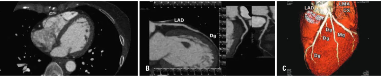
Figure 2. Coronary computed tomographic angiography examination with AIDR-3D. Axial image (A), curved-MIP (B) and 3D volume-rendering (C) reconstructions. Estimated effective radiation dose from coronary computed tomographic angiography only and full examination were 0.43mSv and 1.02mSv, respectively
A B C
CONCLUSION
In conclusion, coronary computed tomography angiography radiation dose can be dramatically reduced, following the ALARA (“as low as reasonable achievable”) principle, combining the exam indication with well-documented technics in coronary computed tomography angiography, such as beta blockers, low-kV, and the use of dose reduction software, as AIDR-3D.
REFERENCES
1. Taylor AJ, Cerqueira M, Hodgson JM, Mark D, Min J, O’Gara P, Rubin GD. American College of Cardiology Foundation Appropriate Use Criteria Task Force; Society of Cardiovascular Computed Tomography; American College of Radiology; American Heart Association; American Society of Echocardiography; American Society of Nuclear Cardiology; North American Society for Cardiovascular Imaging; Society for Cardiovascular Angiography and Interventions; Society for Cardiovascular Magnetic Resonance. ACCF/ SCCT/ACR/AHA/ASE/ASNC/NASCI/SCAI/SCMR2010 Appropriate Use Criteria for Cardiac Computed Tomography. A Report of the American College of Cardiology Foundation Appropriate Use Criteria Task Force, the Society of Cardiovascular Computed Tomography, the American College of Radiology, the American Heart Association, the American Society of Echocardiography, the American Society of Nuclear Cardiology, the North American Society for Cardiovascular Imaging, the Society for Cardiovascular Angiography and Interventions, and the Society for Cardiovascular Magnetic Resonance. J Cardiovasc.Comput Tomogr. 2010;4(6):407.e1-33.
2. Zhang C, Zhang Z, Yan Z, Xu L, Yu W, Wang R. 320-row CT coronary angiography: effect of 100-kV tube voltages on image quality, contrast volume, and radiation dose. Int J Cardiovasc Imaging. 201;27(7):1059-68.
3. Dewey M, Zimmermann E, Deissenrieder F, Laule M, Dübel HP, Schlattmann P, et al. Noninvasive coronary angiography by 320-row computed tomography with lower radiation exposure and maintained diagnostic accuracy: comparison of results with cardiac catheterization in a head-to-head pilot investigation. Circulation. 2009;120(10):867-75.
4. Fleischmann D, Boas FE. Computed tomography--old ideas and new technology. Eur Radiol. 2011;21(3):510-7.
5. Maurer MH, Zimmermann E, Schlattmann P, Germershausen C, Hamm B, Dewey M. Indications, imaging technique, and reading of cardiac computed tomography: survey of clinical practice. Eur Radiol. 2012;22(1):59-72. 6. Hsieh J, Londt J, Vass M, Li J, Tang X, Okerlun D. Step-and-shoot data acquisition
and reconstruction for cardiac x-ray computed tomography. Med Phys. 2006;33(11):4236-48.
7. Lee CH, Goo JM, Ye HJ, Ye SJ, Park CM, Chun EJ, et al. Radiation dose modulation techniques in the multidetector CT era: from basics to practice. Radiographics. 2008;28(5):1451-9.
8. Yoo RE, Park EA, Lee W, Shim H, Kim YK, Chung JW, et al. Image quality of Adaptive Iterative Dose Reduction 3D of coronary CT angiography of 640-slice CT: comparison with filtered back-projection. Int. J Cardiovasc Imaging. 2013; 29(3):669-76.
9. Singh S, Kalra MK, Gilman MD, Hsieh J, Pien HH, Digumarthy SR, et al. Adaptive statistical iterative reconstruction technique for radiation dose reduction in chest CT: a pilot study. Radiology. 2011;259(2):565-73. 10. Hara AK, Paden RG, Silva AC, Kujak JL, Lawder HJ, Pavlicek W. Iterative
reconstruction technique for reducing body radiation dose at CT: feasibility study. AJR Am J Roentgenol. 2009;193(3):764-71.