Flocculation in
Schizosaccharomyces pombe
Eun-Joo Gina Kwon1,2, Amy Laderoute1,2, Kate Chatfield-Reed1,2, Lianne Vachon1,2, Jim Karagiannis3, Gordon Chua1,2*
1Institute for Biocomplexity and Informatics, University of Calgary, Calgary, Alberta, Canada,2Department of Biological Sciences, University of Calgary, Calgary, Alberta, Canada,3Department of Biology, University of Western Ontario, London, Ontario, Canada
Abstract
In the fission yeastSchizosaccharomyces pombe, the transcriptional-regulatory network that governs flocculation remains poorly understood. Here, we systematically screened an array of transcription factor deletion and overexpression strains for flocculation and performed microarray expression profiling and ChIP–chip analysis to identify the flocculin target genes. We identified five transcription factors that displayed novel roles in the activation or inhibition of flocculation (Rfl1, Adn2, Adn3, Sre2, and Yox1), in addition to the previously-known Mbx2, Cbf11, and Cbf12 regulators. Overexpression ofmbx2+and
deletion ofrfl1+resulted in strong flocculation and transcriptional upregulation ofgsf2+/pfl1+and several other putative
flocculin genes (pfl2+–pfl9+). Overexpression of thepfl+genes singly was sufficient to trigger flocculation, and enhanced
flocculation was observed in several combinations of doublepfl+overexpression. Among thepfl1+genes, only loss ofgsf2+
abrogated the flocculent phenotype of all the transcription factor mutants and prevented flocculation when cells were grown in inducing medium containing glycerol and ethanol as the carbon source, thereby indicating that Gsf2 is the dominant flocculin. In contrast, the mild flocculation of adn2+ or
adn3+ overexpression was likely mediated by the
transcriptional activation of cell wall–remodeling genes includinggas2+,psu1+, and SPAC4H3.03c. We also discovered that
Mbx2 and Cbf12 displayed transcriptional autoregulation, and Rfl1 repressedgsf2+expression in an inhibitory feed-forward
loop involvingmbx2+. These results reveal that flocculation inS. pombe is regulated by a complex network of multiple
transcription factors and target genes encoding flocculins and cell wall–remodeling enzymes. Moreover, comparisons between the flocculation transcriptional-regulatory networks ofSaccharomyces cerevisiaeandS. pombeindicate substantial rewiring of transcription factors and cis-regulatory sequences.
Citation:Kwon E-JG, Laderoute A, Chatfield-Reed K, Vachon L, Karagiannis J, et al. (2012) Deciphering the Transcriptional-Regulatory Network of Flocculation in Schizosaccharomyces pombe. PLoS Genet 8(12): e1003104. doi:10.1371/journal.pgen.1003104
Editor:Anuj Kumar, University of Michigan, United States of America
ReceivedJune 1, 2012;AcceptedOctober 3, 2012;PublishedDecember 6, 2012
Copyright:ß2012 Kwon et al. This is an open-access article distributed under the terms of the Creative Commons Attribution License, which permits unrestricted use, distribution, and reproduction in any medium, provided the original author and source are credited.
Funding:This research was funded by grants from the Canadian Institutes of Health Research to GC, National Sciences and Engineering Research Council of Canada to JK, and Canada Foundation for Innovation to GC and JK. The funders had no role in study design, data collection and analysis, decision to publish, or preparation of the manuscript.
Competing Interests:The authors have declared that no competing interests exist. * E-mail: gchua@ucalgary.ca
Introduction
Flocculation is an inherent characteristic of yeasts involving asexual aggregation of cells into flocs that separate rapidly from the medium (reviewed recently in [1,2]). Individual yeast cells transition into this morphological state as an adaptation to various environmental stresses by shielding the inner cells of the flocs [3]. The flocculent trait has also proven highly beneficial in industrial yeast applications by allowing efficient and cost-effective removal of cells [4]. The ability of yeast strains to flocculate is dependent on the expression of specific cell surface glycoproteins known as flocculins. Cell-to-cell adhesion occurs via binding between the flocculin and surface carbohydrates in a calcium-dependent manner [5]. The bound carbohydrates consist of various sugars including mannose, glucose, and galactose that are specific to the type of flocculin and yeast species [6–8]. There has been considerable interest in elucidating the genetic control of flocculation to better understand this phenomenon and generate biotechnological advances in yeast-based industries.
In Saccharomyces cerevisiae, a transcriptional-regulatory network composed of interactions between transcription factors and their
flocculin gene targets is central in controlling flocculation. The primary flocculins that function in flocculation are encoded by the FLO1,FLO5,FLO9, andFLO10genes [9–11]. Overexpression of the individualFLOgenes is sufficient to trigger flocculation [8,12]. However, the degree of flocculation byFLOoverexpression varies from FLO1 to FLO10 exhibiting the strongest to weakest flocculation, respectively. The flocculinFLO11also exhibits weak flocculation when overexpressed [8], but its function is mainly in cell-to-surface adhesion [13], diploid pseudohyphal growth [14], and haploid invasive growth [15]. The transcription factors required for flocculation include Flo8p and Mss11p, which primarily activate FLO1 transcription [16]. The Sacc. cerevisiae laboratory strainS288Ccontaining a nonfunctionalFLO8gene is not able to flocculate, but flocculation is restored in this strain by the overexpression ofFLO8orMSS11[16,17]. In addition, Sfl1p has been shown to inhibit transcription ofFLO1in theW303-1A strain and not in S288C, likely through interactions with the Ssn6p-Tup1p global repressor and components of Mediator [18,19].
972 h2and975 h+
to flocculate has not been observed presumably because the inducing environmental conditions have not been identified. Phenotypic analysis of constitutive flocculent mutant strains show that flocculation is dependent on the presence of calcium, but unlike Sacc. cerevisiae, the flocculin-carbohydrate interactions involve galactose rather than mannose and glucose residues [7]. Moreover, the transcriptional-regulatory network governing flocculation in S. pombe remains poorly characterized. Only a single interaction between the Mbx2 MADS box transcription factor and thegsf2+
flocculin gene is currently known [20,21]. The gsf2+
gene was initially identified as highly upregulated in response to heterologous expression of FLO8 [20]. Overexpression of gsf2+ is sufficient to trigger flocculation
while its deletion abrogates the flocculent phenotype of tup12D, lkh1D, andgsf1mutants. In addition,gsf2+
displays additional roles in cell-to-surface adhesion and invasive growth [20]. The induction of gsf2+
during flocculation and invasive growth is mediated by Mbx2 [21]. Two other transcription factors implicated in flocculation have been reported. The CSL transcription factors Cbf11 and Cbf12 play opposing roles in flocculation where mutant strains lackingcbf11+
or overexpressing cbf12+
flocculate [22]. The direct targets of these transcription factors functioning in flocculation have not been identified, but could be several putative flocculin genes that show protein sequence homology to other yeast-related proteins [23]. Indeed, these putative flocculin genes, as well asgsf2+
are transcriptionally upregulated in certain Mediator mutants that flocculate indicating that these genes are likely repressed by Mediator [24]. Similar to Sacc. cerevisiae, the global transcriptional regulators Tup11 and Tup12 function in flocculation but their influence on the expression of these flocculin genes has not been addressed [25]. Importantly, it has not been directly demonstrated that these putative flocculin genes in S. pombe actually play a role in flocculation and the identity of the transcription factors that regulate them remains unknown.
In this study, we have initiated an extensive characterization of the transcriptional-regulatory network ofS. pombe flocculation by identifying the relevant transcription factors and their flocculin
gene targets. Importantly, we have also determined that hetero-thallic wild-typeS. pombeis able to flocculate when grown in rich medium containing ethanol and glycerol as a carbon source. A screen of transcription factor deletion and overexpression strains for flocculent phenotypes revealed five novel transcriptional regulators of flocculation (Rfl1, Adn2, Adn3, Sre2, Yox1) in addition to our independent finding of Mbx2, Cbf11, and Cbf12. The strongest flocculation was observed upon overexpression of mbx2+ and deletion of rfl1+ (SPBC15D4.02) which encodes an
uncharacterized fungal Zn(2)-Cys(6) transcription factor. Micro-array expression profiling of thembx2OEandrfl1Dstrains revealed good overlap in the upregulation of several flocculin genes, while ChIP-chip analysis of HA-tagged Mbx2 and Rfl1 under control of the nmt41 promoter indicated that these transcription factors bound to some of the flocculin gene promoters. Nine flocculin gene targets (pfl1+
–pfl9+
) includinggsf2+
/pfl1+
were identified. The single overexpression of these genes triggered flocculation to varying degrees and cumulative effects on flocculation were observed in double overexpression experiments. Only loss ofgsf2+
could abrogate the flocculent phenotype of all the transcription factor mutants indicating thatgsf2+
encodes the dominant flocculin in S. pombe. Interestingly, we discovered that certain cell wall-remodeling enzymes can also function in flocculation, and some of these genes are likely regulated by the LisH transcription factors Adn2 and Adn3. In addition to the identification of target genes within the transcriptional-regulatory network, autoregulatory and inhibitory feed-forward loops involving several transcription factors were also detected. These results provide a significant insight into the transcriptional control of flocculation inS. pombe.
Results
Screening for novel transcription factors functioning in fission yeast flocculation
Our understanding of the transcriptional-regulatory network that governs flocculation inS. pomberemains limited. To further decipher this network, we sought to systematically identify transcription factors that play a role in flocculation. A list of 101 genes encoding sequence–specific transcription factors containing a bona-fide DNA-binding domain was assembled from [26] and GeneDB [27]. From this gene list, we constructed 101 nmt1-driven overexpression strains and 92 nonessential deletions in which the entire ORF was replaced with the KanMX6/NatMX6 cassette. A detailed description of the construction and phenotypic character-ization of this transcription factor mutant collection will be described elsewhere (unpublished data). The transcription factor array of overexpression and deletion strains were screened for flocculation in EMM lacking thiamine and YES media, respectively. We recovered a total of eight transcription factors in which four overexpression strains (mbx2OE,adn2OE,adn3OEandcbf12OE) and four deletions (rfl1D,sre2D,yox1Dandcbf11D) exhibited flocculation. These transcription factors represent positive and negative regula-tors of flocculation, respectively. Among these transcription facregula-tors, only the overexpression ofcbf12+
andmbx2+
and deletion ofcbf11+
have been reported to cause flocculation [20,22].
The strongest flocculation was observed in the mbx2OE and rfl1Dstrains. The flocs of therfl1Dstrain in YES medium were larger and sedimented faster than the flocs produced in the mbx2OE strain after 48 hour induction (Figure 1A). The mbx2+
gene encodes a MADS-box transcription factor which was originally isolated in a screen for genes functioning in the biosynthesis of cell surface pyruvated galactose residues [28]. Recently, Mbx2 has been shown to function in flocculation and invasive growth by regulating the flocculin genegsf2+
[20,21]. The Author Summary
rfl1+
(repressor of flocculation) gene encodes an uncharacterized fungal Zn(2)-Cys(6) transcription factor.
The flocculation exhibited by these overexpression and deletion transcription factor mutants recovered from our screens could be abolished with the addition of galactose, but not mannose or glucose (data not shown). The amount of galactose required to completely deflocculate cells depended on the degree of floccula-tion. For example,mbx2OEstrain could be deflocculated with 2% galactose whilerfl1Dstrain required 5–10 times more galactose to completely deflocculate. Reflocculation of these strains was achieved in CaCl2or in YES medium (data not shown).
The growth conditions that trigger flocculation in heterothallic wild-typeS. pombeare not well known. To identify the inducing
conditions, 972 h2 and 975 h+
cells were tested on different carbon sources at different cell densities for flocculation. We determined that heterothallic wild-type cells were able to flocculate when cultured for five days at an initial concentration of 16106
cells/ml in medium containing 1% yeast extract, 3% glycerol and, 4% ethanol (referred to as flocculation-inducing medium, Figure 1B). The degree of flocculation was slightly enhanced in strains auxotrophic for leucine, uracil, and/or adenine indicating that nutrient status may also play a role in triggering flocculation (data not shown). However, these wild-type strains flocculated significantly less in flocculation-inducing medium than thembx2OE and rfl1D mutants in EMM and YES media, respectively. The weaker flocculation in these strains was more easily observed in Figure 1. Flocculation induction byrfl1+deletion,
mbx2+overexpression, or wild type grown in flocculation-inducing medium. (A) The flocculation ofrfl1Dmutant was visualized after culturing in YES medium for 24 hours at 30uC. The flocs of therfl1Dstrain in YES medium were larger and sedimented faster than the flocs produced in thenmt1-drivenmbx2OEstrain after 48 hour induction. Due to fast settling of flocs, the culture tubes were shaken vigorously immediately prior to image capture. (B) Heterothallic wild-type cells (972 h2) flocculate when cultured in flocculation-inducing medium (1% yeast extract, 3% glycerol and 4% ethanol). However, deletion ofmbx2+
orgsf2+
abolishes flocculation. Cells were inoculated in inducing medium at 106cells/ml and cultured for 5 days at 30uC, followed by petri dish assay (see materials and methods). (C) Therfl1D
mutant exhibits enhanced adhesion to agar and invasive growth. Wild type (972 h2) and therfl1Dmutant were grown on LNB medium overlaid on YE+ALU medium without glucose for 10 days as per procedure outlined by Dodgsonet al.[29]. Adhesion and invasive growth were determined by the amount of cells resistant to removal from the agar by gentle washing and more rigorous washing by rubbing cells off the agar with a finger under a stream of water, respectively.
petri-dishes incubated on an orbital rotator than in test tubes. In contrast to wild type, deletion ofmbx2+
did not produce any visible flocs in the flocculation-inducing medium (Figure 1B).
Fungal genes that function in flocculation are usually associated with filamentous invasive growth [17,20]. We hypothesized that the rfl1D strain would exhibit hyperfilamentous invasive growth because of its strong flocculent phenotype. Indeed, the amount of cells resistant to removal from the agar by washing in the invasive assay on LNB medium with an underlayer of YE+ALU was much greater in therfl1Dstrain than in wild type (Figure 1C). Under the microscope, the filamentous growth like those detected by Dodgsonet al.[29] was observed below the agar surface for both wild type andrfl1Dstrain with the latter showing much larger and more frequent formation of filamentous growth (data not shown). Similarly,adn2+andadn3+which were previously observed to have
defects in invasive growth when deleted were recovered in our screens as flocculent when overexpressed [29].
Mbx2 and Rfl1 are opposing transcription factors that regulate putative flocculin genes
The strongest flocculation observed in thembx2OE and rfl1D
strains indicated that these two genes encode the major regulators of flocculation. Therefore, we initially focused on the character-ization of these two transcription factors and proceeded to identify their target genes involved in flocculation. Thenmt41-driven mbx2-HAstrain was subjected to microarray expression profiling with a custom-designedS. pombe8615 K Agilent expression microarray
(Table S2). The intermediate strength nmt41 promoter was sufficient for mbx2OE flocculation and was utilized in the microarray experiments in order to reduce possible secondary transcriptional effects compared to the strongnmt1promoter. To better distinguish the direct target genes, ChIP-chip was also carried out concurrently on the same strain using the S. pombe 4644 K Agilent Genome ChIP-on-chip microarray (Table S3).
For therfl1+
expression profiling and ChIP-chip experiments, the flocculent deletion mutant andnmt41-drivenrfl1-HA strain were used, respectively (Tables S4 and S5). The highly-induced putative target genes identified by microarray expression profiling of these transcription factor mutant strains were validated by qPCR (Table S13).
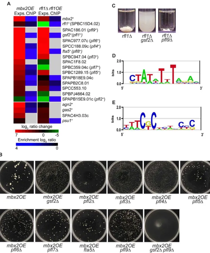
The list of genes that were induced at least two fold in the mbx2OE or rfl1D strain was subjected to gene ontology analysis using the Princeton GO Term Finder (http://go.princeton.edu/ cgi-bin/GOTermFinder). These induced genes were highly enriched in cell wall components with p-values of 9.0e-9 and 6.3e-6 for thembx2OE and rfl1D strains, respectively. Strikingly, the most-induced genes in thembx2OEstrain encoded cell surface glycoproteins. The cell surface glycoprotein genes up-regulated above two-fold were SPAC186.01, gsf2+, SPAC977.07c/
SPBC1348.08c, SPCC188.09c,fta5+
, SPBC947.04, SPBC359.04c, SPBC1289.15, SPAPB2C8.01, SPAC1F8.02c, SPAPB18E9.04c, SPCC553.10, and SPBPJ4664.02, which all butgsf2+
and the last 4 genes were predicted to be pombe adhesins based on BLAST sequence analysis (Figure 2A; [23]). SPAC977.07c and SPBC1348.08c are gene duplications with 100% sequence identity. To our knowledge, these genes with the exception of gsf2+
have not been characterized further. The induction of these genes in thembx2OEstrain ranged from 2 to 112-fold relative to the empty vector control (Figure 2A, Table S13). In addition, several genes (agn2+
,psu1+
, SPAC4H3.03c andgas2+
) encoding cell wall-remodeling enzymes such as glucan glucosidases and a betaglucanosyltransferase were induced up to 91-fold compared to the empty vector control when mbx2+
was overexpressed (Figure 2A). In the rfl1D expression data, a similar set of cell
surface glycoprotein genes were upregulated at a comparable level as the mbx2OE expression data except for SPAC1F8.02, SPBC359.04c, SPAPB18E9.04c and SPBPJ4664.02 (Figure 2A, Table S13). In contrast to the mbx2OE strain, the same genes encoding the cell wall-remodeling enzymes were not highly upregulated in therfl1Dstrain (Figure 2A).
Of the thirteen highly-induced cell surface glycoprotein genes in the mbx2OE expression data, nine of them were detected with ChIP-chip indicating that these genes are very likely the direct transcriptional targets of Mbx2 (Figure 2A). Four of the nine highly-induced cell surface glycoprotein genes in therfl1Dstrain were detected with ChIP-chip confirming that these genes are probably direct transcriptional targets of Rfl1 (Figure 2A). For both Mbx2 and Rfl1,gsf2+
,fta5+
and SPAPB2C8.01 were detected in the expression microarray and ChIP-chip experiments (Figure 2A).
Next, we sought further evidence that these cell surface glycoprotein genes were targets of Mbx2 and Rfl1 by epistasis studies. We decided to study a subset of these genes, which included the majority of the gene sequences analyzed by Linder and Gustafsson [23,24]. Thembx2+
gene was overexpressed in single deletions of these putative target genes and their degree of flocculation was determined visually in petri-dishes, as well as quantitatively (Table S14). The putative glycoprotein gene SPAPB15E9.01c was included in these studies, because even though the transcript was downregulated in both mbx2OE and rfl1D strains, ChIP-chip analysis detected Mbx2 and Rfl1 association with its promoter (Figure 2A). Deletion of gsf2+
decreased mbx2OEflocculation to the greatest extent while the reduction of flocculation was less extensive in the other single deletion mutants (Figure 2B, Table S14). The degree of reduction in mbx2OE flocculation roughly corresponded to the pfl numbers, which were assigned based on the degree of flocculation when overexpressed (see below). Moreover,mbx2OE flocculation was completely abrogated in thegsf2Dpfl9Ddouble mutant indicating that the reduction ofmbx2OEflocculation in these mutants were additive in some cases (Figure 2B). Similar experiments were performed forrfl1+
in which flocculation was assayed in the same putative target deletions in the rfl1D
background. The flocculation exhibited in therfl1D strain was completely abolished by the deletion of gsf2+
, but not by the deletion ofpfl9+
(Figure 2C).
To further analyze the expression microarray datasets of Mbx2 and Rfl1, the promoter regions of the differentially-expressed genes were subjected to the motif-finding algorithms RankMotif++
and MEME to identify their binding specificities [30,31]. Mbx2 is a member of the MEF2-MADS box transcription factor family which has been shown to bind to the consensus sequence 59-(C/ T)TA(T/A)4TA(G/A)-39[28,32,33]. The Mbx2 binding specific-ity obtained by RankMotif++
closely resembled this known consensus sequence (Figure 2D). Similarly, RankMotif++generated
an Rfl1 binding specificity that resembled known consensus sequences of several members of the fungal Zn(2)-Cys(6) transcription factor family (Figure 2E). The binding specificity of Zn(2)-Cys(6) DNA-binding domains is composed of conserved GC-rich trinucleotides spaced by a variable sequence region differing in length among members of the transcription factor family [34]. Analyses of the Mbx2 and Rfl1 expression microarray and ChIP-chip datasets by MEME did not generate any candidate DNA motifs.
The putative flocculin gene targets of Mbx2 and Rfl1 are sufficient to induce flocculation when overexpressed
Besidesgsf2+, the other putative target genes of Mbx2 and Rfl1
that encode for cell surface glycoproteins share some amino acid sequence homology with domains found in other fungal adhesins [23]. However, the role of these glycoprotein genes in flocculation has not been demonstrated. Overexpression studies were em-ployed to the aforementioned set of putative flocculin target genes of Mbx2 and Rfl1 to determine whether they function directly in flocculation. Each single overexpression of these flocculin genes was able to induce flocculation to varying degrees with the strongest flocculation observed in the gsf2OE strain which produced visible flocs within one day (Figure 3A; Table S14). Weaker flocculation was observed from the overexpression of the other flocculin genes after total incubation of 2–7 days in EMM minus thiamine medium with sub-culturing into fresh medium in Day 3. The flocculation images of these overexpression strains
shown in Figure 3A were captured after total of 7 days of induction. As a result of these observations, we named these genes pfl+
for Pombe Flocculins and numbered them according to their degree of flocculation when overexpressed:pfl1+
/gsf2+
(referred as gsf2+
hereafter),pfl2+
/SPAPB15E9.01c,pfl3+
/SPBC947.04,pfl4+
/ SPCC188.09c, pfl5+
/SPBC1289.15, pfl6+
/SPAC977.07c, pfl7+
/ SPBC359.04c,pfl8+
/fta5+
(referred as fta5+
hereafter) andpfl9+
/ SPAC186.01. Furthermore, we overexpressed some double combinations of the weaker flocculin genes to determine whether flocculation could be additive. Indeed, thepfl4+
pfl9+
,pfl6+
pfl9+
, and fta5+
pfl9+
double overexpression strains flocculated earlier and formed larger flocs than their corresponding single over-expressors, thus, demonstrating the additive effect of these flocculins (Figure 3B, Table S14). We next tested the single deletions of the pfl+
genes for their ability to flocculate in flocculation-inducing medium. No visible flocculation was ob-served in thegsf2Dstrain while wild type was flocculent (Figure 1B).
Promoter analysis of differentially-expressed genes inmbx2OE(D) andrfl1D(E) strains. Putative DNA motifs were identified for Mbx2 and Rfl1 by RankMotif++
.
doi:10.1371/journal.pgen.1003104.g002
Figure 3. Overexpression of putative flocculin target genes of Mbx2 and Rfl1 induces flocculation.(A) Single overexpression of the Mbx2 and Rfl1 flocculin target genes (gsf2+
/pfl1+
andpfl2+ –pfl9+
) with the nmt1promoter induces flocculation to varying degrees. The number assigned to thepfl+genes corresponds roughly to the relative strength of flocculation upon overexpression (i.e.gsf2+/pfl1+topfl9+showing strongest to mildest flocculation, respectively). The overexpression strains were cultured for total of 7 days (sub-cultured into fresh medium on third day) in EMM minus thiamine medium at 30uC. (B) The flocculin genes exhibit additive effects on flocculation. Double overexpression of various flocculin genes resulted in greater flocculation than the overexpression of the single corresponding genes.
In contrast, flocculation still occurred in thepfl2D–pfl9Dstrains in the inducing medium indicating thatgsf2+
encodes the dominant flocculin and the other flocculin genes are dispensable for flocculation (data not shown).
These observations revealed that the contribution in floccula-tion by these pfl+
genes varied and certain combinations of pfl+
were additive. The strength of flocculation by the single overexpression of pfl+
genes was directly correlated with the reduction of mbx2OE flocculation in the corresponding deletion strains (Figure 2B and Figure 3A, Table S14). For example, the pfl2OEstrain which produced larger flocs than thepfl3OE–pfl9OE strains exhibited a greater inhibition ofmbx2OEflocculation when deleted. Similarly, the flocculation of the rfl1D strain was completely abrogated by the deletion of gsf2+
, but not at all by the deletion ofpfl9+(Figure 2C). Consistent with the above results,
the deletion of bothgsf2+
andpfl9+
led to a greater abrogation of mbx2OEflocculation compared to each deletion alone (Figure 2B). In summary, we have demonstrated that thesepfl+
genes encode forS. pombeflocculins and Gsf2 is the dominant flocculin.
Positive and negative autoregulation ofmbx2+andrfl1+, respectively
Interestingly, ChIP-chip analysis also detected binding of Mbx2 and Rfl1 to their own promoters, as well as Rfl1 binding to the mbx2+
promoter (Figure 2A), indicating autoregulation andmbx2+
regulation by Rfl1 within the transcriptional-regulatory network of S. pombeflocculation. Mbx2 also appeared to be associated with the rfl1+promoter, but this interaction was marginal as it was found
just above the detection threshold for ChIP-chip (Figure 2A). To investigate the autoregulation ofmbx2+
, the gene was C-terminal tagged with GFP at its native locus (mbx2-GFP). However, the GFP-tagged strain resulted in a hypermorphic allele that displayed constitutive flocculation and nuclear localization of Mbx2-GFP (see below). We speculated that the removal of the 39-untranslated region ofmbx2+
during the C-terminal tagging may be the cause of the hypermorphic allele. To bypass this potential problem, we created an N-terminal GFP-tagged allele (GFP-mbx2) with an intact 59-untranslated region and approximately 1 kb of native promoter sequence. In contrast to the C-terminal tagged hypermorphic allele, the N-terminal tagged GFP-Mbx2 expression was compa-rable to background levels and the strain did not exhibit constitutive flocculation (Figure 4A). Moreover, the GFP-mbx2 strain flocculated when grown in glycerol-inducing medium indicating that the tagged protein is functional (Table S14). When nmt1-drivenmbx2+
expression was induced for 9 hours in theGFP -mbx2strain, nuclear GFP-Mbx2 expression was detected, indicat-ing that Mbx2 can activate its own expression (Figure 4A). As expected, this strain was now flocculent. Longer induction ofnmt1 -drivenmbx2+
expression resulted in greater GFP-Mbx2 expression with multi-nucleated GFP foci (data not shown). The positive autoregulation ofmbx2+is likely to be direct as several putative
MEF2-binding sequences (e.g. 59-TTAAAAATAG-39) are located within 1000 bp upstream from the mbx2+
start codon (data not shown).
To determine whether negative autoregulation occurs withrfl1+
, a C-terminal GFP-tagged strain under native control was generated (rfl1-GFP). The localization of Rfl1-GFP was nuclear in the rfl1-GFP strain (Figure 4B). The induction ofnmt1-driven rfl1+expression for 18 hours in therfl1-GFPstrain led to a reduced
nuclear GFP signal and a slightly increased cytoplasmic Rfl1-GFP signal (Figure 4B). However, overall Rfl1-Rfl1-GFP expression in the cell was reduced when Rfl1 was overexpressed compared to the empty vector control (Figure 4B; two-tailed t-test; p value,0.01). In contrast to our observations with the Rfl1-GFP
protein expression, we found that there was no decrease of the Rfl1-GFP transcript whenrfl1+
was overexpressed (Table S13). These results indicate that although Rfl1 can bind to its own promoter, negative autoregulation appears marginal or may not be occurring.
Rfl1 repressesmbx2+expression
The observation that Rfl1 is associated with thembx2+
promoter by ChIP-chip suggests that Rfl1 may oppose Mbx2 function in flocculation by repressing its expression. To test this hypothesis, we first examined the genetic interactions betweenmbx2+
and rfl1+
. The mbx2D rfl1D double mutant did not display flocculation indicating thatmbx2+
is epistatic torfl1+
(Figure 5A). In addition, the flocculation associated with mbx2OE was abrogated by co-overexpression of rfl1+
(Figure 5A). These results are consistent withmbx2+
being downstream ofrfl1+
and thatrfl1+
opposesmbx2+
function in flocculation.
We next utilized the C-terminal and N-terminal GFP-tagged mbx2+
strains to further determine if Rfl1 represses mbx2+
expression. First, Rfl1 was overexpressed in the hypermorphic C-terminal tagged mbx2-GFP allele which shows constitutive nuclear Mbx2-GFP expression and flocculation. This resulted in the near-abolishment of both the GFP signal (Figure 5B) and flocculation (data not shown) in the hypermorphic mbx2 allele. Second, when the N-terminal taggedGFP-mbx2strain was crossed into therfl1Dbackground, the resulting strain displayed dramatic increase in nuclear GFP-Mbx2 expression (Figure 5C) and flocculation strength equivalent to the rfl1D strain (data not shown). These results support the hypothesis thatmbx2+
expression is repressed by Rfl1 in non-flocculent cells.
Overexpression ofcbf12+causes flocculation due to up-regulation ofgsf2+
Cbf12, a member of the CSL transcription factor family has previously been reported to trigger flocculation when overex-pressed [22]. However, the target genes of Cbf12 that function in flocculation have not been identified. To further elucidate the role ofcbf12+
in flocculation, we took a similar approach to identify its direct target genes by concurrent expression microarray profiling and ChIP-chip analysis of the nmt41-driven cbf12-HA strain (Tables S6 and S7, respectively).
Whencbf12+
was deleted and cultured in flocculation-inducing medium, flocculation was abolished (Figure 6A). In contrast, overexpression of cbf12+
by thenmt1 promoter triggered floccu-lation (Figure 6C) and produced a bowling pin–shaped phenotype after 24 hours in medium lacking thiamine (data not shown). Further induction of thenmt1-drivencbf12+
caused the strain to become sick and granulated, eventually leading to growth arrest (data not shown). To reduce the toxic effects of cbf12+
overex-pression, annmt41-drivencbf12-HAstrain was used for concurrent expression profiling and ChIP-chip analysis.
Gene ontology analysis was carried out separately on the top 50 most highly-induced genes and all 160 promoter-occupied genes by Cbf12 with the Princeton GO Term Finder. Functional enrichment of genes in cell surface (p = 1.8e-7) and plasma membrane (p = 5.7e-4) was detected for the highly-induced and promoter-occupied genes, respectively. These genes included several flocculin genes, (Figure 6B). Both gsf2+
and pfl7+
were among the five highest induced genes (18.1 and 27.6-fold, respectively) in the cbf12OE strain and were also detected by ChIP-chip (Figure 6B) suggesting that Cbf12 directly activates the transcription ofgsf2+and pfl7+for flocculation. The flocculation
triggered bycbf12+
overexpression was completely abrogated in thegsf2D background, whereas deletion of pfl7+
(Figure 6C, Table S14). This was consistent with the hypothesis thatgsf2+
encodes the dominant flocculin. In addition, loss ofgsf2+
orpfl7+did not alter the bowling-pin cell shape or the reduced
fitness phenotypes of thecbf12OEstrain indicating that these two phenotypes were not due to the upregulation of the flocculin genes (data not shown). The much weaker flocculation observed in the cbf12OEstrain in comparison to the mbx2OE and gsf2OEstrains may be attributed to additional defects in cell and nuclear division, which would cause early growth arrest before the full flocculation potential could be reached [22].
Consistent with previous findings, C-terminal GFP-tagged Cbf12 under native control was expressed predominantly in the nucleus in stationary phase cells while expression in logarithmic cells was comparable to background (Figure 6D; [22]). Compared to logarithmic growth in rich medium, Cbf12-GFP nuclear expression increased in cells grown in flocculation-inducing medium, thus supporting its role in flocculation (Figure 6D). Interestingly, Cbf12 was also detected by ChIP-chip to bind to its own promoter (Figure 6B). Indeed, positive autoregulation appears to occur as native Cbf12-GFP expression increased greater than three-fold when nmt1-driven cbf12+was ectopically expressed in
logarithmically growing cells (Figure 6E).
Recently, it was demonstrated that an N-terminal-truncated Cbf12 bound to probes containing a canonical CSL binding motif (59-GTGGGAA-39) by gel mobility shift assay [35]. We next searched for a similar DNA binding sequence for Cbf12 from the expression microarray and ChIP-chip cbf12OE datasets by RankMotif++
and MEME. RankMotif++
and MEME analyses of the expression microarray and ChIP-chip data, respectively, did not identify a binding specificity for Cbf12. However, when the promoters of up-regulated genes in thecbf12OEstrain belonging to the cell surface GO category were subjected to MEME analysis, a motif closely matching the canonical CSL binding motif (6/7 nucleotide match) was recovered (Figure 6F).
These results demonstrate Cbf12 as part of the transcriptional-regulatory network of fission yeast flocculation by controlling the transcription of several flocculin genes includinggsf2+
.
The flocculation function ofyox1+,sre2+, andcbf11+is dependent ongsf2+
From our transcription factor screens, the deletion of yox1+
, sre2+
, orcbf11+
also resulted in flocculation, although the size of the flocs were smaller than observed in mbx2OE, cbf12OE and rfl1D
strains (Figure 7A, Table S14). Yox1 has been implicated in a negative autoregulatory loop to prevent inappropriate transcrip-tional expression of MBF gene targets, while the function of Sre2, which shows homology to the human sterol regulatory element binding protein SREBP-1A remains largely unknown [36,37]. A role of Yox1 and Sre2 in flocculation has not been reported. In contrast, cbf11+
encodes a CSL transcription factor that plays a role in flocculation, but its target genes are not known [22].
To elucidate the transcriptional flocculation program ofyox1+
, sre2+
andcbf11+
, expression microarray profiling was conducted on
the corresponding flocculent deletion strains in rich medium (Tables S8, S9, S10). The expression microarray profiles ofyox1D
and sre2D most resembled each other compared to the other strains described in this study (Figure 7B). Genes upregulated by at least two-fold in theyox1Dandsre2Dstrains showed enrichment for ribosomal subunits (p = 2.8e-31 and 7.4e-25 foryox1Dandsre2D, respectively) and mitochondrial membrane transporters (p = 7.5e-5 and 1.2e-3 foryox1Dandsre2D, respectively). These findings did not intuitively answer our questions as to how these two transcription factors might be related or associated with the flocculation pathway. We next examined whether any of the flocculin genes and their putative regulators were induced in the yox1Dandsre2Dstrains. In thesre2Dstrain,gsf2+
,pfl3+
andfta5+
transcripts were upregulated 3.7, 2.5 and 3.1-fold, respectively, indicating that the expression of these genes could be contributing to the flocculent phenotype (Figure 7C). In contrast,mbx2+
and cbf12+
transcripts were downregulated approximately 2-fold suggesting that the elevated levels of gsf2+
, pfl3+
and fta5+
transcripts in thesre2D strain were not mediated by Mbx2 and Cbf12 (Figure 7C). Similarly in theyox1Dstrain, we observed that gsf2+
andpfl3+
transcripts were upregulated although less than in thesre2Dstrain, and mbx2+and cbf12+were also downregulated
(Figure 7C). Therefore, this suggests thatsre2+
andyox1+
may be involved in the repression of flocculation through a pathway independent frommbx2+andcbf12+.
The microarray expression profile of thecbf11Dstrain revealed greater than 2-fold increase ofgsf2+
andpfl3+
transcripts and a 60-fold increase of the SPAC1F8.02c transcript suggesting that these two flocculin genes and this uncharacterized glycoprotein gene may be responsible for the flocculent phenotype in this mutant. In contrast to the yox1D and sre2D mutants, mbx2+
did not show differential expression in thecbf11Dstrain compared to wild type. However, the cbf12+ transcript was upregulated 1.8-fold in the
cbf11Dstrain. This suggests thatcbf11+
may regulate flocculation through cbf12+
, in agreement with previous reports of the antagonistic functions ofcbf11+andcbf12+in this process [22].
We next determined whether the flocculation caused by the deletion ofyox1+
,sre2+
orcbf11+
was also dependent ongsf2+
. The absence ofgsf2+
was sufficient to abolish the flocculation inyox1D, sre2D, andcbf11D strains, even thoughgsf2+ was not always the
most highly-expressed flocculin gene (Figure 7A and 7C, Table S14). Taken together, these results suggest that the expression of the dominant flocculin Gsf2 is responsible for the bulk of flocculation observed inyox1D,sre2Dandcbf11Dstrains.
The role of flocculation by Adn2 and Adn3 is influenced by genes encoding cell wall–modifying enzymes and gsf2+
The transcription factor genesadn2+
andadn3+
are orthologous to Sacc. cerevisiae FLO8 (http://www.pombase.org/) and exhibit defects in invasive growth and cell-to-surface adhesion when deleted during nitrogen starvation [29]. From our screens, we discovered that the overexpression of adn2+
and adn3+
triggered Figure 4.mbx2+and
rfl1+undergo positive and negative autoregulation, respectively.
(A) Positive autoregulation ofmbx2+
. A strain containing N-terminal GFP-taggedmbx2+under the control of its native promoter displayed increased GFP expression whenmbx2+was ectopically expressed with thenmt1promoter. Nuclear GFP-Mbx2 signal and flocs in liquid culture were detected at the 9 hour induction ofnmt1-drivenmbx2+
in EMM minus thiamine medium. Cells were deflocculated in 2% galactose prior to fluorescence microscopy to facilitate image acquisition. The presence of galactose does not affect the GFP signal (data not shown). The bar graph compares the mean and standard deviation of cellular GFP-Mbx2 signal resulting from
nmt1-driven mbx2+
and empty vector control with a significant difference of p,0.001 (Welch’s two tailed t-test; n = 50, df = 52). (B) Negative autoregulation ofrfl1+. A strain containing C-terminal GFP-taggedrfl1+under native control exhibited nuclear expression (empty vector). Ectopic expression ofnmt1-drivenrfl1+
minor flocculation while loss of adn2+
and adn3+
prevented flocculation in flocculation-inducing medium (Figure 8A and 8B, respectively). The flocculent phenotype of adn2OE and adn3OE strains was disrupted by the addition of galactose (data not shown). To identify the target genes of Adn2 and Adn3 that are involved in flocculation, expression microarray profiling was performed on nmt1-driven adn2OE and adn3OE strains (Tables S11 and S12). Surprisingly,gsf2+transcript levels were relatively unchanged and
the majority of pfl+ genes were downregulated in both
overex-pression strains (Figure 8C). Consistent with these results were the observations thatmbx2+
andcbf12+
transcripts were downregulated greater than 2-fold in bothadn2OEandadn3OEstrains, whereas rfl1+
transcript levels were not differentially regulated (Figure 8C). Therefore, it appeared that the flocculent phenotype ofadn2+
and adn3+
overexpression could not be attributed to the pfl+
genes identified in this study. These results led us to consider that perhaps the expression of other genes besides these encoding for flocculins could be responsible for triggering flocculation in adn2OEandadn3OEstrains.
Interestingly, some of the aforementioned cell wall-remodeling enzymes (gas2+
, psu1+
and SPAC4H3.03c) were also highly upregulated in bothadn2OEandadn3OEstrains (Figure 8C, Table S13). For example, gas2+
and SPAC4H3.03c were the highest induced genes in the adn2OE strain (17.9 and 36.8-fold, respectively) and also appeared within the top 20 most induced genes in theadn3OEstrain. These genes were also induced in the mbx2OE strain except for psu1+ (Figure 2A). Overexpression
analysis was subsequently carried out to determine if these genes possessed some role in flocculation. Although agn2+
was not upregulated in theadn2OEandadn3OEstrains, it was included in the overexpression analysis because it was the second most induced gene (91-fold), as well as detected by ChIP-chip in the mbx2OE strain. Indeed, the single overexpression of these four genes resulted in flocculation after 5-days (including 3rdday sub-culturing into fresh medium) in medium lacking thiamine, implicating the involvement of these cell wall-remodeling enzymes in flocculation (Figure 8D, Table S14). Since deletion ofadn2+
and adn3+ results in defects of invasive growth and cell-to-surface
adhesion in response to nitrogen starvation, we wanted to determine if the single overexpression ofgas2+, agn2+, psu1+and
SPAC4H3.03c could cause enhancement of these processes. We discovered that the single overexpression of these four cell wall-remodeling genes increased cell-to-surface adhesion, but not invasive growth relative to wild type under the nitrogen-deprivation condition (Figure S1). Because gsf2+
encodes the dominant flocculin, we also investigated whether the flocculation caused byadn2+
andadn3+
overexpression was dependent ongsf2+
. Deletion ofgsf2+
completely abrogated the flocculation inadn2OE andadn3OEstrains (Figure 8A, Table S14).
In addition, the adn2OE and adn3OE strains exhibited cell separation defects such as the formation of multisepta and
forkhead phenotypes (Figure 8E). The cell separation defect was more severe whenadn3+
was overexpressed. We next determined whether the putative target genes involved in the flocculation of adn2OEand adn3OEstrains also played a role in the multisepta phenotype. Overexpression of adn2+
and adn3+
in the gsf2D
background did not alter the multisepta phenotype (Figure 8E), while the overexpression ofgas2+
, SPAC4H3.03c,psu1+
andagn2+
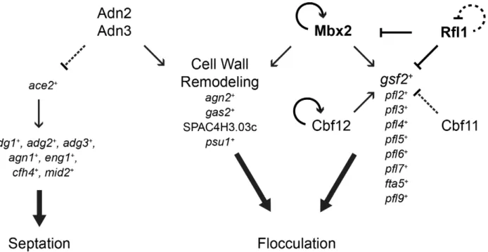
did not lead to formation of multisepta (data not shown). These results suggest that Adn2 and Adn3 may regulate cell separation and flocculation independently through different sets of target genes. Our microarray expression data suggests that Adn2 and Adn3 may control cell separation throughace2+, which encodes a
major transcriptional activator of this process (Alonso-Nun˜ezet al., 2005). Overexpression ofadn2+and adn3+resulted in the
down-regulation oface2+and many of its known target genes such as
adg1+,adg2+,adg3+,cfh4+,agn1+,eng1+, andmid2+by 1.5 to 3.4-fold
(Figure 8C).
In summary, the regulation of flocculation byadn2+andadn3+is
likely mediated by the induction of genes encoding the cell wall-remodeling enzymes Gas2, SPAC3H3.03c, and Psu1. The regulation of these genes is independent from Mbx2 because mbx2+ was downregulated in the adn2OE and adn3OE strains.
Althoughgsf2+transcript level was not significantly upregulated by
adn2+and adn3+overexpression, it was sufficient to abrogate the
flocculation when deleted. However, it is possible that other cell surface glycoprotein genes not investigated in this study but were upregulated may also play a significant role in the flocculation function ofadn2+
andadn3+
.
Discussion
In this study, we have deciphered a significant portion of the transcriptional-regulatory network governing flocculation in S. pombe. To date, few transcription factors and their target genes that function in flocculation have been identified. The MADS box transcription factor Mbx2 positively regulates flocculation by induction of the flocculin genegsf2+, while the CSL transcription
factors Cbf11 and Cbf12 repress and activate flocculation, respectively, but their target genes are not known [21,22]. We have substantially expanded our limited knowledge of the flocculation transcriptional-regulatory network by the identifica-tion of several novel transcripidentifica-tional activators (Adn2 and Adn3) and repressors (Rfl1, Yox1 and Sre1), and their putative target genes that function in flocculation. In addition, novel target genes of Mbx2, Cbf11 and Cbf12 were identified. The putative target genes of the transcription factors implicated in flocculation encode for several cell surface glycoproteins (gsf2+
andpfl2+
–pfl9+
) and cell wall-remodeling enzymes (agn2+
,psu1+
, SPAC4H3.03c andgas2+
). These target genes were sufficient to trigger flocculation when overexpressed. Moreover, instances of regulation between tran-scription factors (Rfl1 repression of mbx2+
), as well as positive Figure 5. Rfl1 repressesmbx2+expression.
(A) Genetic interactions betweenmbx2+ andrfl1+
. The flocculation phenotype of therfl1Dmutant is abolished by deletion ofmbx2+(left panel). In addition, the flocculation caused by overexpression ofmbx2+is abolished byrfl1+overexpression (right panel). The deletion and overexpression strains were cultured in YES and EMM minus thiamine media, respectively, for 24 hours at 30uC. (B) The expression of C-terminal GFP-taggedmbx2+
under native control is reduced by ectopic expression ofrfl1+
. The C-terminal GFP-taggedmbx2+ strain is a hypermorphic allele that exhibits constitutive flocculation and nuclear Mbx2-GFP expression. The C-terminal GFP-taggedmbx2+strain containing nmt1-drivenrfl1+
or empty vector was inoculated at approximately 104cells/ml in EMM minus thiamine medium and cultured for 20–30 hours at
30uC until late log phase. The strain containing the empty vector was deflocculated in 2% galactose prior to fluorescence microscopy. This procedure was not required for therfl1OEstrain because it was no longer flocculent. (C) Deletion ofrfl1+
results in the expression of native-controlled N-terminal GFP-taggedmbx2+. The N-terminal GFP-taggedmbx2+strain in a wild type andrfl1Dbackground were cultured to mid-log phase in YES medium. The bar graph compares the mean and standard deviation of overall cellular Mbx2-GFP (B) and GFP-Mbx2 (C) signals between the experimental and control strains with significant difference of p,0.001 (Welch’s two tailedt-test; n = 50, df = 50 for each experiment). Cells were stained with DAPI to visualize nuclei. Scale bar, 10mm.
(mbx2+ and cbf12+) autoregulation were detected within the
flocculation network.
Mbx2 and Rfl1 appeared to be the major positive and negative regulators of flocculation, respectively, based on the largest flocs observed in thembx2OEandrfl1Dstrains compared to the other flocculent mutants in this study. Our initial efforts to identify the target genes of Mbx2 and Rfl1 revealed several putative flocculin genes that were strikingly upregulated in thembx2OE and rfl1D
flocculent mutants. Previously, Gsf2 was the onlyS. pombeflocculin demonstrated to be directly involved in flocculation, and its transcription was influenced by the activity of Mbx2 [20,21]. Similar to these studies, we also found that overexpression ofgsf2+
triggers flocculation while loss of gsf2+
abrogates the flocculent phenotype of several mutants including mbx2OE. Here, we identified an additional eight flocculin genes (pfl2+
–pfl9+
) as putative target genes of Mbx2. Seven of these target genes (pfl3+
–pfl9+
) were reported to contain tandem repeats found in fungal adhesins, while pfl2+ is a sequence orphan predicted to
encode a GPI-anchored protein [23,27]. Seven pfl+
genes (gsf2+
/ pfl1+
, pfl3+
, pfl4+
and pfl6+
–pfl9+
) have been reported to be upregulated in loss-of-function flocculent mutants of Cdk8 module genes (cdk8+/srb10+, med12+/srb8+) suggesting that the
transcrip-tional repression of these putative flocculin genes may be controlled by Mediator [24]. The transcriptional repression of flocculin genes by Mediator may not be direct, but could be throughmbx2+since its expression is highly upregulated in thecdk8
kinase-mutant and med12D strain (9 and 13-fold increase, respectively, within top 11 up-regulated genes, found in supple-mentary data [24]). This proposed role of Mediator appears conserved inSacc. cerevisiaeasFLOgenes are similarly upregulated in cdk8 mutants [38]. Despite these observations, no direct evidence has been shown aside from thegsf2+
study by Matsuzawa et al.[20] that thepfl+gene products are actually flocculins. We
have shown that this is indeed the case as single and double overexpression of thepfl+
genes is sufficient to trigger flocculation and that this flocculation is galactose-specific.
The degree of flocculation triggered by single overexpression of thepfl+
genes varied, withgsf2+
andpfl9+
producing the largest and smallest flocs, respectively (thepflnumbers correspond roughly to the degree of flocculation upon overexpression). This result indicates thatgsf2+
encodes the most dominant flocculin compared to the otherpfl+
genes. In agreement are the observations that only deletion ofgsf2+and not the otherpfl+genes prevented flocculation
in flocculation-inducing medium, and reduced the constitutive flocculent phenotype to the greatest extent of all the transcription factor mutants examined in this study. Moreover, the strength of the flocculins was directly correlated with the amount of reduction in mbx2OE flocculation observed in the various pfl deletion backgrounds (Figure 2B). These observations are similar inSacc.
cerevisiae, where overexpression of FLO1 produces the strongest flocculation compared toFLO5,FLO9,FLO10andFLO11[8,12]. Furthermore, the flocculation mediated bypfl+
genes was additive as observed in our double deletion and co-overexpression experiments (Figure 2B and Figure 3B). These results suggest that the varying strengths of flocculation exhibited byS. pombestrains could be attributed to the upregulation of different combinations ofpfl+genes.
We identified Rfl1, an uncharacterized Zn(2)-Cys(6) transcrip-tion factor as a novel repressor of flocculatranscrip-tion in fission yeast. The repression of flocculation by Rfl1 appears to be primarily mediated by the inhibition ofgsf2+
expression since loss ofgsf2+
can abrogate the constitutive flocculent phenotype of the rfl1D mutant. Rfl1 repressesgsf2+either directly by association with its promoter or
indirectly by inhibition ofmbx2+
transcription, thereby forming an inhibitory feed-forward loop (coherent type 2) within the transcriptional-regulatory network (Figure 9). These results indi-cate that Mbx2 and Rfl1 are opposing transcription factors, and the latter inhibitsmbx2+
andgsf2+
expression under non-inducing conditions of flocculation.
Aside from its role in flocculation, Rfl1 may have a role in regulating genes involved in carbohydrate metabolism such as glycolysis and gluconeogenesis. Rfl1 appeared to be associated with promoters of genes enriched in glucose catabolic and metabolic processes (p-values = 0.00092 and 0.00269, respectively) includingadh1+
,hxk2+
,pfk1+
,tpi1+
,adh4+
,pgi1+
,gpd3+
,tdh1+
,pgk1+
, fba1+, eno101+, pyr1+, SPCC794.01c (predicted
glucose-6-phos-phate 1-dehydrogenase), SPBC2G5.05 (predicted transketolase) and SPBC660.16 (phosphogluconate dehydrogenase). Most of these genes with the exception offba1+,eno101+, SPCC794.01, and
SPBC660.16 were upregulated 1.2 to 26-fold in the rfl1Dstrain (Table S4). From these data, we speculate that Rfl1 could serve as a negative transcriptional regulator of several enzymes involved in the glycolysis and gluconeogenesis. Because flocculation and invasive growth are associated with nutritional limitation, Rfl1 may coordinate the expression of genes involved in flocculation and carbohydrate metabolism in fission yeast.
Previously, the CSL proteins Cbf11 and Cbf12 were shown to exhibit antagonistic roles in flocculation [39]. Overexpression of cbf12+or loss ofcbf11+triggers flocculation. However, none of their
target genes have been identified. We present supportive evidence that Cbf12 induces flocculation by directly activating the transcription ofgsf2+. In addition,gsf2+expression is up-regulated
approximately 2.4-fold in the cbf11D strain suggesting that the repressive flocculation function of Cbf11 may also be directly mediated through gsf2+
. The activation and repression of gsf2+
transcription by Cbf12 and Cbf11, respectively, may occur by competitive binding to promoter sites since both transcription factors have been shown to interact with a canonical CSL Figure 6. Regulation of flocculation by Cbf12.(A) Loss ofcbf12+
prevents flocculation under inducing conditions. Wild type and thecbf12Dmutant were cultured in flocculation-inducing medium for 5 days at 30uC. (B) Cbf12 regulates putative flocculin genes. The heat map shows induction of several flocculin genes and their promoter occupancy by Cbf12 from microarray expression profiling and ChIP-chip analysis, respectively, of annmt41-driven
cbf12-HAstrain. The color bars reflect relative expression and ChIP enrichment ratios between experimental and control. (C) The absence ofgsf2+ , but not
pfl7+abolishes the flocculation triggered bycbf12+overexpression. Strains containingnmt1-drivencbf12+in wild type,gsf2Dorpfl7
Dbackgrounds were cultured for 24 hours in EMM minus thiamine medium at 30uC. Approximately 1/16 of the petri-dish was magnified to reveal more details of the flocs. (D)
cbf12+is expressed in wild-type cells grown in rich medium at stationary phase and in flocculation-inducing medium, but not in rich medium at log phase. A C-terminal GFP-taggedcbf12+
strain under the control of its native promoter was grown to log or stationary phase in YES or in the inducing medium at 30uC. The bar graph compares the mean and standard deviation of cellular Cbf12-GFP signals. (E) Positive autoregulation ofcbf12+. Ectopic expression ofnmt1-drivencbf12+
results in the upregulation of native-controlled Cbf12-GFP expression in log-phase cells. The bar graph compares the mean and standard deviation of cellular Cbf12-GFP signals between induced and uninducedcbf12OEcells with significant difference of p,0.001 (Welch’s two tailedt-test; n = 19, df = 25). (F) A DNA motif closely matching the binding specificity of CSL transcription factors was retrieved from the
cbf12OEmicroarray expression data. The promoter region (1000 base pairs upstream of the start codon) of 11 highly-induced genes encoding for cell surface proteins as identified by the Princeton GO Term Finder was applied to MEME using default settings. The orange line indicates bases that match with the known binding site of CSL transcription factors. Cells were stained with DAPI to visualize nuclei. Scale bar, 10mm.
consensus sequencein vitro[39]. Several putative sites with six out of seven nucleotide match to the canonical CSL consensus sequence are located within 900 base pairs of thegsf2+
promoter (data not shown). Further experimentation would be required to verify this proposed mechanism ofgsf2+
transcriptional regulation by Cbf11 and Cbf12. It is likely thatcbf12+
plays a lesser role in activating flocculation compared to mbx2+
since the floc size resulting fromcbf12+
overexpression is considerably smaller than the mbx2OE strain. Also unlike mbx2+
, deletion of cbf12+
is not sufficient to abrogate the flocculation of therfl1Dstrain (data not shown). These data suggest that the flocculent phenotype of the cbf12Drfl1Ddouble mutant is probably caused by the presence of mbx2+
activity.
CSL transcription factors are components of the conserved Notch signaling pathways in metazoans which primarily function in cell-to-cell communication during development [40]. Although multiple fungal CSL proteins have been discovered, their exact roles remain unclear in unicellular organisms [39]. Flocculation has been described as a manifestation of social behaviour in yeast with a purpose of enhancing survival under stressful conditions [3]. Therefore, it is conceivable that CSL transcription factors originated as regulators of this primitive form of cell-to-cell communication, and later evolved into the metazoan Notch signaling pathway.
We also discovered novel functions of the Yox1 and Sre2 transcription factors in the repression of flocculation. Loss ofyox1+
or sre2+ results in a mild flocculent phenotype. The Yox1
homeodomain transcription factor functions as a repressor of MBF (Mlu1 binding factor) target genes to prevent their inappropriate expression at the end of S-phase [36]. Transcrip-tional repression of MBF target genes is mediated by the direct interaction of Yox1 and Nrm1 to the MBF complex [41]. Deletion ofyox1+
causes a cell cycle delay and results in elevated constitutive expression of MBF gene targets [36]. Similarly, these genes (e.g. cdc18+
,cdc22+
,cdc10+
,cdt1+
,cdt2+
,cig2+
andnrm1+
) were also found to be upregulated 2.2 to 6.4-fold in our yox1D microarray expression data (Table S8). We found that the flocculent phenotype of the yox1D strain is also dependent upon gsf2+
. However, thepfl+
genes includinggsf2+
were not highly expressed in theyox1Dstrain. One possible explanation whypfl1+
genes were not highly expressed in theyox1D strain is that our experiments were performed under asynchronous culturing conditions, and therefore, the upregulation ofpfl+
genes includinggsf2+
could have been obscured if their expressions were periodically controlled in the vegetative cell cycle. However, it is unlikely that Yox1 regulates thepfl+genes directly because previous chIP-chip analysis did not
detect binding of Yox1 to the promoters of pfl+ genes [36].
Although there has been solid evidence linking yeast morphogen-esis events such as pseudohyphal and hyphal growth to cell cycle regulators [42–47], the relationship between cell cycle control and flocculation remains unclear. A flocculation function foryox1+
has not been reported in other yeasts. However, disruption ofYOX1in the Sacc. cerevisiae g1278b strain inhibited filamentous invasive growth, a process usually associated with flocculation during nutritional limitation, while deletion ofC. albicans NRM1reduced flocculation [48,49].
Sre2 is an uncharacterized membrane-tethered helix-loop-helix transcription factor predicted to be an ortholog of mammalian SREBP-1a, which is responsible for the transcriptional activation of genes needed for uptake and synthesis of cholesterol, fatty acids, triglycerides, and phospholipids [50]. While sre1+
, a paralog of sre2+
has been shown to function in the transcriptional activation of sterol-biosynthetic and hypoxic-adaptation genes, there has been no direct evidence that sre2+
plays similar biological roles [37]. Loss ofsre2+
results in the upregulation of gsf2+
, pfl3+
and fta5+
transcripts (3.76, 2.51 and 3.08-fold, respectively) (Figure 7C, Table S9) which may contribute to its flocculent phenotype. The sre2Dflocculent phenotype requiresgsf2+
activity and is indepen-dent ofmbx2+
andcbf12+
since these transcripts are downregulated in the deletion mutant.
In addition, the microarray expression profiles of yox1D and sre2Dstrains displayed similar differential gene expression despite the supposedly different functions of these transcription factors (Figure 7B). Mitochondrial genes were found to be highly upregulated in both deletion mutants (Tables S8 and S9). This occurrence may not be unexpected for Sre2 if it has a similar role in hypoxia as Sre1 where mitochondrial function is probably impaired [37]. It is currently not clear whether the mitochondrial genes are direct targets of Yox1 and Sre2 or induced in response to an altered physiological state in the deletion mutants. Interestingly, mitochondrial activity has been reported to be important for flocculation and invasive growth in Sacc. cerevisiae [48,51]. Disruption of mitochondrial activity has been shown to alter the synthesis and structure of the cell wall, possibly by interfering with the interactions of flocculins and their substrates [52]. Based on these observations, the flocculent phenotype of yox1Dand sre2D
strains could be partially the result of enhanced mitochondrial activity from the upregulation of mitochondrial genes.
A genome-wide systematic deletion screen previously uncovered a cell-to-surface adhesion function that is sensitive to the presence of galactose for the Adn2 and Adn3 transcription factors [29]. Here, we discovered that adn2+
and adn3+
have additional functions in flocculation. Overexpression of adn2+
and adn3+
induced minor flocculation while loss of these genes prevented flocculation in inducing glycerol medium (Figure 8A and 8B). However, the flocculent phenotype of the adn2OE and adn3OE strains appeared to be primarily caused by the differential regulation of genes encoding cell wall-remodeling enzymes rather than flocculins. Several genes encoding cell wall-remodeling enzymes (gas2+
, agn2+
, psu1+
and SPAC4H3.03c) were highly induced when mbx2+, adn2+or adn3+ was overexpressed. In the
adn2OE strain, gas2+ and SPAC4H3.03c were the most highly
induced genes (17.9 fold and 36.8-fold, respectively) (Figure 8C, Table S11) while in theadn3OEstrain, these two genes andpsu1+
appeared within the top 20 up-regulated genes (Table S12). Similarly, in thembx2OEstrain, gas2+
, agn2+
, and SPAC4H3.03c appeared within the top 100 up-regulated genes (greater than 3.7-fold increase, Figure 2A). We found that the single overexpression of these four genes could trigger flocculation (Figure 8D). Cell wall remodeling is an essential process for proper growth and adaptation to environmental stresses in yeast cells. Part of the cell wall-remodeling process involves the dissolution of sugar Figure 7. Regulation of flocculation by Yox1, Sre2, and Cbf11.(A) The regulation of flocculation by Yox1, Sre2 and Cbf11 is dependent on
gsf2+.yox1
D,sre2Dandcbf11Dcells flocculate in late log and stationary phase when grown in YES medium (left panels). The flocculation of these mutant strains was abrogated in thegsf2Dbackground (right panels). (B) Clustergram of microarray expression profiles of flocculent transcription factor mutants. The microarray expression profiles of the yox1Dand sre2D strains are most similar and show upregulation of ribosomal and mitochondrial genes. (C) Theyox1D,sre2Dandcbf11Dflocculent strains show upregulation of several flocculin genes. The color bars reflect relative expression between experimental and control.
moieties in the glucan layer and elongation of glucan chains by glycoside hydrolases and glycosyltransferases, respectively. Among these four genes, three (agn2+
,psu1+
and SPAC4H3.03c) encode for glycoside hydrolases while the fourth (gas2+
) encodes for a glycosyltransferase. Agn2 is an endo-(1,3)-a-glucanase that hydro-lyzes (1,3)-a-glucans of the ascus wall for ascospore release [53,54]. Althoughagn2+
function appears only specific for sporulation, its ectopic expression could alter the cell wall structure during vegetative growth by inappropriate hydrolysis of (1,3)-a-glucan. Similarly, inappropriate glucan hydrolysis of the cell wall could be occurring as a result of ectopic expression of SPAC4H3.03c which encodes a putative (1,4)-a-glucanase (Hertz-Fowleret al., 2004). Psu1, which exhibits close homology to the members of the SUN family inSacc. cerevisiae and C. albicans, as well as the BglA beta glucosidase ofC. wickerhamii, has an essential function in cell wall synthesis [55]. Loss ofpsu1+
activity conferred resistance to (1,3)-b -glucanase suggesting that Psu1 may influence the amount or structure of (1,3)-b-glucan in the cell wall [55]. In addition, the (1,3)-b-glucanosyltransferase Gas2 has been shown to lengthen glucan chains during cell wall assembly and its overproduction is able to suppress the cell wall defect and lethality ofgas1Dcells [56]. Then how does the overexpression of these cell wall-remodeling genes trigger flocculation in S. pombe cells? The expression of flocculin genes during vegetative growth is not well characterized
in yeasts, but studies in Sacc. cerevisiae indicate that flocculin synthesis and insertion into the cell wall initiate in early exponential phase prior to the onset of flocculation during stationary phase [57]. This suggests that the flocculins are already present in the cell wall, but cannot induce flocculation because of inaccessibility to cell surface oligosaccharides. We speculate that the restructuring of theb-glucan layer during cell wall remodeling may result in the rearrangement of flocculins that enhances galactose oligosaccharide binding, thereby promoting flocculation. Several lines of evidence inSacc. cerevisiaesupport this hypothesis. First, alteration of cell wall structure by disruption ofPKC1activity results in flocculation [58]. Second, heat shock induces flocculation and regulation of cell wall-remodeling genes via the Hsf1 transcription factor [57,59]. Currently, we cannot rule out that agn2+
, psu1+
, SPAC4H3.03c and gas2+
are the only cell wall remodeling enzymes that can trigger flocculation when overex-pressed. Other genes with potential functions in cell wall modification and integrity such as gas4+
, gma12+
, meu7+
, agl1+
, meu10+
and mde5+
were also detected as putative target genes of Mbx2 (Table S2). In contrast, there was little change in gsf2+
transcript levels in theadn2OEoradn3OEstrains compared to the empty vector control. However, the flocculation triggered byadn2+
and adn3+
overexpression was abrogated in agsf2D background indicating thatgsf2+
was indispensible for this process (Figure 8A). Figure 8. Regulation of flocculation by Adn2 and Adn3 is dependent ongsf2+and cell wall–remodeling genes.
(A) Overexpression of
adn2+oradn3+triggers weak flocculation, which is abrogated in thegsf2Dbackground. The mutant strains were cultured for 3 days in EMM minus thiamine medium. Approximately 1/8 of the petri-dish was magnified and shown for each strain. (B) Deletion ofadn2+
oradn3+
prevents flocculation in flocculation-inducing medium. (C) Several cell wall-remodeling genes were upregulated while the majority ofpfl+
genes and known target genes of Ace2 (bottom panel) were downregulated uponadn2+or adn3+overexpression. Microarray expression profiling was performed onnmt1-driven adn2OEandadn3OEstrains and induced in EMM minus thiamine medium. The color bar reflects relative expression ratios between experimental and control strains. (D) Overexpression of the cell wall-remodeling genesagn2+,psu1+,gas2+and SPAC4H3.03c triggers flocculation. These overexpression strains were cultured for total of 5 days (subcultured on third day into fresh medium) in EMM minus thiamine medium. (E) The multiseptation phenotype resulting fromadn2+oradn3+overexpression is not dependent ongsf2+. The strains were induced for 34 hours in EMM minus thiamine medium and stained with calcofluor white. Scale bar, 10mm.
doi:10.1371/journal.pgen.1003104.g008