Available online at www.ijpsdr.com
International Journal of Pharmaceutical Sciences and Drug Research 2011; 3(4): 338-341
338
Research Article
ISSN 0975-248X
Pharmacognostical Evaluation and Qualitative Analysis of
Saccharum
spontaneum
(L.) Root
Mohammad Khalid
*, Hefazat H. Siddiqui
Faculty of Pharmacy Dasauli Kursi road, Integral University, Lucknow 226 026, Uttar Pradesh, India
ABSTRACT
Saccharum spontaneumL. known as Kasa (Family: Poaceae) is a traditional herb, it has excellence medicinal value; has been advocated in the treatment gynaecological troubles, respiratory disease. Roots are used as galactagogue and diuretic and in ayurveda system roots are also used as astringent, emollient, refrigerant, diuretic, purgative, tonic, aphrodisiac and useful in treatment of dyspepsia, burning sensation, piles and sexual weakness. Various parameters like macroscopy, microscopy, fluorescence analysis as well as extractive value and quantitative phytochemical screening of different extractives were studied. The major components of the extractives like total phenolic, total flavonoids were also estimated respectively. The characteristic of microscopy, physicochemical, fluorescence analysis and quantitative chemical screening were performed in root extractives of the plant material as a mean of authentication.
Keywords: Saccharum spontaneum, phenolic and flavonoid content, fluorescence analysis, physicochemical, galactagogue.
INTRODUCTION
The several types of plant materials such as vegetables fruits leaves oil seeds cereals crops bark and roots spices and herbs and crude plant drugs are potential sources of antioxidants compounds. Most of the isolated compounds with antioxidants activity are phenolic compounds. [1] Reactive oxygen species singlet oxygen and hydrogen peroxide are often generated as by products of biological reaction or from exogenous factors.[2]The reactive species play an important role in cell metabolism, phagocytosis and intercellular signaling. [3] However, these reactive species produced by sunlight, ultraviolet rays, ionizing radiation, chemical reactions and metabolic processes have a wide variety of pathological effects such as DNA damage, carcinogenesis and various diseases such as cardiovascular diseases, aging and neuro-degenerative diseases. [4-5] In foods, the reactive species can cause lipid peroxidation, which leads to the deterioration of the food. [6]The oxidative deterioration of the lipid-containing food is responsible for the rancid odours and flavours during processing and storage, consequently decreasing the nutritional quality and safety of foods, due to the formation of secondary, potentially toxic compounds. The addition of antioxidant is a method for increasing the shelf life of foods.[7]The studies have been shown that a
*Corresponding author: Mr. Mohammad Khalid,
Assistant Professor, Faculty of Pharmacy, Integral University Dasauli, Kursi Road, Lucknow 226 026, Uttar Pradesh, India;
Tel.:+91-9919239289; E-mail:[email protected]
number of plant products containing polyphenols, flavonoids, terpenes and various plant extracts exerted an antioxidant action. [8] There is currently immense interest in natural antioxidants and their role in human health and nutrition.[9]
Saccharum spontaneum L. (S. spontaneum)
(Family-Poaceae) locally known as Kasa is a tall erect reed-like perennial grass. It is distribute throughout India [10] and tropical Asia [11] Leaves and stalks contain lignin, carbohydrates, proteins and amino acids.[12]Roots and root-stocks contain starch and polyphenolic compounds. Aerial parts possess laxative and aphrodisiac properties, and are useful in burning sensations, strangury, phthisis, vesical calculi, blood diseases, biliousness and haemorrhagic diathesis.[13] The stems are useful in vitiated conditions of pitta and vata burning sensation strongly and dyspepsia, haemorrhoids, menorrhagia dysentery, agalactia phthisis and general debility.[14]
MATERIAL AND METHODS Extract preparation
Khalidet al./ Pharmacognostical Evaluation and Qualitative Analysis of Saccharum spontaneum…..……
IJPSDR October-December, 2011, Vol 3, Issue 4 (338-341)
339
Microscopy
Transverse section (TS) of the aerial parts and root was cut by free hand sectioning and stained with different safranin and aniline blue. The various histological parts examined and drawn with the help of camera Lucida. [15] Histochemical colour reactions of powdered drug was carried out with Ruthenium red, iodine solution, Millon’s reagent and Dragendorff’s reagent for the detection of mucilage, starch, protein and alkaloids respectively. The other compounds are also reported by this method.[16]
Physico-chemical and fluorescence analysis
Loss on drying, total ash, acid insoluble ash, water soluble ash and crude fibres contents were performed as per Indian Pharmacopoeia. [17] The extract of the powdered fruit was prepared with different polar and non-polar solvents for the study of successive extractive values. Fluorescence analysis of the powder drug was carried out with different chemical reagents in day (254 nm) and UV light (365 nm). The dry powder drug was studied on glass slide whereas the different extracts were studied by adsorbing the extracts on Whatmann filter paper.[18]
Qualitative estimation
For the quantitative estimation 100 g of powdered drug was successively extracted in a soxhlet apparatus with various solvents like petroleum ether, chloroform, ethyl acetate, methanol and water. [19] The extracts were dried on water bath, weighed and colour of the extracts was also observed. The different extracts were subjected to qualitative estimation for the presence of various phytoconstituents.[20]
Determination of total phenolic content
A total phenolic content in the S. spontaneum extract was determined by the modified Folin-Ciocalteu method. [21] An aliquot of the extracts was mixed with 5 ml Folin-Ciocalteu reagent (previously diluted with water 1:10 v/v) and 4 ml (75 g/l) of sodium carbonate. The mixtures were allowed to stand for 30 min at 40°C for colour development. Reagent blank using distilled water was prepared. The total phenolic content was calculated with the help of calibration curve prepared by repeating the operation using 1ml of gallic acid solutions at concentrations (50,100, 150, 200, 250, 300 μg/ml) in distilled water.
Determination of total flavonoid content
Total flavonoid content of S. spontaneum was estimated by colorimetric method. [22] The extract was added in a volumetric flask (1 ml containing 10mg/ml) of each fallowed by distil water. The extract was mixed with 5% solution of sodium nitrite. After 5 min 0.3 ml of 10% AlCl3and after 6
minute 2 ml of 1M-NaOH was added. Made up the volume to 10 ml with distilled water and the mixture of the volumetric flask were mixed thoroughly. The Absorbance of mixture was measured at 510 nm against blank. The total flavonoid content was calculated with the help of calibration curve and prepared standard rutin solutions at concentrations (50, 100, 200, 300, 400, 500μg/ml) in distilled water.
RESULTS AND DISCUSSION Macroscopic Characters
Drug occurs in the form of root stock with attached stem potion having dark brown roots cylindrical, surface smooth, yellowish brown to brown in colour, 2-25 cm length and 0.2-1 cm thick, fracture splintery.
Microscopic examination
The root stock showed the single layered epidermis consisting oval, thin walled cells, a few elongated pointed aseptate some long unicellular epidermal hairs was present on epidermal layers. Cortex composed of thin walled parechymatous cells. Vascular bundle composed of sclerenchymatous cells, phloem and metaxylem. Below the metaxylem schizolysigenous cavity was found (Fig. 1). Cut the transverse section of leaves have an upper and lower epidermal cells. The upper epidermal cells made up of tubular or rectangular shape and thick walled. Numerous hairs are present on upper epidermal cells. It was unicellular, thin walled and uniseriate. On the lower epidermal cells present stomata. Mesophyll cells contain palisade cells and spongy cells. The palisade cells were present upper epidermis elongated compactly arranged. The spongy cells were present in midrib region polygonal in shape. Vascular bundles were present between below and upper epidermal cells. Stoma was present on the lower epidermal cells (Fig. 2).
Powder characteristics and histochemical colour reactions
The powdered root was yellowish in colour sweet in taste. When the powdered drug was pressed between filter paper mechanically no greasy stains were observed indicating the absence of fatty oil. When powdered drug was mixed with water in a test tube and shake frothing was not observed for one minute indicating saponins was absent. Powdered root were pass through 60 mesh and mounted with different chemical reagents ruthenium red solution, Dragendorff reagent, conc NaOH, anisaldehyde, chloral hydrate, iodine and phloroglucinol + HCl were used for detection of colour of the powdered drug respectively (Table 1).
Table 1: Histochemical Colour reaction of Powder drug of S. spontaneumroot
S. No. Reagents + Powder Drug Colour
1. Phloroglucinol + conc HCl Pink colour lignified cell.
2. Anisaldehyde Bright yellow colour lignified
sclerides.
3. Ruthenium red solution Pink colour fibers.
4. Iodine solution Blue colour presence of starch.
5. Dragendorff ’s reagent Reddish brown colour.
6. Conc NaOH Golden yellow colour flavonoids.
Khalidet al./ Pharmacognostical Evaluation and Qualitative Analysis of Saccharum spontaneum…..……
IJPSDR October-December, 2011, Vol 3, Issue 4 (338-341)
340
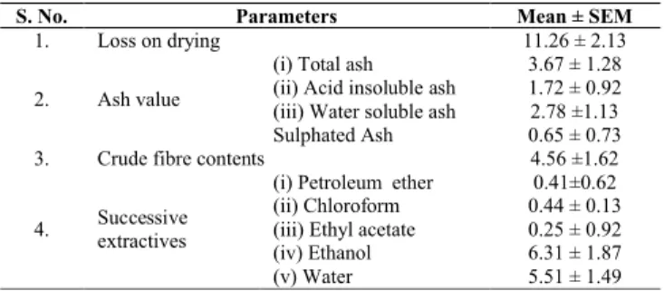
Physicochemical analyses of powdered drug like loss on drying, ash values, crude fibres and successive extractive values with different solvents of powdered root were analysed. The percentage all values in triplicate and their mean values ± SEM were calculated with reference to the air dried drug (Table 2). The changes in the colour of S. spontaneumroot powder under UV radiation in reference to day light were observed with different chemical reagents, it showed different colours of the powder in the presence or absence of chemical constituents (Table 3). The fluorescence analyses of powdered drug play a vital role in the determination of quality and purity of drug.
Qualitative analysis
The presence or absence of different phytoconstituents viz. carbohydrate, glycoside, protein, tannins, saponins, flavonoids and terpenoids were detected by the phytochemical screening methods with different chemical reagents.[23]Ethanolic and water extracts of the roots powder showed positive results for carbohydrate, glycoside, protein, tannins, flavonoids and terpenoids (Table 4). The chloroform and ethyl acetate extract show positive results for terpenoids. Petroleum ether extracts have resinous matter which was not dissolved in other solvents.
Determination of total phenolic contents
The total phenolic content (351.25 ± 1.31µg) was present in the ethanolic extract and water in fraction (254.42 ± 1.82µg gallic acid was equivalent to per 10 mg of the extract of the
S. spontaneum. The aqueous fraction was found to be
maximum phenolic content whereas in other fractions like petroleum ether, chloroform, ethyl acetate, acetone fractions respectively, was not found the phenolic content.
Table 2: Quantitative standards of powdered of Saccharum spontaneum
root
S. No. Parameters Mean ± SEM
1. Loss on drying 11.26 ± 2.13
2. Ash value
(i) Total ash 3.67 ± 1.28
(ii) Acid insoluble ash 1.72 ± 0.92
(iii) Water soluble ash 2.78 ±1.13
Sulphated Ash 0.65 ± 0.73
3. Crude fibre contents 4.56 ±1.62
4. Successive
extractives
(i) Petroleum ether 0.41±0.62
(ii) Chloroform 0.44 ± 0.13
(iii) Ethyl acetate 0.25 ± 0.92
(iv) Ethanol 6.31 ± 1.87
(v) Water 5.51 ± 1.49
Determination of Total flavonoids contents
The flavonoid contents of S. spontaneum root in ethanolic and water extract to be found 48.60 ± 2.17μg and 38.59 ± 2.10μg rutin was equivalent to per 10 mg of the extract. The highest flavonoid content was found to be in ethanolic extract whereas in petroleum ether, chloroform, ethyl acetate and acetone fractions flavonoidal content was not found.
The present study of the S. spontaneum root powdered indicated the presence of carbohydrate, glycoside, protein, tannin, flavonoid and terpenoid respectively. Pharmacognostical studied of the root stock, preliminary phytochemical analysis will help for authentication of plant and for further pharmacological investigation. In this study it was found that the S. spontaneum root had minerals, organic acids, flavonoids and phenolic compounds which has to found possesses antioxidant, mast cells stabilizing effects.[24]
S. spontaneumroot is utilized as food or parts of food may provide medical health benefits including the prevention and or treatment of diseases.
Table 3: Fluorescence Analysis of Powder Sacchrum spontaneum
S. No. Treatment Colour in day light Colour in shorter UV(254nm) Coloure in longer UV(365nm)
1. Dry powder White Light yellow Particles gives brown colour
2. Powder +Alcohlic HCl Faint green Light green Black
3. Powder + Aqueous 0.1NHCl Light yellow White Black
4. Powder+ Aqueous NaOH White Greenish yellow Black
5. Powder + Alcohlic NaOH Light green Green Blackish brown
6. Powder + 50% H2So4 Green Light green Black
Table 4: Qualitative analysis of Sacchrum spontaneumroot
S. No. Test Pet ether extract Chloroform extract Ethyl acetate extract Alcohlic extract Aqueous extract
1. Carbohydrate - - - + +
2. Glycoside - - - + +
3. Tannin - - - + +
4. Flavonoid - - - + +
5. Terpinoid - + + + +
6. Steroid - - + +
Khalidet al./ Pharmacognostical Evaluation and Qualitative Analysis of Saccharum spontaneum…..……
IJPSDR October-December, 2011, Vol 3, Issue 4 (338-341)
341
ACKNOWLEDGMENT
Author have great thankful to vice chancellor and Prof. (Dr.) H.H. Siddiqui, Intergral University, Lucknow (India) for providing research facilities in University premises for research.
REFERENCES
1. Odukoya OA, Jenkins MO, Ilori MOO, Sofidiya MO, The use of
selected Nigerian natural products in management of
environmentally induced skin damage. Pakistan Journal of Biological Science. 2005; 8: 1074-1077.
2. Kikuzaki H, Nakatani N, Antioxidant effects of some ginger constituents. Journal of Food Science. 1993; 58: 1407-1410. 3. Ottolenghi A, Interaction of ascorbic acid and mitochondria lipids.
Arch. Biochem Biophy. 1959; 79: 355-363.
4. Osawa T. Postharvest biochemistry. In: Uritani I, Garcia VV, Mendoza, EM, editors. Novel neutral antioxidant for utilization in food and biological systems. Japan: Japan Scientific Societies Press, 1994, pp: 241-251.
5. Noda Y, Anzai-Kmori A, Kohono M, Shimnei M. L, Packer,
Hydroxyl and superoxide anion radical scavenging activities of natural source antioxidants using the computerized JES-FR30 ESR spectromoter system. Biochem Mol Biol Inter. 1997; 42: 35-44. 6. Miller NJ, Rice-Evans CA, The relative contributions of ascorbic
acid and phenolic antioxidants to the total antioxidants to the activity of orange and apple fruit juices and black currant drink. Food and Chemistry. 1997; 60: 331-337.
7. Cook NC, Samman S, Flavonoids-chemistry, metabolism,
cardioprotective effect and dietary sources. Journal of Nutrition and Biochemistry, 1996.7: 66-76.
8. Zhou YC, Zheng RL, Phenolic compounds and an analog as
superoxide anion scavengers and antioxidants. Bioch Pharm. 1991; 2: 1177-1179.
9. Aruoma OI, Nutrition and health aspects of free radicals and antioxidants. Food Chemistry and Toxicology, 1994; 32: 671-683.
10. Kirtikar KR, Basu BD. Indian medicinal plants. International Book Distributor, Dehradun, India, 2005, pp. 2668.
11. Parrotta JA. Healing Plants of Peninsular India. CABI publishing, USA, 2001, pp. 591.
12. Ghani A. Medicinal plants of Bangladesh with chemical
constituents and uses, 2nd
edition, The Asiatic society of Bangladesh, Dhaka, 2003, pp: 369
13. Chopra RN, Nayar SL, Chopra IC. Glossary of Indian Medicinal Plants.CSIR, New Delhi, 1956, pp. 1-259
14. Yoganarashimhan SN. Medicinal Plants of India, Vol. 2, 2002, pp. 474-475.
15. Johansen DA. Plant Microtechnique. McGraw Hill Book Co. Inc. New York & London, 1940, pp. 95-102.
16. Khandelwal KR. Practical Pharmacognosy Techniques and
Experiments. 12th
edition, Nirali Prakashan, 2004, pp. 21-36.
17. Anonymous. Indian Pharmacopoeia. Government of India, Ministry
of Health and Family Welfare. The Controller of Publications, Civil Lines, Delhi, Vol. I & II 1996.
18. Kokoski CJ, Kokoski RJ, Sharma M, Fluorescence of powdered vegetable drugs and ultraviolet radiation. Journal of American Pharmaceutical Association. 1958; 47: 715-717.
19. 25 Mukherjee PK. Quality control of herbal drugs, an approach to evaluation of botanicals. Business Horizons Pharmaceutical Publishers, New Delhi, 2002, pp. 356-358.
20. Kokate CK. Practical Pharmacognosy. 4th
edition. Vallabh Prakashan, New Delhi, 1994, pp. 20-27.
21. Wolfe K, Wu X, Liu RH, Antioxidant activity of apple peels. Journal of Agriculture and Food Chemistry. 2003; 51: 609-614. 22. Chang C, Yang M, Wen H, Chern J, Estimation of total flavonoid
content in propolis by two complementary colorimetric methods. Journal of Food Drug Analysis. 2002; 10: 178-182.
23. Khandelwal KR. Practical Pharmacognosy. Techniques and
experiments, Nirali Prakashan. 9th
edition, 2008, pp. 149-156.
24. Cotelle N, Bemier JL, Catteau JP, Pommery J, Wallet JC, Gaydou