Modulated by Serotonin and Octopamine Receptors and
Affect Social Behavior
Jiangnan Luo1, Oleh V. Lushchak1, Philip Goergen2, Michael J. Williams2, Dick R. Na¨ssel1* 1Department of Zoology, Stockholm University, Stockholm, Sweden,2Department of Neuroscience, Uppsala University, Uppsala, Sweden
Abstract
A set of 14 insulin-producing cells (IPCs) in theDrosophilabrain produces three insulin-like peptides (DILP2, 3 and 5). Activity in IPCs and release of DILPs is nutrient dependent and controlled by multiple factors such as fat body-derived proteins, neurotransmitters, and neuropeptides. Two monoamine receptors, the octopamine receptor OAMB and the serotonin receptor 5-HT1A, are expressed by the IPCs. These receptors may act antagonistically on adenylate cyclase. Here we investigate the action of the two receptors on activity in and output from the IPCs. Knockdown of OAMB by targeted RNAi led to elevatedDilp3transcript levels in the brain, whereas 5-HT1Aknockdown resulted in increases ofDilp2and5. OAMB-RNAi in IPCs leads to extended survival of starved flies and increased food intake, whereas 5-HT1A-RNAi produces the opposite phenotypes. However, knockdown of either OAMB or 5-HT1Ain IPCs both lead to increased resistance to oxidative stress. In assays of carbohydrate levels we found that 5-HT1Aknockdown in IPCs resulted in elevated hemolymph glucose, body glycogen and body trehalose levels, while no effects were seen after OAMB knockdown. We also found that manipulations of the two receptors in IPCs affected male aggressive behavior in different ways and 5-HT1A-RNAi reduced courtship latency. Our observations suggest that activation of 5-HT1Aand OAMB signaling in IPCs generates differential effects onDilptranscription, fly physiology, metabolism and social interactions. However the findings do not support an antagonistic action of the two monoamines and their receptors in this particular system.
Citation:Luo J, Lushchak OV, Goergen P, Williams MJ, Na¨ssel DR (2014)DrosophilaInsulin-Producing Cells Are Differentially Modulated by Serotonin and Octopamine Receptors and Affect Social Behavior. PLoS ONE 9(6): e99732. doi:10.1371/journal.pone.0099732
Editor:Susan Broughton, Lancaster University, United Kingdom
ReceivedFebruary 12, 2014;AcceptedMay 19, 2014;PublishedJune 12, 2014
Copyright:ß2014 Luo et al. This is an open-access article distributed under the terms of the Creative Commons Attribution License, which permits unrestricted use, distribution, and reproduction in any medium, provided the original author and source are credited.
Funding:The study was supported by: The Swedish Research Council (D.R.N) and The Carl Trygger Foundation (D.R.N., M.J.W.), Stiftelsen Olle Engkvist Byggma¨stare and Stiftelsen Lars Hiertas Minne (both to M.J.W.). The funders had no role in study design, data collection and analysis, decision to publish, or preparation of the manuscript.
Competing Interests:The authors have declared that no competing interests exist.
* E-mail: [email protected]
Introduction
Insulin and insulin-like growth factors (IGFs) are evolutionary conserved peptides that regulate development, growth and aspects of physiology in a broad range of animals [1–9]. InDrosophila, eight insulin-like peptides (DILP1–8) have been identified as likely ligands of a single insulin tyrosine kinase receptor [5,6,8,10,11]. In adult Drosophila the different DILPs, and thus insulin/IGF signaling (IIS), are of vital importance in the regulation of reproduction, metabolic homeostasis, resistance to stress and life span [11–15]. Additionally, attraction to food odors and feeding behavior are modulated by DILPs [16–18]. A cluster of 14 insulin-producing cells (IPCs) in the pars intercerebralis of the brain express DILP2, 3 and 5, which are secreted into the circulation via axon terminations in the corpora cardiaca, anterior aorta, foregut and anterior midgut as well as the crop [11,12,19].
In adult flies the activity in IPCs and thus production and release of DILPs is under control by fat body-derived diffusible molecules such as DILP6 and the leptin-like cytokine Unpaired 2 (Upd2) [20,21]. Systemic release of these factors from the fat body is nutrient-dependent. Hence, when the fly feeds the increased levels of circulating carbohydrate and amino acids are sensed by adipocytes in the fat body, which induces signaling to the IPCs. In addition several neurotransmitters such as GABA and serotonin,
as well as the neuropeptides corazonin, short neuropeptide F and
Drosophilatachykinin [22–27] act on the brain IPCs. Except for the inhibitory transmitter GABA it is, however, not known what triggers the signaling by these substances to the IPCs. A portion of the GABAergic system in the pars intercerebralis seems to be inactivated by circulating Upd2 after feeding and thereby tonic inhibition of the IPCs is lifted (via the action of Jak/Stat signaling) which facilitates DILP release [20].
Previously we demonstrated a role of one of the serotonin receptors, 5-HT1A, in regulation of Drosophila IPCs [22]. This receptor commonly inhibits adenylate cyclase (AC), and thus decreases levels of cyclic AMP (cAMP) and thereby diminishes activity of protein kinase A (PKA) (See reviews [31–33]). The OAMB receptor (K3 splice form) can both increase intracellular Ca2+
and activate adenylate cyclase and thus elevate cAMP and activate PKA [28,34,35]. The possible convergence of the two monoamine receptors on adenylate cyclase signal transduction lead us to compare the action of OAMB and 5-HT1Aon IPCs. Do the two receptors mediate antagonistic activity in IPCs via opposite actions on adenylate cyclase or do they act on independent intracellular systems?
To test this we employed the Gal4-UAS system [36] to direct OAMB and 5-HT1A-RNAi to IPCs and analyzed the effect on transcript levels ofDilp2,3and5and on carbohydrate metabolism and stress responses. We found that manipulations of the two receptors had differential effects onDilptranscription, and mostly also in the other assays. Since both serotonin and octopamine are known to regulate social behavior in flies [37–42] we furthermore investigated the role of IPCs on aggressive and courtship behaviors by manipulating OAMB and 5-HT1Ain IPCs.
Our results do not support that octopamine and serotonin act antagonistically on the IPCs but suggest that activation of OAMB and 5-HT1A in these cells induce differential effects on Dilp transcription, metabolism, stress resistance as well as male-male and male-female interactions.
Results
Processes from octopaminergic neurons superimpose IPC branches
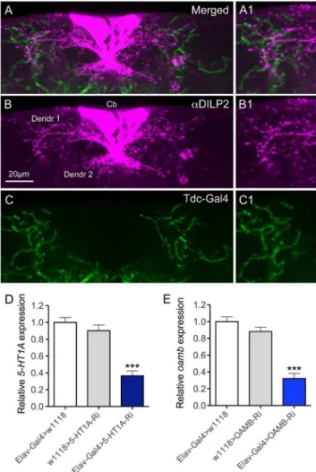
In a recent study it was shown that the IPCs express the OAMB-K3 receptor splice form, as determined by RT-PCR on RNA extracted from single neurons, and that a small set of octopamine-producing neurons, designated ASM, send axon processes to the IPCs [28]. The ASMs are a subpopulation of theTdc2-Gal4 expressing neurons [28]. However, the octopamine distribution in relation to the presumed dendrites of IPCs was not revealed in detail. With application of DILP2 antiserum to brains with octopaminergic and tyraminergic neurons marked byTdc2 -Gal4 driven GFP, we found that some GFP-labeled branches superimpose those of the IPC dendrites in the pars intercerebralis of the brain (Fig 1). In the following we rely on published data [28] that the octopaminergic ASM neurons mediate activation of IPCs. Expression of the 5-HT1A receptor in IPCs, and the possible innervation of IPCs by serotonergic neurons were shown previously [22].
Knockdown of 5HT1Aor OAMB receptors in IPCs does not change cell size
To study the roles of the 5HT1Aand OAMB receptors in IPCs, we targeted RNAi for the two receptors to these cells using the Gal4-UAS technique [36]. First we determined the efficiency of the UAS-5-HT1A-RNAi (referred to as 5HT1A-RNAi) and the UAS-OAMB-RNAi GD lines (referred to as OAMB-RNAi), by crossing these flies to the pan-neuronal elav-Gal4 driver, and performing qPCR to measure the level of 5-HT1A and OAMB
transcript (Fig. 1D, E). These flies were raised at 18uC until they eclosed, at which point the newly eclosed flies were collected and kept at 29uC for 5-7 days to obtain maximal expression from the Gal4-UAS system [36]. Compared with elav-Gal4 heterozygous controls (SEM60.05), 5HT1A-RNAi flies had 0.36-fold (SEM60.06, P,0.005) of the normal 5-HT1A RNA expression
levels, while OAMB-RNAi flies had 0.32-fold (SEM60.06, P,
0.005) of the normalOAMBRNA levels (Fig. 1D, E).
We next used aDilp2-Gal4 driver to specifically target receptor RNAi to the IPCs, using the same two RNAi lines. Since manipulations of IPC activity can lead to alterations of cell growth [43] we determined the effects of receptor-RNAi on the size of cell bodies of IPCs. No changes in cell size were observed after knockdown or either receptor (Fig. S1A,B). Therefore, we can exclude the possibility that physiological or behavioral phenotypes observed in subsequent experiments are due to gross develop-mental effects on cell morphology.
Figure 1. Branches of octopaminergic neurons superimpose those of insulin producing cells (IPCs). A–CDissected adult brains were stained with rabbit anti-DILP2 (magenta) and octopaminergic neurons were visualized by Tdc2-Gal4 driving GFP (green). Cb, cell bodies of IPCs; Dendr1 and dendr2, presumed dendrites of IPCs. Scale bar = 20mm.A1–C1Magnification of regions of interest in A–C.Dand EThe efficiencies of the UAS-5-HT1A- and UAS-OAMB-RNAi (UAS-OAMB-RNAi GD) were determined by using the pan-neuronal Elav-Gal4 driver and monitoring extracted RNA by qPCR. The transcript value for Elav-Gal4.w1118was set as 1 in both assays. A significant knockdown (about 60–70% reduction) of 5-HT1Aand OAMB transcript was seen (***p, 0.001 to both parental controls, One-way ANOVA, n = 2 samples for each genotype).
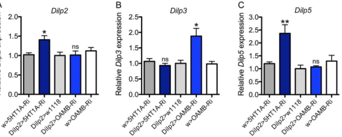
Knockdown of 5-HT1Aor OAMB affects Dilp transcription in IPCs
To test for effects of 5-HT1Aand OAMB knockdown on IPCs function we utilized qPCR to analyze Dilp2, 3 and 5 brain transcript levels after targeted receptor RNAi in IPCs. Knockdown of 5-HT1Ain IPCs resulted in a significant increase in brainDilp2 and Dilp5, but not Dilp3 transcript levels (Fig. 2). This is partly consistent with our earlier data where DILP2 immunoreactivity increased after 5-HT1Aknockdown both in fed and starved flies. On the other hand, knockdown of OAMB in IPCs (usingOAMB -RNAi GD line) caused a significant increase inDilp3, but notDilp2
and Dilp5(Fig. 2). These data indicate that each receptor would upon activation reduce specificDilptranscription. However, it is important to note that the targeted RNAi does not mimic inactivation of the IPCs (or the two receptors), but induces a partial downregulation of receptor expression levels, which probably renders the IPCs slightly less responsive to octopamine or serotonin. The differential actions of serotonin and octopamine on IPCs inDilptranscription might explain the different effects of OAMB and 5-HT1Aknockdown in some assays described in the following sections. It should be noted that we have no data to show effects on DILP release and therefore no clear readouts for alterations of IIS. Furthermore, these findings do not suggest clear-cut antagonistic actions of the two receptors since no decrease in
DilpRNA was detected for either receptor.
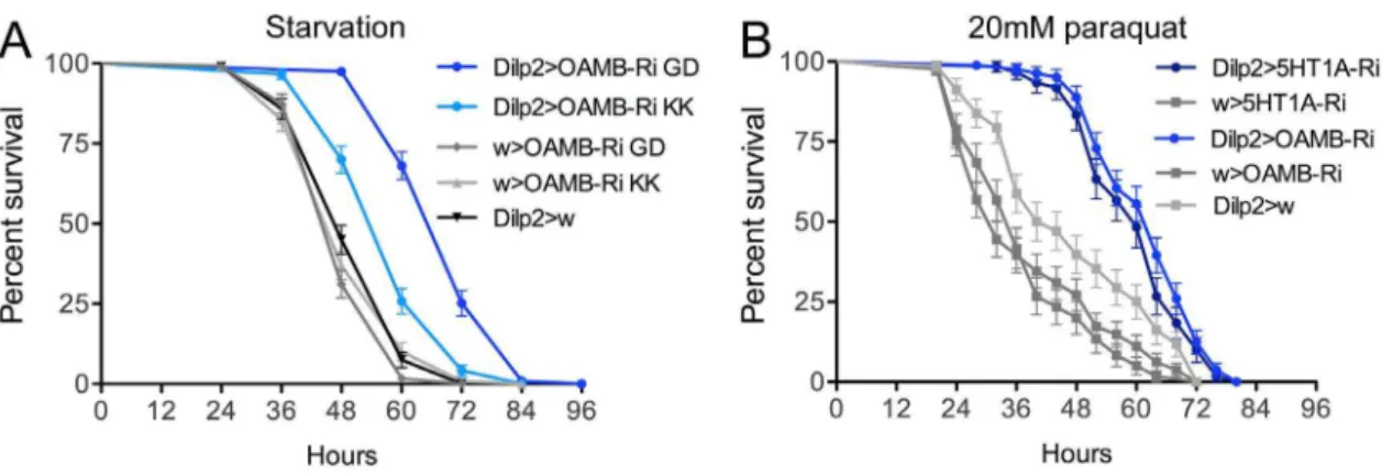
Knockdown of OAMB in IPCs causes increased resistance to starvation
Since previous evidence (see [22]) suggests that modulation of activity in IPCs results in changes in stress tolerance, we measured the starvation resistance in flies with diminished IPC OAMB receptor levels. Compared to controls, the flies with reduced OAMB expression displayed a significant increase in survival when exposed to starvation (Fig. 3A). Two independent UAS-OAMB-RNAi fly lines were tested in this assay and the same phenotypes were obtained. One of these RNAi lines (UAS-OAMB-RNAi GD), used in all experiments described above, produced a more prominent effect when crossed with the Dilp2 -Gal4, and was therefore used also in subsequent experiments. Previously similar experiments were performed after 5-HT1A RNAi in IPCs and resulted in decreased starvation resistance [22].
Thus OAMB and 5-HT1Aknockdowns in IPCs produce opposite effects on starvation resistance.
Knockdown of OAMB or 5-HT1Ain IPCs both cause increased resistance to oxidative stress
It was shown that insulin signaling plays a role in resistance to oxidative stress; flies with ablated IPCs are more stress resistant [14]. We found that flies with diminished OAMB in IPCs were more resistant to oxidative stress induced by feeding 20mM paraquat in standard food (Fig. 3B). Also flies where 5-HT1Awas knocked down in IPCs displayed increased resistance to oxidative stress (Fig. 3B). Based on earlier findings [14] these effects on oxidative stress resistance suggest that both OAMB and 5-HT1A activation in IPCs stimulate insulin signaling, or that the stress resistance phenotype is caused by other mechanisms mediated by the IPCs.
Knockdown of OAMB or 5-HT1A in IPCs results in opposite effects on food intake
Insulin signaling is known to modulate food search and feeding behavior in flies [16–18,44,45]. We determined whether food intake is affected by diminishment of OAMB or 5-HT1Ain IPCs by testing flies in a capillary feeding (CAFE) assay over 96 h. After 5-HT1A-RNAi a slight, but significant, decrease in food consump-tion was observed the third day (Fig. 4A). On the other hand OAMB knockdown resulted in increased food intake days 2–4 (Fig. 4B).
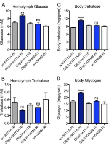
Knockdown of 5-HT1A or OAMB in IPCs affect circulating and stored carbohydrate levels
Previous studies have shown that DILPs produced by the brain IPCs are important in the regulation of carbohydrate and lipid homeostasis [12,14,46]. Therefore we assayed carbohydrate levels to determine whether interference with OAMB and 5-HT1A in IPCs affects systemic insulin signaling and metabolism.
We measured the hemolymph levels of glucose and trehalose in
ad lib fed male flies of different genotypes. Flies with 5-HT1A knocked down in the IPCs displayed significantly increased hemolymph glucose levels, but not trehalose compared to parental controls (Fig. 5A, B). Knocking down OAMB did not influence circulating glucose or trehalose levels (Fig. 5A, B). Next we
Figure 2.Dilptranscript levels in the brain are affected by 5-HT1Aand OAMB knockdown in IPCs. A–CRelative expression ofDilp2, 3and 5transcripts after5-HT1A-RNAi andOAMB-RNAi (UAS-OAMB-RNAi GD) in IPCs was measured from head extracts of fed flies. The value for w1118. 5-HT1A-RNAi was set to 1. TheDilp2 and5transcripts increase significantly after 5-HT1A knockdown, and Dilp3transcript increases after OAMB knockdown (one way ANOVA; **p,0.01, *p,0.05, ns, not significant; n = 3 samples for each genotype).
measured whole body trehalose and glycogen in the same experimental flies (normally fed) and found that 5-HT1A knockdown in IPCs increase both stored carbohydrates, but OAMB-RNAi had no effect (Fig. 5C, D). Thus OAMB knockdown in IPCs does not seem to affect carbohydrate homeostasis in fed flies.
Knockdown of 5HT1Aor OAMB in IPCs has no effect on body weight
Insulin signaling was shown to regulate larval development and growth inDrosophila[11,12]. We therefore tested whether knocking down 5-HT1A or OAMB in IPCs affected the body weight as measured in adult male flies. No effect was seen on body weight (Fig. S1C), similar to effects of GABAB receptor knockdown in IPCs [27]. For the serotonin receptor this could be explained by the finding that 5-HT1Aappears to be absent in larval IPCs when growth occurs [22]. It is not known when OAMB starts to be expressed in IPCs.
5-HT1AandOAMBregulate social behavior
It is known that serotonin and octopamine regulate aggression and mating behaviors inDrosophila[37–41,47]. It was furthermore suggested that octopamine could modulate aggressive behavior via neurons releasing the cholecystokinin-like peptide drosulfakinin (DSK) [40]. One set of neurons that express DSK are the brain IPCs [45]. Thus, we tested he effects of 5-HT1A and OAMB knockdown in IPCs on social behavior in male flies.
Aggression analysis experiments were executed by placing pairs of 5–7 day old males, raised in isolation, in a behavioral assay chamber, containing 1% agarose, and their interactions were monitored over a 20 min period. The total number of interactions for each fly was recorded, whether it involved aggressive or courtship behavior. The assayed male-male interactions consisted of eight distinct behaviors. Aggressive interactions were scored as either low or high-intensity engagements. Low intensity fighting (LIF) was scored as side-by-side pushing with a leg (side-fencing), face-to-face pushing with one leg (fencing), or quick wing flicking (wing flick); high intensity fighting (HIF) was graded as lunging (lunging), boxing face-to-face with the two front legs (boxing), as Figure 3. Stress responses after knockdown of octopamine receptor OAMB and serotonin receptor 5-HT1Ain IPCs. AReduction of octopamine receptor OAMB level in IPCs increases resistance to starvation (seen by extended survival). OAMB was diminished in IPCs by driving two different OAMB-RNAi lines withDilp2-Gal4. The Dilp2-Gal4.OAMB-RNAi GD flies showed about 50% percent increase of median lifespan compared with controls (***p,0.001 to both parental controls; Log rank test, Mantel-Cox; n = 120 for each genotype). We used the OAMB-RNAi GD lines hereafter for all other experiments. In an earlier study it was shown that 5-HT1Aknockdown decreased starvation resistance [22].BReduction of octopamine receptor OAMB or serotonin receptor 5-HT1Alevels in IPCs increases resistance to oxidative stress induced by feeding male flies 20mM paraquat in standard food. Both receptor knockdowns result in flies that display significantly increased survival under paraquat exposure (***p,0.001 to both parental controls, Log rank test, Mantel-Cox; n = 60–90 for each genotype).
doi:10.1371/journal.pone.0099732.g003
Figure 4. Knockdown of OAMB or 5-HT1Ain IPCs resulted in opposite phenotypes in a capillary feeding assay (CAFE). ADiminishing 5-HT1Ain IPCs (Dilp2-Gal4.5-HT1A-RNAi) significantly decreases food intake only during day 2. The units in the Y-axis are mm food consumed from the calibrated capillary tube (total volume 5mL; see methods). The graph shows cumulative intake of food over 4 days (*p,0.05, n = 5-10 flies for
each genotype in 3 replicates, One-way ANOVA).BReduction of OAMB level in IPCs (Dilp2-Gal4.OAMB-RNAi) significantly increases food intake from the second day onwards (**p,0.01, ***p,0.001, n = 5–10 flies for each genotype in 3 replicates, One-way ANOVA).
well as holding the wings up at a 30–45u angle (wing threat). Courtship behavior was marked as one-wing extended at a 90u
angle (singing), circling to the posterior (circling), or bending the abdomen towards the other fly (abdomen bending).
For the HIF behaviors there was significant difference between controls,5-HT1AandOAMBknockdown males (P,0.05). Unlike controls, OAMB-RNAi males did not perform any wing threats (Table S1), while 5HT1A-RNAi males performed significantly fewer lunging behaviors than controls (Table S1). Interestingly, when LIF behaviors were compared, 5HT1A-RNAi males performed significantly more wing flicks and side fencing over a 20 min fighting bout than control males (Table S1). While the percentage of LIF behaviors performed byOAMB-RNAimales was significantly lower than controls (Fig. 6A), this was most likely due
to the significant increase observed in courtship behaviors, since the actual number of LIF behaviors was not significantly different from controls (Table S1). Finally, knocking down OAMB had a significant effect on all scored mating behaviors, especially singing (Fig. 6A and Table S1).
Next we determined whether 5-HT1AandOAMBalso regulate male behavior towards virgin females. To test this, males were paired with wild-type virgin females and two aspects of male-female courtship were measured: latency and courtship index (see Materials and Methods). No effect was observed on courtship index for either receptor knockdown (Fig. 6B). When5-HT1Awas knocked down there was a substantial decrease in latency (45.4 seconds, SEM628.4, P,0.005) compared to either Dilp2-GAL4+/2 (179.9 seconds, SEM629.4) or5-HT1ARNAi+/2(172.8 seconds, SEM616.4) controls (Fig. 6C). No significant effect was observed when OAMB was knocked down.
Figure 5. Levels of carbohydrates were affected by knockdown of 5-HT1Ain IPCs while no changes were observed for OAMB
knockdown.A Hemolymph glucose levels in normally fed flies were higher after 5-HT1A-RNAi in the IPCs (Dilp2.5HT1A-Ri) than controls, while no changes were observed for OAMB knockdown (Dilp2. OAMB-Ri). Data were analyzed with one way ANOVA, **p,0.01, n = 60-90 for each genotype, and experiments were performed in 6 independent replicates. B No significant changes of hemolymph trehalose levels were observed after5HT1A-RNAi orOAMB-RNAi in the IPCs. One-way ANOVA, ns, not significant; n = 60-90 for each genotype, and experiments were performed in 6 independent replicates. C Body trehalose levels in normally fed flies are higher after5HT1A-RNAi in the IPCs (Dilp2.5HT1A -Ri) than controls, but were not significantly changed afterOAMB-RNAi in the same cells (Dilp2.OAMB-Ri). One way ANOVA, ***p,0.001, n = 60-90 for each genotype, and experiments were performed in 6 independent replicates. D Glycogen levels (whole body) in normally fed flies are higher after 5HT1A-RNAi in the IPCs (Dilp2.5HT1A-Ri) than controls, but were not significantly changed afterOAMB-RNAi in the same cells. One way ANOVA, ***p,0.001, n = 60–90 for each genotype, and experiments were performed in 6 independent replicates. doi:10.1371/journal.pone.0099732.g005
Discussion
Our study shows that octopamine and serotonin differentially modulate the activity of brain IPCs inDrosophilavia the receptors OAMB and 5-HT1A. The physiological readout of diminishing expression of either receptor in IPCs is complex and does not suggest convergence of the two receptors on the same downstream signaling cascade and also indicates involvement of IPC outputs additional to systemic IIS. Also the effects of targeted receptor-knockdown on social behavior may suggest that the two receptors act on independent pathways. We first discuss the effects of diminishment of OAMB and 5-HT1Aon physiology that may be regulated by IIS.
We find that knockdown of OAMB in IPCs affects some aspects of physiology that may be diagnostic of decreased systemic IIS, such as increased starvation and oxidative stress resistance and increased food intake (see Table 1). Surprisingly, however, OAMB-RNAi induced increased Dilp3 transcription (and no effects on Dilp2 and 5). These findings would suggest that the activation of OAMB in IPCs in normally fed flies decreases DILP3 production, but stimulates some aspects of systemic IIS or produces phenotypes reminiscent of increased IIS. We did not detect any effect of OAMB knockdown on carbohydrate metabolism. We did not test triacylglyceride (TAG) levels here, but using a crude technique [22] to estimate of lipid levels in flies with OAMB knocked down in IPCs, we recorded a significant lipid decrease both in fed flies and flies starved for 24 h (not shown). A previous study found that OAMB mutants display decreased TAG levels, and activation of octopamine producing neurons increases TAG [30]. These authors also propose that the effect on TAG levels is via the IPCs since activation of octopamine neurons in aDilp2, 3mutant background leads to less increase in TAG. In our earlier study we found that 5-HT1Aknockdown in IPCs results in decreased levels of stored lipid in fed and starved flies; the same was found in 5-HT1Amutant flies [22]. Although the lipid assays performed in the two studies were different, it seems that knockdown of both OAMB and 5-HT1A lead to decreased lipid levels. Possibly this suggests that activation of these receptors triggers increases stored lipids.
Although the role of OAMB in regulation of IIS remains somewhat unclear, our present data from manipulations of the 5-HT1Ain IPCs, combined with those of an earlier report [22], are more consistent with effects on systemic IIS. We show that 5-HT1A knockdown increasesDilp2and5transcripts, reduces resistance to starvation, increases oxidative stress resistance, diminishes food intake and elevates levels of circulating and stored carbohydrates (Table 1). Taken together these findings indicate that targeted 5-HT1Aknockdown increases systemic IIS, and thus the activation of this receptor should decrease IIS. However, as seen in Table 1 our data from physiological readouts may suggest more complex effects of the receptor manipulations in IPCs.
One question raised in the present study was whether octopamine and serotonin act antagonistically on a shared downstream signal cascade to regulate IPC function. This was prompted by the possibility that both OAMB and 5-HT1Aact on adenylate cyclase, cAMP and PKA [28,31–35]. We therefore compared the effects of receptor RNAi in several assays. In three of these we saw distinct effects of knocking down the two receptors: starvation resistance, food intake and transcription ofDilp2, 3and
5(Table 1). In assays of carbohydrates only 5-HT1Aknockdown increased levels and no effect was seen with OAMB. Actually only in two assays, food intake and starvation resistance, did we observe opposite phenotypes after diminishing the two receptors. None of the receptor knockdowns diminishedDilptranscript levels, instead they increased RNAs of mutually exclusiveDilps(Table 1). Thus it is not possible to assign clear antagonistic effects of the two receptors in IPCs. One dilemma is that for each receptor we obtained incomplete or conflicting results. The effects of 5-HT1A -RNAi on metabolism suggest decreased IIS, which indicates that activation of the receptor should stimulate DILP release and increase IIS. However, the starvation resistance assay produced a phenotype opposite to the expected one after diminishment of 5-HT1A [22]. In this context it should be noted that starvation resistance in the experimental flies could be caused by factors other than diminished IIS, such as increased locomotion or altered feeding [48,49].
In the CAFE assay we noted opposite effects on food intake for the different receptor knockdowns. The effects of IIS on feeding
Table 1.Effects of OAMB and 5-HT1Aknockdown in various assays.
Assays with adult flies Effect Likely effect on insulin signaling1(output) Different effects
OAMB-Ri 5-HT1A-Ri OAMB-Ri 5-HT1A-Ri
Starvation resistance Increase Decrease Decrease Increase Yes*
Food intake (CAFE) Increase Decrease Increase Decrease Yes*
Glucose (in circulation) NE Increase NE Decrease Yes
Trehalose (in circulation) NE NE NE NE No
Glycogen (stored) NE Increase NE Decrease Yes
Trehalose (stored) NE Increase NE Decrease Yes
Dilp2transcript NE Up NE Increase Yes
Dilp3transcript Up NE Increase NE Yes
Dilp5transcript NE Up NE Increase Yes
Oxidative stress resistance Increase Increase Decrease Decrease No
IPC cell body size NE NE NE NE No
Body weight NE NE NE NE No
NOTES:1This assumes that targeted receptor RNAi is similar to inactivating the receptor, which of course is not entirely correct. NE, no effect; * indicates opposite effect for the two knockdowns.
are not clearly established inDrosophila. Some experiments suggest that silencing of the IPCs diminishes appetite and food intake when food has low calorie content [44,48]. We showed in a recent study that silencing IPCs by expression of a constitutively active hyperpolarizing K-channel (Ork) increased intake of caffeine-spiked or sugar-free food [45], and conversely conditional activation of the IPCs with a temperature inducible TrpA1 channel diminishes food intake (J. Luo, Y. Liu and Na¨ssel, in prep). In our present experiments 5-HT1Aknockdown in IPCs decreased food intake slightly. Thus, our data might indicate that serotonin receptor knockdown increases IIS and thereby diminishes feeding, and OAMB deficiency causes the opposite phenotype.
The effects on social behavior of OAMB and 5-HT1A knockdown in IPCs are partly differential. Among the male interactions high intensity fighting is reduced by both knockdowns, low intensity fighting is up-regulated after 5-HT1A-RNAi and male courtship is up-regulated by OAMB-RNAi. In male-female courtship we only recorded a decrease in courtship latency for 5-HT1A-RNAi. There is no direct evidence that IIS affects aggression or courtship behaviors, although sexual receptivity and sexual attraction in female flies was shown to involve IPCs and IIS, respectively [50,51], and aggression may depend on activity in neurons of the pars intercerebralis [52]. However octopamine was shown to regulate aggression inDrosophilavia the cholecystokinin-like peptide drosulfakinin (DSK) [40]. DSK was detected in a subpopulation of the larval and adult IPCs [45,53]. Thus, it is possible that the effects of manipulating OAMB in IPCs are caused by changes in DSK signaling rather than IIS, both in social behavior and physiology.
The action of OAMB and 5-HT1Aon IPCs may be complicated by the fact that OAMB-K3 is known to also activate intracellular Ca2+
in Drosophila [34,35]. Thus, OAMB activation may cause more complex responses in IPCs including both Ca2+
and adenylate cyclase. Another factor that induces additional com-plexity is the role of OAMB in IPCs in regulation of sleep/wake activity, independent of IIS [30]. Also serotonin is implicated in sleep [54,55] and the possible role of IPCs in mediating this action has not been investigated. Thus, if the IPCs play roles in regulatory activities other than systemic IIS, we might expect that phenotypes obtained after manipulating the two monoamine receptors are complex.
There are several important questions for the future. What are the triggers of octopamine and serotonin action on IPCs and how are the IPCs integrated into octopaminergic and serotonergic modulatory pathways? Octopamine has been extensively investi-gated in insects and crustaceans and is known to act as a neuromodulator and neurohormone with pleotropic functions, including roles in modulation of muscle and neurons, appetite, ovulation, aggression, sleep, and learning and memory [27,34,39,56,57,58]. Octopamine is also involved in modulation of flight and escape jumping in Drosophila [56,59]. Since octopamine seems central in modulation of energy demanding activities a role in regulation of IPCs and IIS is not surprising. Serotonin is also known to play multiple roles in Drosophila
physiology and behavior: visual and olfactory learning [60,61], courtship and mating [38,62], aggression [37,47], sleep and circadian activity [54,55], olfaction [63] and feeding [64]. It is therefore important to design experiments to reveal which octopaminergic and serotonergic pathways that act on the IPCs and under what conditions they do so.
In summary, we established differential effects of OAMB and 5-HT1Aactions on IPCs in flies that suggest that octopamine and serotonin play distinct roles in modulation of these important neurosecretory cells. However, our data indicate that each
receptor may trigger complex activity in the IPCs, some of which seem not to affect IIS.
Materials and Methods
Fly strains and husbandry
All flies were reared at 25uC on standard yeast, corn meal agar medium [according to Bloomington Drosophila Stock Center (BDSC), Bloomington, IN] under 12:12 h light:dark conditions. Flies with other original genetic backgrounds were backcrossed into w1118 background for four generations before experiments, and w1118flies were used as controls in crosses in all experiments. Fly stocks for behavior experiments were maintained on Jazz-mix
Drosophila food (Fisher Scientific) containing sucrose, corn meal, 10% yeast, agar, benzoic acid, methyl paraben and propionic acid, and maintained at 25uC, 60% humidity, on a 12:12 light:dark cycle. To delay Gal4 expression flies crossed to a Gal4 driver were kept at 18uC, and once the progeny eclosed they were shifted to 29uC for at least 5–7 days before any behavior assays were performed.
The following Gal4 lines were used:Tdc2-Gal4 [65] (J. Hirsh, Charlottesville, VA),Dilp2-Gal4 [12] (E. Rulifson, Stanford, CA) andelav-Gal4 (BDSC). We used the UAS lines: UAS-OAMB-RNAi (two different lines with stock ID 2861 and 106511) and UAS- 5-HT1A-RNAi [stock ID 106094; see [22]] from the Vienna
Drosophila RNAi Center (VDRC), Vienna, Austria, and
UAS-mcd8-gfpare from BDSC. For mating behavior females of aCSORC
strain was used. This is a lab wild type strain created by crossing Canton-S andOregon-Rwild type strains (BDSC).
Antisera and immunocytochemistry
For immunocytochemistry adultDrosophilaheads were dissected in 0.1 M sodium phosphate buffer (PB), pH 7.4 and fixed in ice-cold 4% paraformaldehyde in 0.1 M PB for 2-4 h and dissected adult brains were used for whole mount immunocytochemistry.
Incubation with primary antiserum for whole mount tissues was performed for 48 h at 4uC. The following primary antisera were used: rabbit antiserum to Drosophila insulin-like peptide 2 (anti-DILP2) at 1:4000 (gift from M. Brown, Athens, GA), rabbit and mouse anti-GFP (Invitrogen) were used at 1:1000. For detection of primary antisera Cy3-tagged goat anti-rabbit antiserum (Jackson Immuno Research) and Alexa-488 tagged goat anti-mouse (Invitrogen) were used at 1:1000.
Body weight
For each genotype at least three groups of 10 male flies were weighed on a Mettler MT5 Microbalance (Mettler Toledo, Switzerland) to obtain wet weight, and the average weight was calculated.
Stress assays
Male and female flies (4–6 d old) were collected for starvation experiments. Flies were placed individually in 2 ml glass vials with 500ml of 0.5% aqueous agarose and dead flies were monitored every 12 h. These starvation experiments were run in three replicates with at least 30 flies of each genotype per replicate.
Assays of carbohydrates
Male flies (4–6 days old) were used to measure concentrations of circulating glucose and trehalose together with stored glycogen and whole body trehalose. Pre-weighed flies were decapitated and hemolymph was collected by centrifugation (3000 g, 6 min). Hemolymph was used to measure circulating glucose and trehalose whereas whole bodies were used for determination of glycogen and stored trehalose. All parameters were measured with a glucose assay kit involving glucose oxidase and peroxidase (Liquick Cor-Glucose diagnostic kit, Cormay, Poland). Trehalose was converted to glucose by porcine kidney trehalase (Sigma T8778) and glycogen by amyloglucosidase from Aspergillus niger
(Sigma 10115). Glucose and trehalose are expressed as concen-tration in hemolymph whereas glycogen and body trehalose are given as amount per wet weight. All genotypes were tested in 3 independent replicates (15–20 flies of each genotype in each sample) and one-way ANOVA was used to compare differences between genotypes.
Capillary feeding (CAFE) assay
The capillary feeding (CAFE) assay was conducted according to Ja and others [66] with slight changes. Male flies were placed into 1.5 ml Eppendorf tubes with an inserted 5ml capillary tube with 5% sucrose, 2% yeast extract and 0.1% propionic acid. Three food-filled capillaries were inserted as controls in identical tubes without flies. The final consumption of food was determined as the diminished food level (in mm) minus the average diminishment in control capillaries (due to evaporation). Daily food consumption was measured every 24 h and calculated cumulatively over 4 consecutive days. These experiments were run in three replicates with 10 flies of each genotype for each replicate.
Quantitative real-time PCR (qPCR)
Relative amounts ofDilp2, 3 and 5 RNA in heads of male flies were measured by qPCR. RNA was isolated from 4 biological replicate samples of each genotype tested. Onemg of total RNA was used for cDNA synthesis. cDNA was synthesized in triplicates, which were subsequently pooled and diluted for qPCR. Expression of genes of interests was measured relative to that of the housekeeping gene Actin88 (Act) using an ABI Prism 7000 instrument (Applied Biosystems) and a SensiFAST SYBR Hi-ROX Kit (Bioline) under conditions recommended by manufac-turer. Each analytical and standard reaction was performed in three technical replicates. The levels ofDilp2,3and5andActwere measured with the following primer pairs (all 59to 39):
Dilp2F: AGCAAGCCTTTGTCCTTCATCTC and Dilp2R: ACACCATACTCAGCACCTCGTTG; Dilp3F: TGTGTGTATGGCTTCAACGCAATG and Dilp3R: CACTCAACAGTCTTTCCA-GCAGGG; Dilp5F: GAGGCACCTTGGGCCTATTC and Dilp5R: CATGTGGTGAGATTCG-GAGC; Act88F: AGGGTGTGATGGTGGGTATG and Act88R: CTTCTCCATGTCGTCCCAGT.
For analysis of the efficiency of the two RNAi lines by qPCR we used a slightly different protocol. Relative expression levels of three housekeeping genes (EF-1, Rp49 & RpL11) and of the genes of interest were determined with qPCR. Each reaction, with a total volume of 20ml, contained 20 mM Tris/HCl pH 9.0, 50 mM KCl, 4 mM MgCl2, 0.2 mM dNTP, DMSO (1:20) and SYBR Green (1:50000). All qPCR experiments were performed in duplicates; for each primer pair a negative control with water and a positive control with 5 ng/ml of genomic DNA was included on
each plate. Analysis of qPCR data was performed using MyIQ 1.0 software (Bio-Rad) as previously reported [67]. Differences in gene
expression between groups were analyzed with ANOVA followed by Fisher’s PLSD test where appropriate. P,0.05 was used as the criterion of statistical significance. The following primers were used to amplify reference housekeeping genes:
EF-1F: 59-GCGTGGGTTTGTGATCAGTT-39, EF-1R: 59-GATCTTCTCCTTGCCCATCC-39; Rp49F: CACACCAAATCTTACAAAATGTGTGA-39, Rp49R: 59-AATCCGGCCTTGCACATG-39;
RpL11F: 59-CCATCGGTATCTATGGTCTGGA-39, RpL11R: 59-CATCGTATTTCTGCTGGAACCA-39. To amplify 5-HT1A the primers were as follows:
F: 59-GTGGCCAATACC-39, R: 59 -ATCTGGTTGCCA-GAAGTGCT-39.
To amplify OAMB the primers were as follows: F: 59- TTGGCCGTCCTACCCTTCT-39, R: 59-CGGTCCAGTGATATGGCACAC-39.
Image analysis
Specimens were imaged with Zeiss LSM 510 META and Zeiss LSM 780 confocal microscopes (Jena, Germany) using 20x, 406 oil or 636 oil immersion objectives. Confocal images were processed with Zeiss LSM software for either projection of z-stacks or single optical sections. Images were edited for contrast and brightness in Adobe Photoshop CS3 Extended version 10.0. For cell size determination, the outline of cell body was delineated manually and its area determined using Image J [from NIH, Bethesda, MD, USA (http://rsb.info.nih.gov/ij/)]. For each genotype neurons of 8–15 male flies from 3 independent crosses were measured.
Aggression assay
Newly emerged male flies were collected and isolated for 5 to 7 days at 29uC, 60% humidity, on a 12:12 light:dark cycle. Behavioral tests were carried out at room temperature with 60% humidity in cylindrical behavioral chambers (2 cm by 2.5 cm; height6diameter), filled with 1% agarose to 1.5 cm in height to maintain proper humidity. Two male flies were anesthetized using an ice-water bath before being transferred to a behavioral chamber. After a recovery period of at least 3 minutes, a camera (Panasonic HDC-SD90), positioned above the chamber, was used to record activity for a minimum of 30 minutes. After the 3 min recovery period the behavioral interactions between the males was scored for 20 minutes. Distinct stereotypic aggressive interactions were scored as described by Nilsenet al.[15] and Chenet al.[68]. Aggressive interactions were further scored as either low or high-intensity engagements. Low high-intensity fighting (LIF) was scored as side-by-side pushing with a leg (side-fencing), face-to-face pushing with one leg (fencing) or quick wing flicking (wing flick); high intensity fighting (HIF) was graded as frontal lunging (lunging) or boxing face-to-face with the two front legs (boxing), holding the wings at a 30uangle (wing threat), as well as chasing one another (chasing). Courtship behavior (CB) was marked as one-wing extended at a 90uangle (singing), circling to the posterior (circling), tapping the abdomen (tapping). At least 10 replicates were conducted for each genotype.
Mating behavior assay
3 min recovery period the behavioral interactions between the males and females was scored for 20 minutes or until copulation occurred. Scoring of the courtship behaviors was performed as described by Becnelet al. [38]. Latency, courtship index as well as the frequency of mating behaviors were measured. Latency was calculated by counting the time it took a male to initiate mating and courtship index is calculated as the percentage of time a male spends actively courting a female over a 20 minute period (Seconds spent actively courting/(1200 seconds – Latency seconds). At least 10 replicates per genotype were conducted.
Statistical Analysis
All statistical analyses were performed using GraphPad Prism 5.0. Survival data were analyzed by Log rank test with Mantel-Cox post test, for quantification of immunofluorescence, lipid values and body weights we used One-way ANOVA with Tukey’s comparison or two way ANOVA depending on analysis (see Figure legends for details). Data are presented as means and standard error of means (SEM). For behavior analysis we used ANOVA with appropriate post hoc analysis for multiple comparisons.
Supporting Information
Figure S1 Knockdown of 5HT1Aor OAMB in IPCs does not affect of cell sizes of IPCs, or body weight.AIPCs were visualized by anti-DILP2 labeling after 5-HT1Aand OAMB knockdown in
IPCs (using Dilp2-Gal4). No difference in immunolabeling intensity or cell body size was noted. Scale bar 10mm. B
Quantification of cell body size of IPCs after 5-HT1Aand OAMB knockdown in IPCs. Adult male flies of 4-6 d age were used (ns, not significant; Student’s T-test, n = brains of 7-9 flies for each genotype). C Adult body weight after 5-HT1A and OAMB knockdown in IPCs. Adult 4-6d old male flies were weighed as described in Materials and Methods. (ns, not significant; one-way ANOVA; n = 40 flies for each genotype).
(TIF)
Table S1 Effects of OAMB- and 5-HT1A-RNAi on social behavior. Experimental conditions and statistics are described in legend of Fig. 6.
(TIF)
Acknowledgments
We thank M. Brown, J. Hirsh, E. Rulifson, The BloomingtonDrosophila
Stock Center and the ViennaDrosophilaRNAi Center for providing fly stocks and reagents.
Author Contributions
Conceived and designed the experiments: JL OVL PG MJW DRN. Performed the experiments: JL OVL PG. Analyzed the data: JL OVL PG MJW DRN. Wrote the paper: JL DRN. Edited manuscript (wrote behavior parts): MJW.
References
1. Geminard C, Arquier N, Layalle S, Bourouis M, Slaidina M, et al. (2006) Control of metabolism and growth through insulin-like peptides inDrosophila. Diabetes 55: S5–S8.
2. Kaletsky R, Murphy CT (2010) The role of insulin/IGF-like signaling inC. eleganslongevity and aging. Dis Model Mech 3: 415–419.
3. Kenyon CJ (2010) The genetics of ageing. Nature 464: 504–512.
4. Taguchi A, White MF (2008) Insulin-like signaling, nutrient homeostasis, and life span. Annu Rev Physiol 70: 191–212.
5. Colombani J, Andersen DS, Leopold P (2012) Secreted peptide Dilp8 coordinatesDrosophilatissue growth with developmental timing. Science 336: 582–585.
6. Garelli A, Gontijo AM, Miguela V, Caparros E, Dominguez M (2012) Imaginal discs secrete insulin-like peptide 8 to mediate plasticity of growth and maturation. Science 336: 579–582.
7. Garofalo RS (2002) Genetic analysis of insulin signaling inDrosophila. Trends Endocrinol Metab 13: 156–162.
8. Gro¨nke S, Clarke DF, Broughton S, Andrews TD, Partridge L (2010) Molecular evolution and functional characterization ofDrosophila insulin-like peptides. PLoS Genet 6: e1000857.
9. Hansen M, Flatt T, Aguilaniu H (2013) Reproduction, fat metabolism, and life span: what is the connection? Cell Metab 17: 10–19.
10. Fernandez R, Tabarini D, Azpiazu N, Frasch M, Schlessinger J (1995) The
Drosophilainsulin receptor homolog: a gene essential for embryonic development encodes two receptor isoforms with different signaling potential. EMBO J 14: 3373–3384.
11. Brogiolo W, Stocker H, Ikeya T, Rintelen F, Fernandez R, et al. (2001) An evolutionarily conserved function of theDrosophilainsulin receptor and insulin-like peptides in growth control. Curr Biol 11: 213–221.
12. Rulifson EJ, Kim SK, Nusse R (2002) Ablation of insulin-producing neurons in flies: growth and diabetic phenotypes. Science 296: 1118–1120.
13. Teleman AA (2010) Molecular mechanisms of metabolic regulation by insulin in
Drosophila. Biochem J 425: 13–26.
14. Broughton SJ, Piper MD, Ikeya T, Bass TM, Jacobson J, et al. (2005) Longer lifespan, altered metabolism, and stress resistance inDrosophilafrom ablation of cells making insulin-like ligands. Proc Natl Acad Sci U S A 102: 3105–3110. 15. Nilsen SP, Chan YB, Huber R, Kravitz EA (2004) Gender-selective patterns of
aggressive behavior in Drosophila melanogaster. Proceedings of the National Academy of Sciences of the United States of America 101: 12342–12347. 16. Root CM, Ko KI, Jafari A, Wang JW (2011) Presynaptic facilitation by
neuropeptide signaling mediates odor-driven food search. Cell 145: 133–144. 17. Wu Q, Zhang Y, Xu J, Shen P (2005) Regulation of hunger-driven behaviors by
neural ribosomal S6 kinase inDrosophila. Proc Natl Acad Sci U S A 102: 13289– 13294.
18. Wu Q, Zhao Z, Shen P (2005) Regulation of aversion to noxious food by
Drosophilaneuropeptide Y- and insulin-like systems. Nat Neurosci 8: 1350–1355.
19. Cao C, Brown MR (2001) Localization of an insulin-like peptide in brains of two flies. Cell Tissue Res 304: 317–321.
20. Rajan A, Perrimon N (2012) Drosophila cytokine unpaired 2 regulates physiological homeostasis by remotely controlling insulin secretion. Cell 151: 123–137.
21. Bai H, Kang P, Tatar M (2012) Drosophila insulin-like peptide-6 (dilp6) expression from fat body extends lifespan and represses secretion of Drosophila insulin-like peptide-2 from the brain. Aging Cell 11: 978–985.
22. Luo J, Becnel J, Nichols CD, Na¨ssel DR (2012) Insulin-producing cells in the brain of adultDrosophilaare regulated by the serotonin 5-HT1A receptor. Cell Mol Life Sci 69: 471–484.
23. Lee KS, You KH, Choo JK, Han YM, Yu K (2004) Drosophila short neuropeptide F regulates food intake and body size. J Biol Chem 279: 50781– 50789.
24. Kapan N, Lushchak OV, Luo J, Na¨ssel DR (2012) Identified peptidergic neurons in theDrosophilabrain regulate insulin-producing cells, stress responses and metabolism by coexpressed short neuropeptide F and corazonin. Cell Mol Life Sci 69: 4051–4066.
25. Lee KS, Kwon OY, Lee JH, Kwon K, Min KJ, et al. (2008)Drosophilashort neuropeptide F signalling regulates growth by ERK-mediated insulin signalling. Nat Cell Biol 10: 468–475.
26. Birse RT, So¨derberg JA, Luo J, Winther A˚ M, Na¨ssel DR (2011) Regulation of insulin-producing cells in the adultDrosophilabrain via the tachykinin peptide receptor DTKR. J Exp Biol
27. Enell LE, Kapan N, So¨derberg JA, Kahsai L, Na¨ssel DR (2010) Insulin signaling, lifespan and stress resistance are modulated by metabotropic GABA receptors on insulin producing cells in the brain ofDrosophila. PLoS ONE 5: e15780.
28. Crocker A, Shahidullah M, Levitan IB, Sehgal A (2010) Identification of a neural circuit that underlies the effects of octopamine on sleep:wake behavior. Neuron 65: 670–681.
29. Crocker A, Sehgal A (2008) Octopamine regulates sleep inDrosophilathrough protein kinase A-dependent mechanisms. J Neurosci 28: 9377–9385. 30. Erion R, DiAngelo JR, Crocker A, Sehgal A (2012) Interaction between sleep
and metabolism inDrosophilawith altered octopamine signaling. J Biol Chem 287: 32406–32414.
31. Polter AM, Li X (2010) 5-HT1A receptor-regulated signal transduction pathways in brain. Cell Signal 22: 1406–1412.
32. Nichols DE, Nichols CD (2008) Serotonin receptors. Chem Rev 108: 1614– 1641.
34. Kim YC, Lee HG, Lim J, Han KA (2013) Appetitive learning requires the alpha1-like octopamine receptor OAMB in the Drosophila mushroom body neurons. J Neurosci 33: 1672–1677.
35. Han KA, Millar NS, Davis RL (1998) A novel octopamine receptor with preferential expression inDrosophilamushroom bodies. J Neurosci 18: 3650– 3658.
36. Brand AH, Perrimon N (1993) Targeted gene expression as a means of altering cell fates and generating dominant phenotypes. Development 118: 401–415. 37. Alekseyenko OV, Lee C, Kravitz EA (2010) Targeted manipulation of
serotonergic neurotransmission affects the escalation of aggression in adult male
Drosophila melanogaster. PLoS One 5: e10806.
38. Becnel J, Johnson O, Luo J, Na¨ssel DR, Nichols CD (2011) The serotonin 5-HT7Dro receptor is expressed in the brain ofDrosophila, and is essential for normal courtship and mating. PLoS One 6: e20800.
39. Certel SJ, Leung A, Lin CY, Perez P, Chiang AS, et al. (2010) Octopamine neuromodulatory effects on a social behavior decision-making network in
Drosophilamales. PLoS One 5: e13248.
40. Williams MJ, Goergen P, Rajendran J, Klockars A, Kasagiannis A, et al. (2014) Regulation of aggression by obesity-linked genes TfAP-2 and Twz through octopamine signaling inDrosophila. Genetics 196: 349–362.
41. Zhou C, Huang H, Kim SM, Lin H, Meng X, et al. (2012) Molecular genetic analysis of sexual rejection: roles of octopamine and its receptor OAMB in
Drosophilacourtship conditioning. J Neurosci 32: 14281–14287.
42. Baier A, Wittek B, Brembs B (2002) Drosophila as a new model organism for the neurobiology of aggression? The Journal of experimental biology 205: 1233– 1240.
43. Luo J, Liu Y, Na¨ssel DR (2013) Insulin/IGF-regulated size scaling of neuroendocrine cells expressing the bHLH transcription factor Dimmed in
Drosophila. PLoS genetics 9: e1004052.
44. Cognigni P, Bailey AP, Miguel-Aliaga I (2011) Enteric neurons and systemic signals couple nutritional and reproductive status with intestinal homeostasis. Cell Metab 13: 92–104.
45. So¨derberg JA, Carlsson MA, Na¨ssel DR (2012) Insulin-producing cells in the
Drosophila brain also express satiety-inducing cholecystokinin-like peptide, drosulfakinin. Frontiers Endocrinol 3: 109.
46. Ikeya T, Galic M, Belawat P, Nairz K, Hafen E (2002) Nutrient-dependent expression of insulin-like peptides from neuroendocrine cells in the CNS contributes to growth regulation inDrosophila. Curr Biol 12: 1293–1300. 47. Johnson O, Becnel J, Nichols CD (2009) Serotonin 5-HT(2) and 5-HT(1A)-like
receptors differentially modulate aggressive behaviors inDrosophila melanogaster. Neuroscience 158: 1292–1300.
48. Broughton SJ, Slack C, Alic N, Metaxakis A, Bass TM, et al. (2010) DILP-producing median neurosecretory cells in the Drosophila brain mediate the response of lifespan to nutrition. Aging Cell 9: 336–346.
49. Mattaliano MD, Montana ES, Parisky KM, Littleton JT, Griffith LC (2007) The
DrosophilaARC homolog regulates behavioral responses to starvation. Mol Cell Neurosci 36: 211–221.
50. Kuo TH, Fedina TY, Hansen I, Dreisewerd K, Dierick HA, et al. (2012) Insulin signaling mediates sexual attractiveness inDrosophila. PLoS Genet 8: e1002684. 51. Sakai T, Watanabe K, Ohashi H, Sato S, Inami S, et al. (2014) Insulin-Producing Cells Regulate the Sexual Receptivity through the Painless TRP Channel inDrosophilaVirgin Females. PLoS ONE 9(2): e88175.
52. Davis SM, Thomas AL, Nomie KJ, Huang L, Dierick HA (2014) Tailless and Atrophin control Drosophila aggression by regulating neuropeptide signalling in the pars intercerebralis. Nature communications 5: 3177.
53. Park D, Veenstra JA, Park JH, Taghert PH (2008) Mapping peptidergic cells in
Drosophila: where DIMM fits in. PLoS ONE 3: e1896.
54. Yuan Q, Lin F, Zheng X, Sehgal A (2005) Serotonin modulates circadian entrainment inDrosophila. Neuron 47: 115–127.
55. Yuan Q, Joiner WJ, Sehgal A (2006) A sleep-promoting role for theDrosophila
serotonin receptor 1A. Curr Biol 16: 1051–1062.
56. Zumstein N, Forman O, Nongthomba U, Sparrow JC, Elliott CJ (2004) Distance and force production during jumping in wild-type and mutant
Drosophila melanogaster. The Journal of experimental biology 207: 3515–3522. 57. Zhang T, Branch A, Shen P (2013) Octopamine-mediated circuit mechanism
underlying controlled appetite for palatable food inDrosophila. Proceedings of the National Academy of Sciences of the United States of America 110: 15431– 15436.
58. Kim YC, Lee HG, Seong CS, Han KA (2003) Expression of a D1 dopamine receptor dDA1/DmDOP1 in the central nervous system of Drosophila melanogaster. Gene Expr Patterns 3: 237–245.
59. Brembs B, Christiansen F, Pfluger HJ, Duch C (2007) Flight initiation and maintenance deficits in flies with genetically altered biogenic amine levels. J Neurosci 27: 11122–11131.
60. Sitaraman D, LaFerriere H, Birman S, Zars T (2012) Serotonin is critical for rewarded olfactory short-term memory inDrosophila. J Neurogenetics 26: 238– 244.
61. Sitaraman D, Zars M, Laferriere H, Chen YC, Sable-Smith A, et al. (2008) Serotonin is necessary for place memory inDrosophila. Proc Natl Acad Sci USA 105: 5579–5584.
62. Lee G, Villella A, Taylor BJ, Hall JC (2001) New reproductive anomalies in fruitless-mutantDrosophilamales: extreme lengthening of mating durations and infertility correlated with defective serotonergic innervation of reproductive organs. J Neurobiol 47: 121–149.
63. Dacks AM, Green DS, Root CM, Nighorn AJ, Wang JW (2009) Serotonin modulates olfactory processing in the antennal lobe ofDrosophila. J Neurogenet 23: 366–377.
64. Neckameyer WS, Coleman CM, Eadie S, Goodwin SF (2007) Compartmen-talization of neuronal and peripheral serotonin synthesis inDrosophila melanogaster. Genes, brain, and behavior 6: 756–769.
65. Cole SH, Carney GE, McClung CA, Willard SS, Taylor BJ, et al. (2005) Two functional but noncomplementing Drosophila tyrosine decarboxylase genes: distinct roles for neural tyramine and octopamine in female fertility. J Biol Chem 280: 14948–14955.
66. Ja WW, Carvalho GB, Mak EM, de la Rosa NN, Fang AY, et al. (2007) Prandiology ofDrosophilaand the CAFE assay. Proc Natl Acad Sci U S A 104: 8253–8256.
67. Lindblom J, Johansson A, Holmgren A, Grandin E, Nederga˚rd C, et al. (2006) Increased mRNA levels of tyrosine hydroxylase and dopamine transporter in the VTA of male rats after chronic food restriction. The European journal of neuroscience 23: 180–186.