(Turk Patoloji Derg 2013, 29:127-133)
Received : 22.06.2012 Accepted : 10.01.2013
Correspondence:Ercan AKBAY
Mustafa Kemal Üniversitesi Tıp Fakültesi, Kulak Burun Boğaz Anabilim Dalı, HATAY, TURKEY
ABSTRACT
Objective: h e aim of this study was to assess the relationship between clinical, radiological and histopathological findings in nasal polyposis.
Material and Method: h is was a retrospective study of 85 patients with nasal polyposis who were treated by endoscopic sinus surgery between the years of 2005 and 2010. All patients underwent preoperative paranasal sinus Computerized Tomography scan in the coronal plane and these images was evaluated with Lund-Mackay scores. h e biopsy materials diagnosed as nasal polyp were identified by archive scanning at the pathology department. h e Haematoxylin-Eosin stained preparations of the biopsy materials were re-evaluated. All of the results were analyzed by using the SPSS 13.0 sot ware program. Statistical significance was determined as p-value<0.05.
Results: When the patients with (16.4%) and without (83.6%) bronchial asthma were compared regarding the eosinophil, lymphocyte, neutrophil, macrophage, mast cells and goblet cells/ epithelial cell rate, no statistical significance was found (p>0.05). h ere was a positive relationship between the preoperative complaint durations and the total Lund-Mackay scores (r=0.270, p=0.012). h e neutrophil amount (16.53±19.87) on the preparations of the patients with metaplasia was higher than the neutrophil amount (8.71±23.98) on the preparations of the patients without metaplasia (p=0.001). Radiologically, there was a positive correlation between the infiltration of anterior-posterior sinuses by the polyp tissues and the Lund-Mackay score of the nasal cavity.
Conclusion: h ere was a positive correlation between preoperative complaint duration and squamous metaplasia; and there was also a positive correlation between the preoperative complaint duration and the total Lund-Mackay score. h ere was no histopathological dif erence between the polypoid infiltration of the front and rear group sinuses.
Key Words: Nasal polyps, Pathology, Eosinophils, Tomography
ÖZ
Amaç: Bu çalışmanın amacı nazal polipli hastalarda klinik, radyolojik ve postoperatif histopatolojik bulgular arasındaki ilişkiyi değerlendirmektir.
Gereç ve Yöntem: 2005-2010 yılları arasında nazal polip tanısı konularak endoskopik sinüs cerrahisi uygulanan 85 hastanın verileri retrospektif olarak incelendi. Tüm hastaların cerrahi öncesi çekilen koronal plan paranazal Bilgisayarlı Tomografi görüntüleri Lund-Mackay ölçülerine göre skorlandı. Patoloji bölümünde, postoperatif nazal polip tanısı alan olguların Hematoksilen-Eosin boyalı preparatları yeniden değerlendirilerek klinik ve radyolojik bulgular ile karşılaştırıldı. Tüm sonuçlar SPSS 13.0 yazılımı kullanılarak analiz edildi. p<0.05 istatistiksel olarak anlamlı kabul edildi.
Bulgular: Astımı olan (%16,4) ve olmayan (%83,6) hastalar histopatolojik açıdan kıyaslandığında eosinofil, lenfosit, nötrofil, makrofaj, mast hücreleri ve goblet hücresi/epitel hücresi oranlarında anlamlı bir fark bulunamadı (p>0.05). Lund-Mackay skoru ile hastaların preoperatif şikayetlerinin süresi arasında anlamlı ilişki saptandı (r=0,270, p=0,012). Metaplazi bulunan hastaların preparatlarında gözlenen nötrofil sayısı (16,53±19,87) metaplazi bulunmayan (8,71±23,98) hastalardan daha yüksek bulundu (p=0,001). Radyolojik olarak Lund-Mackay skoruna gore ön ve arka grup sinüslerin tutulumları ile nazal kavitenin tutulumu da paralellik gösterdi.
Sonuç: Preoperatif şikayet süresi ile skuamöz metaplazi varlığı ve total Lund-Mackay skorlaması arasında pozitif korelasyon saptandı. Ön ve arka grup sinüslerin polip dokuları arasında histopatolojik olarak fark görülmedi.
Anahtar Sözcükler: Nazal polipler, Patoloji, Eozinofiller, Tomografi
Is There Any Relationship Between the Clinical,
Radiological and Histopathologic Findings in Sinonasal
Polyposis?
Sinonazal Polipozisde Klinik, Radyolojik ve Histopatolojik Bulgular
Arasında İlişki Var mı?
Ercan AKBAY1, Tümay ÖZGÜR2, Yaşar ÇOKKESER1
INTRODUCTION
Nasal polyposis (NP) is a chronic inl ammatory disease originating mainly in the ethmoid sinus (1). h e disease has been known for more than 3000 years and is the most common reason for a nasal cavity mass. h e incidence, which increases as the age of the population studied increases, is about 2%-4% (2). h is mass is the prolapse of inl amed mucosa within the nose, and can be pediculated with a smooth surface, gelatinoid, spherical, or in the shape of grape. h e incidence in males is twice that in females. Several mechanisms have been proposed for the formation of nasal polyps. h ese mechanisms include allergy, infection, enzyme abnormality, mechanical obstruction, autonomic imbalance, abnormal transepithelial ion transport, and epithelial rupture (3). Allergic and inl ammatory theories regarding the etiopathogenesis are particularly emphasized. Aspirin-sensitive asthma (ASA) also known as “aspirin triad” (nasal polyposis, asthma, and sensitivity to aspirin) is a well-recognized clinical entity. h e management of nasal polyps remains primarily medical, with oral and topical nasal steroids (3). Even though there are medical and surgical treatment options, relapses have been reported.
Radiological Scoring: h e Lund-Mackay scoring is a system of grading changes in Computerized Tomography (CT) scans of patients with chronic rhinosinusitis (4). h is system relies on a score of 0-2 that is assigned based on the absence, partial, and complete opacii cation of each sinus and reaching the ostiomeatal complex, deriving a maximum total score of 12 for both sides (4,5). In this study, we re-scaled the Lund-Mackay scoring of the nasal cavity to a 1-3 point-scale. In this new modii ed scale, no opacii cation in the cavity is equal to “0”; opacii cation limited to the ostiomeatal complex, and opacii cation upon the inferior turbinate are equal to “1”; opacii cation extending under the inferior turbinate, but not reaching to nasal base is equal to “2”; and lastly, opacii cation reaching the nasal base is equal to “3” points. In this study, the Lund-Mackay scoring system produced a maximum score of 13 per side of nasal cavity. Considering only the nasal cavity, the total Lund-Mackay score for both sides is 6 points. h e number of epithelial mast cells in nasal polyp is high (6). Most of the polyp surface area is covered by a ciliated pseudostratii ed epithelium. However, while a transitional and squamous epithelium is found, squamous metaplasia is not found in ordinary polyp tissue. Gross epithelial defects do not seem to appear in polyp specimens. h e glands of NP dif er markedly from normal nasal glands. NP’s glands are long, tubular, and of varying shape, size, and type. h eir density is at least 10 times less than the density in the normal
nasal mucosa. Pathologic studies show major variations in both types of epithelium and goblet cell density in dif erent locations on a single polyp (2). In inl ammatory polyps, neutrophils are observed in large numbers. In allergic polyps, eosinophils are observed more than the other cells. However, lymphocytes can be observed in chronic sinusitis, especially with NP.
Many studies have reported inl ammatory cells in nasal polyps (2,7-9). However, very few studies simultaneously analyzed eosinophils, neutrophils, lymphocyte, and epithelial metaplasia in nasal polyps (10,11). h ere are some studies in the literature considering the correlation between symptoms and radiological i ndings or clinical measures and histopathological i ndings for patients with chronic rhinosinusitis (12-14).
Preoperative CT scan was performed before the surgery and sinonasal distribution assessment with the Lund-Mackay score system, (5). h e diagnosis of the postoperative surgical sample was NP as identii ed during the archive scanning conducted at the pathology department. To detect the presence of mucosal inl ammation and cellular, epithelial, and stromal design we carried out a histopathological review. Finally, the goal of this study was to evaluate the relationships between the preoperative Lund – Mackay score and histopathological i ndings in NP.
MATERIAL and METHODS
Clinical Evaluation:We randomly selected the participants among patients with sinonasal polyp who had undergone endoscopic sinus surgery in our clinic within the last i ve years. h en, we retrospectively evaluated them. We noted the patients’ clinical i ndings, ages, gender distribution, systemic diseases, and accompanying sinonasal pathologies. Preoperative CT scans of the patients were examined and scored. h e pathology preparations were re-evaluated and the correlations between the pathological, radiological and clinical i ndings were determined.
Histopathological review was performed to assess the presence of mucosal inl ammation including cellular, epithelial, and stromal markers (Figure 1A,B). h e exact amount of eosinophils, neutrophils, lymphocytes, mast cells, plasma cells, and macrophages were evaluated as cellular markers. h e most dense cellular ini ltrate was selected for each specimen and each cellular marker was counted as an absolute number at 400X power in i ve i elds. h e presence of squamous metaplasia and goblet cells were evaluated as epithelial markers. Goblet cells were quantii ed at 400X as a percentage of epithelial cells within a 0.44 mm length mucosal fragment. h e epithelial lining was scanned for the presence and degree of squamous metaplasia. h e scoring was accomplished by using a Likert scale from 0 to 3 with 0=not present, 1=focal/mild, 2=patchy/moderate, and 3=extensive/marked. Subepithelial edema and i brosis were also assessed as stromal markers and scaled by a Likert scale from “0” to “3” with 0=not present, 1=focal/mild, 2=patchy/moderate, and 3=extensive/marked. Dysplasia was detected in none of the patients.
Radiological Evaluation: All the related screenings of the patients were conducted in the same clinic using the same devices. CT scanning was carried out by a Toshiba
Aquilion64® system at 120 KV and 75 mA with 5 mm
intervals and a gantry tilt, in 2-second interval and in the coronal plane (Bone window: WW= 2700, WL=350). All patients underwent preoperative paranasal sinus CT scan in the coronal plane and the CT images were adapted to Lund-Mackay scores. We evaluated the polypoid ini ltration of the nasal cavity by using Lund-Mackay scoring. We assigned zero point for cases in which polyp tissue did not involve the nasal cavity; we assigned one point
when nasal tissue held the ostiomeatal complex, but did not hold the sub-concha. Failure to reach nasal base although the inferior turbinate was involved was represented by 3 points. h e nasal cavity was scored as at most 6 points for each side. We obtained a numerical “total Lund-Mackay score” by summing the let and right nasal cavity scores and dividing by two. h is numerical quantity served as a base for comparing pathological data and intranasal clinical i ndings.
We separately evaluated the maxillary, frontal, sphenoid, anterior ethmoid and posterior ethmoid sinus cells, and assigned three dif erent points for every sinus. When the polyp tissue did not ini ltrate the sinus, a “0” point was assigned. “1” point was given in the case that the polyp tissue ini ltrated the sinus, but did not i ll itcompletely. Finally, “2” points were designated for the situation when the tissue was completely ini ltrated. We graded the right and let nasal cavities and the right and let sinuses separately. We also grouped the sinuses as anterior and posterior. Maxillary, frontal, and anterior ethmoid sinuses constituted the anterior group; posterior ethmoid, and sphenoid sinuses composed the posterior group. Dividing the sum of 6 bilateral anterior groups’ sinus points by 6, we obtained “the
average point of anterior group sinus”. In the same manner,
we summed 4 posterior sinus points and divided by 4 to get
“the average point of posterior group sinuses”. To compare
the radiological grading and histopathological i ndings of both anterior and posterior group sinuses we used these average points.
Statistical Analysis: Data were collected with the Excel Microsot ® sot ware program and all the results were analyzed using the SPSS® 13.0 sot ware. Continuous variables
Figure 1:(A) Polypoid lesion with edema, inl ammatory cell infiltrate rich with eosinophils, mononuclear cells and goblet cells in the
surface epithelium (HE x40), (B) High power view of the polypoid lesion seen in Figure 1A (HEx100).
were analyzed by the Kolmogorov-Smirnov test in terms of normal distribution. h e Mann-Whitney U test was used to compare dif erences between the groups, and Spearman correlation test was run to see the relationship between the continuous variables. Linear regression analysis was used to compare the ef ects of the means of anterior and posterior sinus groups on Lund-Mackay scores. Statistical signii cance was determined as p<0.05.
RESULTS
h e average age of the patients diagnosed as NP including 54 (63.5%) males and 31 (36.5%) female (total 85 patients) was 38.05±16.64 (min. 11 – max. 70) years. Besides their main diagnosis, we determined the accompanying sinonasal pathologies. Two of the patients had aspirin hypersensitivity; 1 of them had adenoid vegetation; 1 of them was suf ering from osteoma of frontal sinus; 1 had fungal sinusitis; and 1 patient had rhinolithiasis. Once we examined them for systemic diseases, we saw that 60 (70.6%) of them did not have any kind of systemic disease; however, 10 had bronchial asthma, 4 had diabetes mellitus, 3 had hypertension, 4 had asthma and hypertension, 3 had diabetes mellitus and hypertension, and 1 patient was diagnosed with Kartagener’s syndrome.
h e time-interval calculated for preoperative complaint was 63.17±51.14 (min. 2 – max. 240) months; and the average number of previously surgeries was 0.45±1.01. h e maximum number of surgeries performed on a patient was counted as 5 operations. h e mean of the sum of bilateral Lund-Mackay scores with respect to nasal cavity polypoid involvement was 4.21±1.44 out of 6. Once the sinuses were categorized into anterior and posterior; anterior sinus involvement for each sinus was determined as an average of 1.10±0.62 out of 2; similarly the average for each sinus of posterior sinus involvement was 0.69±0.71 out of 2. Considering the age and gender, neither radiological nor pathological i ndings revealed a dif erence. Nonetheless, the male/female ratio was close to the general data corresponding with the NP diagnosis (54/31).
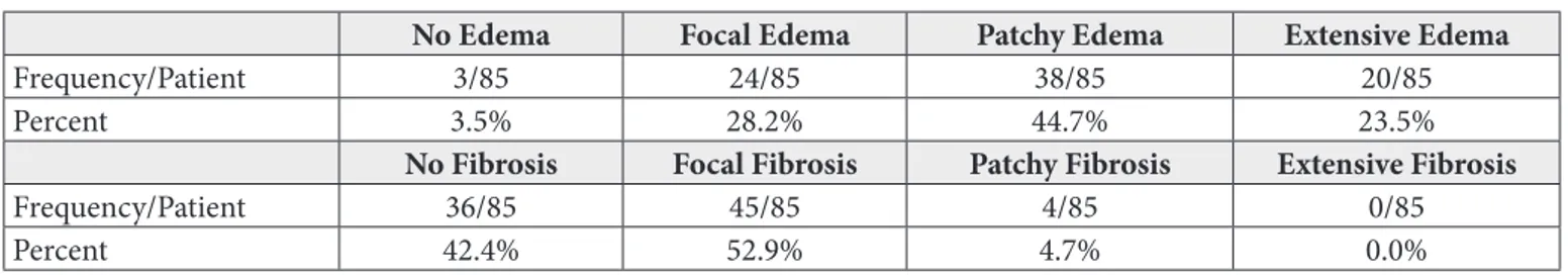
Histopathologically, average rates of eosinophil, neutrophil, lymphocyte, macrophage, mast cells and goblet cells over epithelial cells were obtained respectively as 32.95±38.51 (min. 0 – max. 160), 11.10±22.9 (min. 0 – max. 160), 49.72±34.76 (min. 10 – max. 150), 0.43±0.66 (min. 0 – max. 3), 1.47±1.88 (min. 0 – max. 8), 0.18±0.17 (min. 0 – max. 0.78) (Table I). Fit y-nine (69.4%) patients did not have squamous metaplasia (not present); however, 13 (15.33%) of them were +1 (focal / mild) positive; 10 (11.8%) were +2 (patchy/moderate) positive; and 3 (3.5%) were +3 (extensive/marked) positive. h e other parameters of epithelial data are presented in the table (Table II).
In the statistical comparison of 14 (16.4%) patients with bronchial asthma and 71 (83.6%) without bronchial asthma, there was no statistical signii cance with respect to eosinophil, lymphocyte, neutrophil, macrophage, mast cell and goblet cells / epithelial cells rate (p>0.05). h e comparison of these same groups’ radiological data also showed no statistically signii cant dif erence with respect to the Lund-Mackay scores (p=0.169).
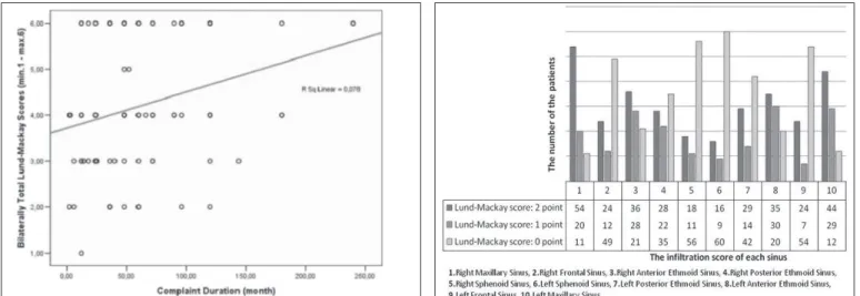
We have observed a positive relationship between the preoperative complaint period and total Lund-Mackay scores (r=0.270, p=0.012) (Figure 2). We have also seen that averages of both anterior and posterior group sinuses’ radiological scores were parallel to average Lund-Mackay
Table I: Average rates of cellular dispersion of NP: At 400X
power the i eld is 0.44 mm x 0.44 mm, yielding an area of
approximately 0.19 mm2 histologically. h e densest cellular
ini ltrate in each case at 400X power was obtained and each cellular marker was counted as an absolute number
Cells Min. Max. Mean Std. Dev.
Eosinophil 0 160 32.95 38.51
Neutrophil 0 160 11.10 22.97
Lymphocyte 10 150 49.72 34.76
Macrophage 0 3 0.43 0.66
Mast cell 0 8 1.47 1.88
Table II: Distribution of subepithelial edema and i brosis were assessed by using a Likert scale from 0 to 3 (0=not present, 1=focal/mild,
2=patchy/moderate, and 3=extensive/marked)
No Edema Focal Edema Patchy Edema Extensive Edema
Frequency/Patient 3/85 24/85 38/85 20/85
Percent 3.5% 28.2% 44.7% 23.5%
No Fibrosis Focal Fibrosis Patchy Fibrosis Extensive Fibrosis
Frequency/Patient 36/85 45/85 4/85 0/85
scores of the nasal cavity (Table III). h is i nding shows that nasal cavity polyps and sinus polyps grow in correlation. In this study it was also observed that anterior group cells were ini ltrated more than posterior group cells (Figure 3). Looking at the Lund-Mackay scoring and average sinus scoring, the neutrophil increase was signii cant. On the other hand, no signii cant dif erence determined for other inl ammatory cells, epithelial cells, sub-mucosal i brosis raise, goblet cell / epithelial cell rate.
We also compared the patients with and without metaplasia based on their pathological examination with respect to other pathological i ndings; and no signii cant result was drawn for eosinophil, lymphocyte, macrophage, mast cell, goblet cell / epithelial cell rate (p>0.05). Preoperative complaint period of the patients with metaplasia was longer (80.11±57.08 months) than of those without metaplasia (55.71±46.88 months) (p=0.03). h e neutrophil amount (16.53±19.87) on the preparation of the patients with metaplasia was also higher than the neutrophil amount (8.71±23.98) on the preparation of the patients without
metaplasia (p=0.001). Considering the metaplasia, we could not specify any meaningful relation between other inl ammatory and allergic cells.
From the radiological aspect, by looking at the polypoid tissue distribution of the patients with and without the squamous metaplasia, neither Lund-Mackay scoring nor anterior-posterior sinus polypoid involvements suggested a signii cance dif erence.
DISCUSSION
NP can be dei ned as chronic inl ammatory disease of a paranasal sinus and especially the nasal mucosa (7). h e most severe form of chronic rhinosinusitis is nasal polyposis (8). A variety of inl ammatory cells may be found inside the polypoid tissue, in the mucus or nasal mucosa. h is study was designed to i nd out the correlation between clinical i ndings, preoperative CT scan’s opacii cation and their histopathologic i ndings.
NP is common between the ages of 40-70 years and is predominant in males with a ratio of 2:1 (15). In our study,
Table III: (a) Dependent Variable: Total Score of Lund-Mackay.(b) Positive correlation between Lund-Mackay score of nasal cavity and
ini ltration of anterior-posterior sinuses by the polyp tissues
Coei cients(a)
Model
Unstandardized Coei cients
Standardized
Coei cients t Sig. 95% Coni dence Interval for B B Std. Error Beta Lower Limit Higher Limit
(Constant)
Anterior Cell’s Mean Score Posterior Cell’s Mean Score
2.760 .261 10.590 .000 2.241 3.278
1.045 .293 .449 3.570 .001 .463 1.627
.433 .254 .215 1.707 .092 -.072 .938
Figure 2: Positive correlation between the complaint duration of
like the literature data, 63.5 % of the patients were male and the mean age was 38.05±16.64 (11-70) years.
NP’s are mainly located in the middle meatus, maxillary sinus, frontal recess; and originate from the nasal mucous membrane ostia in the paranasal sinus (2,16). In this study, regarding Lund-Mackay scores, it is inferred that polyp tissues more ot en ini ltrate the anterior sinuses, maxillary sinuses, and nasal cavity’s ostiomeatal complex areas than other areas.
NP is ot en observed with other disorders such as cystic i brosis, asthma, ASA hypersensitivity, Kartagener’s syndrome, Young syndrome and diabetes mellitus. In this study, 60 (70.6%) patients did not have any kind of systemic disease; however, 10 of them were diagnosed as bronchial asthma, 4 of them had diabetes mellitus, 3 patients had hypertension, 4 of them asthma and hypertension, 3 diabetes mellitus and hypertension, and 1 patient was diagnosed with Kartagener’s syndrome.
h e association between asthma and NP has been revealed since the 19th century (17). h e exact cause of this relation is still not well understood. According to an epidemiologic standpoint, the frequency of polyps ranges between 10% and 15% in asthmatic patients (18). However, the foregoing report shows that the frequency of asthma in the setting of NP is between 30% and 70%. As for our study, only 14 (16.4%) out of 85 patients had asthma. Asthma symptoms were assessed using objective measures, specii cally pulmonary function testing and steroid usage data. Haro et al., by using the Lund-Mackay system in their study, found the average score of 10.9 in the tomographic evaluation of non-asthmatic patients; yet, they found the average score of 17.6 in the tomography of asthmatic patients. h ere was statistical signii cance between asthmatic and non-asthmatic patients (19). h ere was no correlation between the stages of NP and the presence of asthma as also reported by Garrel et al (20). Moreover, our study also did not show clinical and radiological stage dif erence between the patients with and without asthma.
Patients with cystic i brosis and patients with primary cilliary dyskinesia (Kartagener’s syndrome) frequently
develop NP (2). h e polyps associated with these
diseases are not characterized by tissue eosinophilia but characterized by lymphocytes in the tissue and neutrophils in the secretion (21). Only one patient was diagnosed with Kartagener’s syndrome in this study. For this particular patient, opacii cation was observed within all sinuses and the bilateral nasal cavity with the CT scans. Regarding the pathological examination; there were 20 lymphocytes in
every unit square (specii ed in the material and method section). We did not observe other inl ammatory cells in this study. h ere is no study on the comparison of the clinical, radiological, and histopathological data in patients with NP. Soler et al. showed that mucosal eosinophilia correlated with objective disease severity as dei ned by CT for patients with chronic sinusitis. h e other inl ammatory cells did not correlate with CT scans in that study (12). In this study, with respect to Lund-Mackay scores, we found that all inl ammatory cells including eosinophilia were not correlated with CT scanning. Eosinophils are crucial ef ector cells in the inl ammatory reactions associated with asthma, allergic inl ammatory disease, parasitic diseases, granulomatous disorders, i brosis and several sinonasal diseases (7,8). Since all of the patients in our study had NP, we compared the patients according to presence of asthma and there was no dif erence between two groups in terms of eosinophil levels.
Number of neutrophil cells for patients with and without metaplasia dif ered. Neutrophils are the markers found in the higher numbers with acute inl ammatory cases. However, metaplasia may occur as a result of chronic irritation. Considering these facts, it could be inferred that metaplasia occurs in recurrent acute inl ammatory phase rather than chronic period. h e time duration until making the i nal decision for an operation for those patients with metaplasia turned out to be longer than of those patients without having metaplasia. h is result shows that metaplasia comes into existence in a long period of time. We also uncovered that metaplasia does not depend upon Lund-Mackay scores.
Baird et al., in a short period of time such as eight months, showed the existence of atypia in i t een nasal polypectomy specimens containing squamous metaplasia (22). h e study shows that preoperative complaint period of patients with metaplasia is longer than those without metaplasia. Furthermore, the number of neutrophils in cases that have metaplasia was higher than of those without metaplasia. No dysplasia was determined for any preparation. We did not detect any signs of dysplasia in any patient in our study as in this report.
CONCLUSION
period and squamous metaplasia, and the preoperative complaint period and Lund-Mackay scoring. Looking at this result, a meaningful correlation between Lund-Mackay score and metaplasia might be expected to exist; however, no statistically signii cant correlation was identii ed. h is result could also be af ected by the small sample size. No histopathological dif erence was found between polypoid tissues of anterior group and posterior group sinuses.
REFERENCES
1. Dufour X, Bedier A, Ferrie JC, Gohler C, Klossek JM: Dif use nasal polyposis and endonasal endoscopic surgery: Long-term results, a 65-case study. Laryngoscope 2004, 114:1982-1987 2. Mygind N, Dahl R, Bachert C: Nasal polyposis, eosinophil
dominated inl ammation, and allergy. h orax 2000, 55:79-83 3. Assanasen P, Naclerio RM: Medical and surgical management of
nasal polyps. Curr Opin Otolaryngol Head Neck Surg 2001, 9: 27-36
4. Mortuaire G, Bahij J, Maetz B, Chevalier D: Lund-Mackay score is predictive of bleeding in ethmoidectomy for nasal polyposis. Rhinology 2008, 46:285-288
5. Lund VJ, Mackay IS: Staging in rhinosinusitis. Rhinology 1993, 31:183-184
6. Ruhno J, Howie K, Anderson M, Andersson B, Vanzieleghem M, Hitch D, Lapp P, Denburg J, Dolovich J: h e increased number of epithelial mast cells in nasal polyps and adjacent turbinates is not allergy-dependent. Allergy 1990, 45:370-374
7. Shin SH, Lee SH, Jeong HS, Kita H: h e ef ect of nasal polyp epithelial cells on eosinophil activation. Laryngoscope 2003, 113:1374-1377
8. Eliashar R, Levi-Schaf er F: h e role of the eosinophil in nasal diseases. Curr Opin Otolaryngol Head Neck Surg 2005, 13: 171-175
9. Wise SK, Ahn CN, Schlosser RJ: Localized immunoglobulin E expression in allergic rhinitis and nasal polyposis. Curr Opin Otolaryngol Head Neck Surg 2009, 17:216-222
10. Morinaka S, Nakamura H: Inl ammatory cells in nasal mucosa and nasal polyps. Auris Nasus Larynx 2000, 27:59-64
11. Van Drunen CM, Reinartz S, Wigman J, Fokkens WJ:
Inl ammation in chronic rhinosinusitis and nasal polyposis. Immunol Allergy Clin North Am 2009, 29:621-629
12. Soler ZM, Sauer DA, Mace J, Smith TL: Relationship between clinical measures and histopathologic i ndings in chronic rhinosinusitis. Otolaryngol Head Neck Surg 2009, 141:454-461 13. Basu S, Georgalas C, Kumar BN, Desai S: Correlation
between symptoms and radiological i ndings in patients with chronic rhinosinusitis: An evaluation study using the Sinonasal Assessment Questionnaire and Lund-Mackay grading system. Eur Arch Otorhinolaryngol 2005, 262:751-754
14. Moghadasi H, Saneei Taheri M, Vazir Nezami M, Mohammadpour H, Jalali AH, Delavari H, Peyravi Chashnasar M: Association between clinical symptoms and CT i ndings in chronic rhinosinusitis. Iran J Radiol 2008, 5:145-149
15. Dursun E, Samim E, Korkmaz H, Bayızü, Uygur K, Akmansu H, Özeri C: Nazal polipozisli olgularda endoskopik sinüs cerrahisi. KBB ve BBC Dergisi 1998, 6:71-80
16. Larsen PL, Tos M: Origin of nasal polyps: An endoscopic autopsy study. Laryngoscope 2004, 114:710-719
17. Uri N, Cohen-Kerem R, Barzilai G, Greenberg E, Doweck I, Weiler-Ravell D: Functional endoscopic sinus surgery in the treatment of massive polyposis in asthmatic patients. J Laryngol Otol 2002, 116:185-189
18. Batra PS, Kern RC, Tripathi A, Conley DB, Ditto AM, Haines GK 3rd, Yarnold PR, Grammar L: Outcome analysis of endoscopic sinus surgery in patients with nasal polyps and asthma. Laryngoscope 2003, 113:1703-1706
19. Haro JI, Gavioli F, Junior VM, Crespo CC: Clinical aspects of patients with nasal polyposis. Int Arch Otorhinolaryngol 2009, 13:259-263
20. Garrel R, Gardiner Q, Khudjadze M, Demoly P, Vergnes C, Makeief M, Guerrier B, Crampette L: Endoscopic surgical treatment of sinonasal polyposis-medium term outcomes (mean follow-up of 5 years). Rhinology 2003, 41:91-96
21. Sorensen H, Mygind N, Tygstrup I, Winge Flensborg E: Histology of nasal polyps of dif erent etiology. Rhinology 1977, 15:121-128 22. Baird AR, Hilmi O, White PS, Robertson AJ: Epithelial atypia