Effects of vacuum sealing drainage on the treatment
of cranial bone-exposed wounds in rabbits
X.J. Chen*, S. Liu*, G.Z. Gao, D.X. Yan and W.S. Jiang
Department of Burn and Plastic Surgery, the 253rd Hospital of People’s Liberation Army, Xincheng District, Hohhot, Inner Mongolia, China
Abstract
This study was designed to assess the efficacy of vacuum sealing drainage (VSD) on skull exposure wounds in rabbits and to investigate the underlying mechanism of the process. Full-thickness excisional circular wounds 22 cm with or without
periosteum involvement were created in 88 New Zealand white rabbits (mean body weight: 3.0±0.65 kg). Animals were randomly divided into 4 groups: periosteum-intact wounds treated with traditional dressing (p+control), periosteum-intact wounds treated with VSD (p+VSD), periosteum-lacking wounds treated with traditional dressing (p–control) and periosteum-lacking wounds treated with VSD (p–VSD). The wounds treated with traditional dressing were covered with Vaseline gauze, while VSD treatment was accompanied with continuous –120 mmHg pressure. Finally, wound tissues were harvested for analysis of hydroxyproline content and histologic detection. VSD hastened the wound healing process significantly (Po0.05) compared to the corresponding control groups. VSD alleviated the inflammation reaction, accelerated re-epithelialization and facilitated the organization of collagenfibers into neat rows. During the wound healing process, the hydroxyproline content increased overtime [i.e., postoperative days (POD) 7, POD 10 and POD 15] in all four groups, and it peaked in the p+VSD group. VSD also promoted angiogenesis via increasing number and quality of collagen. We concluded that VSD can promote healing in bone-exposed wounds via increasing hydroxyproline content and vessel density, reducing inflammatory responses and generating ordered collagen arrangement.
Key words: Cranial bone-exposed wound; Vacuum sealing drainage; Periosteum; Wound healing; Efficacy
Introduction
The number of patients with large-area soft tissue defects accompanied by bone exposure has been increas-ing due to frequently-occurrincreas-ing road accidents, high-energy explosives, deep burns, work-related injuries and tumor resection (1). Such wounds are often associated with severe avulsion, infection, and poor blood supply, there-fore requiring a long healing time. The traditional wound dressing methods often cause complications such as wound infection, non-healing bone fracture, non-union, osteonecrosis, osteomyelitis andfistula formation. Serious infections can lead to amputation. Hence, it is critical to resolve certain problems, including prevention of patho-logic microorganisms, temporary cover of wound surface, among others.
The vacuum sealing drainage (VSD) technique (2) has been widely applied for effectively treating diverse wound surfaces, especially for injuries that could not achieve debridement and coverage in a one-stage procedure. The technique involves usage of polyvinyl alcohol/alginate
blend foam, drainage tube, semi-permeable membranes (3) and a negative pressure source. Gases generated from decomposition of necrotic tissues within the wound sur-face can smoothly infiltrate into outer space of the semi-permeable membrane, yet air and bacteria outside the membrane cannot enter the wound surface successfully. Nonetheless, the healthy skin around the wound surface can breathe in a normal way.
Overall, VSD can improve partial blood circulation, thus reducing edema and bacteria (4), promoting growth of granulation tissue and enhancing the healing rate. The inhibitory effects on bacteria reproduction could largely result from the negative pressure of VSD, which creates a relatively anoxic environment on the wound surface, without affecting the surrounding healthy tissues. Owing to these merits of VSD, surgeons have broadly accepted VSD for treatment of soft tissue trauma (1,5,6) and bone-exposed wounds (7–9). Nonetheless, few comprehensive analyses have investigated the effectiveness and inherent
Correspondence: X.J. Chen:<[email protected]> *These authors contributed equally to this study.
mechanism of VSD treatment for bone-exposed wounds. Thus, we used the rabbit skull-exposed wound model to compare the efficacy of VSD with traditional dressing therapy by histological and vascular density analyses, and hydroxyproline content.
Material and Methods
Grouping of animals
A total of 88 New Zealand white rabbits (mean body weight: 3.0±0.65 kg) were provided by the 253rd Hospital of People’s Liberation Army of China (PLA). The rabbits were randomly divided into 4 groups of periosteum-intact traditional dressing therapy (p+control), periosteum-intact VSD-treated (p+VSD), periosteum-lacking traditional dressing therapy (p–control), and periosteum-lacking VSD-treated (p–VSD). The experiment protocol for this study was approved by the Ethics Committee for Animal Research of PLA 253rd Hospital SCXK-(Huhhot) 2015-0021.
Model preparation
After anesthetizing the rabbits through intra-peritoneal injection of pentobarbital sodium (concentration: 30 mg/kg), their heads were shaved and smeared with sodium sulfide (6%). Subsequently, rabbits in the prone position were disinfected with 75% ethyl alcohol. Specific operations relevant to successive removal of skin, subcutaneous tissue and galea aponeurotica, as well as exposure of periosteum were performed according to a previously reported investigation (10). Finally, full-thickness wounds (22 cm) were created in either periosteum-intact or periosteum-lacking forms.
In the traditional dressing therapy group, wounds were covered with Vaseline gauze every other day. In the VSD group, VSD foam (Weisidi Medical Technology Corpora-tion, China) was cut43 mm beyond the wound margins. Then, we inserted a porous silica gel tube around the foam, and connected it to a negative pressure device (Weisidi Medical Technology Corporation). The negative pressure source was switched on, and a biological semi-permeable membrane (Smith & Nephew Corporation, UK) was pasted. The device was set to negative pressure (i.e.,
–120 mmHg) continuously for 16 days (i.e., the 15th postoperative day (POD) since the surgery), and the seal of the device was strictly checked every 6 h. Vacuum sealing drainage was conducted daily, and healing was measured over time. The wound tissues were harvested on POD 7, 10, and 15 from at least three animals for the detection of hydroxyproline content and histological analysis.
Gross assessment of wounds and histological analysis
Daily photographs were taken to visually track wound healing progress. The wounds were traced on transparent
graph paper and then their sizes were analyzed with Image-Pro Plus 6.0 software (Version X; Media Cyber-netics, USA). The rate of closure was calculated as daily change of the wound area relative to the initial wound area. Wound healing time was defined as the time required for epithelial healing.
For histologic analyses, wounds were harvested within a 2–3-mm rim from rabbits that were euthanized at POD 7, 10, and 15. Half of the wounds were placed in 10% formalin, while the other half were frozen in liquid nitrogen and stored at–80°C. The formalin-fixed wounds were embedded in paraffin and cut into 5-mm sections, then placed onto glass slides and stained with hematoxy-lin and eosin (H&E) (Sigma-Aldrich Corporation, Germany). Each slide was measured objectively and automatically at least three times.
Collagen quantification
The remaining tissues were stained with Masson’s trichrome (Sigma-Aldrich) to visualize collagen fibers. As hydroxyproline is a basic constituent of collagen struc-ture, its content can serve as indicator of collagen synthesis. Hydroxyproline contents were analyzed on POD 7, 10, and 15, in line with manufacturers’instructions of hydroxy-proline analysis kit (Jiancheng Technology Corporation, China, catalog number: A030-2). To be specific, tissue samples were successively mixed with 10 mM CuSO4 (1 mL), 2.5 M NaOH (1 mL), and 6% H2O2(1 mL). Sub-sequently, the mixture was incubated at 80°C for 5 min with frequent vigorous shaking, and then 3 M H2SO4 (4 mL) was added with agitation. Finally, after addition of 5% p–dimethyl amino benzaldehyde (2 mL), the mixed solution was incubated at 60°C for 15 min. The absorbance (optical density) was evaluated at 550 nm under the microplate reader (Tecan Genios Corpora-tion, Australia).
Statistical analysis
The statistical analyses were performed using SPSS17.0 software (SPSS, USA). The measurement data are reported as means±SD, and statistical comparisons
were performed using either unpaired Student’st-test or multivariate analysis of variance (ANOVA). The P value was considered statistically significant when it was less than 0.05.
Results
VSD therapy accelerated wound healing
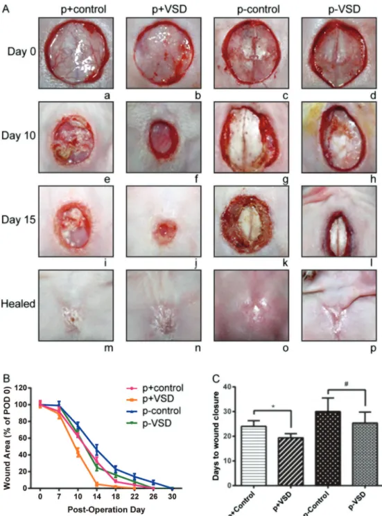
Whether the wounds were periosteum-intact or peri-osteum-lacking, the wounds treated with VSD exhibited a significantly faster healing rate than those treated with traditional dressing therapy (Figure 1). During the healing process, wounds of p+control and p–control groups displayed purulent discharge, whereas wounds of p+VSD and p–VSD groups were cleaner. Furthermore, the cortical bone surfaces of periosteum-lacking wounds (i.e., p–control and p–VSD groups) looked smooth and dry, while those of the periosteum-intact wounds (i.e., p+control and p+VSD groups) appeared bright red and moist (Figure 1A).
Application VSD significantly accelerated wound heal-ing at each time point of POD 7 to POD 15 compared with utilization of traditional dressing. The periosteum-intact group was also associated with more desirable healing rate compared to the periosteum-lacking group, though eventually the wound healing rates were all shown as 100% (Figure 1B).
It took only 19.40±1.65 days for wounds in the p+VSD group to heal completely, which was much shorter than
24.00±2.31 days in the p+control group (Po0.05). Obviously, the periosteum-lacking wounds demanded a longer healing period, with 30.00±5.50 days in the p–control group. Nonetheless, p–VSD shortened the wound healing time to 25.40±4.43 days (Figure 1C). Interestingly, there was no significant difference of healing time between p–VSD and p+control groups (P40.05).
Histological observations
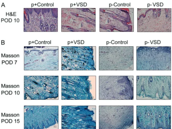
In accordance with results of HE staining, wounds treated with traditional dressing therapy showed stronger inflammatory responses and more poorly re-epithelialized epidermis than wounds treated with VSD on POD 10 (Figure 2A). Improved collagen organization was observed in the VSD-treated group as early as POD 7, compared with traditional dressing therapy groups. In spite of the time points, the VSD-treated groups presented more well-organized collagen fibers and faster fibroblast prolifera-tion than the tradiprolifera-tional dressing therapy-treated groups (Figure 2B).
Determination of hydroxyproline content
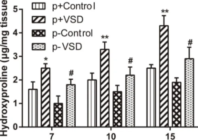
VSD treatments generally induced higher levels of hydroxyproline than traditional treatment on POD 7, 10 and 15
(Po0.05). The hydroxyproline level of the p+VSD group peaked, while the p–control group bottomed (Figure 3). During the period from POD 7 to POD 15, hydroxyproline content showed a significant increasing trend after treat-ment with VSD (Po0.05), yet no significant differences of
hydroxyproline levels were found after traditional treat-ments (P40.05).
VSD-treated wounds showed increased blood vessels The wounds treated with VSD were correlated with significantly higher vessel density than those treated with traditional dressing on POD 7 (Po0.05; Figure 4A). Similarly, the VSD-treated wounds possessed larger calibers and more well-developed vessels, compared with traditionally treated wounds on POD 10 and POD 15 (Figure 4A and B). From POD 7 to POD 10, a significant increasing tendency of vessel density was only found in wounds of the p+control group (Po0.05).
Discussion
It is widely known that wound surfaces, though receiving treatments of debridement and active anti-infection, are still readily subjected to severe infection. For example, parental antibiotics usually do not reach effective bactericidal concentration within the local wound microenvironment.
The VSD technique has been expanded dramati-cally since it was popularized in 1997 by Argenta and Morykwas (2) and further studied in 2010 by Kairinos et al. (13). The application of VSD has been widely recognized as effective in treating soft tissue trauma, however, usage of VSD for treatment of bone-exposed wounds has rarely Figure 3. Quantification of hydroxyproline content during the
course of wound healing, on post-operative days 7, 10, and 15. The vacuum sealing drainage (VSD)-treated groups had sig-nificantly higher levels of hydroxyproline content than controls (traditional treatment). p+: periosteum-intact; p–: periosteum-lacking. Data are reported as means±SD. *Po0.05, **Po0.01 compared to p+Control,#Po0.05 compared to p–Control.
been comprehensively and thoroughly investigated. For instance, skin graft combined with VSD has been employed to treat large soft tissue defects and bone exposure in the lower leg, which might eliminate the need for amputation and complex surgeries (1). However, that research was limited to the clinical setting, and we designed rabbit models to explore the underlying mechan-isms of VSD therapy. Although the rabbit bone-exposed wound is a study model easy to reproduce and inexpen-sive, the size of the wound is finite. Thus, we would require larger animal models to investigate whether VSD therapy could promote wound healing without skin grafting and skinflap transplantation (14).
It was documented that VSD therapy could improve removal of edema fluid, blood flow, granulation tissue formation and bacterial clearance from wounds (10). Our study consistently showed that wounds undergoing treatment with VSD were clean and without exudate. Moreover, the periosteum (osteogenic tissues surrounding bones) contains multiple stem cells that secrete large quantities of VEGF and BMP2, thereby playing critical roles in boosting bone healing (15–17). In our investiga-tion, the wound healing time showed no notable distinction between p–VSD and p+control groups, further suggest-ing that VSD therapy could enhance wound healsuggest-ing. The fact that VSD-treated wounds had little inflammatory cell infiltration and well-organized collagen fibers also confirms that VSD therapy could inhibit inflammatory responses and could enhance formation of epidermis.
Hydroxyproline is as a basic constituent of collagen structure, implying that its increased concentration might contribute to improved wound healing activity (18,19). Our results also showed that VSD therapy augmented hydroxy-proline concentration both in the periosteum-lacking and periosteum-intact groups, and p+VSD group possessed the highest level of hydroxyproline. In fact, hydroxyproline was crucial at specific moments of healing, for it maintained the equilibrium between synthesis and degradation of extra-cellular matrix proteins, achieving remodeling of the wounds and reduced formation of scars (20).
Rat wounds treated with negative pressure showed relatively high MVD on day 3, which could be due to the negative pressure functioning as promotor of angio-genesis (21). Also with a rodent model, Jacobs et al. (10) concluded that treatment with vacuum would advance formation of blood vessels by POD 3. In our study, VSD treatment increased blood vessel density in the early days of wound healing through increasing cell number or improving cell quality.
In conclusion, VSD promoted the healing of bone-exposed and periosteum-lacking wounds in a rabbit model by increasing hydroxyproline content and MVD, reducing inflammation, and inducing ordered collagen arrangements.
Acknowledgments
This study is supported by Army Medical and Health Care Research Fund (No. CBJ15J007).
References
1. Qu J, Yan R, Wang L, Wu J, Cao L, Zhao G, et al. Free dermatoplasty combined with vacuum sealing drainage for the treatment of large-area soft tissue defects accompanied by bone exposure in the lower leg.Exp Ther Med2013; 5: 1375–1380, doi: 10.3892/etm.2013.999.
2. Argenta LC, Morykwas MJ. Vacuum-assisted closure: A new method for wound control and treatment: Clinical experi-ence. Ann Plast Surg 1997; 38: 563–576, doi: 10.1097/ 00000637-199706000-00002.
3. Semsarzadeh NN, Tadisina KK, Maddox J, Chopra K, Singh DP. Closed incision negative-pressure therapy is associated with decreased surgical-site infections: A meta-analysis. Plast Reconstr Surg 2015; 136: 592–602, doi: 10.1097/ PRS.0000000000001519.
4. Milcheski DA, Chang AA, Lobato RC, Nakamoto HA, Tuma P Jr, Ferreira MC. Coverage of deep cutaneous wounds using dermal template in combination with negative-pres-sure therapy and subsequent skin graft.Plast Reconstr Surg Glob Open 2014; 2: e170, doi: 10.1097/GOX.000000000 0000108.
5. Li RG, Ren GH, Tan XJ, Yu B, Hu JJ. Free flap transplantation combined with skin grafting and vacuum seal-ing drainage for repair of circumferential or sub-circumferential soft-tissue wounds of the lower leg.Med Sci Monit2013; 19: 510–517, doi: 10.12659/MSM.883963.
6. Li RG, Yu B, Wang G, Chen B, Qin CH, Guo G, et al. Sequential therapy of vacuum sealing drainage and free-flap transplantation for children with extensive soft-tissue defects below the knee in the extremities.Injury2012; 43: 822–828, doi: 10.1016/j.injury.2011.09.031.
7. Bohac M, Mikusz K, Fedeles J Sr. Application of negative pressure wound therapy in scalp reconstruction.Bratisl Lek Listy2015; 116: 719–721.
8. Chiummariello S, Guarro G, Pica A, Alfano C. Evaluation of negative pressure vacuum-assisted system in acute and chronic wounds closure: our experience.G Chir2012; 33: 358–362.
9. Gumus N. Negative pressure dressing combined with a traditional approach for the treatment of skull burn. Niger J Clin Pract 2012; 15: 494–497, doi: 10.4103/1119-3077. 104539.
10. Jacobs S, Simhaee DA, Marsano A, Fomovsky GM, Niedt G, Wu JK. Efficacy and mechanisms of vacuum-assisted closure (VAC) therapy in promoting wound healing: a rodent model.J Plast Reconstr Aesthet Surg2009; 62: 1331–1338, doi: 10.1016/j.bjps.2008.03.024.
12. Goddard JC, Sutton CD, Berry DP, O’Byrne KJ, Kockelbergh RC. The use of microvessel density in assessing human urological tumours.BJU Int2001; 87: 866–875, doi: 10.1046/j.1464-410x.2001.02181.x. 13. Kairinos N, Solomons M, Hudson DA. The paradox of
negative pressure wound therapy--in vitro studies.J Plast Reconstr Aesthet Surg 2010; 63: 174–179, doi: 10.1016/ j.bjps.2008.08.037.
14. Zhang YG, Yang Z, Zhang H, Liu M, Qiu Y, Guo X. Negative pressure technology enhances bone regeneration in rabbit skull defects. BMC Musculoskelet Disord 2013; 14: 76, doi: 10.1186/1471-2474-14-76.
15. Bielby R, Jones E, McGonagle D. The role of mesenchymal stem cells in maintenance and repair of bone.Injury2007; 38 (Suppl 1):S26–S32, doi: 10.1016/j.injury.2007.02.007. 16. Mattar T, Friedrich PF, Bishop AT. Effect of rhBMP-2 and
VEGF in a vascularized bone allotransplant experimental model based on surgical neoangiogenesis. J Orthop Res 2013; 31: 561–566, doi: 10.1002/jor.22277.
17. Deckers MM, van Bezooijen RL, van der Horst G, Hoogendam J, van Der Bent C, Papapoulos SE, et al. Bone morphogenetic proteins stimulate angiogenesis through osteoblast-derived vascular endothelial growth factor A.Endocrinology2002; 143: 1545–1553, doi: 10.1210/endo.143.4.8719.
18. Upadhyay A, Chattopadhyay P, Goyary D, Mitra Mazumder P, Veer V. Ixora coccinea enhances cutaneous wound healing by upregulating the expression of collagen and basic fibroblast growth factor. ISRN Pharmacol 2014; 2014: 751824, doi: 10.1155/2014/751824.
19. Panda V, Thakur T. Wound healing activity of the infl ores-cence of Typha elephantina (Cattail). Int J Low Extrem Wounds2014; 13: 50–57, doi: 10.1177/1534734613516859. 20. Raghavan KV, Babu M, Rajaram R, Purna Sai K. Efficacy of frog skin lipids in wound healing.Lipids Health Dis 2010; 9: 74, doi: 10.1186/1476-511X-9-74.