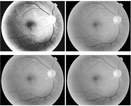
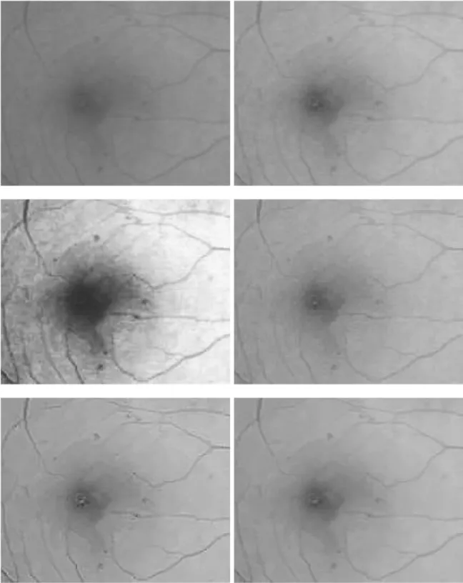
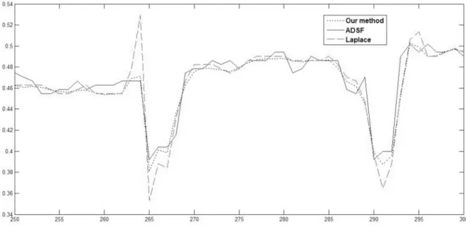
Retinal Image Enhancement Using Robust Inverse Diffusion Equation and Self-Similarity Filtering.
Texto
Imagem


Documentos relacionados
The filtering procedures tested for the removal of the multiple reflections were: inverse filtering for least-squares using the software GRADIX; Wiener predictive filtering using
Axial T1- weighted image with gadolinium shows enhancement without mass effect in left lenticular nucleus, suggestive of subacute ischemic injury (B). Axial diffusion-weighted
a) VEGFs and Retinopathy: Retinal neovascularisation is the pathognomonic feature of all the ischaemic retinal diseases namely diabetic retinopathy, retinal
In recursively separated and weighted histogram equalization (RSWHE) method preserves the image brightness and enhances the image contrast. RSWHE first segments the histogram
If figures have the letter B symmetry, then the top is a mirror image of the bottom, but the right is not a mirror image of the left. In so doing, the top and the bottom side of
Figure 6A shows some simple geometric shapes (cross, triangle, square and wheel) with different sizes on a step-edge background with 30% additional Gaussian noise. Figures 6B – 6E
Barman et al., “Automatic detection of diabetic retinopathy exudates from non-dilated retinal images using mathematical morphology methods,” Computer Medical Imaging and Graphics
The present study showed that the major retinal complications in transplanted patients were deterioration of diabetic retinopathy (or new diabetic retinopathy due to onset


