Pathogen (
Metarhizium acridum
) Enables It to Infect
Caterpillars
Sibao Wang1¤, Weiguo Fang1, Chengshu Wang2, Raymond J. St. Leger1*
1Department of Entomology, University of Maryland, College Park, Maryland, United States of America,2Institute of Plant Physiology and Ecology, Shanghai Institutes for Biological Sciences, Chinese Academy of Sciences, Shanghai, China
Abstract
An enduring theme in pathogenic microbiology is poor understanding of the mechanisms of host specificity.Metarhiziumis a cosmopolitan genus of invertebrate pathogens that contains generalist species with broad host ranges such asM. robertsii (formerly known asM. anisopliaevar.anisopliae) as well as specialists such as the acridid-specific grasshopper pathogenM. acridum. During growth on caterpillar (Manduca sexta) cuticle,M. robertsiiup-regulates a gene (Mest1) that is absent inM. acridumand most other fungi. DisruptingM. robertsii Mest1reduced virulence and overexpression increased virulence to caterpillars (Galleria mellonellaand M. sexta), while virulence to grasshoppers (Melanoplus femurrubrum) was unaffected. WhenMest1was transferred toM. acridumunder control of its nativeM. robertsiipromoter, the transformants killed and colonized caterpillars in a similar fashion toM. robertsii. MEST1 localized exclusively to lipid droplets inM. robertsiiconidia and infection structures was up-regulated during nutrient deprivation and had esterase activity against lipids with short chain fatty acids. The mobilization of stored lipids was delayed in theMest1disruptant mutant. Overall, our results suggest
that expression of Mest1 allows rapid hydrolysis of stored lipids, and promotes germination and infection structure
formation byM. robertsiiduring nutrient deprivation and invasion, whileMest1expression inM. acridumbroadens its host range by bypassing the regulatory signals found on natural hosts that trigger the mobilization of endogenous nutrient reserves. This study suggests that speciation in an insect pathogen could potentially be driven by host shifts resulting from changes in a single gene.
Citation:Wang S, Fang W, Wang C, St. Leger RJ (2011) Insertion of an Esterase Gene into a Specific Locust Pathogen (Metarhizium acridum) Enables It to Infect Caterpillars. PLoS Pathog 7(6): e1002097. doi:10.1371/journal.ppat.1002097
Editor:Barbara Jane Howlett, University of Melbourne, Australia
ReceivedFebruary 14, 2011;AcceptedApril 15, 2011;PublishedJune 23, 2011
Copyright:ß2011 Wang et al. This is an open-access article distributed under the terms of the Creative Commons Attribution License, which permits unrestricted use, distribution, and reproduction in any medium, provided the original author and source are credited.
Funding:This work was supported by NSF Grant MCB-0542904 and USDA-CSREES grant 2010-65106-20580. The funders had no role in study design, data collection and analysis, decision to publish, or preparation of the manuscript.
Competing Interests:The authors have declared that no competing interests exist. * E-mail: [email protected]
¤ Current address: Department of Molecular Microbiology and Immunology, Johns Hopkins School of Public Health, Baltimore, Maryland, United States of America.
Introduction
An enduring theme in pathogenic microbiology is poor under-standing of the mechanisms of host specificity. That is, what factors limit a pathogens host range, how is host specificity linked with virulence and what changes in pathogens or hosts can open new host ranges? These are fundamental questions that relate both to the co-evolution of host susceptibility and pathogen virulence, as well as to factors underlying host switching and the emergence of new pathogens that originate in different host species. The molecular mechanisms controlling host selectivity in fungi are particularly poorly understood. A number of plant pathogenic fungi produce secondary metabolites with biological specificities that correspond with the host range of the producing fungi [1,2], and small secreted proteins produced by the pathogen can trigger resistance in some plants limiting host range [3]. However, to date these have not been found in animal pathogenic fungi, many of which seem to be broadly opportunistic. This has allowed resear-chers on emergent human pathogenic fungi to employ insects as model systems [4]. Most human pathogens are not normally transmitted between insect hosts. However, specialization to
entomopathogenicity is a major fungal lifestyle with ,1000
known species that can be highly infectious. In contrast to the opportunistic pathogens, many of these have evolved narrow host ranges by as yet unknown mechanisms.
Metarhiziumis a cosmopolitan genus of Ascomycetes (class Sor-dariomycetes) comprising species that exhibit varied lifestyles. Metarhizium robertsii(formerly known asM. anisopliaevar.anisopliae [5]) is a generalist able to infect hundreds of insect species. It has been at the forefront of efforts to develop biocontrol alternatives to chemical insecticides in agricultural and human disease-vector control programs [6–8].M. robertsiihas also been used to study the interactions between invertebrate model hosts and pathogenic fungi as host innate immune responses are broadly conserved across many phyla [9]. In contrastM. acridumis a specialist with a narrow host range for certain locusts and grasshoppers [10]. This specificity is one of the reasons it is being mass produced as an environmentally safe alternative to pesticides [10–12].
quantity are not conducive for differentiation of infection stru-ctures. On a host, however, apical elongation terminates and germ tubes produce infection structures, called appressoria, which pro-mote the localized production of cuticle degrading enzymes and also build up turgor providing a mechanical component to pe-netration [13–15]. The formation of appressoria by broad host range strains ofM. robertsiisuch as ARSEF2575 (Mr2575) can also be induced efficiently by low levels of complex nitrogenous nutrients [13]. However, pathogens with a narrow host range such asM. acridumARSEF324 (Ma324) ( = CSIRO FI 485: the active ingredient of ‘Green Guard’) germinate poorly under these conditions and only produce abundant appressoria in the presence of a lipid extract from host insects [16].
M. robertsiiMr2575 sharply up-regulates theMest1gene when germinating on insect cuticles [17]. This gene is absent in Ma324 [18], which suggests that it is unlikely to have an essential function in the relatedM. robertsii but we speculated that it might have a niche role in pathogenicity, perhaps facilitating an opportunistic life-style. In this study, we functionally characterized MEST1 and demonstrated that inserting Mest1intoM. acridum is sufficient to expand its host range to include lepidopterans.Mest1is thus the first gene identified in an entomopathogenic fungus that encodes a determinant of specificity and is to our knowledge the first example where a single metabolic protein assumes such a crucial role for host selectivity in a animal pathogenic fungus. We speculate that speciation in insect pathogens can be driven by host shifts that become fixed in populations due to the gain or loss of a pathogen gene that confers wide host specificity.
Results
Gene cloning, molecular characterization and gene disruption ofMest1
The M. robertsii Mest1 is 1188 bp long and lacks introns. It encodes a predicted protein of 395 amino acids, with a deduced molecular weight of 42244 Da and a pI of 5.34. The SignalP 3.0 program (http://www.cbs.dtu.dk/services/SignalP/) revealed no signal sequence suggesting that MEST1 is a cell-bound protein. MEST1 contains the sequence GGS341VG which conforms to the
motif G-X1-S-X2-G, commonly observed in serine esterases and many lipases [19–21]. According to M. robertsii genome sequence data [22],Mest1(MAA_03283) is downstream of three secreted cuticle degrading subtilisins (Pr1F (MAA_03280), Pr1E (MAA_03281) and a subtilisin-like protease (MAA_03282). The M. robertsii genome also contains a paralog (MAA_08059) with 42% identity to MEST1 that is downstream of three hypothe-tical proteins and upstream of a glutathione S-transferase. The genome of M. acridum strain CQMa 102 lacks an ortholog of MEST1 but contains a sequence (MAC_02852) with 90.2% iden-tity to MAA_08059 (the median sequence ideniden-tity of orthologs is 89.8%). MAC_02852 is downstream of four hypothetical proteins and upstream of glutathione S-transferase indicating that it is syn-tenic with its ortholog MAA_08059.
Homologs to MEST1 and MAA_08059 were identified in four other ascomycete fungi, but only the relatedNectria haematococcahad a sequence (EEU_38198) that was highly similar (82% identity) to MEST1. The relatedGibberella zeae(XP_380200) as well asAspergillus niger (XP_001397035) andPenicillium chrysogenum (XP_002567997) contain single copy sequences resembling MAA_08059 (Figure 1). The functions of these genes have not been reported and contain a putative penicillin-binding domain characteristic ofb-lactamase class C proteins. Homologs of MEST1 were absent inNeurospora crassa, Magnaporthe grisea, Schizosaccharomyces japonicus, Trichoderma harzianum and T. reesei, as well as Basidiomycetes, Zygomycetes and Chytridiomycetes. However, MEST1 shows up to 42% identity with bacterial sequences, including known esterases [19]. A phylo-genetic tree (Figure 1) confirmed that MEST1 and the fungal homo-logs formed a separate well supported clade distinct from bacterial clades containing actinomycete and pseudomonad sequences.
MEST1 regulates virulence and appressorial differentiation inMetarhizium robertsii
To study the function of Mest1, Mest1null mutants (DMest1), were generated in Mr2575 by homologous replacement (Figure 2 and Table 1). Pathogenicity assays againstGalleria mellonella and Manduca sexta caterpillars revealed a significant reduction in mortality and speed of kill by DMest1 relative to wild type M. robertsiiMr2575 (Figure 3A and 3B), while virulence against acridid grasshoppers (Melanoplus femurrubrum) was unaltered (Figure 3C). To demonstrate that the altered phenotype ofDMest1 was speci-fically due to gene inactivation, theMest1gene was reintroduced intoDMest1 in single copy. Six isolates of the resulting comple-mented strain (Mest1-Com) infectedGalleriain an identical fashion as the wild type indicating successful complementation. Overex-pression ofMest1under control of the constitutive glyceraldehyde-3-phosphate dehydrogenase (gpd) promoter (Mr2575-gpd::Mest1) resulted in a significant increase in virulence against both cater-pillars (Manduca and Galleria) and grasshoppers (Figure 3C). Mr2575 transformed with an additional copy of Mest1 under control of its native promoter increased pathogenicity to lepidop-terans but not grasshoppers, suggesting thatMest1is not activated by the wild type fungus on grasshoppers.
To elucidate whether Mest1is needed for developmental pro-cesses, wild type Mr2575,DMest1 and Mest1-Com strains were grown on SDA (Sabouraud dextrose agar) or SDB (Sabouraud dextrose broth) (nutrient rich conditions), in 0.01% YE (Yeast extract) plusManducacuticular lipids (0.25 mg/ml) and on ground grasshopper or Manduca cuticles. There was no significant diffe-rence in sporulation, germination and growth rates betweenM. robertsii Mr2575,DMest1 and Mest1-Com on SDA or SDB, in-dicating thatMest1does not facilitate these processes in nutrient rich conditions. Germination, germ tube formation and appres-sorial formation by Mr2575 on intact grasshopper andManduca Author Summary
cuticles were similar (Figure 4). The germination rate ofDMest1 was significantly (P,0.01) lower than that of wild typeM. robertsii in 0.01% YE with or withoutManducacuticular lipids, and 24 h post-inoculation only 2.761.5 and 26.562.4% ofDMest1 germ-lings had appressoria in YE and YE+lipids, respectively, as compared to 25.561.5 (YE) and 81.663.2% (YE+lipids) of wild type Mr2575 and 24.761.2% (YE) and 80.763.5% (YE+lipids) of complemented strain Mest1-Com (Table 2). Furthermore,DMest1 appressoria (3.660.1mm63.960.8mm) were significantly smaller (P,0.01) than those of the wild type (4.160.2mm612.760.4mm) on insect cuticles (Figure 4A and 4B). Conversely, overexpression ofMest1in Mr2575-gpd::Mest1 resulted in multiple lobed appre-ssoria on branched hyphae (Figure 4C).
Heterologous expression ofMetarhizium robertsiiMEST1 enablesM. acridumto infect caterpillars
Wild typeM. acridumMa324 forms appressoria in locust cuticle lipid extracts and on locust wings [16]. We expressed a 2.7 kb clone encoding M. robertsii Mest1under native control in Ma324 and studied its impact on pathogenicity and growth on cater-pillar cuticle lipid extracts. Compared to the wild type Ma324, Ma324-Mest1 conidia exhibited significantly faster germination in Manduca cuticle lipids with or without YE (P,0.01). Approxi-mately 7% of Ma324-Mest1 germlings produced appressoria in Manduca lipids compared to 35% of germlings in lipids+YE (Table 3). In contrast, wild type Ma324 did not form appressoria
in either YE orManduca lipid extracts, and ,10% of germlings formed appressoria inManducalipid extracts+YE (Table 3). These appressoria were atypically small (2.860.2mm63.660.7mm), whereas Ma324-Mest1 formed compound appressoria (3.96 0.3mm610.260.6mm) at the end of germ tubes similar to those produced by Mr2575 (Figure 4D). Neither wild type Ma324 nor Ma324-Mest1 formed appressoria in water or YE, even though there was hyphal growth in the latter (Table 3). Thus heterologous expression ofMest1byM. acridumis not sufficient by itself to trigger differentiation of appressoria.
Unlike wild type Ma324, Ma324-Mest1 produced appressoria within 24 hours of inoculation onto G. mellonella and M. sexta cuticles (Figure 4F and 4G). Pathogenicity assays showed that Ma324-Mest1 killsM. sextaand G. mellonellalarvae, even though these are very poor hosts for the wild type at the spore dose tested (Figure 5A and 5B). The LT50(time required to kill 50%) values
of M. robertsii Mr2575 and Ma324-Mest1 against M. sexta were similar, being 5.1 days and 6.2 days, respectively. Inoculation of caterpillars with Ma324-Mest1 caused localized melanization (in-dicating cuticle penetration) and sluggishness similar to Mr2575, whereas caterpillars developed no symptoms with the wild type Ma324 8 days post-inoculation. Ma324-Mest1 was able to com-plete the full pathogenic life cycle on caterpillars as cadavers quickly became covered in spores. In contrast, wild type Ma324 only produced spores on the cadavers of a preferred acridid host M. rubrum(Figure S1). The LT50values of Ma324 (4.960.2 d) and
Figure 1. Phylogenetic relationship of MEST1 with its homologs.The amino acid sequences were aligned with Clustal X and a neighbor-joining tree was generated with 1,000 boot strap replicates using the program MEGA v4.0.
Ma324-Mest1 (4.760.6 d) againstM. rubrumwere not significantly different (Figure 5C), suggesting that the unknown esterases/ lipases used by Ma324 for mobilizing internal nutrients are as efficient as MEST1, but only expressed on its specific hosts.
Mr2575 expresses MEST1 during nutrient deprivation Transcript levels of Mest1 gene and two reference genes gpd (glyceraldehyde 3-phosphate dehydrogenase) and tef (translation elongation factor 1-a) were measured using SYBR dye technology (Applied Biosystems, CA) and quantitative real-time PCR (qRT-PCR) analysis. Real-time PCR analysis demonstrated stronger expression ofMest1by wild type Mr2575 in nutrient-poor media
Figure 2. Disruption of theMest1gene inMetarhizium robertsii
Mr2575. (A) Schematic representation of theMest1 wild type (WT) locus and the plasmid pBarGFP-Mest1 (containing two regions homologous to the Mest1 reading frame), that was used for gene disruption through double crossover recombination. Replacement- and WT-specific primer combinations and expected fragments are shown as grey lines. (B) Replacement-specific PCR analysis. Confirmation of the predicted gene targeting conducted with primer combinations that only amplify a signal in the recombinant locus (mu). The absence of a WT-specific signal in the clonal disruptants DMest1 (D1, D2) and plasmid pBarGFP-Mest1 (P) confirms the genetic homogeneity of the mutant isolates. (C) MEST1 expression inM. acridumMa324 was verified by RT-PCR using cDNA from wild type Mr2575, Ma324, mutant (DMest1) and transgenic strain Ma324-Mest1. (D) MEST1 expression inM. acridum Ma324-Mest1,E. coli(pYES2-Mest1), and yeast cells (pET28a-Mest1) was confirmed by Western blot using a 6-Histidine Epitope Tag Antibody. doi:10.1371/journal.ppat.1002097.g002
Table 1.List ofMetarhiziumstrains used in this study.
Strains Characterization
Mr2575 Wild type of the generalist strainM. robertsii2575 Ma324 Wild type of the specialist acridid strainM. acridum324 DMest1 M. robertsii2575 mutant disrupted inMest1
Mest1-Com DMest1 strain complemented withMest1
Mr2575-Mest1 Mr2575 over-expressingMest1under control of its native promoter
Mr2575-gpd:: Mest1
Mr2575 over-expressingMest1under control of the glyceraldehyde-3-phosphate dehydrogenase (gpd) promoter
Mr2575-Mest1:GFP
Mr2575 over-expressing a MEST1-EGFP fusion protein under control of the native Mr2575Mest1promoter.
Ma324-Mest1 Ma324 over-expressing the Mr2575Mest1gene under control of the native Mr2575Mest1promoter
doi:10.1371/journal.ppat.1002097.t001
Figure 3. Pathogenicity of Metarhizium robertsii strains. (A) Survival of Manduca sexta larvae following topical application with suspensions of 107 conidia/ml of Mr2575, mutant DMest1, or overexpression strain Mr2575-gpd::Mest1 under control of the consti-tutivegpdpromoter. (B) Survival ofGalleria mellonellalarvae following topical application with suspensions of 107 conidia/ml of Mr2575, mutant DMest1, or Mr2575-gpd::Mest1. (C) Survival of grasshopper Melanoplus femurrubrumfollowing application of 3ml of 108conidia/ml of Mr2575, mutant DMest1, Mr2575-Mest1 under native promoter control, or Mr2575-gpd::Mest1 per grasshopper pronotum. Control insects were treated with 0.01% Tween-80. Error bars indicate standard errors.
including water, basal medium and 1%Manducacuticle relative to nutrient-rich media such as insect hemolymph or SDB (Figure 6A). We also analyzed Mest1expression in time course studies.Mest1 was activated within 2 h of conidia being incubated in H2O but
activation took up to 4 h when cultured in 1% groundManduca cuticle medium (Figure 6B and 6C).
Catabolite repression is a common mechanism by which easily available carbon sources decrease the expression of enzymes required for the use of other more complex nutrients such as lipids [23–25]. Real-time PCR analysis demonstrated that Mest1 expression was repressed in Mr2575 when grown on glucose, galactose, sorbose, fructose, trehalose or sucrose as sole carbon
Figure 4. MEST1 regulates appressorial differentiation.Growth of (A) wild typeMetarhizium robertsiiMr2575, (B) mutantDMest1, (C) over-expressing strain Mr2575-gpd::Mest1, (D) transgenic strain Ma324-Mest1, and (E) wild typeMetarhizium acridumMa324 in YE+Manduca sexta cuticular lipids. Appressoria formed by transgenic strain Ma324-Mest1 on (F)Galleria mellonellaand (G)M. sextacuticles compared to differentiation of wild type Ma324 on (H)G. mellonellaand (I)M. sextacuticles 24 h post inoculation. CO, conidia; AP, appressorium. Bar, 10mm. The fungal hyphae and infection structure were stained by Lactophenol Cotton Blue stain.
doi:10.1371/journal.ppat.1002097.g004
Table 2.Effects ofManduca sextacuticular lipids on conidial germination and appressorial differentiation ofMetarhizium robertsii strains.
TreatmentsaPercentage germination (%) Appressorial formation (%)
6 h 10 h 24 h
Mr2575 DMest1 Mest1-Com Mr2575 DMest1 Mest1-Com Mr2575 DMest1 Mest1-Com
Water 0 0 0 0 0 0 0 0 0
YE 45.562.4 22.660.8 43.663.5 88.560.5 61.561.5 87.761.2 8.661.2 0 6.761.2 Lipids 32.261.8 13.861.7 31.561.2 64.761.0 37.061.0 62.863.2 25.561.5 2.761.5 24.761.2 YE+lipids 85.263.8 42.061.0 82.160.6 99.561.6 82.061.0 98.561.2 81.663.2 26.562.4 80.763.5
aCuticular lipids were extracted in dichloromethane. Fungal conidia (2
6107spores ml21) were induced to germinate in 5.5 cm polystyrene petri dishes containing 2 ml
sources (Figure 6D). These results suggest that Mest1expression occurs when Mr2575 needs to access nutrient reserves. However Mest1expression was increased by 1% alanine, a common com-ponent of insect cuticles and not by locust cuticle, suggesting that the availability of easily accessible nutrients is not the only con-trolling factor forMest1expression.
MEST1 localizes exclusively to lipid droplets
To visualize the intracellular targeting of MEST1 in vivo, MEST1 tagged at its C-terminus with the green fluorescent protein (GFP) was analyzed by fluorescence microscopy of living Mr2575 cells. We also determined whether lipid droplet loca-lization is a general quality of MEST1 by expressing it in the yeast S. cerevisiae, as these lack an endogenous MEST1-like protein. The GFP signal co-localized with lipid droplets stained with the neutral lipid stain Nile red in Mr2575 and the transformed yeast cells, confirming that MEST1 is binding to lipid droplets (Figure 7). No additional diffuse cytoplasmic signal was seen with either GFP or Nile Red.
The expression patterns and the intracellular localization of MEST1 are therefore consistent with the protein playing a part in mobilizing global triacylglycerol storage by acting at the level of lipid droplets.
MEST1 plays an important role in lipid hydrolysis In spite of possessing endogenous nutrient reserves, germination ofM. robertsiirequires external nutrients, albeit these can be at very low levels. When conidia are incubated in water they swell but do not germinate [13]. To test the involvement of MEST1 in lipid metabolism, we compared the lipid content ofM robertsiiMr2575, DMest1, Mest1-Com, Mr2575-gpd::Mest1,M. acridumMa324 and Ma324-Mest1. Except for the reduced lipid content of Mr2575-gpd::Mest1, which constitutively expressesMest1, conidia from the tested strains showed no significant differences in their lipid content, indicating that MEST1 is not required for lipid storage (Figure 8A). As expected, total lipid content in all strains fell significantly (P,0.01) as nutrient reserves were mobilized during nutrient stress (conidia incubated in water) and during germination on 1% alanine. However, germlings of the wild typeM. robertsii Mr2575 contained only 44.4% of the original lipid present in conidia as compared to 65.2% in the mutantDMest1. The com-plemented strain Mest1-Com had similar lipid content as wild type strain Mr2575. Ma324-Mest1 contained 50.3% of the original lipid as compared to 70% in wild type M. acridum Ma324, demonstrating that the transgenicMest1was hydrolyzing lipids in Ma324-Mest1 (Figure 8A).
Triglycerides are degraded into fatty acids and glycerol. Con-sistent with more rapid hydrolysis of triglycerides, the glycerol content ofM. robertsiiMr2575 was 1.98-fold higher (P,0.01) than in the disruptantDMest1-2575, while the over-expression strain Mr2575-gpd::Mest1 had 1.35-fold higher glycerol than the wild type strain Mr2575 (Figure 8B), suggesting that MEST1 contri-butes to the generation of turgor pressure in M. robertsii. The complemented strain had similar glycerol content as WT Mr2575. Similarly, heterologous expression of M. robertsii MEST1 in M. acridumMa324 resulted in a 1.95-fold increase in glycerol content compared to wild type Ma324 (Figure 8B).
To determine if addition of exogenous nutrients overcomes the inability of wild typeM. acridiumto infect lepidopterans, we inoculatedG. mellonellacaterpillars with Ma324 spores suspended in 1% nutrient solutions (SDB, glucose, glycerol, or N-acetylglu-cosamine), or we topically applied pre-germinated conidia 6 exogenous nutrients. Neither exogenous nutrients nor pregermi-nation triggered differentiation of infection structures on insect cuticle, and consequentlyM. acridum was unable to infect cater-pillars (Table S2).
MEST1 hydrolyzes typical esterase substrates
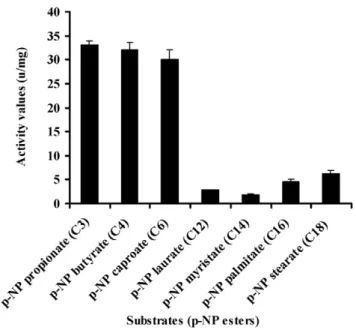
To investigate the substrate specificity of MEST1, we ex-pressed Mest1 in E. coli Rosetta (DE3) cells. SDS-PAGE and western blot analysis confirmed that a novel 47 kDa band in the transformed E. coli (ED3) cell lysates was MEST1-(His)6 fusion (Figure 2D and Figure S2). Attempts to purify six-His-tagged MEST1 expressed in E. coli Rosetta (DE3) cells failed, which could be because the six-His tag is inaccessible in this protein. Therefore, esterase activity was measured in crude extracts, with similar extracts from E. coli Rosetta (DE3) transformed with the corresponding empty vector used as control. The substrate specificity of the expressed MEST1 was determined against p-nitrophenyl esters with different carbon chain-lengths (Figure 9). MEST1 exhibited a marked preference for short-chain fatty acids, with highest activity against p-NP propionate (C3), p-NP butyrate (C4) and p-NP caproate (C6). As is typical for esterases [26], the enzyme was much less active against p-NP esters with longer-chain lengths.
Discussion
The genusMetarhiziumprovides a novel model system to study evolutionary processes as it includes species such asM. robertsiiwith broad host ranges, as well asM. majus,M. flavovirideandM. acridum that are specific for scarabs, hemipterans and acridids, respectively [5].M. acridumMa324 in particular has been widely used for locust
Table 3.Effects ofManduca sextacuticular lipids on conidial germination and appressorial differentiation ofMetarhizium acridum strains.
Treatmentsa Percentage germination (%) Appressorial formation (%)
8 h 12 h 24 h
Ma324 Ma324-Mest1 Ma324 Ma324-Mest1 Ma324 Ma324-Mest1
Water 0 0 0 0 0 0
YE 23.562.0 49.562.5 71.061.2 86.061.6 0 0
Lipids 16.561.2 31.861.5 35.561.9 62.562.0 0 6.560.7
YE+lipids 61.560.5 89.062.2 83.560.5 97.562.5 8.561.5 34.562.6
aCuticular lipids were extracted in dichloromethane. Fungal conidia (2
6107spores ml21) were induced to germinate in 5.5-cm polystyrene petri dishes containing 2 ml
control. The genetic distinctness of M. acridum from generalist strains implies evolutionarily conserved host use patterns. However, being a generalist does not precludeM. robertsiistrains from showing adaptations to nutrients on frequently encountered hosts. For example, nutrients on Hemiptera (e.g., aphids) are supplemented by insect secretions rich in sugars while beetles carry low levels of nitrogenous nutrients. Consistent with this, many hemipteran-derived lines produce appressoria in glucose medium whereas coleopteran-derived lines do not [27]. Closely related strains show these differences indicating that there are genetic mechanisms allowing rapid adaptation [28].
Lipids are the main nutrient reserve in fungal spores [16,25]. In addition, lipid bodies are transported to the developing appressoria and degraded to release glycerol, which contributes to the hydrostatic pressure that provides a driving force for mechanical penetration [15]. This process is controlled by a perilipin that surrounds the lipid droplets and is predominantly expressed when M. robertsii is engaged in accumulating lipids in nutrient rich conditions. The perilipin layer is broken down during nutrient deprivation which presumably allows esterases/lipases to hydro-lyze the lipid [15]. In this paper we show that unlike perilipin, Mest1 is predominantly expressed when lipids are being broken down. Disrupting Mest1 (DMest1) did not interfere with sapro-phytic growth of Mr2575 in nutrient rich conditions showing that its function is only important when it is adaptive for the fungus to mobilize endogenous nutrient reserves. Compared to DMest1, wild type Mr2575 grown in nutrient poor media or with cuticular lipids germinated faster and the germlings contained less lipid and more glycerol, while overexpression of Mest1 further boosted germination as well as appressorial differentiation. Intracellular lipid content was also significantly reduced and glycerol content increased in Ma324-Mest1 compared to wild type Ma324. These findings indicate that MEST1 is required byM. robertsiifor rapid hydrolysis of endogenous energy reserves during germination and for infection structure formation. Lipid droplet enzymes have not been studied in filamentous fungi, but in yeasts all lipid droplet associated proteins are involved in the mobilization of triacylgly-cerols under conditions of fatty acid starvation [29]. The nutrient poor conditions that induce expression of Mest1are a necessary trigger for virulence asM. robertsiistrain Mr2575 only produces infection structures on cuticle surfaces with low levels of nutrients [13]. However, inDMest1 the mobilization of stored lipids was delayed, but not abolished, suggesting the existence of additional enzymes involved in breakdown of lipid droplets.
It seems clear from culturing the Ma324-Mest1 in yeast extract medium, that MEST1 expression is not sufficient by itself to induce appressoria production. Consequently, production of appressoria by Ma324-Mest1 onM. sextaandG. mellonellacuticles suggests that these have at least some of the inducers required by Ma324 for appressorial differentiation (Figure 2F and 2H). The incidence of infection and the severity of the disease symptoms shown by Ma324-Mest1 were similar to those observed withM. robertsiiMr2575, an authentic pathogen of caterpillars. We have therefore shown that inserting a single gene able to mobilize nutrient reserves from a broad spectrum pathogen into a specialist is enough to broaden the latter’s host range. This implies that mobilization of nutrient reserves or lack thereof can account for the broad host range ofM. robertsiiand the restricted host range of M. acridum, respectively. Restricted host range could therefore be due essentially to biochemical limitations. Addition of exogenous nutrients (SDB, glucose, N-acetylglucosamine, glycerol) or preg-ermination on rich medium (SDB) did not overcome the inability of M. acridum to infect caterpillars. The implication is that intracellular lipid reserves are not only a nutrient source but also a source of chemical signals triggering infection processes. Consistent with this, disrupting the Mest1 gene in M. robertsii (DMest1) reduces virulence against caterpillars but not grasshop-pers, indicating that the physiological conditions on grasshoppers and caterpillars are different, and there are different mechanisms for linking signals on the host surfaces with differentiation of infection structures. Hydrocarbons comprise over 90% of the wax layer on the surface of grasshoppers, with the balance being composed of wax esters, free fatty acids and triacylglycerides, whereas in larval Lepidoptera, aliphatic alcohols are the most abundant compounds, and triacylglycerides are absent [30].M. Figure 5. Pathogenicity of Metarhizium acridum strains. (A)
Survival ofManduca sexta or (B) Galleria mellonella larvae following topical application with suspensions of 107Ma324 or Ma324-Mest1 conidia/ml. (C) Survival ofMelanoplus femurrubrumfollowing applica-tion of 3ml of 108 conidia/ml of Ma324 or Ma324-Mest1 per grasshopper pronotum. Control insects were treated with 0.01% Tween-80. Error bars indicate standard errors.
Figure 6.Mest1expression in wild type Mr2575, as measured by quantitative real-time RT-PCR.(A) Analysis ofMest1expression by wild type Mr2575 grown for 6 h in Sabouraud dextrose broth (SDB), cell-free hemolymph (HE), basal medium, 1% grasshopper cuticle (Gcut), 1%Manduca sextacuticle (Mcut), 0.1% bean root exudates (BRE), 0.01% yeast extract medium (YEM) and water. Quantitative RT-PCR time course analyses ofMest1 expression by wild typeM. robertsiiMr2575 incubated in (B) water for up to 8 h, or (C) 1% (w/v) groundM. sextacuticle for up to 12 h. (D) Effect of sugars onMest1expression by Mr2575. Quantitative RT-PCR analysis ofMest1expression by wild type Mr2575 grown in SDB or basal medium (BM) complemented with 1% glucose (Glu), 1% galactose (Gal), 1% sorbose (Sor), 1% trehalose (Tre), 1% sucrose (Suc) or 1% alanine (Ala). Culture conditions and RNA extraction were as described previously [38].gfpandtefwere used as reference genes.
doi:10.1371/journal.ppat.1002097.g006
Figure 7. Intracellular localization of MEST1 in Metarhizium robertsii Mr2575 and budding yeast Saccharomyces cerevisiae. Co-localization of MEST1 and neutral lipids demonstrated by Nile red staining of GFP-MEST1 expressing cells in (A) an ungerminatedM. anisopliae conidium, (B) a germinatedM. anisopliaeconidium and (C) a budding yeast (S. cerevisiae) cell. BF, bright field microscopy. GFP, fluorescence filter. NR, Nile red staining.
acridum extensively hydrolyzes surface lipids and waxes during germination and pre-penetration growth on locust cuticles [31]. A possible explanation for the different responses to grasshopper and caterpillars may be that breakdown products of triglycerides on grasshoppers provide signaling molecules to trigger infection processes by Ma324, but on caterpillars MEST1 provides these signals from stored triglycerides.
As the transgenicM. acridumMa324-Mest1 is able to kill cater-pillars, wild typeM. acridummust have retained most of the genetic machinery required for parasitism of insects outside its natural host range. This is consistent with the developmental processes withinM. anisopliaeandM. acridumbeing very similar, e.g. formation of germ tubes, appressoria, penetration pegs unicellular blastospores, and multi-cellular hyphal bodies that facilitate the infection of target insects, proliferation within haemolymph, and eventual eruption through the host cadaver. Besides novel lineage specific proteins such as MEST1, host recognition may therefore be determined by regulatory controls that allow expression of pathogenicity genes that are not expressed on non-hosts. Expression ofMest1under its native M. robertsiipromoter inM. acridummay have broadened host range by bypassing its need for esterases that are regulated by specific locust-related stimuli.
The impact of a single gene on host range suggests that host shifts may have occurred during Metarhizium speciation by the acquisition or loss of novel pathogenicity factors. The presence of a MEST1 ortholog in the relatedN. haematococcais consistent withM. acridumandM. robertsiihaving inherited MEST1 from a common ancestor. Their patchy distribution could be explained by rapid mutation or multiple lineage specific gene loss events inM. acridum and other fungi. An important question is whether utilizing
MEST1-like esterases during infection processes is the ancestral state. There are 16 esterases inM. robertsii(6 of which are secreted) and 18 inM. acridum(6 secreted) [22], andMetarhiziumspp. express multiple esterases on insect cuticles [32]. It seems likely that Ma324 uses the same esterases for mobilizing lipids as other fungi, and theMest1gene inM. robertsiihas acquired unique functions in this species as it is clearly highly dispensable. The new functions could potentially have turned the recipient into a novel pathogen or allowed it to infect new hosts. Expression ofMest1in Ma324 does not change virulence against its preferred grasshopper host suggesting that acridids do not represent a specialized ecological niche in which aM. robertsii-like MEST1 activity is detrimental, and there is no evidence in this study for any conditional benefits in losing MEST1’s function. This is consistent with the infection-related functions of MEST1 arisingde novoinM. robertsii.
This study has important safety implications for field applica-tions ofM. acridumas it shows that Ma324 lacks a gene important for opportunism and this should severely constrain the possibility of host switching to non-target beneficials. In addition, an understanding of how Mest1 affects fungal responses to hosts, and identification of the signaling cascades involved in regulating the mobilization of nutrient reserves will provide fundamental new insights into the initial steps that are required for the establishment of a compatible interaction between fungi and their hosts.
Materials and Methods
Strains and culture conditions
M. robertsiiMr2575 andM. acridumMa324 are wild type strains that were obtained from the U.S. Department of Agriculture Entomopathogenic Fungus Collection (ARSEF) in Ithaca, N.Y. Strain Mr2575 can infect caterpillars (Manduca sexta), beetles (Cucurlio caryae) [33], grasshoppers (Melanoplus femurrubrum) and locusts (Schistocerca gregaria) [34].M. acridumMa324 ( = CSIRO FI 485) is the active ingredient of ‘‘Green Guard’’ used for locust
Figure 8. Determination of lipid content. (A) Lipid content in conidia freshly harvested from PDA plates (black bars), nutrient-stressed conidia soaked in water for 36 h (grey bars) and conidia germinated in 1% alanine for 12 h (white bars). (B) Glycerol content in the six strains grown for 48 h in BM plus 0.25 mg/mlManducacuticle lipids. doi:10.1371/journal.ppat.1002097.g008
Figure 9. Esterase activity of MEST1 is highest in the presence of short-chain esters (C4–C8).Substrate specificity of MEST1 was determined by hydrolysis of p-nitrophenyl esters with different carbon chain lengths. The activity values are reported as means6standard errors from three independent assays using crude cell extracts ofE. coli Rosetta (DE3) expressing MEST1 as compared to extracts fromE. coli transformed with the corresponding empty vector.
control in Australia. In the field it is found exclusively in acridids, and is only infectious to caterpillars in laboratory conditions at very high spore concentrations. Fungal strains were maintained on Potato Dextrose Agar (PDA) at 27uC. Conidia were obtained from 10 day old PDA cultures. For preparation of genomic DNA and RNA, fungal spores were cultured in Sabouraud dextrose broth (SDB) (26106conidia/ml) at 27uC.
Mest1gene cloning, disruption, complementation and over-expression
Mest1was originally identified as an EST expressed when strain Mr2575 was grown on insect cuticle [17]. The full-length sequence of theMest1cDNA was obtained from the EST using RACE, and a genomic clone was obtained using the DNA Walking Speed Up Kit II (Seegene Inc., Rockville, Maryland, USA). The primers are listed in Supplementary Information, Table S1.
For targeted deletion ofMest1, the 59and 39flanking regions of the Mest1ORF were amplified by PCR from Mr2575 genomic DNA, and then subcloned into theXbaI andSpeI sites of the binary vector pBarGFP [35]. The gene disruption construct (pBarGFP-Mest1) was then transformed into Agrobacterium tumefaciensAGL-1 for targeted gene disruption by homologous recombination as described previously [36]. Replacement-specific PCR amplifica-tions of theMest1locus were performed with specific primer pairs (primers are listed in Table S1) that amplify either the wild type or the mutant gene locus.
To revert disruptantDMest1, the full-lengthMest1gene with its native promoter and terminator sequences was amplified by PCR and cloned into the XbaI site of pBenGFP [36] to generate complementation vector pBenGFP-Mest1. The complementary strain Mest1-Com was generated by reintroduction of pBenGFP-Mest1 into the disruptantDMest1 using Agrobacterium tumefaciens -mediated transformation.M. robertsii MEST1 was heterologously expressed in specialistM. acridumMa324 using vector pBenGFP-Mest1 to generate Ma324-pBenGFP-Mest1. Four putative transformants were chosen and verified forMest1gene expression by RT-PCR (data now shown).
A transformation vector was constructed by amplifying the coding region ofMest1 from the cDNA clone with primer pairs Mest1F and Mest1R (Table S1) containing a 59BamHI site and a 39EcoRI site plus a 66His-tag. The resulting PCR fragment was
cloned into the pGEM-T/A cloning vector (Promega), and the sequence of the Mest1 amplicon was confirmed by sequencing. The Mest1 gene fragment was released with BamH I andSmaI, and subcloned into pBarGPE1 [37] downstream of a constitutive Aspergillus nidulans gpdApromoter to obtain pBarGPE1-Mest1. The Mest1 cassette was released by cleavage with BglII, and then inserted into the BglII site of pBarGFP to generate pBarGFP-gpdA::Mest1 for A. tumefaciens-mediated transformation into Mr-2575. The resulting strain was designated as Mr2575-gpd::Mest1. Genomic DNA was extracted from putative transformants for Southern blot analysis as previously described [18]. Mest1 gene expression in transformants was verified by RT-PCR [38].
GFP fusion construct and subcellular localization of MEST1
To determine the subcellular localization of MEST1, the pro-moter region (,1.5 kb) together with theMest1ORF minus the 39
TAA stop codon was amplified with primer pairs Mest1F2 and Mest1R2 (Table S1) and inserted into theBamHI andEcoRI sites of pBarGPE1 [37] to generate pBarGPE1-Pmest1::Mest1. An enhanced Green Fluorescent gene (eGFP) was amplified from pEGFP (Clontech) with primers gfpF1 and gfpR1 containing a 59
EcoRI site and a 39XhoI site, and integrated into theEcoRI and XhoI sites of the plasmid pBarGPE1-Pmest1::Mest1 to generate pBarGPE1-Pmest1::Mest1:GFP. The construct was restricted with Pml1 and BamHI, and the released cassette Pmest1::Mest1:GFP was subcloned into the BamHI and EcoRV sites of the binary vector pPK2 [39] to generate pBar-Pmest1::Mest1:GFP. The final construct was transformed into wild type Mr2575 using A. tumefaciensAGL-1 to generate Mr2575-Mest1:GFP.
We also determined whether MEST1 localizes to lipid droplets in Saccharomyces cerevisiae, a fungus unrelated to Metarhizium and lacking an endogenous MEST1 protein. The Mest1 ORF was amplified with primers Mest1yesF and Mest1yesR (Table S1) and the product integrated into theEcoRI and NotI sites of pYES2 (Invitrogen). The resulting pYES2-Mest1 or the parent plasmid pYES2 (used as a control) were transformed intoS. cerevisiaestrain INVSc1 according to the manufacturer’s instructions (Invitrogen). Nile red, (9-diethylamino-5H-benzo [alpha] phenoxazine-5-one) was used to stain intracellular lipid droplets, which were viewed by fluorescence microscopy as previously described [15].
Reverse transcription and real-time RT-PCR
To monitor Mest1 expression in different growth conditions, fungal spores were incubated (6 hrs) in 10 ml of fresh SDB, Manduca sexta hemolymph [17], basal medium [BM, 0.02% KH2PO4, 0.01% MgSO4 (pH 6)], water or water supplemented
with either 0.1% bean root exudate [40]) or 1% insect cuticle as described [38]. Fungal cells were also incubated in BM supp-lemented with 1% glucose, 1% galactose, 1% sorbose, 1% tre-halose, 1% sucrose or 1% alanine. Total RNA was extracted using RNeasy Plant Mini Kit (Qiagen). First strand cDNA was syn-thesized using Verso cDNA Kit (ABgene) according to manu-facturer’s instructions. Real-time quantitative reverse transcription PCR (qRT-PCR) reactions were performed using a Quantitative real-time SYBR Green MasterMix Kit (Applied Biosystems) on an Applied Biosystems 7300 real-time instrument and ABI Prism SDS 1.2.2. software. The qPCRs were performed using the following conditions: 50uC for 2 min, then denaturation at 95uC for 10 min followed by 40 cycles of denaturation at 95uC for 20 s, annealing and extension at 60uC for 1 min. The primers used for geneMest1 and the reference genesgpdandtefare listed in Table S1.
Lipid extraction and quantification
To test the involvement of MEST1 in lipid metabolism, the cell lipid content was quantified by the sulfo-phospho-vanillin method as previously described [15]. A reference standard curve was generated using triolein (Sigma). To determine whether starvation stress induces the hydrolysis of residual stored lipids, conidia from wild type M. robertsii Mr2575, mutant DMest1, complementary strain Mest1-Com, over-expression strain Mr2575-gpd::Mest1, wild type M. acridum Ma324 and transgenic Ma324-Mest1were incubated in H2O for 36 h, which causes spores to swell but not
germinate, or induced to germinate in basal medium plus 1% alanine (wt/vol) for up to 12 h. The total lipid content of the conidia was assayed as described above.
Intracellular glycerol measurements
to the manufacturer’s instructions (Sigma). The data represent the average of three independent experiments.
Conidial germination and appressorium differentiation The germination rate of conidia was measured by inoculating 20ml of spore suspension (26107 spores ml21) in 5.5 cm
polys-tyrene Petri dishes containing either 2 ml of water, 0.01% yeast exact (YE) and/or insect cuticle lipids (0.25 mg/ml). Three hun-dred spores from each of three replicates were recorded micro-scopically to assess germination and appressorial differentiation against the hydrophobic surface of the Petri dish. Appressoria were also induced against locust (Schistocerca gregaria) hind wings, Galleria mellonellaandManduca sextacuticle as described previously [16].
Prokaryotic expression and Western blotting of MEST1 For prokaryotic expression of MEST1, the full length cDNA of Mest1was cloned by RT-PCR using specific primers Mest1EexF and Mest1EexR (Table S1), restricted withEcoRI andNotI, and subcloned into the prokaryotic expression vector pET28a at the EcoRI andNotI sites to form pET28a-Mest1.E. coliRosetta (DE3) cells (Novagen) were transformed with the recombinant expre-ssion vector. His–tagged MEST1 production was induced by 0.2 mM IPTG in LB medium for 16 h at 28uC, and cells were lysed with the B-PER Bacterial Protein Extraction Reagent (Thermo Scientific) according to the manufactor’s instructions. The His-tagged MEST1 was detected by Western blot analysis using rabbit Anti 6-Histidine Epitope Tag monoclonal antibody and anti-rabbit IgG (Fc), conjugated to alkaline phosphatase (Invitrogen).
Esterase activity assay
Ester hydrolase activities were determined in E. coli Rosetta (DE3) transformed with pET28a-Mest1 or the empty vector pET28a. Cell extract (0.1 ml) containing,50mg of protein and 0.1 ml of ap-NP-derivative substrate in 50 mM potassium pho-sphate buffer (pH 8) was incubated at 37uC. Enzyme activity was spectrophotometrically determined by measuring the liberation of p-nitrophenol as previously described [42]. One unit of activity was defined as the amount of enzyme that released 1mmol ofp-NP per min per mg of protein under the assay conditions. Substrate specificity towards various p-nitrophenyl esters (Sigma-Aldrich) was determined usingp-NP propionate (C3),p-NP butyrate (C4),p
-NP caproate (C6),p-NP laurate (C12),p-NP myristate (C14),p-NP
palmitate (C16), andp-NP stearate (C18) as substrates.
Virulence bioassay
The virulence of the wild type M. robertsii Mr2575, mutant DMest1, complementary strain Mest1-Com, over-expression strain Mr2575-gpd::Mest1, wild typeM. acridumMa324 and transgenic strain Ma324-Mest1 were assayed against wild caught 5th instar Melanoplus femurrubrumgrasshoppers (College Park, Maryland) and newly molted fifth-instar larvae of Manduca sexta and Galleria mellonella(Carolina Biological supplies). An aliquot of 3ml of fungal spore suspension was applied to the pronotum of each grasshopper as previously described [43]). Grasshoppers were then placed in
clear plastic boxes at 28uC under an 18:6-h photoperiod in humid conditions (.80%), and supplied daily with fresh wheat seedlings. Each box contained 10 grasshoppers; three containers were used for each dosage of fungus tested (36105or 56105spores/insect). Manduca sexta and Galleria mellonella were inoculated by topical immersion in conidial suspensions (16107conidia/ml) as
previ-ously described [38]. Mortality was recorded every 12 h. After death, cadavers were surface sterilized [35], and incubated in Petri dishes with a sterile wet cotton ball to promote fungal emergence, and thus confirm cause of death. Each treatment was replicated three times with 30 insects per replicate, and the bioassays were repeated twice. LT50 values were calculated with the SPSS
program [44].
Nucleotide sequence accession numbers
Sequence data reported here have been deposited in the GenBank database under the following accession numbers: Metarhizium robertsii Mest1 mRNA (HM747114), Mest1 genomic DNA (HM747115).
Supporting Information
Figure S1 Insects infected with Metarhizium strains showing sporulation on cadavers.(A)Melanoplus femurrubrum infected with wild type Metarhizium acridum Ma324. Galleria mellonellainfected with transgenic Ma324-Mest1 (B) and wild type Metarhizium robertsii Mr2575 (C). Manduca sexta infected with transgenicM. acridumMa324-Mest1 (D) and wild typeM. robertsii Mr2575 (E). Dead insects were surface sterilized in 1% bleach for five minutes, rinsed five times with sterile distilled water and placed in sterile Petri dishes containing a wet filter paper to encourage fungal emergence and sporulation.
(TIF)
Figure S2 Expression ofMest1inE. coliRosetta (DE3) and yeast Saccharomyces cerevisiae INVSc1. (A) SDS-PAGE of total cellular proteins from E. coli Rosetta (DE3) cell lysate harboring empty plasmid pET28a, pET28a-Mest1 without IPTG induction, or pET28a-Mest1 with IPTG induction (4 h). (B) SDS-PAGE separation of total cellular proteins fromS. cerevisiae strain INVSc1 harboring pYES2 or pYES2-Mest1. The size of molecular mass markers is indicated. Arrowheads indicate the target bands of expressed MEST1.
(TIF)
Table S1 Primer sequences used for PCR amplification.
(DOC)
Table S2 Effect of exogenous nutrients on the ability of
Metarhizium acridum Ma324 to infect Galleria mello-nella.
(DOC)
Author Contributions
Conceived and designed the experiments: SW RJSL. Performed the experiments: SW WF RJSL. Analyzed the data: SW CW RJSL. Contributed reagents/materials/analysis tools: CW. Wrote the paper: SW RJSL.
References
1. Walton JD (2006) HC-toxin. Phytochemistry 67: 1406–1413.
2. Wolpert TJ, Dunkle LD, Ciuffetti LM (2002) Host-selective toxins and avirulence determinants: What’s in a name? Annu Rev Phytopathol 40: 251–285. 3. van der Does HC, Rep M (2007) Virulence genes and the evolution of host
specificity in plant-pathogenic fungi. Mol Plant Microbe Interact 20: 1175–1182.
4. Mylonakis E, Casadevall A, Ausubel FM (2007) Exploiting amoeboid and non-vertebrate animal model systems to study the virulence of human pathogenic fungi. PLoS Pathog 3: e101.
6. Blanford S, Chan BH, Jenkins N, Sim D, Turner RJ, et al. (2005) Fungal pathogen reduces potential for malaria transmission. Science 308: 1638–1641. 7. de Faria MR, Wraight SP (2007) Mycoinsecticides and Mycoacaricides: A comprehensive list with worldwide coverage and international classification of formulation types. Biol Control 43: 237–256.
8. Kanzok SM, Jacobs-Lorena M (2006) Entomopathogenic fungi as biological insecticides to control malaria. Trends Parasitol 22: 49–51.
9. Gottar M, Gobert V, Matskevich AA, Reichhart JM, Wang C, et al. (2006) Dual detection of fungal infections inDrosophilavia recognition of glucans and sensing of virulence factors. Cell 127: 1425–1437.
10. Driver F, Milner RJ, Trueman JWH (2000) A taxonomic revision ofMetarhizium based on a phylogenetic analysis of rDNA sequence data. Mycol Res 104: 134–150.
11. Peng GX, Wang ZK, Yin YP, Zeng DY, Xia YX (2008) Field trials of Metarhizium anisopliaevar. acridum(Ascomycota: Hypocreales) against oriental migratory locusts,Locusta migratoria manilensis(Meyen) in Northern China. Crop Prot 27: 1244–1250.
12. Thomas MB, Read AF (2007) Can fungal biopesticides control malaria? Nat Rev Microbiol 5: 377–383.
13. St Leger RJ, Butt TM, Goettel MS, Staples RS, Roberts DW (1989) Production in vitro of appressoria by the entomopathogenic fungusMetarhizium anisopliae. Exp Mycol 13: 274–288.
14. St Leger RJ, Joshi L, Bidochka MJ, Rizzo NW, Roberts DW (1996) Characterization and ultrastructural localization of chitinases fromMetarhizium anisopliae,M. flavoviride, andBeauveria bassianaduring fungal invasion of host (Manduca sexta) cuticle. Appl Environ Microbiol 62: 907–912.
15. Wang C, St Leger RJ (2007) TheMetarhizium anisopliaeperilipin homolog MPL1 regulates lipid metabolism, appressorial turgor pressure, and virulence. J Biol Chem 282: 21110–21115.
16. Wang C, St Leger RJ (2005) Developmental and transcriptional responses to host and nonhost cuticles by the specific locust pathogenMetarhizium anisopliae var.acridum. Eukaryot Cell 4: 937–947.
17. Freimoser FM, Screen S, Bagga S, Hu G, St Leger RJ (2003) Expressed sequence tag (EST) analysis of two subspecies ofMetarhizium anisopliaereveals a plethora of secreted proteins with potential activity in insect hosts. Microbiology 149: 239–247.
18. Wang S, Leclerque A, Pava-Ripoll M, Fang W, St Leger RJ (2009) Comparative genomics using microarrays reveals divergence and loss of virulence-associated genes in host-specific strains of the insect pathogen Metarhizium anisopliae. Eukaryot Cell 8: 888–898.
19. Berger R, Hoffmann M, Keller U (1998) Molecular analysis of a gene encoding a cell-bound esterase fromStreptomyces chrysomallus. J Bacteriol 180: 6396–6399. 20. Bornscheuer UT (2002) Microbial carboxyl esterases: classification, properties
and application in biocatalysis. FEMS Microbiol Rev 26: 73–81.
21. Kugimiya W, Otani Y, Hashimoto Y, Takagi Y (1986) Molecular cloning and nucleotide sequence of the lipase gene fromPseudomonas fragi. Biochem Biophys Res Commun 141: 185–190.
22. Gao Q, Jin K, Ying SH, Zhang Y, Xiao G, et al. (2011) Genome sequencing and comparative transcriptomics of the model entomopathogenic fungiMetarhizium anisopliaeandM. acridum. PLoS Genet 7: e1001264.
23. Ebbole DJ (1998) Carbon catabolite repression of gene expression and conidiation inNeurospora crassa. Fungal Genet Biol 25: 15–21.
24. Gancedo JM (1998) Yeast carbon catabolite repression. Microbiol Mol Biol Rev 62: 334–361.
25. Requena N, Fuller P, Franken P (1999) Molecular characterization of GmFOX2, an evolutionarily highly conserved gene from the mycorrhizal
fungusGlomus mosseae, down-regulated during interaction with rhizobacteria. Mol Plant Microbe Interact 12: 934–942.
26. Verger R (1997) Interfacial activation of lipases: Facts and artifacts. Trends Biotechnol 15: 32–38.
27. St Leger RJ, May B, Allee LL, Frank DC, Staples RC, et al. (1992) Genetic differences in allozymes and information of infection structures among isolates of the entomopathogenic fungus Metarhizium anisopliae. J Invertebr Pathol 60: 89–101.
28. St Leger RJ, Bidochka MJ, Roberts DW (1994) Germination triggers of Metarhizium anisopliae conidia are related to host species. Microbiology 140: 1651–1660.
29. Jandrositza A, Petschnigga J, Zimmermanna R, Nattera K, Scholzeb H, et al. (2005) The lipid droplet enzyme Tgl1p hydrolyzes both steryl esters and triglycerides in the yeast,Saccharomyces cerevisiae. Biochimica et Biophysica Acta 1735: 50–58.
30. Chapman RF (1998) The Insects: structure and function. New York: Cambridge University Press. 788 p.
31. Jarrold SL, Moore D, Potter U, Charnley AK (2007) The contribution of surface waxes to pre-penetration growth of an entomopathogenic fungus on host cuticle. Mycol Res 111: 240–249.
32. St Leger RJ, Goettel M, Roberts DW, Staples RC (1991) Pre-penetration events during infection of host cuticle byMetarhizium anisopliae. J Invertebr Pathol 58: 168–179.
33. Moon YS, Donzelli BG, Krasnoff SB, McLane H, Griggs MH, et al. (2008) Agrobacterium-mediated disruption of a nonribosomal peptide synthetase gene in the invertebrate pathogenMetarhizium anisopliaereveals a peptide spore factor. Appl Environ Microbiol 74: 4366–4380.
34. Dillon RJ, Charnley AK (1991) The fate of fungal spores in the insect gut. In: Cole GT, Hoch HC, eds. The fungal spore and disease initiation in plants and animals. New York: Plenum Press. pp 129–156.
35. Fang W, Pava-Ripoll M, Wang S, St Leger R (2009) Protein kinase A regulates production of virulence determinants by the entomopathogenic fungus, Metarhizium anisopliae. Fungal Genet Biol 46: 277–285.
36. Fang W, Pei Y, Bidochka MJ (2006) Transformation ofMetarhizium anisopliae mediated byAgrobacterium tumefaciens. Can J Microbiol 52: 623–626.
37. McCluskey K (2003) The Fungal Genetics Stock center: from molds to molecules. Adv Appl Microbiol 52: 245–262.
38. Wang C, St Leger RJ (2006) A collagenous protective coat enablesMetarhizium anisopliaeto evade insect immune responses. Proc Natl Acad Sci U S A 103: 6647–6652.
39. Covert SF, Kapoor P, Lee M, Briley A, Nairn CJ (2001)Agrobacterium tumefaciens mediated transformation ofFusarium circinatum. Mycol Res 105: 259–264. 40. Welte MA, Cermelli S, Griner J, Viera A, Guo Y, et al. (2005) Regulation of
lipid-droplet transport by the perilipin homolog LSD2. Curr Biol 15: 1266–1275.
41. Philips J, Herskowitz I (1997) Osmotic balance regulates cell fusion during mating inSaccharomyces cerevisiae. J Cell Biol 138: 961–974.
42. Prim N, Blanco A, Martinez J, Pastor FIJ, Diaz P (2000) EstA, a gene coding for a cell-bound esterase from Paenibacillus sp.BP-23, is a new member of the bacterial subclass of type B carboxylesterases. Res Microbiol 151: 303–312. 43. Nowierski RM, Zeng Z, Jaronski S, Delgado F, Swearingen W (1996) Analysis
and modeling of time-dose-mortality ofMelanoplus sanguinipes,Locusta migratoria migratorioides, and Schistocerca gregaria (Orthoptera: Acrididae) from Beauveria, Metarhizium, andPaecilomycesisolates from Madagascar. J Invertebr Pathol 67: 236–252.