SLEEP DISORDER
A possible cause of attention deicit in children and
adolescents with Chiari malformation type II
Paulo Sérgio Azeredo Henriques Filho
1, Riccardo Pratesi
2Abstract – Background: Attention deficit may be related to sleep disorders in Chiari malformation type II (CMII). Our aim is identify sleep disorders and their specific contribution in attention deficit. Method: We selected 24 patients with CM II and 24 without CM II. DSM-IV criteria and a neuropsychological analysis were applied in all. All patients underwent full night polysomnography. Results: 14 CM II patients presented sleep apnea syndrome, REM sleep behavior disorder and periodic limb movement in sleep; six patients without CM II presented sleep apnea syndrome. Among these patients, 12 (six with CM II and six without CM II) presented attention deficit related to the sleep disorders. Conclusion: Sleep disorders may impair cognitive functions, as attention, and contribute to poor quality of learning also in patients with CM II.
KEy WORDS: polysomnography, REM sleep behavior disorder, apnea, cognition, attention deficit disorder.
Distúrbios do sono: possível causa de déficit de atenção em crianças e adolescentes com malformação de Chiari tipo II
Resumo – Introdução: Déficits de atenção podem estar relacionados a distúrbios do sono em indivíduos com malformação de Chiari tipo II (CM II). Nosso objetivo é identificar distúrbios do sono e sua contribuição para a ocorrência de déficit de atenção. Método: Foram selecionados 24 pacientes com CM II e 24 sem CM II. Todos foram submetidos à avaliação neuropsicológica, aos critérios do DSM-IV e a polissonografia. Resultados: 14 pacientes com CM II apresentaram síndrome da apnéia do sono, distúrbio do comportamento da fase do sono REM e movimentos periódicos dos membros em sono; seis pacientes sem CM II apresentaram síndrome da apnéia do sono. Entre estes pacientes, 12 (seis com CM II e seis sem CM II) apresentaram déficit de atenção relacionado a distúrbios do sono. Conclusão: Distúrbios do sono podem prejudicar funções cognitivas, como a atenção, contribuindo para a piora da qualidade de aprendizado também em pacientes com CM II. PAlAVRAS-ChAVE: polissonografia, distúrbio do comportamento da fase do sono com movimentos oculares rápidos, apnéia, cognição, transtorno do déficit de atenção.
1Graduation Program in Medical Sciences, Brasilia University School of Medicine, Brasília DF, Brazil; 2Senior Fellow Researcher.
Received 11 August 2008, received in inal form 6 October 2008. Accepted 18 November 2008.
Dr. Paulo Sérgio Azeredo Henriques Filho – MILN TR 06, CH 232, C2, Lago Norte - 71540-065 Brasilia DF - Brasil. E-mail: [email protected] The central nervous system (CNS) abnormality
charac-terized by herniation of cerebellar tonsils below the plane of foramen magnum was initially described by Chiari in 1891. Subsequently, the same author expanded the spec-trum of abnormalities of the craniocervical junction in-cluding disorders that are currently recognized as Chiari malformation (CM) type I, II, III and IV. These congenital disorders disclose complex physical and neuropsychologi-cal symptomatology and are associated with developmen-tal anomalies of both the spine and brain1. The CM type
II (CM II) is characterized by the descent of the inferior vermis and cerebellar hemispheres through the foramen magnum with a displacement of the brain stem
(medul-la, fourth ventricle, and lower portion of the pons) inside the spinal canal, resulting in aqueduct and fourth ventri-cle elongation1,2. Myelomeningocele is present in almost
all children with CM II and hydrocephalus is concomitant-ly seen in more than 70% of cases1. The increased
preva-lence of a variable degree of encephalic developmental anomalies observed in children with CM II may negative-ly affect their cognition3. Although presently, mostly due
to improved diagnostic methods and precocious treat-ment, children with myelomeningocele and hydroceph-alus are more likely to have an IQ within normal range4.
attend and follow a normal school program showing sat-isfactory performance and a socially acceptable degree of continence and behavior4. Besides the potential
pres-ence of cognitive dificulties, CM II patients are, mostly due to their brain stem anomalies, often affected by sleep disorders, the more frequently observed being sleep ap-neas, central hypoventilation and pulmonary restrictive disease3,5. Periodic limb movement in sleep (PlMS) may
appear in association to spinal anomalies6, and would be
another sleep disorder which could result in further cog-nitive impairment7. PlMS in CM II patients were never
pre-viously described, although Kaplan and Oksuz8 reported
this disorder in a CM I patient. Another sleep disorder that could eventually contribute to a poor school perfor-mance would be the REM sleep behavior disorder (RBD). Also this disorder has been never observed previously in CM II patients, although lapierre and Montplaisir9
report-ed a single case of RBD in a CM I patient.
In general, sleep disorders have been referred as a cause of behavioral and school performance dificulties leading to a possible diagnosis of attention deicit with or without associated hyperactivity10 and, in the speciic
case of children with CM II, they may aggravate the pre-existing cognitive impairment consequent to a variable degree of CNS dysfunction. Unfortunately, sleep disor-ders are rarely considered as potential source of inatten-tive behavior in children with CM II, since all their hand-icaps are generally considered to be secondary to their malformations and sleep disorders, as cause of inatten-tion and hyperactivity, seldom receive a careful though. Davidovitch et al.11 described high prevalence of attention
deicit in CM II patients. Dise and lohr12 described deicits
in conceptual reasoning abilities, considering the coexist-ing brain malformations as the responsible for the disor-der, but, to the present time, no studies have been made focusing the possible association between of sleep disor-ders and attention deicit in CM II patients.
The purpose of the present study was to assess the frequency of sleep disorders in a group of school chil-dren and adolescents affected by CM II, comparing their attention capacity with a group of controls paired by age, attempting to ascertain a possible correlation between sleep disorders and attention deicits in patients affect-ed by CM II.
MEthOD Study group
This case-control study was carried out between March 2005 and February 2007, after the approval of the Institutional Ethic Committee of the Sarah Rehabilitation hospital of Brasil-ia, as well as the approval of relatives and/or patients involved in the study. The study group was selected among patients seen at the hospital outpatient clinic due to varied complaints and
di-agnosed as having CM II and was selected after a complete med-ical history and physmed-ical examination, including the assessment of the body mass index (BMI) and neck circunference (NC) 24 children and adolescent were selected according to the follow-ing criteria: (a) befollow-ing born with spina biida, and havfollow-ing under-gone corrective spinal surgery in neonatal period; (b) having an age comprised between seven and 16 years; (c) attending a nor-mal school and a corresponding grade for their age; (d) disclosing a T1-weighted magnetic resonance imaging (MRI) conclusive for presence of CM II; (e) being considered of normal intelligence on neuropsychological evaluation. Children reported as being af-fected by respiratory, cardiovascular and neurological disorders or other ailments aside CM II, or being in use of medication that could interfere with their behavior, or with their sleep pattern, were excluded from the study. The control group was composed by 24 children without CM II, paired by age, of the same socio-economic level and pertaining to the same school.
Sleep study
Full night polysomnography evaluation was carried out uti-lizing a polygraphic digital system (Sleepscan Bio-logical Sys-tem Corporation, San Diego, CA, USA). Each subject underwent one or two full night polysomnography (depending on his or her adaptation to the sleep laboratory settings). Digital recordings included electroencephalogram (EEG) adopting identical mon-tage (C3–A2, C4–A1, O1–A2 and O2–A1, according to the Inter-national 10–20 system), electro-oculogram, chin electromyo-gram (EMG) electrocardioelectromyo-gram, upper airway sound, respiratory effort using piezoelectric belts over the chest and abdomen, and airlow through the nose and mouth (monitored through a can-nula-pressure transducer and oronasal thermocouples). We al-so recorded bilateral surface EMG from arms (biceps muscles) and legs (anterior tibial muscles). Oxyhaemoglobin saturation was monitored by pulse oximetry and subjectsí behavior during sleep was monitored by continuous video and sound recordings. All patients disclosing any type of movement disorders during sleep or with history of possible RBD underwent EEG recordings with a complete 21 electrode montage.
Scoring
arousals during this interval and if the interval is less than three minutes long. During the REM sleep phase, each 30-second och was scored and analyzed to establish the proportion of ep-ochs showing a predominance of abnormally elevated chin mus-cle background tone (tonic component), and was also scored and analyzed the proportion of three-second mini-epochs (within the 30-second REM sleep epochs) showing bursts of EMG activ-ity (phasic component). Epochs were scored as tonic or aton-ic depending on whether tonaton-ic chin EMG activity (2 mV above EMG background) was present for more or less than 50% of the epoch. We utilized thirty percent of REM sleep epochs with ab-normalities to establish as the cut-off value.
In children, the hypopnea and apnea duration cut-off uti-lized was two or more respiratory cycles15. These events were only considered signiicant when associated to an oxyhaemo-globin desaturation equal to or greater than 4% independently from the onset of subsequent arousal, partial arousal or awak-ening15. The total duration of arterial desaturation was quanti-ied as the accumulated span of arterial oxyhaemoglobin satu-ration below 90%. The apnea-hypopnea index (AhI) was deined as the sum of apneic and hypopneic episodes per hour. AhI were evaluated according to the patientís age. In children and adoles-cents, between one and ive episodes per hour were considered mildly abnormal, between six and 10 as moderate, and above 10 as severely abnormal. Periodic limb movements were scored ac-cording Zucconi et al.16 classiication. The presence of attention deicit was determined in agreement with the DSM-IV criteria and by the application of the composite teacher rating scale (ComTRS)17 which assesses ive factors: hyperactivity, indepen-dent functioning, inattention, socialization and anxiety.
Statistical analysis
Variables showing normal distribution were expressed as mean value±standard deviation and an abnormal distribution
as median values. Differences among mean of continuous vari-ables were calculated by Student t test . A signiicance level of p<0.05 was used18.
RESuLtS
The CM II group was composed by 15 boys (age range: seven to 14 years) and nine girls (age range: seven to 14 years) mean age of the group being: 10.19±0.54 years. The control group comprised 13 boys (age range: seven to 11 years) and 11 girls (age range: seven to 13 years) mean age 9.82±2.09 years. No signiicant difference was found neither BMI values (19.13±6.62 in controls patients and 16.19±1.56 in CM II patients), nor in neck circunference (30.42±3.45 in controls patients and 27.35±3.10 in CM II patients).
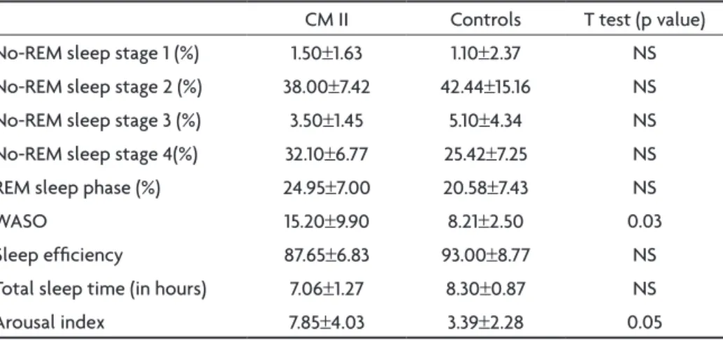
Mean values and standard deviations of sleep phases, wake after sleep onset, sleep eficiency, total sleep time in hours and arousal index can be seen in Table 1.
Fourteen of the 24 (58%) CM II patients disclosed ep-isodes of central sleep apnea, while in controls six out of 24 (25%) showed episodes of obstructive sleep apnea. The central AhI in CM II was 5.10±4.53, and in controls was 0.40±0.28 (p<0.05). The obstructive AhI in CM II was 0.21±0.08, and in controls was 3.72±3.63 (p<0.05). In CM II patients, the increase AhI was mainly consequent to the presence of central apnea and hypopnea, while in controls it was due to obstructive apneas and hypopnea. The AhI values among CM II patients and controls that did not ful-ill criteria to characterize a sleep apnea syndrome were respectively 0.62±0.13 and 0.29±0.22 (p<0.05). PlMS were observed among 11 of the 24 (45.8%) CM II patients. No abnormal PlMS index was observed in the control group. PlM index was 8.02±18.24 in CM II, and 0.36±0.68 in con-trols (p<0.05). RBD was present in six of the 24 (25%) CM II patients and in none of the controls.
Table 1. Mean values (in percentage) and standard deviations of sleep stages, total sleep time, wake after sleep onset (WASO), sleep eficiency, total sleep time in hours and arousal index in CM II patients and controls.
CM II Controls T test (p value) No-REM sleep stage 1 (%) 1.50±1.63 1.10±2.37 NS No-REM sleep stage 2 (%) 38.00±7.42 42.44±15.16 NS No-REM sleep stage 3 (%) 3.50±1.45 5.10±4.34 NS No-REM sleep stage 4(%) 32.10±6.77 25.42±7.25 NS REM sleep phase (%) 24.95±7.00 20.58±7.43 NS
WASO 15.20±9.90 8.21±2.50 0.03
Sleep eficiency 87.65±6.83 93.00±8.77 NS Total sleep time (in hours) 7.06±1.27 8.30±0.87 NS
Arousal index 7.85±4.03 3.39±2.28 0.05
An assessment and comparison between the CM II pa-tients and controls of the mean of apnea and hypopnea duration, the longest apneic and hypopneic event and the apneic event type were also performed and the results can be seen in Table 2.
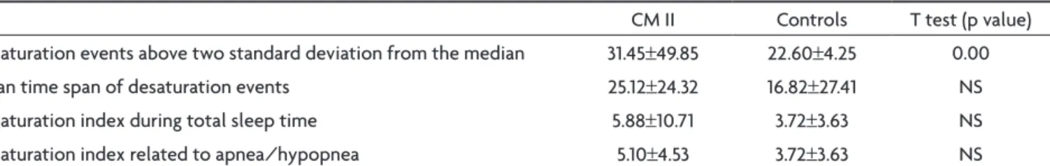
Oxy-haemoglobin saturation abnormalities can be seen in Table 3.
The relationship between the presence of sleep disor-ders (RBD, central sleep apnea, PlMS in the CM II patients and obstructive apneas in controls patients) and the
co-existence of attention deicit assessed by DSMI-V and EA-CI-P scales can be appreciate in Table 4.
DISCuSSIOn
No relationship could be found between increased body mass index and neck circumference, and the pres-ence of obstructive sleep apneas (OSA) both in CM II pa-tients and in controls. This can be explained by the fact that none of the individuals included in the study was overweight or obese. Another probable explanation could
Table 2. Types of apneas and hypopneas, mean value and standard deviation of the apneas/hypopneas duration in CM II patients and controls
CM II Controls T test (p value) Central apnea
More than two standard deviation from the median More or equal to two respiratory cycles
20.02±22.00 11.37±5.54
14.73±3.51 13.13±3.43
NS NS Obstructive apnea
More than two standard deviation from the median More or equal to two respiratory cycles
44.56±55.56 16.70±10.75
12.91±2.80 11.72±2.86
0.00 NS Mixed apnea
More than two standard deviation from the median More or equal to two respiratory cycles
9.50±0.84 7.80±1.27
11.20±0.0 7.70±0.0
NS NS hypopnea
More than two standard deviation from the median More or equal to two respiratory cycles
49.34±46.93 19.13±8.33
15.40±1.70 14.72±2.43
0.00 NS Signiicance level of p<0.05; NS, not signiicant.
Table 3. Mean values and standard deviation of the longest oxy-haemoglobin desaturation events, duration of the events, index of isolated events and index related to apnea/hypopnea events in CM II patients and controls.
CM II Controls T test (p value) Desaturation events above two standard deviation from the median 31.45±49.85 22.60±4.25 0.00 Mean time span of desaturation events 25.12±24.32 16.82±27.41 NS Desaturation index during total sleep time 5.88±10.71 3.72±3.63 NS Desaturation index related to apnea/hypopnea 5.10±4.53 3.72±3.63 NS Desaturation index, number of desaturation per hour; longest oxy-haemoglobin desaturation events, events above two standard deviation from the median; Signiicance level of p<0.05; NS, not signiicant.
Table 4. Analysis of attention deicit in CM II patients and controls with and without sleep disorders (RBD, central sleep apnea, PLMS in CM II patients, obstructive apneas in controls patients).
CM II T test Controls T test
With SD Without SD p value With SD Without SD p value
With AD 6 – 0.01 6 – 0.01
Without AD 8 10 NS – 18 0.00
Total 14 10 6 18
be that among CM II patients sleep apneas are generally caused by brain stem malformations being consequently of central origin5,19. Among controls the main cause for
sleep apneas was the presence of allergic rhinitis. In CM II patients, even in those suffering from isolat-ed RBD, was observisolat-ed an increase of NREM stage 4 and of REM sleep phases (Table 1), which was probably con-sequent to sleep deprivation. In the presence of sleep fragmentation, the sleep generators try to preserve slow wave sleep, increasing the delta band frequency and the percentage of NREM slow wave sleep20. The concomitant
increase in wake after sleep onset (WASO) and arousal in-dex and the consequent decrease in sleep eficiency and total sleep time, mirror the deleterious effects that sleep disorders, mainly SAS, RBD and PlMS, may cause on the sleep architecture and result in the appearance of atten-tion disorders in these patients10.
Sleep disorders were detected in 14 CM II patients and in six controls (Table 4). The most prevalent disor-ders among CM II patients were central apneas (12/14), RBD (5/14) and PlMS (3/14). In contrast, controls showed an increased frequency of obstructive apneas, predomi-nantly caused by airway pathologies, allergic rhinitis be-ing the most common (6/24). The increased frequency of sleep disorder in CM II patients, may be explained by the variable degree of brain stem abnormalities in CM II patients21,22, generally involving midbrain, pons and
medul-la oblongata, where are localized neurons of the respira-tory regulation, and neurons of the REM sleep and of the movement control during sleep23-25. Analyzing these data,
it is possible to conclude that sleep disorders are more prone to cause attention deicit than the presence of CM II alone, since in CM II patients without sleep disorders no attention deicit could be detected (Table 4).
Sleep apneas may cause and increased fragmentation of sleep both in CM II and in controls, mainly due to the coexisting hypoxemia or to intermittent oxy-haemoglobin desaturarion (Table 3), which would be most deleterious to sleep architecture, than another sleep disorder without hypoxemia26.
CM II patients disclosed longest apnea and or hypo-pnea events when compared to controls patients (Table 2). This difference is probably due to a functional and or anatomical disconnection between the aortic and carotid chemoreceptors and the glossopharyngeal nerve27.
Anoth-er possibility would be the presence of functional deicits of the retrotrapezoideus nucleus (RTN), which is localized in the medulla oblongata, and is frequently involved in CM II brain stem abnormalities. The RTN nucleus is re-sponsible to the respiratory control from changes in ph, in the presence of hypoxemia, and from CO2 elevation28.
The increased time of oxy-haemoglobin desaturation, among CM II patients (Table 3), consequent to episodes of apnea and or hypopnea, can probably be explained, as in the case of apneic events, by functional abnormalities in the chemoreceptor activity of the peripheral (aortic, carot-id) and or in the central nucleus. In CM II, these chemore-ceptors are probably affected by the malformations27,29,30.
The behavioral evaluation of CM II children, applying the ComTRS and the DSM-IV criteria, disclosed 12 cases of attention deicit (Table 4). hyperactivity was concomi-tantly diagnosed in three children. These abnormalities were more frequently seen among CM II patients with sleep apneas (SAS) than in those without this sleep disor-der or in patients with RBD alone. The difference between CM II patients with SAS and patients with RBD alone was probably due to the greater impairment of sleep archi-tecture in CM II patients with SAS, which could be con-sequence of the deleterious effect of chronic or intermit-tent hypoxia on cognition in these children26.
The relative lesser repercussion on sleep architecture and the scarce clinical expression observed in CM II pa-tients with RBD when compared to papa-tients with RBD, but without CM II, is probably due to the frequent impairment of movements present in CM II patients4 that preclude
intensive and energetic dream out movements.
With the exception of one patient, that disclosed a severe PlMS index, PlMS were found in association with abnormal respiratory events in all the remaining CM II pa-tients. This difference probably was related to degree of spinal cord malformation with hydrosiringomyelia, which involved propiospinal neurons, presented in 40 a 88% of the CM II patients30. In accordance with Nogués et al.6, in
CM II patients there is a relationship between the pres-ence of PlMS and spinal cord abnormalities. Abnormali-ties of the spinal cord may result in hyperexcitability of spinal regions affected by malformations, leading to the appearance of PlMS6. The CM II patient with severe PlMS
index was also affected by attention deicit, reinforcing the hypothesis that some sleep disorders, as PlM, may impair cognitive performance7.
All these sleep disorder presented among cases (CM II) and controls may affect sleep architecture, and conse-quently impair brain functions during infant critical de-velopmental stages10. In the case of CM II patients, sleep
disorders can provoke attention deicit, and further im-pair the cognitive functions already damaged by the CM II abnormalities1,11.
In conclusion, sleep disorders may impair cognitive functions, as attention, and contribute to poor quality of learning also in patients with CM II.
patients and controls, in the frequency of RBD, PlMS and SAS events that are frequently responsible to the devel-opment of attention deicits. These indings emphasized the necessity of always evaluate the sleep of CM II pa-tients, avoiding to exclusively crediting the presence of attention deicits and cognitive impairments to the cen-tral nervous system malformations.
REfEREnCES
1. Gilbert JN, Jones KL, Rorke LB, Chernoff GF, James HE. Central ner-vous system anomalies associated with meningomyelocele, hydroceph-alus, and the Arnold-Chiari malformation: reappraisal of theories re-garding the pathogenesis of posterior neural tube closure defects. Neu-rosurgery 1986;18:559-564.
2. Koehler PJ. Historical vignette. Chiariís description of cerebelar ecto-py (1891), with a summary of Clelandís and Arnoldís contributions and some early observations on neural-tube defects. J Neurosurg 1991; 75:823-826.
3. Vinck A, Maassen B, Mullaart R, Rotteveel J. Arnold-Chiari II malfor-mation and cognitive functioning in spina biida. J Neurol Neurosurg Psychiatry 2006;77:1083-1086.
4. Mirzai H, Erşahin Y, Mutluer S, Kayahan A. Outcome of patients with meningomyelocele: the Ege University experience. Childs Nerv Syst 1998;14:120-123.
5. Waters KA, Forbes P, Morielle A et al. Sleep-disordered breathing in children with myelomeningocele. J Pediatr 1998;132:672-681. 6. Nogués M, Cammarota A, Leiguarda R, Rivero A, Pardal A, Encabo H.
Periodic limb movements in syringomyelia and syringobulbia. Mov Disord 2000;15:113-119.
7. Crabtree VM, Ivanenko A, OíBrien LM, Gozal D. Periodic limb move -ment disorder of sleep in children. J Sleep Res 2003;12:73-81. 8. Kaplan Y, Oksuz E. Association between restless legs syndrome and
Chiari type 1 malformation. Clin Neurol Neurosurg 2008;110:408-410. 9. Lapierre O, Montplaisir J. Polysomnographic features of REM sleep behavior disorder: development of a scoring method. Neurology 1992;42:1371-1374.
10. O’Brien LM, Mervis LMO, Holbrook CR et al. Neurobehavioral correlates of sleep-disordered breathing in children. J Sleep Res 2004;13:165-172. 11. Davidovitch M, Manning-Courtney P, Hartmann LA, Watson J, Lut-kenhoff M, Oppenheimer S. The prevalence of attentional problems and the effect of methylphenidate in children with myelomeningocele. Pediatr Rehabil 1999;3:29-35.
12. Dise JE, Lohr ME. Examination of deficits in conceptual reasoning
abilities associated with spina biida. Am J Phys Med Rehabil 1998;77: 247-251.
13. Rechtschaffen A, Kales A. A manual of standardized terminology; tech-niques and scoring system for sleep stages of human subjects. Los Ange-les: UCLA Brain Information Service / Brain Information Institute, 1968. 14. Consens FB, Chervin RD, Koeppe RA et al. Validation of a polisomno-graphic score for REM sleep behavior disorder. Sleep 2005;28:993-997. 15. Uliel S, Tauman R, Greenfeld M, Sivan Y. Normal polysomnography re -spiratory values in children and adolescents. Chest 2004;125:872-878. 16. Zucconi M, Ferri R, Allen R et al. The official World Association of
Sleep Medicine (WASM) standards for recording and scoring periodic leg movements in sleep (PLMS) and wakefulness (PLMW) developed in collaboration with a task force from the International Restless Legs Syndrome Study Group (IRLSSG). Sleep Med 2006;7:175-183. 17. Brito GNO de. Escala de Avaliação do Comportamento Infantil para o
Professor: EACI-P. 1. ed. Rio de Janeiro: Entreletras; 1999. 18. SPSS for Windows. Standard version. Release 10.0.1. SPSS Inc.1999. 19. Dauvilliers Y, Stal V, Abril B et al. Chiari malformation and sleep related
breathing disorders. J Neurol Neurosurg Psychiatry 2007;78:1344-1348. 20. Banks S, Dinges DF. Behavioral and physiological consequences of sleep
restriction. J Clin Sleep Med 2007;3:519-528.
21. Henriques Filho PS, Pratesi R. Abnormalities in auditory evoked po -tentials of 75 patients with Arnold-Chiari malformations type I and II. Arq Neuropsiquiatr 2006;64:619-623.
22. Henriques Filho PS, Pratesi R. Sleep apnea and REM behavior disor -der in patients with Chiari malformations. Arq Neuropsiquiatr 2008;66: 344-349.
23. Berger AJ, Mitchell RA, Severinghaus JW. Regulation of respiration. N Engl J Med 1977;297:92-97.
24. Netick A, Orem J, Dement W. Neuronal activity speciic to REM and its relationship to breathing. Brain Res 1977;120:197-207.
25. Hendricks JC, Morrison AR, Mann GL. Different behaviors during par-adoxical sleep without atonia depend on pontine lesion site. Brain Res 1982;239:81-105.
26. Bass JL, Corwin M, Gozal D et al. The effect of chronic or intermittent hypoxia on cognition in childhood: a review of the evidence. Pediatrics 2004;114:805-816.
27. Bullock R, Todd NV, Easton J, Hadley D. Isolated central respirato -ry failure due to syringomyelia and Arnold-Chiari malformation. BMJ 1988;297:1448-1449.
28. Guyenet PG, Mulkey DK, Stornetta RL, Bayliss DA. Regulation of ven -tral surface chemoreceptors by the cen-tral respiratory pattern genera-tor. J Neurosci 2005;25:8938-8947.
29. Bokinsky GE, Hudson LD, Weil JV. Impaired peripheral chemosensivi-ty and acute respiratory failure in Arnold-Chiari malformation and sy-ringomyelia. N Engl J Med 1973;288:947-948.