Impact of Body Weight on Radiation Exposure
During Invasive Cardiac Procedures
Francine Gonçalves Vargas
1, Bruna Santos da Silva
1, Cristiano de Oliveira Cardoso
2, Natalia Leguisamo
1,
Cláudio Antônio Ramos de Moraes
2, Cláudio Vasques de Moraes
2, Júlio Vinícius de Souza Teixeira
2,
La Hore Correa Rodrigues
2, Alexandre Schaan de Quadros
2, Carlos Antonio Mascia Gottschall
2ABSTRACT
Background: Invasive cardiac procedures expose patients and physicians to ionising radiation. The aim of this study was to determine the impact of body weight on radiation exposure during cardiac procedures. Methods: A prospective cohort study of patients undergoing cardiac catheterisation or percutaneous coronary intervention (PCI) between August of 2010 and December of 2011. Clinical, angiographic, and radiation exposure characteristics were recorded in a dedicated database. Radiation exposure patterns were established in three groups: A (≤ 79 kg), B (80–99 kg), and C (≥ 100 kg).
Data were analysed using the Statistical Package for Social Sciences (SPSS), version 18.0, and the results were presented as the mean, standard deviation, percentage, percentile, and interquartile interval. The independent predictors of increased radiation exposure were identiied using a multiple logistic regression analysis. Results: The sample included a total of 671 patients, 363 in group A, 252 in group B, and 56 in group C. The mean dose of radiation exposure was 484.29 mGy, 735.69 mGy, and 900.36 mGy for groups A, B, and C, re-spectively (P < 0.001). The median dose area product was 29.327 mGy.cm², 43.319 mGy.cm², and 57.987 mGy.cm² for groups A, B, and C, respectively (P < 0.001). The predic-tors of increased radiation exposure were weight (odds ratio [OR] 1.03, conidence interval [CI] 1.01–1.05, P = 0.003), conidence interval (CI) 1.01–1.05, P = 0.003], elective PCI (OR 11.9, CI 4.26–33.24, P < 0.001), and ad hoc PCI (OR 15.46, CI 5.44–43.87, P < 0.001). Conclusions: Patient weight has a signiicant impact on radiation exposure during invasive cardiac procedures. Overweight patients are signiicantly more exposed to higher doses of ionising radiation.
DESCRIPTORS: Heart catheterisation. Body weight. Radiation ionising. Radiation exposure.
1 Technical Course of Radiology and Diagnostic Imaging – Escola
Proissional da Fundação Universitária de Cardiologia (FUC) – Porto Alegre, RS, Brazil.
2 Instituto de Cardiologia do Rio Grande do Sul – Fundação Universitária
de Cardiologia (IC-FUC) – Porto Alegre, RS, Brazil.
Correspondence to: Cristiano de Oliveira Cardoso. Avenida Francisco
Petuco, 340/805 – Boa Vista – Porto Alegre, RS, Brazil – CEP 90520-620 E-mail: [email protected]
Received on: 1/6/2012 • Accepted on: 3/4/012
Original Article
RESUMO
Impacto do Peso Corporal dos Pacientes na Exposição Radiológica Durante Procedimentos
Cardiológicos Invasivos
Introdução: Procedimentos cardiológicos invasivos expõem pa -cientes e médicos aos riscos da radiação ionizante. É objetivo deste estudo determinar o impacto do peso do paciente na exposição radiológica durante procedimentos cardiológicos. Métodos: Estudo de coorte prospectivo incluindo pacientes submetidos a cateterismo cardíaco ou intervenção coronária percutânea (ICP) entre agosto de 2010 e dezembro de 2011. Características clínicas, angiográicas e de exposição à radiação foram registradas em banco de dados especíico. Os padrões de exposição à radiação foram determinados em três grupos: A (≤ 79 kg), B (80-99 kg) e C (≥ 100 kg). Os dados foram analisados
em programa SPSS 18.0, sendo os resultados apresentados em média, desvio padrão, porcentual, percentil e intervalo interquartil. Preditores independentes de exposição à radiação aumentada foram identiicados por análise de regressão logística múltipla. Resultados: A amostra incluiu 671 pacientes, sendo 363 no grupo A, 252 no B e 56 no C. A dose média de radiação recebida pelos pacientes foi de 484,29 mGy, 735,69 mGy e 900,36 mGy para os grupos A, B e C, respectivamente (P < 0,001). A mediana do produto dose área foi de 29.327 mGy.cm², 43.319 mGy.cm² e 57.987 mGy.cm² para os grupos A, B e C, respectivamente (P < 0,001). Os preditores de exposição radiológica aumentada foram peso [razão de chance (RC) 1,03, intervalo de coniança (IC) 1,01-1,05; P = 0,003], ICP eletiva (RC 11,9, IC 4,26-33,24; P < 0,001) e ICP ad hoc (RC 15,46, IC 5,44-43,87; P < 0,001).
Conclusões: O peso exerce impacto signiicativo na exposição radiológica em procedimentos cardiológicos invasivos. Pacien-tes com peso elevado são signiicativamente mais expostos à radiação ionizante.
DESCRITORES: Cateterismo cardíaco. Peso corporal. Radiação ionizante. Exposição a radiação.
H
aemodynamic monitoring procedures have been widely used to evaluate coronary artery disease. Concurrently, the number of diagnostic and thera-peutic procedures in modern cardiology has increased the exposure to ionising radiation for patients, physicians, and nurses.1,2 Together with increasing technologicaladvances and more potent haemodynamic monitoring equipment,3,4 reports on the harmful effects of ionising
radiation have increased.5,6
It is known that patient weight is directly related to unfavourable outcomes, such as vascular complica-tions, in interventional cardiology.7 Nevertheless, few
reports exist in the Brazilian national literature about the relation between weight and radiation exposure.
The aim of this study was to determine the impact of body weight on the radiation exposure of patients undergoing invasive cardiac procedures.
METHODS
Design
This was an observational study with prospective data collection.
RADIAÇÃO Registry
The RADIAÇÃO registry8 is an institutional registry
aimed at documenting the diagnostic and therapeutic procedures performed with the help of lat detectors in the ield of interventional cardiology. Information regarding radiation exposure and technical details of the procedures are prospectively registered.
Sample
Patients underwent, diagnostic cardiac catheterisa-tion or coronary intervencatheterisa-tion were followed to register the radiation exposure patterns. All patients signed an informed consent, and the protocol was approved by the local research ethics committee.
Analysed characteristics
For the RADIAÇÃO registry, data regarding patient age, gender, risk factors for cardiovascular disease (diabetes, arterial hypertension, tobacco smoking, dys-lipidemia, and family history), clinical presentation and procedure indication, ventricular function, number of vessels affected, treated vessels, lesion characteristics, and success index were collected and analysed. Speciic data concerning radiation exposure (received dose, dose area product, and luoroscopy time) were also collected.
Parameters of radiation exposure
The radiation exposures of the patients were mea-sured with the entrance skin radiation dose (cumulative air KERMA – Kinetic Energy Released per unit MAss).
The luoroscopy times and dose area product were also measured to determine the radiation exposure time and the irradiated area, respectively.
The procedures were performed in the Allura Xper FD10 monoplane lat detector (Philips – Einthoven, Netherlands), with three magnetic ields (15 cm, 20 cm, and 25 cm), double ilter (copper + aluminium) and a standard image acquisition program at 15 frames per second.
Statistical analysis
Three groups were compared. Group A comprised patients weighing ≤ 79 kg; group B, patients weighed 80–99 kg; and group C, patients weighed ≥ 100 kg. Data were prospectively collected and stored in a dedicated database within the ACCESS program. The Statistical Package for Social Sciences (SPSS) version 18.0 for Windows was used for the analyses. The results were shown as mean, standard deviation, me-dian, and interquartile interval. The chi-squared tests, the Kruskal-Wallis test, and the Bonferroni post-hoc test of ANOVA were used for the group comparisons. A multiple logistic regression model was applied to identify the possible predictors of increased radiation exposure (total dose ≥ 2 Gy). In this analysis, statisti-cally significant variables were used in the univariate analysis. A two-tailed P-value of < 0.05 was considered statistically significant.
RESULTS
Between August of 2010 and December of 2011, 671 invasive cardiac procedures were performed: 363 in group A, 252 in group B, and 56 in group C.
The total number of procedures corresponded to 420 diagnostic catheterisations and 251 coronary angioplasties. There was no signiicant difference between the groups regarding the proportion of diagnostic cardiac catheterisa-tions (61.2%, 63.5%, and 67.9% in groups A, B, and C, respectively) and coronary angioplasty (38.8%, 36.5%, and 32.1% in groups A, B, and C, respectively).
Demographic characteristics
In general, it was observed that the patients in group C presented signiicantly greater prevalences of systemic arterial hypertension and diabetes mellitus. Regarding the other risk factors for cardiovascular dis-ease and the prescribed patient medications, there was no signiicant difference between the groups. Table 1 details the clinical characteristics of the three groups.
Procedural angiographic characteristics
two vessels (14%, 17.5%, and 10.7%; P = 0.85), and three or more vessels (8%, 7.9%, and 3.6%; P = 0.9). In all groups, the left anterior descending artery was most affected (group A, 39.7%; group B, 37.3%; and group C, 30.4%; P = 0.76), while the left main coronary artery presented signiicant lesions in 1.1%, 2.8% and 3.6% of the patients in groups A, B, and C, respectively (P = 0.21).
In coronary angioplasties, the preferred access site was the femoral artery (80%, 77.4%, and 55% in
TABLE 1
Patient Clinical Characteristics
Variable
Group A (≤ 79 kg)
n = 363
Group B (80-99 kg)
n = 252
Group C (≥ 100 kg)
n = 56 P
Age, years 64.8 ± 10.8 60.8 ± 10.7 57.2 ± 8.2 < 0.001
Height, cm 162.5 ± 7.8 169.1 ± 7.18 171.5 ± 8.1 < 0.001
BMI, kg/m2 26 ± 3 30 ± 3 37 ± 4 < 0.01
Male gender, n (%) 157 (43.3) 181 (71.8) 38 (67.9) < 0.001
Tobacco, active smoking, n (%) 75 (20.7) 53 (21) 11 (19.6) 0.97
Arterial hypertension, n (%) 276 (76) 205 (81.3) 50 (89.3) 0.04
Diabetes mellitus, n (%) 99 (27.3) 72 (28.6) 24 (41.9) 0.049
Insulin use 49 (13.5) 32 (12.7) 10 (17.9) 0.59
Dyslipidemia, n (%) 186 (51.2) 135 (53.6) 26 (46.4) 0.6
Family history of CAD, n (%) 214 (59) 147 (58.3) 28 (50) 0.44
Prior PCI, n (%) 121 (33.4) 92 (36.5) 16 (28.6) 0.47
Prior CABG, n (%) 40 (11) 32 (12.7) 1 (1.8) 0.059
Prior AMI, n (%) 152 (41.9) 94 (38.3) 16 (28.6) 0.12
Prior Stroke, n (%) 16 (4.4) 14 (5.6) 2 (3.6) 0.73
Associated valve disease, n (%) 8 (2.2) 4 (1.6) 3 (5.4) 0.22
Medications in use, n (%)
ASA 250 (68.9) 167 (66.3) 39 (69.6) 0.76
Clopidogrel/ticlopidine 161 (44.4) 108 (42.9) 19 (33.9) 0.34
Beta blocker 217 (59.8) 163 (64.7) 37 (66.1) 0.38
Nitrate 164 (45.2) 97 (38.5) 19 (33.9) 0.11
Statin 213 (58.7) 149 (59.1) 38 (67.9) 0.41
ACE inhibitor 177 (48.8) 120 (47.6) 34 (60.7) 0.19
Calcium channel blocker 51 (14) 44 (17.5) 12 (21.4) 0.26
Diuretics
Aldosterone antagonist 36 (9.9) 23 (9.1) 7 (12.5) 0.74
ASA = acetylsalicylic acid, CAD = coronary arterial disease, ACE = angiotensin-converting enzyme, AMI = acute myocardial infarction, PCI = percutaneous coronary intervention, BMI = body mass index, CABG = coronary artery bypass graft.
Parameters of radiology exposure
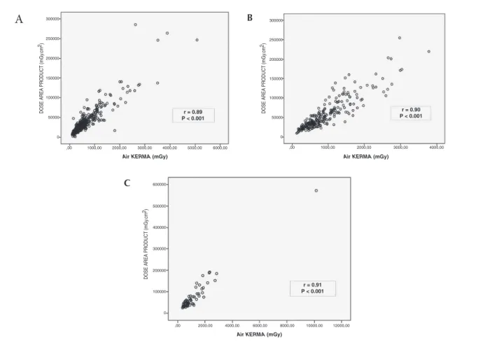
It was observed that patients with higher weight were signiicantly more exposed to ionising radiation. Both the entrance skin radiation (air KERMA) and the dose area product were progressively increased in the heavier patients. Table 2 presents the values for radiation exposure in each group. The correlation between the total dose received and the irradiated area was signii-cant in the three groups studied, as shown by Figure 1.
Predictors of increased radiology exposure
In the present sample, radiation exposure > 2 Gy occurred in 3.6% (13/363) of the procedures in group A, in 16.3% (41/252) of the group B procedures, and in 10.7% (6/56) of the group C procedures. Using the uni- and multivariate analyses, patient’s weight, elective angioplasty and ad hoc angioplasty were determined to be predictors of increased radiation exposure. Table 3 presents the odds ratio and its conidence interval.
DISCUSSION
The present study is the irst in this institution to examine the impact of body weight on the radiation exposure of patients undergoing invasive cardiac proce-dures. The results demonstrated that obese patients were more exposed to radiation than non-obese individuals. Currently available equipment for haemodynamic monitoring has an automatic dose and image quality
TABLE 2
Radiology Exposure Parameters among the Groups
Variable
Group A (≤ 79 kg) n = 363
Group B (80–99 kg)
n = 252
Group C (≥ 100 kg)
n = 56 P
Fluoroscopy time, minutes 4.35 4.53 4.41 0.60
Number of graphs per exam 11 11 11 > 0.9
Total number of frames 779 811 739 0.45
Total number of frames/graphs 67.5 69.5 69.25 0.83
Radiation exposure
Air KERMA for patients, mGy < 0.001
- Inferior quartile (Q1/4) 276.2 476.94 606.34
- Median (Q2/4) 484.29 735.69 900.36
- Superior quartile (Q3/4) 766.19 1,191.93 1,517.48
Dose area product, mGycm2 < 0.001
- Inferior quartile (Q1/4) 17,239 30,401 38,782
- Median (Q2/4) 29,327 43,319 57,987
- Superior quartile (Q3/4) 46,210 71,287 90,856
control (Automatic Bright Control – ABC).3,9,10 Although
the operation system is complex, in practice, every time the equipment detects low-resolution image or great brightness variation, the dose is increased to compensate. In overweight patients, the thickness and density of the chest are increased; therefore, the equipment automatically releases a higher dose to maintain quality standards. Studies using an ionisation chamber have shown that for every 1-cm of thickness, radiation exposure increased by 25%.3 For this reason,
obese patients receive higher dose of radiation during the procedures.
Obesity has been proven to interfere in some cardiovascular outcomes, such as vascular complica-tions7,11 and the incidence of atrial ibrillation.12 Data
from electrophysiological studies have shown that obese patients, when undergoing pulmonary vein abla-tion, received a dose that was two times higher than non-obese patients.13 The present results show that the
patients weighing over 100 kg received 1.8 and 1.2 times more radiation than the patients weighing up to 79 kg and up to 99 kg, respectively. Therefore, obesity has a signiicant impact on radiation exposure.
Concerns related to ionising radiation are com-pletely reasonable and pertinent to all individuals who are exposed to this type of biological effect. Radiation reduction methods are outdated, and new proposals have been presented.14–18 It has been stipulated that ionising
it is inevitable. The “as low as reasonably achievable” (ALARA) principle,19,20 established in 1977, essentially
states that radiation exposure should be kept as low as reasonably achievable. Although the ALARA principle is widely known, Mavrikou et al.21 called attention to the
fact that many radiation and protection concepts are neglected by interventional physicians. Critical exposure doses (2 Gy) are frequently surpassed in the procedures; therefore, the principle is not being respected.8
TABLE 3
Multivariate Analysis for Determining the Predictors of Increased Radiation Exposure
Variable OR CI P
Body weight 1.03 1.01–1.05 0.003
Elective angioplasty procedure
11.9 4.26–33.24 < 0.001
Ad hoc angioplasty
procedure 15.46 5.44–43.87 < 0.001
CI = conidence interval; OR = odds ratio.
Figure 1 – Correlation between the irradiated area and the total dose received.
300000
250000
200000
150000
100000
50000
0
,00 1000,00 2000,00 3000,00 4000,00 5000,00 6000,00
DOSE AREA PR
ODUCT (mGy
.cm
2 )
Air KERMA (mGy) r = 0.89 P < 0.001
r = 0.90 P < 0.001
r = 0.91 P < 0.001
300000 400000 500000 600000
200000
100000
0
,00 2000,00 4000,00 6000,00 8000,00 10000,00 12000,00
DOSE AREA PR
ODUCT (mGy
.cm
2 )
Air KERMA (mGy)
Air KERMA (mGy)
300000
250000
200000
150000
100000
50000
0
,00 1000,00 2000,00 3000,00 4000,00
DOSE AREA PR
ODUCT (mGy
.cm
2 )
A
BC
Radiation over-exposure is becoming more frequent in daily practice. This group has demonstrated that lat detector equipment can add 65% more radiation compared to image intensiier devices,22 and has also
demonstrated that up to 12% of the invasive cardiac procedures surpassed the critical dose of 2 Gy.8
Nowa-days, skin lesions, which were previously rare, have been reported during interventional cardiology procedures.5
More recently, six alarming cases of brain tumors6 in
interventional cardiology patients have again raised concerns about occupational risks. Therefore, medical societies are encouraging more training programs and education measures to reduce the biological risks.1,16,17
The area exposed to biological radiation effects and the potential stochastic risks of neoplasia are as important as the total radiation dose. The present study demonstrated that there is a strong relation between the total received dose and the irradiated area, regardless of the patient’s weight. Patients weighing over 100 kg received approximately 57,987 mGy.cm2 during the
procedures. This measure is higher than the 50,000 mGy-cm2 dose recommended by the International Atomic
Energy Agency (IAEA).23 In obese patients, the
This inding is important because larger irradiated areas have increased risks for stochastic effects and neoplasia.
Although it has not been demonstrated in the present study, other authors have determined that the risk of radiation exposure was attributable to cancer incidence. Using the Biological Effects of Ionising Ra-diation (BEIR VII)24,25 risk model, it can be concluded
that risk of developing a solid tumour after radiation exposure is low. Nevertheless, continuing exposure and higher doses can promote a higher risk that may still be unknown.26,27
Limitations of the study
The present study had several limitations that must be considered. This analysis was conducted in only one centre with a small patient sample. Radiation exposure is only related to the radiation dose received by the patient; therefore, any inference regarding the dose received by the haemodynamicists could not be determined.
CONCLUSIONS
Body weight has a signiicant impact on radiation exposure in invasive cardiac procedures. Overweight patients are signiicantly more exposed to higher doses of ionising radiation.
ACKNOWLEDGMENTS
The authors would like to thank the haemodynamicists Drs. Alexandre Damiani Azmus, André Manica, Carlos Roberto Cardoso, Henrique Basso Gomes, Flávio Celso Leboute, Luis Maria Yordi, Mauro Régis Moura, and Rogério Sarmento-Leite for collaborating in this study.
CONFLICTS OF INTEREST
The authors declare no conlicts of interest.
REFERENCES
1. Miller DL, Vano E, Bartal G, Balter S, Dixon R, Padovani R, et al. Occupational radiation protection in interventional radiolo-gy: a joint guideline of the Cardiovascular and Interventional Radiology Society of Europe and the Society of Interventional Radiology. Cardiovasc Intervent Radiol. 2010;33(2):230-9. 2. Picano E, Vano E. The radiation issue in cardiology: the time
for action is now. Cardiovasc Ultrasound. 2011;9:35. 3. Gurley JC. Flat detectors and new aspects of radiation safety.
Cardiol Clin. 2009;27(3):385-94.
4. Seibert JA. Flat-panel detectors: how much better are they?. Pediatric Radiol. 2006;36 Suppl 2:173-81.
5. Padovani R, Bernardi G, Quai E, Signor M, Toh HS, Morocutti G, et al. Retrospective evaluation of occurrence of skin injuries in interventional cardiac procedures. Radiat Prot Dosimetry. 2005;117(1-3):247-50.
6. Roguin A, Goldstein J, Bar O. Brain tumours among interven-tional cardiologists: a cause for alarm? Report of four new cases from two cities and a review of the literature. Eu roIntervention. 2012;7(9):1081-6.
7. Davis C, VanRiper S, Longstreet J, Moscucci M. Vascular complica-tions of coronary intervencomplica-tions. Heart Lung. 1997; 26(2):118-27. 8. Cardoso CO, Sebben JC, Fischer L, Vidal M, Broetto GG,
Silva BS, et al. Padrão de exposição radiológica e preditores de superexposição dos pacientes submetidos a procedimen-tos cardiológicos invasivos em equipamenprocedimen-tos com detectores planos. Rev Bras Cardiol Invasiva. 2011;19(1):84-9.
9. Lin PJ. Operation logic and functionality of automatic dose rate and image quality control of conventional luoroscopy. Med Phys. 2009;36(5):1486-93.
10. Trianni A, Bernardi G, Padovani R. Are new technologies always reducing patient doses in cardiac procedures?. Radiat Prot Dosimetry. 2005;117(1-3):97-101.
11. Nikolsky E, Mehran R, Dangas G, Fahy M, Na Y, Pocock SJ, et al. Development and validation of a prognostic risk score for major bleeding in patients undergoing percutaneous coronary intervention via the femoral approach. Eur Heart J. 2007;28(16):1936-45.
12. Wang TJ, Parise H, Levy D, D’Agostino RB Sr, Wolf PA, Vasan RS, et al. Obesity and the risk of new-onset atrial ibrillation. JAMA. 2004;292(20):2471-7.
13. Ector J, Dragusin O, Adriaenssens B, Huybrechts W, Willems R, Ector H, et al. Obesity is a major determinant of radiation dose in patients undergoing pulmonary vein isolation for atrial ibrillation. J Am Coll Cardiol. 2007;50(3):234-42.
14. European Society of Radiology (ESR). White paper on radiation protection by the European Society of Radiology. Insights into Imaging. 2011;2(4):357-62.
15. Bernardi G, Padovani R, Trianni A, Morocutti G, Spedicato L, Zanuttini D, et al. The effect of fellows training in invasive cardiology on radiological exposure of patients. Radiat Prot Do simetry. 2008;128(1):72-6.
16. Chambers CE, Fetterly KA, Holzer R, Lin PJ, Blankenship JC, Balter S, et al. Radiation safety program for the cardiac catheterization laboratory. Catheter Cardiovasc Interv. 2011; 77(4):546-56.
17. Carpeggiani C, Kraft G, Caramella D, Semelka R, Picano E. Radioprotection (un)awareness in cardiologists, and how to improve it. Int J Cardiovasc Imaging. 2011 Aug 18. [Epub ahead of print] 18. Chambers CE. Radiation dose: it is more than just “time”.
Catheter Cardiovasc Interv. 2011;78(1):143-4.
19. Hendee WR, Edwards FM. ALARA and an integrated approach to radiation protection. Semin Nucl Med. 1986;16(2):142-50. 20. International Commission on Radiological Protection (ICRP). Recommendations. Oxford: Pergamon Press; 1977 (Publica-tion, 26).
21. Mavrikou I, Kottou S, Tsapaki V, Neofotistou V. High patient doses in interventional cardiology due to physicians’ negli-gence: how can they be prevented?. Radiat Prot Dosimetry. 2008;129(1-3):67-70.
22. Medeiros RF, Sarmento-Leite R, Cardoso CO, Quadros AS, Risso E, Fischer L, et al. Exposição à radiação ionizante na sala de hemodinâmica. Rev Bras Cardiol Invasiva. 2010;18(3):316-20. 23. International Atomic Energy Agency (IAEA). Establishing guidan-ce levels in X-ray guided medical interventional proguidan-cedures: a pilot study. Vienna, Austria; 2009. (Safety Reports Series, 59). 24. Higson D. BEIR VII-2. J Radiol Prot. 2005;25(3):324-5. 25. Huda W, He W. Estimating cancer risks to adults undergoing
body CT examinations. Radiat Prot Dosimetry. 2011 Sep 17. [Epub ahead of print]
26. Einstein AJ, Henzlova MJ, Rajagopalan S. Estimating risk of can-cer associated with radiation exposure from 64-slice computed tomography coronary angiography. JAMA. 2007;298(3):317-23. 27. Vano E, Ubeda C, Leyton F, Miranda P, Gonzalez L. Staff