Quim. Nova, Vol. 35, No. 7, 1375-1380, 2012
Artigo
*e-mail: [email protected]
EVALUATION OF ANTIMICROBIAL ACTIVITY AND TOXIC POTENTIAL OF EXTRACTS AND TRITERPENES ISOLATED FROM Maytenus imbricata
Vanessa G. Rodrigues*, Lucienir P. Duarte, Grácia D. F. Silva, Fernando C. Silva, Jefferson V. Góes, Jacqueline A. Takahashi e Lúcia P. S. Pimenta
Departamento de Química, Instituto de Ciências Exatas, Universidade Federal de Minas Gerais, Av. Antônio Carlos, 6627, 31270-901 Belo Horizonte - MG, Brasil
Sidney A. Vieira Filho
Departamento de Farmácia, Escola de Farmácia, Universidade Federal de Ouro Preto, Rua Costa Sena, 171, 35400-000 Ouro Preto - MG, Brasil
Recebido em 3/10/11; aceito em 26/2/12; publicado na web em 2/7/12
The phytochemical study of hexane/ethyl ether (1:1) extract of the roots of M. imbricata, Celastraceae, resulted in the isolation and characterization of six known triterpenes: 11α-hydroxylup-20(29)-en-3-one, previously isolated from this species besides, 3β,11α -di-hydroxylup-20(29)-ene, 3,7-dioxofriedelane, 3-oxo-29-hydroxyfriedelane, tingenone and 6-oxo-tingenol. The chemical structures of these triterpenes were established by spectrometric data (IR, 1H and 13C NMR) and through comparison with literature data. The hexane/ethyl ether (1:1), ethyl acetate and methanol extracts, and 11α-hydroxylup-20(29)-en-3-one, tingenone and 6-oxo-tingenol, showed antimicrobial properties on in vitro assays. All extracts and triterpenes, except 3β,11α-di-hydroxylup-20(29)-ene, presented toxicity demonstrated by the larvicidal effect test using Artemia salina.
Keywords: Maytenus imbricata; Celastraceae; pentacyclic triterpenes.
INTRODUCTION
From different species of the genus Maytenus, various groups of secondary metabolites have been found, such as triterpenes,1
sesqui-terpenes,2 phenolic glycosides,3 alkaloids,1 flavonoids,4,5 and tannins,5
among others. Members of this genus are important not only in terms of experimentally observed biological activities,6 but also because
they are used in folk medicine for gastric diseases,7 and as antiseptic,
anti-asthmatic, anti-tumor,8 antiviral,9 and anti-inflammatory agents.10
In a recent review, Niero et al.11provided an adequate description of
the ethnopharmacological, chemical and pharmacological knowledge about species of the genera Maytenus, with particular emphasis on those growing in Brazil. Many species of this genus are effective medicines, and represent promising sources of bioactive substances of medicinal interest.
The indiscriminate use of antibiotics has induced an increase in the incidence of infectious diseases caused by pathogenic micro-organisms that have acquired resistance to several antibiotics currently used in clinical treatments. This scenario has led to a continuous, urgent search for new antimicrobial compounds, especially those bearing different chemical structures and having specific mechanisms of action.12 In this context, the compounds isolated from plants have
emerged as a promising alternative. Indeed, around 50% of drugs approved by the Food and Drug Administration (FDA) between 1981 and 2006 were of natural origin.13
It has been reported that the molecular diversity of natural pro-ducts is higher than those derived from chemical synthesis processes. Thus, the biological assays of natural products represent a continuing source of new structural models for compounds with antimicrobial properties and are an important alternative for reducing the incidence of infectious diseases.14
According to the literature, compounds that present brine shrimp (Artemia salina) toxicity, in general also have cytotoxic properties
against cells of solid tumors found in humans. This bioassay has being considered as adequate for initial screening of bioactive molecules, paving the way for subsequent tests of greater complexity such as Aedes aegypti larvicidal assays or for testing the response of cancer cells to anti-cancer drugs.15
Maytenus imbricata Mart, ex. Reissek is a shrub or subshrubs found in Cerrado regions (rupestrian fields) of Minas Gerais and Bahia States, Brazil. Leaves, twigs and stems of this species were subjected to biological assays. The CHCl3, EtOAc and EtOH extracts
of leaves, the hydroalcoholic extract of roots, the EtOAc extract of stems and epicatechin, isolated from M. imbricata showed antioxi-dant activity. The 3,4-seco-friedelan-3-oic acid, isolated from leaves, presented inhibitory activity of ATP synthesis raising possibilities for its potential use in the future development of natural herbicides.16
In this paper, the phytochemical study of the roots of Maytenus imbricata and isolation of the pentacyclic triterpenes (PCTT) 11α-hydroxylup-20(29)-en-3-one (1), 3β,11α -di-hydroxylup-20(29)-ene (2), 3,7-dioxofriedelane (3), 3-oxo-29-hydroxyfriedelane (4), tingenone (5) and 6-oxo-tingenol (6)(Figure 1) were reported.
The extracts, and some constituents from roots, of M. imbricata were submitted to in vitro antimicrobial assays to evaluate their prop-erties against the bacteria Staphylococcus aureus, Bacillus cereus, Salmonella typhimurium, Escherichia coli andthe fungus Candida albicans. The cytotoxic effect of these constituents was screened through their larvicidal effect on Artemia salina.
EXPERIMENTAL
Plant material
Rodrigues et al.
1376 Quim. Nova
Federal de Viçosa (UFV) and M. C. T B. Messias, Departamento de Botânica of the Universidade Federal de Ouro Preto (UFOP). A voucher specimen (No 27780) was deposited in the collection of the
Herbarium of Departamento de Botânica, UFV, Brazil. General procedures
Purification processes by column chromatography (CC) were carried out using silica gel 60 (0.063-0.200 mm) as the stationary phase. Organic solvents or mixtures of increasing polarity were used as mobile phases. Silica gel 60 GF (Merck) was used to perform analytical [(TLC), 0.25 mm] or preparative [(PTLC), 0.75 mm] thin layer chromatographic processes.
The 1H (400 or 200 MHz) and 13C (100 or 50 MHz
3) NMR
spectra were obtained on a Bruker Avance DRX-400 or DPX-200 spectrometer, operating at 300 K. The chemical shifts (δ) were ex-pressed in units of ppm, using TMS as a reference (δH = δC = 0) and the coupling constants (J) were expressed in Hz. CDCl3 was used as
the solvent for the samples.
The IR spectra of constituents in KBr discs were obtained on a Shimadzu IR408 spectrometer and the results reported in reciprocal centimeters (cm-1) in the range 400-4000 cm-1.
The melting point (mp) ranges were determined on an MQAPF-302 apparatus (Microquímica Equipamentos Ltda, Brazil). Extraction and isolation of compounds
The collected roots of M. imbricata were powdered in a mill. The powder (1.5 kg) was then submitted to extractions in a Soxhlet apparatus with hexane-ethyl ether (1:1), ethyl acetate, and finally with methanol. During extraction with hexane/ethyl ether (1:1) a solid was formed and separated by filtration giving the hexane/ethyl ether solid material [HES (22.2 g)]. The solvent of the filtrated product was removed in a rotatory evaporator, giving the hexane/ethyl ether (1:1) extract [HEE (16.1 g)]. During extraction using ethyl acetate, another solid was formed and separated by filtration, giving the ethyl
acetate solid [EAS (56.2 g)]. The resultant product of solvent remo-val was ethyl acetate extract [EAE (21.2 g)]. Finally, the methanol extract [ME (176.7 g)] was obtained. A total of 2 L of hexane/ethyl ether (1:1), 2 L of ethyl acetate and 2 L of methanol were used in the extraction processes.
The extract HEE (3.0 g) was submitted to silica gel (300.8 g) CC eluted with hexane-EtOAc. Three hundred fifty fractions of 100 mL each were obtained and grouped according to the similar profiles observed in the chromatoplates. Fractions 14 to 19 produced a white crystalline solid (12.3 mg), 0.41% yield and mp 247.7-250.1 °C. This solid was identified as 3,7-dioxo-friedelane (3). The fractions 44-49 provided a white solid in the form of flakes (63.1 mg), 2.10% yield, mp 154.2-158.8 °C, subsequently identified as 11α -hydroxylup-20(29)-en-3-one (1). Fractions 84-95 produced a white crystalline solid (7.0 mg), 0.23% yield, mp 146.9-148.4 °C which was identified as 3-oxo-29-hydroxyfriedelane (4). Fractions 107-128 produced an amorphous orange solid (183.3 mg) which was subjected to silica gel CC eluted with Hex, EtOAc and MeOH, pure or in mixtures of increasing polarity, providing 240 fractions of 10 mL each. Solvent evaporation from fractions 211 to 235 (eluted with Hex/EtOAc 6:4) produced an orange solid (64.4 mg), 2.15% yield, mp 145.0-147.9 °C, which was identified as tingenone (5).
Fractions 205-207 produced an amorphous orange solid (146.7 mg) which was submitted to silica gel CC (47.3 mg) eluted with Hex, EtOAc and MeOH, pure or in mixtures of increasing polarity, providing 137 fractions of 10 mL each. After the solvent was wi-thdrawn, fractions 1 to 15 (eluted with Hex/EtOAc 9:1) yielded an orange solid (25.3 mg), which showed three spots when analyzed by TLC. These fractions (1-15) were submitted to silica gel PTLC eluted with Hex/EtOAc 7:3. The less polar compound (Rf ~ 0.9) was isolated as a white solid (11.0 mg), 0.37% yield, mp 212.9-218.6 °C and was identified as 3β,11α-di-hydroxylup-20(29)-ene (2). Fraction 76 yielded a light yellow solid (8.0 mg), 0.27% yield, mp 218.7-221.4 °C that was identified as 6-oxo-tingenol (6). These terpenes were characterized through their NMR spectral data and also by comparing with published data.
The extract HES (2.0 g) was submitted to silica gel (107.9 g) CC eluted with Hex, EtOAc and MeOH, pure or in mixtures of increasing polarity, providing 114 fractions of 25 mL each that were grouped according to the similar profiles observed in the chromatoplates.
Fractions 33-42 produced an amorphous brown solid (55.0 mg) which was subjected to silica gel CC eluted with Hex, EtOAc and MeOH, pure or in mixtures of increasing polarity, providing 117 fractions of 3 mL each. After solvent evaporation from fractions 33 to 47 (eluted with Hex/EtOAc 2:8), a white solid in the form of flakes was obtained (14.0 mg), 0.7% yield and mp 154.2-158.8 °C. This solid was identified as 11α-hydroxylup-20(29)-en-3-one (1).
Fractions 66-85 produced an orange solid (314.0 mg), 15.7% yield, mp 145.0-147.9 °C, which was identified as tingenone (5).
Fractions 94-102produced an amorphous brown solid (300.9 mg) which was subjected to silica gel CC eluted with Hex, EtOAc and MeOH, pure or in mixtures of increasing polarity, providing 294 fractions of 3 mL each. Solvent evaporation from fractions 121 to 133 (eluted with EtOAc) produced a light yellow solid (7.9 mg), 0.4% yield, mp 218.7-221.4 °C which was identified as 6-oxo-tingenol (6).
The extracts EAE, EAS and ME have yet to be submitted to phytochemical studies. However phytochemical prospection17 allowed
the detection of the presence of alkaloids, flavonoids and catechin. Biological assays
Antimicrobial activity
To evaluate antibacterial and antifungal activity, the microdilution
Figure 1. Chemical structures of pentacyclic triterpenes isolated from roots of Maytenus imbricata
HO 1 2 6 11 15 16 19 20 22 23 24 25 26 27 28 29 30 R1 R1 compound 1 2 =O OH 7 O R2 5 3 9 10 11 13 19 21 22 23 25 26 27 30 24 R1 15
R1 R2
compound
3
4 =O
Evaluation of antimicrobial activity and toxic potential of extracts 1377 Vol. 35, No. 7
method was used to determine the average minimal inhibitory con-centrations inhibiting the growth of 50% (MIC50) and 90% (MIC90) of
the microorganisms.18 All extracts and the triterpenes 1, 5 and 6 were
tested against Salmonella typhimurium (ATCC 13311), Escherichia coli (ATCC 25723), Staphylococcus aureus (ATCC 25923), Bacillus cereus (ATCC 11778)and Candida albicans (ATCC 18804). The bacteria and the fungus were maintained in brain heart infusion (BHI) culture medium, at 7 ºC. An initial duplicate screening was carried out using disposable microplates with all samples being tested at a concentration of 100.00 µg/mL. In this step, all extracts and the triterpenes 1, 5 and 6 showed antimicrobial activity. Subsequently, these were tested at concentrations of 250.00, 125.00, 62.50, 31.30, 15.60, 7.81, 3.91, 1.95, 0.98, 0.49, 0.24 and 0.12 µg/mL to determi-ne the minimal inhibitory concentration (MIC50 and MIC90). All 12
microdilution assays were performed in duplicate. Chloramphenicol (MIC50 = 0.24µg/mL and MIC90 = 15.60 µg/mL) was used as a positive
control for bacteria and miconazole (MIC50 = 3.32µg/mL and MIC90
= 600.48 µg/mL) for C. albicans. The inocula of bacteria and fungus used in experiments contained 4.16 x 10³ cells/mL. At the end of
incubation time (24 h), the plates were analyzed using a Microplate TP-Reader (Thermoplate, Brazil).
Evaluation of toxic potential through Artemia salina larvicidal activity
The cysts of Artemia salina for the test were acquired in Belo Horizonte City, MG, Brazil. The A. salina cysts (10 mg) were added to 100 mL of synthetic marine salt solution (38.0 g/L) previously prepared using deionized water, and maintained under artificial light at 28 °C. Larvae hatching occurred after 24 h of incubation time. Lapachol, a compound with proven anti-A. salina effect, was used as the control. The extracts and terpenes 1, 3, 4, 5 and 6 isolated from M. imbricata were submitted to larvicidal assays, at concentrations ranging from 1000.00 to 0.06 µg/mL, to determine their toxic po-tential. The concentration range was chosen based on the highest concentration that showed 100% mortality and at the concentration which induced no death of A. salina. The samples were dissolved in DMSO and their concentration ranges were HESandHEE (15.62-0.48 µg/mL), EAS,EAEandME(1,000.00-62.50 µg/mL), terpene 1 (31.25-1.95 µg/mL), 3 and 4 (125.00-7.81 µg/mL), 5 (0.97-0.06 µg/mL) and 6 (500.00-31.25 µg/mL). Terpene 2 was not submitted to larvicidal assays against A. salina because it was used in another test and an insufficient amount remained for testing.
RESULTS AND DISCUSSION
Phytochemical study
Through the phytochemical study of HEE it was possible to isolate the known pentacyclic triterpenes 1, 2, 3, 4, 5 and 6 (Figure 1). The chemical structures of these constituents were identified based on IR,
1H and 13C NMR spectral data, and by comparing with literature data.
Compound 1 was previously isolated through phytochemical study of the aerial part of M. imbricata.19 In this paper, the isolation of
terpenes 2, 3, 4, 5 and 6 from M. imbricata is reported for the first time. The profile of IR spectra of compounds 1 to 5 were in accordance with the published data for pentacyclic triterpenes.20-23
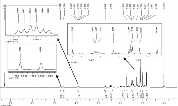
The 1H NMR spectrum of 1 disclosed signals of 7 methyl groups
[δ 1.69 (3H), δ 1.09 (3H), δ 1.07 (6H), δ 1.06 (3H), δ 0.98 (3H) and δ 0.80 (3H)], of methine [triple doublet at δ 3.91 (H11ax-ax, J = 10.8 Hz;
and H11ax-eq, J = 4.8 Hz )] and of olefin hydrogens (δ 4.72 and δ 4.60).
These data, together with the signals at δ 109.95 (C29) and δ 150.22 (C20) observed in the 13C NMR spectrum, suggested that 1 was a
PCTT of lupane series,21 which was then compared with the literature.22
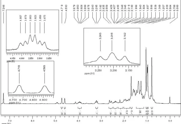
Seven methyl signals [δ 1.69 (3H), δ 1.26 (3H), δ 1.04 (9H), δ 0.96 (3H) and δ 0.79 (3H)], together with the signals of methine hydrogen [δ 3.93 (H11ax-ax, J = 10.8 Hz and H11ax-eq, J = 4.8 Hz) and of methine
hydrogen H3 (δ 3.21), were observed in the 1H NMR spectrum of
2.21 Two singlet signals (δ 4.72 and δ 4.59) were attributed to H29
of terminal double bond. The signals of carbons [δ 109.95 (C29) and δ 150.25 (C20)] confirmed the double bond and suggested 2 as member of the PCTT lupane series.21 The NMR spectral data of 2
were in accordance with literature data.21
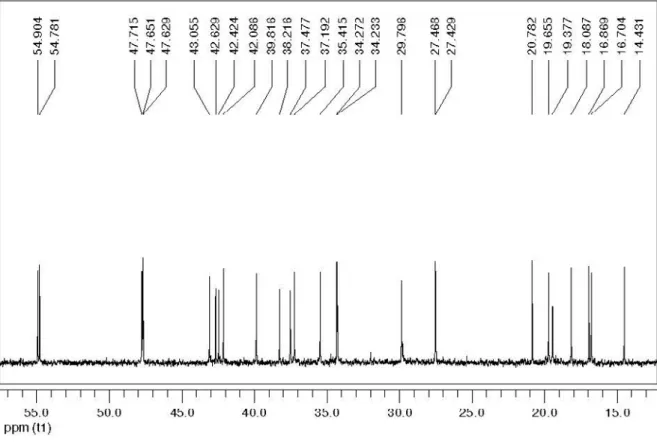
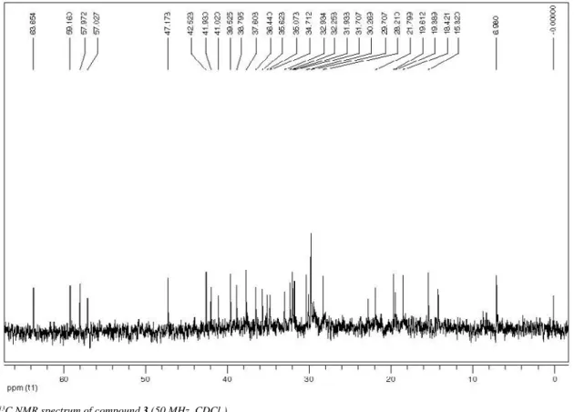
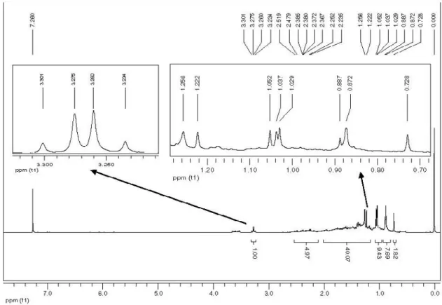
The signals in the 1H NMR spectrum of 3 at δ 0.77; δ 0.88; δ
0.91; δ 0.96; δ1.00; δ1.07 and δ1.18 were attributed to seven methyl groups, and a doublet at δ 0.88 was associated to another methyl group (C23). These signals are commonly related to PCTT of the friedelane series.21 The 13C NMR spectrum of 3 revealed signals at δ 211.12 and
at δ 210.62 corresponding to ketone carbonyls. The chemical shift values found in the 13C NMR spectrum of terpene 3 were compared
with literature data.21
The signals at δ0.73; δ 0.87; δ 1.03; δ 1.04; δ 1.05 and δ 1.22 observed in the 1H NMR spectrum of 4 were associated to 6 methyl
groups, and a doublet signal at δ 0.88 to another methyl group (C23), while a doublet at δ 3.27 was attributed to hydrogen linked to hy-droxylated carbon. The 13C NMR spectrum disclosed signals at δ 6.83
assigned to methyl carbon (C23) of PCTT friedelane,21 at δ 213.18
attributed to carbonyl (C3) and at δ 74.78 assigned to hydroxylated carbon (C29). The NMR data of terpene 4 were in accordance with published data.21
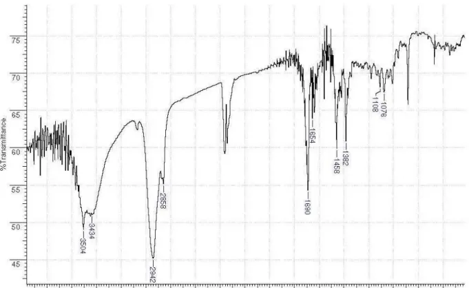
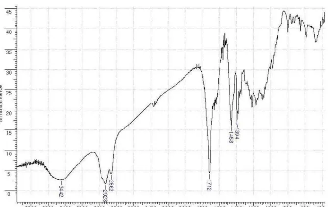
The profile of the IR spectrum, especially in the region between 1500-1708 cm-1, characteristic of C=O and C=C bonds, suggested
compound 5 as a quinone methide PCTT. 20 The signals observed in
the 1H NMR spectrum of 5 at δ 0.98; δ 1.01; δ 1.35; δ 1.51 and at
δ 2.23 corresponded to 5 methyl groups, a doublet signal at δ 1.00 was attributed to another methyl group (C30); and a doublet at δ2.92 (H22) and a multiplet at δ 2.51 were associated to hydrogen linked to C20. These spectral data, together with the doublet signal at δ 7.03 and at δ 6.38; and a singlet at δ 6.55, confirmed 5 as being a quinone methide triterpene.21 The carbon signals at δ 178.43 (C2) and at δ
213.58 (C21) were attributed to carbonyl carbons. The chemical shift values found in the 13C NMR spectrum of terpene 5 were consistent
with the literature data.23
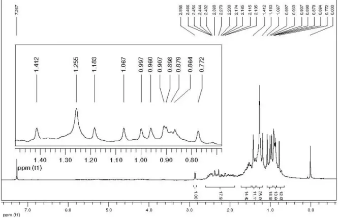
The IR spectrum of 6 revealed bands at 1592 and 1458 cm-1 that
were associated to the C=C bond of aromatic compound.20 Six methyl
signals [singlets at δ 2.63; δ 1.58; δ 1.38; δ 1.00 and δ 0.99, as well as a doublet at δ 0.99 (C30)], were identified in the 1H NMR spectrum
of 6.Two singlets, at δ6.86 (H1) and at δ 6.25 (H7), confirmed the aromatic and olefin hydrogens, respectively. The carbon signals at δ 187.94 (C6) and at δ 214.69 (C21) were associated to carbonyls. The chemical shift values found in the 13C NMR spectrum of terpene
6 were consistent with the literature data.24
Antimicrobial activity
The level of resistance or sensitivity of the bacteria and fungus to the samples was determined by the presence or absence of growth. The results of MIC50 of the extracts and triterpenes from M. imbricata
subjected to antimicrobial assays are shown in Table 1.
In accordance with the results, it was found that all samples sho-wed MIC50 within the concentration range used in the experiments.
The solid HES, extract HEE, and terpene 5 showed better inhibition of S. aureus growth. Azithromycin is a macrolide antibiotic that suppresses the biosynthesis of protein, retards bacterial growth, or causes death of microorganisms, when tested against S. aureus presented a MIC50 of 4.0 µg/mL.
Rodrigues et al.
1378 Quim. Nova
than that found for HES, HEE, and for terpene 5, which showed a lower value for MIC50. All extracts and terpenes 5 and 6 induced a
high rate of growth inhibition of C. albicans (Table 1). Fluconazole is an antifungal used to treat infectious diseases caused by fungus. In assays with C. albicans, fluconazole showed a MIC50 of 1.1 µg/mL.
26
The MIC50 value found using HES, HEE and terpene 5, indicated
its higher activity against C. albicans, evidencing their potential as antifungal agents.
The extract from leaves of Maytenus ilicifolia showed no activity against Salmonellasp.27 However, in this work, it was verified that all
extracts as well as the compounds 1, 5 and 6, obtained from roots of M. imbricata were active against Salmonella typhimurium.
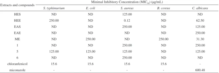
As shown in Table 2, higher antimicrobial activity was found for the extract HEE against S. aureus (MIC90 = 0.12 µg/mL) and for ME
against C. albicans (MIC90 = 31.30 µg/mL).
Vancomycin, an antibiotic highly effective against Gram-positive bacteria, has being adopted as a first choice for the treatments of infectious diseases caused by S. aureus. This is the main pathogen associated to hospital-acquired infections. In assays in vitro against S. aureus, vancomycin presented a value higher (MIC100 = 1.5 µg/mL)
28
value than that found for HEE (MIC90 = 0.12 µg/mL).
Neomycin is an antibiotic used to prevent or treat skin infections caused by bacteria, including S. aureus. Jain et al.29 tested neomycin
against S. aureus, and found MIC100 = 190.0 µg/mL, a value higher
than the MIC90 of HES, HEE and compound 5. These authors also
tested the ethanol extract from roots of Maytenus senegalensis against S. aureus and found MIC100 = 1250.0 µg/mL. This value is higher than
the MIC90 found for the extracts HES, HEE, EAS and for terpenes
1, 5 and 6 (Table 2).
The methanol extract (ME) was active against B. cereus. This bacterium showed high sensitivity when treated with ME extract. In addition, this extract also presented activity against E. coli and C. albicans.
Considering the results of the in vitro assays with bacteria and Candida albicans, a high antibacterial and antifungal activity was attributed to the extracts and terpenes isolated from roots of M. imbricata.
Evaluation of cytotoxic potential
The toxicity assays using Artemia salina, a marine microcrusta-cean, were carried out according to the methodology described by Pimenta et al..30 The experiments were performed in triplicate and
the lethal concentration of sample necessary to induce 50% death (LC50) of brine shrimps, was established using the Probit Method,
a parametric statistical procedure with a 95% confidence interval. Table 1. Minimal inhibitory concentrations that inhibit 50% of the microorganism growth (MIC50) determined for the extracts and compounds isolated from M. imbricata against pathogenic microorganisms
Extracts and compounds Minimal Inhibitory Concentration (MIC50) (µg/mL)
S. typhimurium E. coli S. aureus B. cereus C. albicans
HES 125.00 125.00 0.12 125.00 0.49
HEE 125.00 125.00 0.12 250.00 0.12
EAS 125.00 125.00 31.30 125.00 62.50
EAE 250.00 250.00 31.30 125.00 31.30
ME 125.00 250.00 31.30 125.00 15.60
1 250.00 250.00 62.50 250.00 125.00
5 62.50 125.00 0.12 125.00 0.12
6 31.30 125.00 62.50 250.00 62.50
chloranfenicol 0.24 0.24 0.24 0.24
-miconazole - - - - 3.32
Table 2. Minimal inhibitory concentrations that inhibit 90% of the microorganism growth (MIC90) determined for the extracts and compounds isolated from M. imbricata against pathogenic microorganisms
Extracts and compounds Minimal Inhibitory Concentration (MIC90) (µg/mL)
S. typhimurium E. coli S. aureus B. cereus C. albicans
HES ND ND 125.00 ND ND
HEE 250.00 ND 0.12 ND 62.50
EAS ND ND 250.00 ND 125.00
EAE ND ND ND ND 250.00
ME ND 250.00 ND 250.00 31.30
1 ND ND 250.00 ND 250.00
5 125.00 125.00 125.00 ND 125.00
6 ND ND 250.00 ND ND
chloranfenicol 15.6 15.6 15.6 15.6
-miconazole - - - - 600.48
Evaluation of antimicrobial activity and toxic potential of extracts 1379 Vol. 35, No. 7
Extracts of plants or compounds submitted to assays with A salina are considered active when the LC50 is less than 1000.0 µg/mL.
31
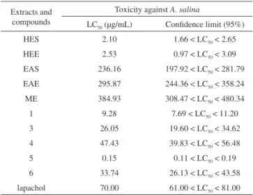
The evaluation of toxicity using A. salina was performed with the extracts HES, HEE, EAS, EAE and ME, and compounds 1, 3, 4, 5 and 6, followed by the respective determination of LC50 (Table 3).
The extracts HES and HEE and all terpenes showed high toxi-city, evidenced by their low LC50 values (Table 3). Tingenone (5) is
a pentacyclic triterpene with known cytotoxic properties.32 In the
present work, this terpene showed higher toxicity when compared to the other samples of M. imbricata tested. This terpene is the main constituent found in the extracts HES (40%) and HEE (9.4%). Thus, it is possible to assign the high cytotoxicity observed for these two extracts to compound 5. Using Artemia franciscana, Macari et al.33
studying Maytenus guyanensis found LC50 = 363 µg/mL for the
he-xane extract of barks. Based on this LC50, these authors considered
this extract as being a potential larvicide product. Through the assay with A. salina, it was found that, except for the extract ME, all other samples presented values of LC50 lower than 363 µg/mL. Based on
these results can be established that substances from M. imbricata also represent potential larvicide agents. Bouzada et al.,34 using a
similar methodology with A. salina,found LC50 > 250 µg/mL for the
methanol extract from leaves of Maytenus ilicifolia. On the other hand, ethanol extract of Maytenus obtusifolia was considered of low toxicity to the larvae of A. salina, with LC50 greater than 1000 mg/mL.
35 The
values of this average lethal dose (LC50) are high when compared with
those found for HES and HEE in the present work (Table 3). Since the extracts and terpenes showed good activity against A. salina it is possible to conclude that they also have potential antitumor, pesticide, trypanosomicide and/or molluscicide activities.15
CONCLUSION
The triterpenes 11α-hydroxylup-20(29)-en-3-one, 3β,11α -di-hydroxylup-20(29)-ene, 3,7-dioxofriedelane, 3-oxo-29-hydroxy-friedelane, tingenone and 6-oxo-tingenol were isolated from roots of Maytenus imbricata. All extracts, and the terpenes 11α -hydroxylup-20(29)-en-3-one, tingenone and 6-oxo-tingenol, were active against S. typhimurium, E. coli, S. aureus, B. cereus and C. albicans. The extracts HES and HEE and the terpenes 1, 3, 4, 5 and 6 showed rep-resentative larvicidal effects against Artemia salina. The results of this work indicate the promising potential of this plant as a larvicide,
or as a source of drugs with antimicrobial or antitumor properties. SUPPLEMENTARY MATERIAL
The Figures 1S-21S present spectra of IR, 1H and 13C NMR of
compounds isolated of hexane/ethyl ether (1:1) extract of the roots of M. imbricata. The Tables 1S-6S present comparison of 13C NMR
data of compounds and of literature. This supplementary material is available free of charge at http://quimicanova.sbq.org.br, as PDF file. ACKNOWLEDGMENTS
The authors are grateful to Conselho Nacional de Desenvolvimento Científico e Tecnológico (CNPq) and Fundação de Amparo à Pesquisa do Estado de Minas Gerais (FAPEMIG) by financial support. REFERENCES
1. Shirota, O.; Tamemura, T.; Morita, H.; Takeya, K.; Itokawa, H.; J. Nat. Prod. 1996, 59, 1072; Orabi, K. Y.; Al-Qasoumi, S. I.; El-Olemy, M. M.; Mossa, J. S.; Muhammad, I.; Phytochemistry 2001, 58, 475.
2. Corsino, J.; Furlam, M.; Bolzani, V. da S.; Pereira, A. M. S.; Franca, S. E.; Phytochemistry 1998, 49, 2181; González, A. G.; Tincusi, B. M.; Bazzocchi, I. L.; Tokuda, H.; Nishino, H.; Konoshima, T.; Jiménez, I. A.; Ravelo, A. G.; Bioorg. Med. Chem. 2000, 8, 1773.
3. Sannomiya, M.; Vilegas, W.; Rastrelli, L.; Pizza, C. A.; Phytochemistry 1998, 49, 237.
4. Tiberti, L. A.; Yariwake, J. H.; Ndjoko, K.; Hostettmann, K.; J. Chroma-togr., B: Anal. Technol. Biomed. Life Sci. 2007, 846, 378; Souza, L. M.; Cipriani, T. R.; Sant’Ana, C. F.; Iacomini, M.; Gorin, P. A. J.; Sassaki, G. L.; J. Chromatogr., A 2009, 1216, 99.
5. De Souza, L. M.; Cipriani, T. R.; Iacomini, M.; J. Pharm. Biomed. Anal. 2008, 47, 59.
6. Souza, S. A. M.; Cattelan, L. V.; Vargas, D. P. V.; Piana, C. F. B.; Bobrowski, V. L.; Rocha, B. H. G.; Ci. Biol. Saúde 2005, 11, 7; Dias, K. S.; Marques, M. S.; Menezes, I. A. C.; Santos, T. C.; Silva, A. B. L.; Estevam, C. S.; Sant’Ana, A. E. G.; Pizza, C.; Antoniolli, A. R.; Marcal, R. M.; Fitoterapia2007, 78, 460.
7. Hussein, G.; Nakamura, N.; Meselhy, M. R.; Hattorim, M.; Phytoche-mistry 1999, 50, 689.
8. Jeller, A. H.; Silva, D. H. S.; Lião, L. M.; Bolzani, V. S.; Furlan, M.; Phytochemistry2004, 6, 1977; Nakagawa, H.; Takaishi, Y.; Fujimoto, Y.; Duque, C.; Garzon, C.; Sato, M.; Okamoto, M.; Oshikawa, T.; Ahmed, S. U.; J. Nat. Prod. 2004, 67, 1919; Perestelo, N. R.; Jiménez, I. A.; Tokuda, H.; Hayashi, H.; Bazzocchi, I. L.; J. Nat. Prod.2010, 73, 127. 9. Baggio, C. H.; Freitas, C. S.; Otofuji, G. M.; Cipriani, T. R.; Souza, L.
M.; Sassaki, G. L.; Iacomini, M.; Rieck, L.; Mesia-Vela, S.; Marques, M. C. A.; J. Ethnopharmacol. 2007, 113, 433; Cipriani, T. R.; Mellinger, C. G.; Souza, L. M.; Baggio, C. H.; Freitas, C. S.; Marques, M. C. A.; Gorin, P. A. J.; Sassaki, G. L.; Iacomini, M.; Carbohydr. Polym. 2009, 5, 361.
10. Sosa, S.; Morelli, C. F.; Tubaro, A.; Cairoli, P.; Speranza, G.; Manitto, P.; Phytomedicine 2007, 14, 109.
11. Niero, R.; Andrade, S. F.; Cechinel Filho, V.; Curr. Pharm. Des. 2011, 17, 1851.
12. Rojas, R.; Bustamante, B.; Bauer, J.; Fernandez, I.; Alban, J.; Lock, O.; J. Ethnopharmacol. 2003, 88, 199.
13. Ferreira, V. F.; Pinto, A. C.; Quim. Nova 2010, 33, 1829.
14. Novais, T. S.; Costa, J. F. O.; David, J. P. L.; David, J. M.; Queiroz, L. P.; França, F.; Giulietti, A. M.; Soares, M. B. P.; Santos, R. R.; Rev. Bras. Farmacogn. 2003, 14, 8; Bolzan, A. A.; Silva, C. M.; Francescato, L. N.; Murari, A. L.; Silva, G. N. S.; Heldwein, C. G.; Heinzmann, B.; Lat. Am. J. Pharm.2007, 26, 619.
Table 3. Average lethal dose (LC50) of extracts and terpenes isolated from roots of Maytenus imbricata against A. salina
Extracts and compounds
Toxicity against A. salina
LC50 (µg/mL) Confidence limit (95%)
HES 2.10 1.66 < LC50 < 2.65
HEE 2.53 0.97 < LC50 < 3.09
EAS 236.16 197.92 < LC50 < 281.79
EAE 295.87 244.36 < LC50 < 358.24
ME 384.93 308.47 < LC50 < 480.34
1 9.28 7.69 < LC50 < 11.20
3 26.05 19.60 < LC50 < 34.62
4 47.43 39.83 < LC50 < 56.48
5 0.15 0.11 < LC50 < 0.19
6 33.74 26.13 < LC50 < 43.58
lapachol 70.00 61.00 < LC50 < 81.00
Rodrigues et al.
1380 Quim. Nova
15. McLaughin, J. L.; Rogers, L. L.; Anderson, J. E.; Drug Information J. 1998, 32, 513.
16. Silva, G. D. F.; Silva, S. R. S.; Barbosa, L. C. A.; Duarte, L. P.; Ri-beiro, S. M. R.; Queiroz, J. H.; Vieira Filho, S. A.; Oliveira, M. L. R.; Rev. Bras. Farmacogn. 2009, 19, 530; Silva, S. R. S.; Silva, G. D. F.; Barbosa, L. C. A.; Duarte, L. P.; King-Diaz, B.; Archundia-Camacho, F.; Lotina-Hennsen, B.; Pest. Biochem. Physiol.2007, 87, 109. 17. Matos, F. J. A.; Introdução à Fitoquímica Experimental, Ed. UFC:
Fortaleza, 1980.
18. Zacchino, A. S.; Gupta, M. P.; Manual de técnicas in vitro para la detección de compuestos antifúngicos, Corpus Editorial y Distribuidora: Rosario 2007, vol. 85.
19. Silva, S. R. S.; Silva, G. D. F.; Barbosa, L. C. A.; Duarte, L. P.; Vieira Filho, A. S.; Helv. Chim. Acta 2005, 88, 1102.
20. Silverstein, R. M.; Webster, F. X.; Kiemle, D. J.; Identificação Espectrométrica de Compostos Orgânicos, 7ª ed., LTC: Rio de Janeiro, 2007.
21. Mahato, S. B.; Kundu, A. P.; Phytochemistry 1994, 37, 1517.
22. Silva, S. R. S.; Tese de Doutorado, Universidade Federal de Minas Gerais, Brasil, 2007.
23. Sotanaphun, U.; Suttisri, R.; Lipipun, V.; Bavovada, R.; Phytochemistry 1998, 49, 1749.
24. Gonzaléz, A. G.; Alvarenga, N. L.; Ravelo, A. G.; Jiménez, I. A.; Bazzochi, I. L.; Canela, N. J.; Moujir, L. M.; Phytochemistry1996, 43, 129.
25. Pereira, I. A.; Soares, L. C.; Coelho, S. M. O.; Balbino, F. A.; Pribul, B. R.; Souza, M. M. S.; Arq. Brasil. de Med. Veterinária Zoot. 2009, 61, 577.
26. Bedout, C.; Ayabaca, J.; Veja, R.; Méndez, M.; Santiago, A. R.; Pabón, M. L.; Tabares, A.; Arango, M.; Restrepo, A.; Newell, V.; Biomédica 2003, 23, 31.
27. Voss-Rech, D.; Klein, C. S.; Techio, V. H.; Scheuermann, G. N.; Rech, G.; Fiorentin, L.; Ciência Rural 2011, 41, 314.
28. França, H. S.; Kuster, R. M.; Rito, P. N.; Oliveira, A. P.; Teixeira, L. A.; Rocha, L.; Quim. Nova 2009, 32, 1103.
29. Jain, N.; Light, M. E.; van Staden, J.; S. Afr. J. Bot. 2008, 74, 163. 30. Pimenta, L. P. S.; Pinto, G. B.; Takahashi, J. A.; Silva, L. G. F.;
Boaventura, M. A. D.; Phytomedicine 2003, 10, 209.
31. Meyer, B. N.; Ferrigni, N. R.; Putnan, J. E.; Jacobsen, L. B.; Nicholas, D. E.; McLaughlin, J. L.; Planta Med. 1982, 45, 31.
32. Ravelo, A. G.; Braun, A. E.; Orellana, H. C.; Sarau, E. P.; Siverio, D. M.; Curr. Top. Med. Chem.2004, 4, 241.
33. Macari, P. A. T.; Portela, C. N.; Pohlit, A. M.; Acta Amaz. 2006, 36, 513. 34. Bouzada, M. L. M.; Fabri, R. L.; Nogueiro, M.; Konno, T. U. P.; Duarte,
G. G.; Scio, E.; Pharm. Biol. 2009, 47, 44.
Quim. Nova, Vol. 35, No. 7, S1-S14, 2012
Supplementary Material
*e-mail: [email protected]
EVALUATION OF ANTIMICROBIAL ACTIVITY AND TOXIC POTENTIAL OF EXTRACTS AND TRITERPENES ISOLATED FROM Maytenus imbricata
Vanessa G. Rodrigues*, Lucienir P. Duarte, Grácia D. F. Silva, Fernando C. Silva, Jefferson V. Góes, Jacqueline A. Takahashi e Lúcia P. S. Pimenta
Departamento de Química, Instituto de Ciências Exatas, Universidade Federal de Minas Gerais, Av. Antônio Carlos, 6627, 31270-901 Belo Horizonte - MG, Brasil
Sidney A. Vieira Filho
Departamento de Farmácia, Escola de Farmácia, Universidade Federal de Ouro Preto, Rua Costa Sena, 171, 35400-000 Ouro Preto - MG, Brasil
Rodrigues et al.
S2 Quim. Nova
Figure 2S. 1H NMR spectrum of compound 1 (400 MHz, CDCl 3)
Evaluation of antimicrobial activity and toxic potential of extracts S3
Vol. 35, No. 7
Figure 4S. 13C NMR spectrum of compound 1 (100 MHz, CDCl 3)
Rodrigues et al.
S4 Quim. Nova
Figure 6S. 1H NMR spectrum of compound 2 (200 MHz, CDCl 3)
Evaluation of antimicrobial activity and toxic potential of extracts S5
Vol. 35, No. 7
Figure 8S. 13C NMR spectrum of compound 2 (50 MHz, CDCl 3)
Rodrigues et al.
S6 Quim. Nova
Figure 10S. 1H NMR spectrum of compound 3 (200 MHz, CDCl 3)
Evaluation of antimicrobial activity and toxic potential of extracts S7
Vol. 35, No. 7
Figure 12S. 13C NMR spectrum of compound 3 (50 MHz, CDCl 3)
Rodrigues et al.
S8 Quim. Nova
Figure 14S. 1H NMR spectrum of compound 4 (400 MHz, CDCl 3)
Evaluation of antimicrobial activity and toxic potential of extracts S9
Vol. 35, No. 7
Figure 16S. IR spectrum of compound 5 (KBr, cm-1)
Rodrigues et al.
S10 Quim. Nova
Figure 18S. 13C NMR spectrum of compound 5 (100 MHz, CDCl 3)
Evaluation of antimicrobial activity and toxic potential of extracts S11
Vol. 35, No. 7
Figure 20S. 1H NMR spectrum of compound 6 (400 MHz, CDCl
3 + CD3OD)
Figure 21S. 13C NMR spectrum of compound 6 (100 MHz, CDCl
Rodrigues et al.
S12 Quim. Nova
Table 1S. Comparison of 13C NMR data of compound 1 with literature for
11α-hydroxylup-20(29)-en-3-one
Nº Type of carbon δC of compound 1 δC ref. 22
1 CH2 42.09 42.07
2 CH2 34.23 34.21
3 C=O 218.78 218.84
4 C 47.63 47.63
5 CH 54.78 54.76
6 CH2 19.66 19.64
7 CH2 34.27 34.27
8 C 42.63 42.41
9 CH 54.90 54.87
10 C 38.22 38.20
11 CHOH 70.50 70.49
12 CH2 37.48 37.44
13 CH 37.19 37.17
14 C 42.42 42.16
15 CH2 27.43 27.41
16 CH2 35.42 35.40
17 C 43.06 43.05
18 CH 47.65 47.63
19 CH 47.72 47.70
20 C 150.22 150.20
21 CH2 29.80 29.78
22 CH2 39.82 39.80
23 CH3 27.47 27.46
24 CH3 20.78 20.77
25 CH3 16.70 16.71
26 CH3 16.87 16.86
27 CH3 14.43 14.42
28 CH3 18.09 18.08
29 =CH2 109.95 109.95
30 CH3 19.38 19.37
Table 2S. Comparison of 13C NMR data of compound 2 with literature for
3β,11α-hydroxylup-20(29)-en-3-one
Nº Type of carbon δC of compound 2 δC ref. 21
1 CH2 39.83 39.00
2 CH2 27.41 27.50
3 CH 78.57 78.60
4 C 39.38 39.40
5 CH 55.52 55.60
6 CH2 18.10 18.10
7 CH2 35.26 35.30
8 C 42.54 41.10
9 CH 55.65 55.70
10 C 38.97 37.70
11 CH 70.52 70.50
12 CH2 27.66 27.70
13 CH 37.02 37.70
14 C 42.60 42.60
15 CH2 27.41 27.50
16 CH2 35.47 35.50
17 C 43.02 43.00
18 CH 47.72 47.70
19 CH 47.72 47.70
20 C 150.26 150.20
21 CH2 29.78 29.90
22 CH2 41.03 39.90
23 CH3 28.27 28.30
24 CH3 15.53 15.60
25 CH3 16.39 16.10
26 CH3 17.23 17.30
27 CH3 14.51 14.50
28 CH3 18.07 18.10
29 CH2 109.91 109.80
Evaluation of antimicrobial activity and toxic potential of extracts S13
Vol. 35, No. 7
Table 3S. Comparison of 13C NMR data of compound 3 with literature for
3,7-dioxo-friedelane
Nº Type of carbon δC of compound 3 δC ref. 21
1 CH2 21.80 21.60
2 CH2 41.02 40.80
3 C=O 211.12 210.60
4 CH 57.97 57.80
5 C 47.18 47.00
6 CH2 57.03 56.90
7 C=O 210.61 210.20
8 CH 63.65 63.40
9 C 42.53 42.40
10 CH 59.16 59.00
11 CH2 35.63 35.50
12 CH2 29.71 29.80
13 C 39.53 39.40
14 C 37.61 37.50
15 CH2 31.94 31.60
16 CH2 36.44 36.30
17 C 30.27 30.10
18 CH 41.93 41.80
19 CH2 35.07 34.90
20 C 28.21 28.00
21 CH2 32.93 32.80
22 CH2 38.80 38.60
23 CH3 6.98 6.80
24 CH3 15.32 15.10
25 CH3 18.42 18.20
26 CH3 19.39 19.20
27 CH3 19.61 19.40
28 CH3 32.26 32.10
29 CH3 31.71 31.80
30 CH3 34.71 34.60
Table 4S. Comparison of 13C NMR data of compound 4 with literature for
3-oxo-29-hydroxyfriedelane
Nº Type of carbon δC of compound 4 δC ref. 21
1 CH2 22.29 22.30
2 CH2 41.53 41.60
3 C 213.18 212.20
4 CH 58.25 58.30
5 C 42.17 42.20
6 CH2 41.30 41.40
7 CH2 18.25 18.30
8 CH 53.42 53.50
9 C 37.45 37.50
10 CH 59.49 59.60
11 CH2 35.65 35.70
12 CH2 29.71 29.80
13 C 39.97 40.00
14 CH 38.25 38.30
15 CH2 32.75 32.80
16 CH2 35.89 36.00
17 C 29.77 29.80
18 CH 41.88 42.00
19 CH2 30.60 30.60
20 C 33.12 33.20
21 CH2 27.81 27.90
22 CH2 39.51 39.60
23 CH3 6.83 6.80
24 CH3 14.67 14.70
25 CH3 17.89 17.90
26 CH3 18.46 18.40
27 CH3 20.77 20.80
28 CH3 32.09 32.10
29 CH2 74.78 74.80
Rodrigues et al.
S14 Quim. Nova
Table 5S. Comparison of 13C NMR data of compound 5 with literature for
tingenone
Nº Type of carbon δC of compound 5 δC ref. 23
1 CH 119.80 119.80
2 C 178.43 178.40
3 C 146.10 146.00
4 C 117.18 117.10
5 C 127.76 127.70
6 CH 133.62 133.60
7 CH 118.15 118.10
8 C 168.69 168.70
9 C 42.73 42.70
10 C 164.72 164.70
11 CH2 33.80 33.80
12 CH2 29.96 29.90
13 C 40.64 40.60
14 C 44.66 44.60
15 CH2 28.53 28.50
16 CH2 35.52 35.50
17 C 38.20 38.20
18 CH 43.55 43.50
19 CH2 32.08 32.00
20 CH 41.92 41.80
21 C 213.58 213.60
22 CH2 52.56 52.50
23 CH3 10.28 10.20
25 CH3 39.07 39.00
26 CH3 21.57 21.50
27 CH3 19.73 19.70
28 CH3 32.58 32.50
30 CH3 15.11 15.10
Table 6S. Comparison of 13C NMR data of compound 6 with literature for
6-oxo-tingenol
Nº Type of carbon δC of compound 6 δC ref. 24
1 CH 108.44 108.19
2 C 148.79 148.87
3 C 141.41 141.42
4 C 126.14 125.87
5 C 122.34 121.71
6 C=O 187.94 187.90
7 CH 126.05 125.53
8 C 170.85 171.02
9 C 40.34 40.07
10 C 151.25 150.99
11 CH2 35.56 35.23
12 CH2 30.26 29.93
13 C 40.00 39.75
14 C 44.34 44.02
15 CH2 28.45 28.14
16 CH2 32.10 31.81
17 C 38.36 38.18
18 CH 43.54 43.23
19 CH2 34.33 33.99
20 CH 42.02 41.74
21 C=O 214.69 215.08
22 CH2 52.67 52.35
23 CH3 13.74 13.23
25 CH3 38.57 38.09
26 CH3 20.77 20.40
27 CH3 19.71 19.30
28 CH3 32.60 32.12