• Nenhum resultado encontrado

The Role of Di-iron Proteins in Pathogen Resistance

N/A
N/A
Protected

Academic year: 2019

Share "The Role of Di-iron Proteins in Pathogen Resistance"

Copied!
260
0
0

Texto

(1)

The Role of Di-iron Proteins

in Pathogen Resistance

Joana Morais Baptista

Dissertation presented to obtain the Ph.D. degree in Biochemistry at the

Instituto de Tecnologia Química e Biológica, Universidade Nova de Lisboa.

(2)

From left to right: João Vicente (4

th

opponent), Ricardo Louro (3

rd

opponent),

Simon Andrews (1

st

opponent), Joana Baptista, Paolo De Marco (2

nd

opponent),

Carlos Romão (President) and Lígia Saraiva (Supervisor).

25

th

July 2012

Second edition, September 2012

Molecular Genetics of Microbial Resistance Laboratory

Instituto de Tecnologia Química e Biológica

Universidade Nova de Lisboa

2780-157

(3)

À memória dos meus avós Norberto, Alice e António.

Aos meus pais.

(4)

Learn from yesterday, live for today, hope for tomorrow.

The important thing is not to stop questioning.

(5)

v

This dissertation would not be possible without the aid, presence, support

and friendship of the people to whom I will dedicate the following pages:

First of all, and most importantly, I would like to thank my supervisor

Dr.

Lígia M. Saraiva, who made all this possible. Without her constant support,

resilience, encouragement, I would not be here now writing this

dissertation. For the trust and believe in me, for always providing new

ideas, for always pushing me to do better and for the 24 hours availability

whenever a question or a problem emerged during experiments, I have to

thank Dr. Lígia for always being there. I hope these 6 years were as good for

You as it were for me... Thank You for the opportunity to work here!

I have also to thank

Prof. Miguel Teixeira for his collaboration in this

work, particularly for the expertise in the analysis of the EPR experiments,

and especially for the very interesting and helpful discussions.

To

Dr.

Ana Melo for the cooperation in this work, for her expertise and

interesting discussions.

(6)

vi

listening to my crazy talks and wild advices and always pretending I was

saying something “smart”. Thank you for being such a good friend and for

all the support whenever I was down. To

Mafalda, as a good friend is the

one that without using words she knows how you feel. For always being

there, for the shoulder when I needed and for understanding me. To Filipa,

for being my lab “husband”, as she was there in the good and in the bad

times, inside and outside the lab, in sunny or rainy days. For providing me

with her good mood and always making me laugh when I thought it would

not be possible. To all my colleagues thank you for their friendship.

To our former group members Vera and Cláudia for their help during my

first months at the lab and for being good friends.

To all the people in the third floor, present and past, especially to

Metalloproteins and Bioenergetics Lab where I always felt welcome

every time I had to purify a protein. To

Ana Paula,

Patrícia,

Sandra,

Liliana,

Miguel e Bruno for always providing me such nice times when I

visit their lab and for all their friendship. To

Filipa Sousa for the good

advices, for being a friend and for all the moments outside ITQB. To

Sofia

Venceslau and

Bárbara Henriques for all the chats and for receiving me

in their lab when I needed their assistance.

(7)

vii

work but also a social place

…. I hope our project continues… A special

Thank You to

João Damas for sharing good musics and concerts and for

being a friend.

To my PhD class colleagues for making those early months easier and for

sharing their experience with me. A big Thank you for

Sofia Leite for not

being only a colleague but also a friend, a

good

friend...

I have also to thank

Prof. Simon Andrews, for the warm welcome at his

lab, for his dedication and for the helpful conversations. To the people in

Reading who helped me to integrate in UK: Joanna and Helen.

Por último não posso esquecer as pessoas que sempre me apoiaram

durante o meu percurso pessoal e profissional. Às minhas amigas Filipa P.,

Xala, Magui, Mimosa, Figuita, Russa, Raquel e Cata por serem as minhas

amigas de sempre e

para sempre

, por poder contar sempre com elas em

todos os momentos, por se preocuparem comigo e por serem como são.

À

Royal Family, por serem a minha segunda família durante estes anos

todos, porque nunca nos fartamos uns dos outros e porque sei que estarão

lá sempre para mim. À

Cristina G., por ser aquela amiga que aparece à

dist

ância de um telefonema…

(8)

viii

todos estes anos, preocupada com o meu bem-estar mesmo quando não

tinha tempo para falar. Obrigada Avó por sempre cuidares de mim…

Aos meus avós que já cá não estão

por contribuírem para o que sou hoje…

Finalmente, às três pessoas mais importantes da minha vida:

Mãe,

Pai e

Pedro. Ao meu Pai, pelo seu apoio incondicional, por todos os momentos

marcantes da minha vida, por querer fazer de mim uma pessoa sempre

melhor e por todo o seu amor. À minha Mãe, por me aturar todos os meus

maus humores, por tornar os meus sonhos realidade, por acreditar em mim

e por estar sempre mas mesmo Sempre lá.

Ao Pedro, por saber sempre o que fazer para me pôr feliz, por toda a sua

Grande paciência, por ser o meu melhor amigo, por todo o seu amor

incondicional, por toda a sua alegria de viver contagiante. Por tudo! ... E por

mais ainda…

(9)

ix

This dissertation is based on original publications, listed by

chronological order:

1.

Overton, T. W., Justino, M. C., Li, Y.,

Baptista, J. M.

, Melo, A. M., Cole,

J. A., Saraiva, L. M. (2008) “Widespread distribution in

pathogenic

bacteria of di-iron proteins that repair oxidative and nitrosative

damage to iron-

sulphur centres” Journal of Bacteriology, 190 (6):

2004-2013

2.

Justino, M. C.,

Baptista, J. M.

, Saraiva, L. M. (2009) “Di

-iron proteins

of the Ric family are involved in iron-sulfur cluster repair

Biometals, 22 (1): 99-108

3.

Baptista, J. M.

, Justino, M. C., Melo, A. M., Teixeira, M., Saraiva, L. M.

(2012) “Oxidative stress modulates the nitric oxide defense

promoted by

E. coli

flavorubredoxin ” Journal of Bacter

iology, 194

(10)
(11)

xi

Reactive oxygen and nitrogen species (ROS and RNS) are produced

by phagocytic cells of the human immune system to attack invading

pathogens due to their ability to damage DNA and the metal centres of

proteins. In order to survive inside the host, bacteria activate genes that

encode detoxifier enzymes, like the

Escherichia coli

nitric oxide-reductase

flavodiiron protein, also known as flavorubredoxin (FlRd), and repairing

proteins, such as the

E. coli

YtfE di-iron protein involved in the recovery of

damaged Fe-S centres. Using

E.

coli

and

Staphylococcus aureus

, the work

presented in this thesis aimed at unravelling: i) the role of

E. coli

FlRd in

bacteria exposed to a combination of oxidative and nitrosative stresses, ii)

the identification and characterisation of

S. aureus

YtfE homologue, iii) the

study of

E. coli

YtfE mechanisms that allow the repair of damaged Fe-S

clusters, and iv) the identification of proteins that interact with

E. coli

YtfE.

To analyse the role of

E. coli

FlRd in cells submitted to both

hydrogen peroxide and nitric oxide, the transcription and expression of

norV

was explored by means of

β

-galactosidase and immunoblotting

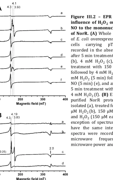
assays, respectively. Under these conditions, it was observed that the

norV

transcription and expression were hindered. To identify if the lack of

norV

(12)

xii

was also examined

in vivo

upon infection of macrophages with the

E. coli

wild type and the

norV

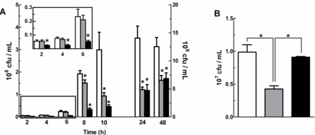
mutant strain. We showed that the contribution of

flavorubredoxin to survival of

E. coli

depends on the stage of macrophage

infection, and that the absence of protection observed at the early phase is

related to the inhibition of the NorR activity by the oxidative burst.

In this dissertation, a homologue of

E. coli

YtfE was found in

Staphylococcus aureus

encoded by the

scdA

gene. To address its role in

S.

aureus

, the

scdA

gene was disrupted and the viability of the mutant

assessed, which resulted in a strain more sensitive to oxidative stress.

Furthermore, upon exposure to nitric oxide and hydrogen peroxide, the

scdA

staphylococcal mutant strain exhibited more pronounced inhibition of

the aconitase activity, an enzyme dependent on the integrity of

[4Fe-4S]

2+/1+

clusters.

S. aureus

scdA

was able to complement the

E. coli ytfE

mutant strain as it rescued the ability of the mutant to repair damaged Fe-S

clusters. In contrast, the Isc (

I

ron-

s

ulphur

c

luster) or Suf (

S

ul

f

ur formation)

systems, which contain proteins involved in the assembly of Fe-S clusters,

were unable to complement the same mutant strain. Hence, it was

concluded that

S. aureus

ScdA is involved in the repair of Fe-S clusters. A

comprehensive search of the amino acid sequence database revealed that

homologues of

E. coli

YtfE and

S. aureus

ScdA are found in the proteomes of

a wide range of bacteria, fungi, protozoa, and in several pathogens

including

Neisseria gonorrhoeae

,

Haemophilus influenzae

and

Bacillus

anthracis

. Thus, this family of proteins was named Ric for

R

epair of

i

ron

c

entres.

(13)

Fe-xiii

desulphurase IscS, the scaffold IscU and the

E. coli

holo-YtfE proteins were

recombinantly produced and purified. UV-visible and resonance Raman

studies demonstrated that holo-YtfE promotes the assembly of Fe-S

clusters in IscU and in the apoform of ferredoxin.

In order to identify the proteins that interact

in vivo

with

E. coli

YtfE, the bacterial adenylate cyclase two-hybrid system (BACTH) was used.

This study was performed in two ways: in the first, the interaction of YtfE

with specific proteins was analysed; in the second, the

E. coli

proteome was

screened for YtfE interactants. Since the assembly of Fe-S clusters requires

cysteine desulphurases as sulphur donors and scaffold proteins to

assemble the centre, it was analysed whether YtfE interacts

in vivo

with

these proteins. Hence, the cysteine desulphurases IscS and SufS, and the

scaffold protein IscU were cloned in the BACTH system plasmids. The

results showed that

E. coli

YtfE is able to interact with both IscS and SufS. In

the second part of this study, using two libraries that covered

approximately 30% of

E. coli

genome, we detected protein interactants that

were confirmed by further BACTH assays to interact with

E. coli

YtfE.

(14)
(15)

xv

As espécies reativas de oxigénio e de azoto (ERO e ERA) são

produzidas por células fagocíticas do sistema imunitário para atacar os

agentes patogénicos devido à sua capacidade de danificar o ADN e os

centros metálicos das proteínas. Para conseguirem sobreviver dentro do

hospedeiro, as bactérias ativam genes que codificam enzimas

destoxificantes, tais como a proteína flavo-di-férrica que reduz o óxido

nítrico em

Escherichia co

li, também conhecida como flavorubredoxina

(FlRd), e proteínas reparadoras, como a proteína di-férrica YtfE de

E. coli

que está envolvida na recuperação de centros ferro-enxofre danificados.

Utilizando

E. coli

e

Staphylococcus aureus

, o trabalho apresentado nesta

tese pretendeu determinar: i) o papel de FlRd de

E. coli

em bactérias

expostas a uma combinação de stress oxidativo e nitrosativo; ii) a

identificação e caracterização do homólogo da YtfE em

S. aureus

; iii) o

estudo de mecanismos usados por YtfE de

E. coli

para reparar os centros

danificados de Fe-S; e iv) a identificação de proteínas que interagem com

YtfE de

E. coli

.

Para avaliar-se o papel de FlRd de

E. coli

em células tratadas com

peróxido de hidrogénio e óxido nítrico, a transcrição e a expressão do gene

norV

foi analisado usando-

se ensaios de β

-galactosidase e immunoblotting,

respectivamente. Nestas condições, observou-se que a transcrição e

expressão de

norV

foram eliminadas. Para identificar se a falta de expressão

de FlRd estava relacionada com o seu regulador, o fator de transcrição

NorR, a proteína foi clonada, expressa, purificada e a ligação de óxido

nítrico à NorR na presença de peróxido de hidrogénio foi estudada.

(16)

xvi

explorámos a atividade ATPase de NorR, um requisito para a ativação de

FlRd, na presença do stress oxidativo e nitrosativo. Os resultados

mostraram que, sob estas condições, a atividade ATPase não foi

despoletada. O papel de FlRd de

E. coli

foi também examinado

in vivo

após a

infeção dos macrófagos com a estirpe selvagem de

E. coli

e com a estirpe

mutada no gene

norV

. Mostrámos assim que a contribuição de

flavorubredoxina para a sobrevivência de

E. coli

depende da fase de infeção

dos macrófagos, e que a ausência de proteção observada na fase inicial está

relacionada com a inibição da atividade de NorR durante o stress oxidativo.

Nesta dissertação, foi encontrado um homólogo de YtfE de

E. coli

em

S. aureus

codificado pelo gene

scdA

. Para estudar a sua função em

S.

aureus

, o gene

scdA

foi interrompido e a viabilidade do mutante avaliada, o

que resultou numa estirpe mais sensível ao stress oxidativo. Para além

disso, após exposição ao óxido nítrico e ao peróxido de hidrogénio, esta

estirpe estafilocócica mutada no gene

scdA

exibiu uma maior inibição da

atividade de aconitase, uma enzima dependente da integridade dos seus

centros [4Fe-4S]

2+/1+

. A proteína ScdA de

S. aureus

conseguiu

complementar a estirpe de

E. coli

mutada no gene

ytfE

resgatando a

capacidade do mutante para reparar os centros Fe-S danificados. Em

contrapartida, os sistemas Isc (

I

ron-

s

ulphur

c

luster) e Suf (

Su

l

f

ur

formation), que contém proteínas envolvidas na montagem dos centros

Fe-S, foram incapazes de complementar a mesma estirpe mutante. Assim,

conclui-se que a proteína ScdA de

S. aureus

participa na reparação dos

centros Fe-S. Uma análise global da base de dados de amino ácidos revelou

(17)

xvii

chamada Ric (

R

epair of

i

ron

c

entres).

O papel de YtfE de

E. coli

na reparação dos centros Fe-S danificados

pelo stress oxidativo e nitrosativo foi também abordado ao analisar-se a

formação de centros Fe-S na proteína molde IscU e na ferredoxina que

contém centros [2Fe-2S]

2+/1+

na presença de YtfE. Com este propósito, a

cisteína desulfurase IscS, a proteína molde IscU e a holo-proteína YtfE

foram

produzidas

recombinantemente

e

purificadas.

Estudos

espectroscópicos de UV-visível e ressonância Raman demonstraram que a

holo-YtfE promove a montagem de centros Fe-S na IscU e na apo-forma de

ferredoxina.

Para se poder identificar as proteínas que interagem

in vivo

com

YtfE de

E. coli

usou-se o sistema BACTH (

B

acterial

A

denylate

C

yclase

T

wo-H

ybrid). Este estudo foi efetuado de duas maneiras: na primeira foi

analisada a interação de YtfE com proteínas específicas; de seguida, o

proteoma de

E. coli

foi sondado para se encontrar as proteínas que

interagem com YtfE. Uma vez que a montagem de centros Fe-S requer

cisteínas desulfurases como dadores de enxofre e proteínas molde para

montar o centro, analisou-se se YtfE interage

in vivo

com estas proteínas.

Assim, as proteínas cisteínas desulfurases IscS e SufS e a proteína molde

IscU foram clonadas nos plasmídeos do sistema BACTH. Os resultados

mostraram que YtfE de

E. coli

é capaz de interagir com IscS e SufS. Na

segunda parte deste estudo, usando duas bibliotecas que cobrem

(18)

xviii

resistência de micróbios patogénicos aos stresses oxidativo e nitrosativo

(19)

xix

Abs

Absorbance

AhpCF Alkyl hydroperoxide reductase

Amp

Ampicillin

Asp

Aspartic acid

ATP

Adenosine tryphosphate

ATPase ATP hydrolase

B.

Bacillus

BACTH Bacterial adenylate cyclase two-hybrid system

Bfr

Bacterioferritin

CLR

C-type lectin receptor

CO

Carbon monoxide

Cya

Adenylate cyclase

Cys

Cysteine

Da

Dalton

DNA

Deoxyribonucleic acid

DNIC

Dinitrosyl-iron complex

Dps

DNA protein from starved cells

DTT

Dithiothreitol

E.

Escherichia

e.g.

exempli gratia, for example

eNOS

endothelial NOS

EPR

Electron paramagnetic resonance

Erm

Erythromycin

et al.

et alia, and other people

(20)

xx

FlRd

Flavorubredoxin

FMN

Flavin mononucleotide

FNR

Fumarate and Nitrate reduction Regulator

Ftn

Ferritin

Fur

Ferric uptake regulator

Glu

Glutamic acid

GMP

Guanosine monophosphate

GTP

Guanosine tryphosphate

H

2

O

Water molecule

H

2

O

2

Hydrogen peroxide

His

Histidine

Hmp

Flavohaemoglobin

HO

-

Hydroxyl anion

HO

Hydroxyl radical

HOCl

Hypochlorous acid

i.e.

id est, that is to say

IFNγ

Interferon γ

IHF

histone-like integration host factor

IL

Interleukin

iNOS

inducible NOS

IPTG

Isopropyl-

β

-D-thiogalactopyranoside

IRP

Iron-regulatory protein

Isc

Iron-sulphur cluster system

Kan

Kanamycin

kDa

kilo Dalton

(21)

xxi

NADH

β

-Nicotinamide adenine dinucleotide, reduced form

NADPH

β

-Nicotinamide adenine dinucleotide phosphate, reduced form

Nfu

NifU-like

Nif

Nitrogen fixation system

NK

Natural killer

NLR

Nod-like receptor

nNOS

neuronal NOS

NO

Nitric oxide

NO

2

-

Nitrite

NOR

Nitric oxide reductase

NorR

Nitric oxide reduction regulator

NOS

Nitric oxide synthase

NrfA

cytochrome c nitrite reductase formate-dependent A

NsrR

Nitrite sensitive repressor regulator

O

2

Oxygen

O

2

-

Superoxide anion

OD

Optical density

ONOO

Peroxynitrite

ONPG o-nitrophenyl-

β

-D-galactopyranoside

OxyR

Oxygen stress regulator

PAMP Pathogen-associated molecular pattern

PerR

Peroxide regulator

(22)

xxii

ROO

Rubredoxin:oxygen oxidoreductase

ROS

Reactive oxygen species

RR

Resonance Raman

S.

Staphylococcus

SOD

Superoxide dismutase

SOR

Superoxide reductase

SoxRS Superoxide regulator and sensor

Suf

Sulfur assimilation

TCA

Tricarboxylic acid

TLR

Toll-like receptor

TNF

Tumor necrosis factor

UV

Ultra-violet

(23)

xxiii

Introduction

Chapter I

Nitric Oxide, an antibacterial molecule of the immune system

I.1

The innate immune system

7

I.1.1

Oxidative stress

10

I.1.2

Nitrosative stress

15

I.2

Bacterial responses to nitrosative stress

18

I.3

Nitric oxide detoxification

21

I.3.1

Flavodiiron family of proteins

21

I.3.2

Globins family: Flavohaemoglobin

27

I.3.3

Nitrite reductases

30

(24)

xxiv

II.1

Chemical properties and biological role of iron

49

II.2

Iron uptake and homeostasis

50

II.2.1

Iron uptake systems

50

II.2.2

Iron storage proteins

52

II.2.3

Regulation of iron homeostasis

54

II.3

Iron containing proteins

57

II.3.1

Haem proteins

57

II.3.2

Non-haem iron proteins:

Mono, dinuclear iron and mixed metal proteins

58

II.3.3

Iron-sulphur containing proteins

59

II.3.3.1

Fe-S clusters types and biological relevance

60

II.3.3.2

Fe-S clusters assembly mechanisms

62

II.3.3.3

Damage and repair of Fe-S clusters

77

II.3.3.4

YtfE, a protein that repairs Fe-S clusters

81

(25)

xxv

Chapter III

Oxidative stress modulates the nitric oxide defense promoted by

E. coli

III.1

Introduction

105

III.2

Materials and Methods

107

III.3

Results

111

III.4

Discussion

119

III.5

References

122

III.6

Acknowledgments

126

Chapter IV

Widespread distribution in pathogenic bacteria of di-iron

proteins that repair oxidative and nitrosative damage to

iron-sulphur centres

IV.1

Introduction

131

IV.2

Materials and Methods

133

IV.3

Results and Discussion

142

(26)

xxvi

Chapter V

Escherichia coli YtfE is able to promote the formation of

iron-sulphur clusters

V.1

Introduction

169

V.2

Materials and Methods

170

V.3

Results

172

V.4

Discussion

176

V.5

References

177

V.6

Acknowledgments

178

Chapter VI

Searching for YtfE interactions in the Escherichia coli proteome

VI.1

Introduction

183

VI.2

Materials and Methods

184

VI.3

Results

190

VI.4

Discussion

196

(27)

xxvii

Discussion

Chapter VII

Discussion and general conclusions

VII.1

Discussion

207

VII.1.1

The role of

Escherichia coli

flavorubredoxin

207

VII.1.2

The function of the Ric family of proteins

213

VII.2

General conclusions

223

(28)
(29)
(30)
(31)

Chapter I

Nitric Oxide

,

(32)
(33)

I.1

The innate immune system

7

I.1.1

Oxidative stress

10

I.1.2

Nitrosative stress

15

I.2

Bacterial responses to nitrosative stress

18

I.3

Nitric oxide detoxification

21

I.3.1

Flavodiiron family of proteins

21

I.3.2

Globins family: Flavohaemoglobin

27

I.3.3

Nitrite reductases

30

I.4

References

32

(34)
(35)

7

Chapter I

Nitric Oxide,

an antibacterial molecule of the immune system

"Though NO's structure is simple, nitric oxide is now regarded as the

most significant molecule in the body, absolutely crucial to your

well-being.”

Louis Ignarro, Nobel Prize (1998)

I.1

The innate immune system

The immune system is a mechanism involving several biological

structures and processes that protects an organism against disease through

recognition and elimination of the infectious agent (1). The immune system

is divided in three levels:

anatomic and physiological barriers, innate

immunity and adaptive immunity (2).

The anatomic and physiological barriers are the first line of defense

against invading microorganisms and include intact skin, surface of mucous

membranes, mucociliary clearance mechanisms and low stomach pH (1, 2).

In contrast, the adaptive immunity occurs after several days of

infection and the peak of primary adaptive response, which is characterized

by lymphocyte activation and proliferation, only takes place after 5-7 days

post-infection (3). The adaptive immune response exhibits four

immunological attributes: specificity, diversity, memory and self/nonself

(36)

8

classes of specialized lymphocytes, the T and B cells, which display an

extremely diverse repertoire of antigen-specific recognition receptors that

enable specific identification and elimination of pathogens, as well as

adaptive immune methods that ensure tailored immune responses and

long lived immunological memory against reinfection (1). The adaptive

immunity is divided into humoral immunity, which is involved in the

eradication of microbes present in the blood or fluid by generating

antibodies which are produced by B-cells, and cellular immunity, that

involves the suppression of cancer cells and microbes hidden inside cells

mediated by killer T-cells (3).

While the adaptive immune system is a later response found only in

vertebrates, the innate immunity occurs in eukaryotes, from humans to

earthworms, acting within seconds upon encounter with a pathogen,

generating a protective inflammatory response (1). The innate immunity

consists of immunological effectors that provide robust, immediate and

nonspecific immune responses, which include evolutionarily primitive

humoral, cellular, and mechanical processes that play a vital role in the

protection of the host from pathogenic challenge (1).

The pathogen recognition is the first and crucial step in innate

immunity, performed by a limited arsenal of pattern recognition receptors

(PRRs), like Toll-like receptors (TLRs), C-type lectin receptors (CLRs),

NOD-like receptors (NLRs), and RIG-NOD-like helicases (RLH), that are able to

recognize conserved structures of microorganisms called

pathogen-associated molecular patterns (PAMPs) (1-3).

In addition, the ingestion of extracellular particulate material (such

as pathogens) by phagocytosis is one of the most important innate defense

mechanisms (1). Phagocytosis involves binding of the particle to be

(37)

9

(1, 4). This process is performed by specialized cells (the phagocytes), such

as neutrophils, dendritic cells and macrophages, that engulf and kill the

pathogenic microorganisms (1, 3). When bacteria enter into mammalian

cells by phagocytosis, the innate immune sensors, like TLRs or NLRs,

triggered by bacterial ligands, initiate pro-inflammatory responses and

innate immune effector functions (1, 5). Phagocytes internalize

microorganisms into specialized vesicles known as phagosomes which will

acidify and fuse with lysosomes (intracellular vesicles that contain a variety

of antimicrobial factors), forming a phagolysosome (5, 6). The antimicrobial

factors generated within the phagolysosome include reactive oxygen and

nitrogen species (ROS and RNS, respectively) (1, 7). Pathogens in this stage

of infection can be divided in two types: the extracellular pathogens that

survive outside host cells because phagocytosis will eliminate them, and

intracellular pathogens that invade host cells within which they survive

and replicate (5).

The phagocytic cells macrophages are the main tools of innate

immunity being found in almost every tissue of the host, and are

responsible for the initial killing of the majority of the engulfed bacteria.

Upon infection, macrophages secrete TNF

α (

Tumor Necrosis Factor α) and

IL-12 (Interleukin

12) that lead NK (Natural

Killer) cells

to produce IFNγ

(InterFeroN-γ), which will result in the increase of macrophage bactericidal

activity (8-10). These specialized phagocytes are implicated in other

physiological processes, such as development of the haematopoietic

system, bone remodeling and wound healing (11, 12).

Note that only vertebrates have the adaptive immune system,

leaving most eukaryotic organisms to survive with innate immunity alone.

Interestingly, new discoveries have been made regarding innate immunity

(38)

10

system but rather as a critical regulator of human inflammatory disease (2).

Now, it is clear that innate immunity is specific as it is able to discriminate

self-molecules from pathogens through the evolutionary conserved

receptors TLRs that also act as adjuvant receptors creating a bridge

between innate and adaptive immunity (3). More important, innate

immune responses are crucial for the initiation of adaptive immune

responses in vertebrates, conferring a specific and long lasting protection

(1).

I.1.1

Oxidative Stress

Reactive oxygen species produced by phagocytes are necessary for

efficient host defense against bacterial and fungal infections (13). However,

the oxygen toxicity is also a problem for non-pathogen aerobic organisms

as ROS are inevitable byproducts of aerobic metabolism (14). Reactive

oxygen species are derivatives of molecular oxygen, that include singlet

oxygen, superoxide anion, hydrogen peroxide and hydroxyl radicals (Figure

I.1). The generation of all these reactive oxygen species is designated as

oxidative burst (15).

Figure I.1

The red ox states of oxygen with reduction potentials.

O

2

molecular dioxygen; O

2

-

- Superoxide anion; H

2

O

2

hydrogen peroxide; HO

hydroxyl radical; OH

-

- hydroxyl anion; e

-

- electrons ; and H

+

- protons. Adapted

from (

14

).

O

2

O

2

-

H

2

O

2

HO

OH

-e

-

e

-

, 2H

+

e

-

, 2H

+

e

-

, H

+

(39)

11

Upon phagocytosis in macrophages, a membrane-bound oxidase is

activated that reduces oxygen to superoxide anion (O

2

-

), a reactive oxygen

intermediate extremely toxic to ingested microorganisms. This

membrane-bound oxidase, designated as Phox (NADPH oxidase), pumps electrons into

the phagolysosome catalyzing the following reaction: 2O

2

+ NADPH

2O

2

-+ NADP

+

+ H

+

(16). This enzyme is dormant in the resting phagocyte but

becomes activated when the cell receives stimuli, like infectious bacteria

and certain inflammatory polypeptides (17). Phagocytes produce

superoxide in the order of 500 nM/h (18, 19). Phox contains haem as

prosthetic group and is an important defense mechanism in microbial and

parasite killing as mice deficient in Phox have enhanced bacterial burden

when compared to wild type (10, 20, 21). Further, humans who

don’t

possess a functional Phox suffer from immunodeficiency due to recurrent

bacterial and fungal infections (22). Superoxide anion is a reactive

compound capable of acting as an oxidant or reductant in biological

systems, and can diffuse for considerable distances before it exerts toxic

effects. Furthermore, extracellularly generated O

2

-

can gain access to

intracellular targets via cellular channels (23). The superoxide anion also

generates other powerful oxidizing agents, including hydroxyl radicals

(OH

) and hydrogen peroxide (H

2

O

2

, through superoxide dismutase

enzyme) (Figure I.1) (2).

Another compound that is involved in microorganism killing is

hypochlorous acid (HOCl) (24-26). This ROS is produced by

myeloperoxidase (MPO), a haem-containing enzyme present in activated

macrophages and neutrophils (26). Mice deficient in MPO are more

susceptible to infection (27). In addition, HOCl reacts with ferrous iron to

(40)

12

form hydrogen peroxide which, subsequently, reacts with Fe(II) to form the

hydroxyl radical (Fenton reaction: H

2

O

2

+ Fe(II)

OH

+ OH

+ Fe(III))

(Figure I.1) (28). All these highly toxic products are capable of killing

pathogens (16, 29).

Hydrogen peroxide is an uncharged species and readily diffuses

across cell membranes (23). Therefore, H

2

O

2

stress emerges inside cells

whenever this species is present in the extracellular space (15). During

respiratory burst, phagocytic cells produce hydrogen peroxide in the order

of 750 nM/h (18, 19).

Reactive oxygen species are involved in the killing of pathogens by

directly reacting with lipids, proteins, DNA and cell carbohydrates (Figure

I.2) (15, 30). The lipid peroxidation occurs essentially in biological

membranes which serve as impermeable barriers and in cellular transport

processes (Figure I.2). The superoxide anion and the hydroxyl radical are

the ROS known to initiate the process of autocatalytic lipid peroxidation in

eukaryotes, but

apparently

not

in bacteria as

most

bacterial

membranes lack

polyunsaturated

fatty acids (18,

30,

31).

This

process leads to

the

conversion

of

unsaturated

lipids into polar lipid hydroperoxides, which causes increased membrane

Figure I.2

Major targets of reactive oxygen species.

Adapted from (

30

).

ROS

Respiration

Environmental oxidants,

phagocytes

Lipid peroxidation

DNA oxidation

Protein oxidation

Loss of

function

Fe

ROS

(41)

13

fluidity, efflux of cytosolic solutes and loss of membrane-protein activities.

Ultimately, lipid peroxidation results in the damage of the membrane and

cell death (18, 30). Also, products of oxidized lipids initiate further

oxidative damage (14, 30).

Although DNA does not react with ROS at physiological pH, the

negatively charged phosphodiester backbone of DNA binds to metal ions,

especially iron (Figure I.2). Consequently, hydroxyl radical is generated,

attacking sugars, purines and pyrimidines of DNA and generating multiple

products (32, 33). Interestingly, DNA seems to be a more important target

of ROS in bacteria as the membrane lipid peroxidation through ROS is

unlikely in these organisms (30).

Protein susceptibility to oxidative damage depends on several

factors, such as the relative content of oxidation-sensitive amino acid

residues (methionine, cysteine or tryptophan), the presence of

metal-binding sites (e.g. Fe-S clusters, chapter II), protein localization in the cell

and solvent exposition that depends upon molecular conformation (Figure

I.2). The newly synthesized proteins are the most prone to oxidative

damage, indicating that complete folding and incorporation into protein

complexes confers protection from oxidative-driven degradation (30).

Although the oxidative damage affects proteins of important metabolic

pathways, the fact is that protein oxidation may not lead to cell death as the

importance of a metabolic pathway for cell vitality can depend strongly on

the environmental situation, hence some oxidation-sensitive proteins may

be dispensable under some conditions, while required in other (30). The

oxidized amino acid residues can form irreversibly carbonyl products.

Carbonylated proteins are degraded by the proteosome, however there is a

limit to the cell capacity to process these modified proteins, as the

(42)

14

carbonylated proteins that are not degraded form toxic aggregated species

leading ultimately to loss of cell viability (30).

Pathogenic microorganisms subvert or resist the action of oxidative

burst through a range of strategies that also involve the up-regulation of

antioxidant proteins that transform ROS into less toxic products. The

scavenger enzymes for superoxide anion are SOD (SuperOxide Dismutase)

and SOR (SuperOxide

Reductase). Superoxide dismutation can occur

spontaneously with a constant rate of 10

5

M

-1

.s

-1

, or under the catalytic

influence of SOD with a constant rate of ~10

9

M

-1

.s

-1

, that varies between

different organisms (34). Four types of prokaryotical SODs exist: the

copper-zinc (Cu/Zn-SOD), the manganese (Mn-SOD), the iron (Fe-SOD) and

the nickel (Ni-SOD) (17). Most bacteria possess Fe-SOD and Mn-SOD in

their cytoplasm. The Fe-SOD is usually the constitutively expressed

enzyme, while Mn-SOD is in general induced by oxygen stress (35). The

periplasmic bacterial Cu/Zn-SOD is constitutively expressed and confers

infection resistance to pathogenic bacteria (36, 37). Superoxide reductases

are used by anaerobic organisms to scavenge superoxide anion, rather than

SODs (15, 38). This enzyme reacts with superoxide anion with a rate

constant of

10

9

M

1

. s

1

, that differs depending on the organism (39).

For hydrogen peroxide, the pathogens possess two families of

scavenger enzymes (15, 40): peroxidases with rate constants of 10

7

-10

8

M

-1

.s

-1

(41, 42), and catalases with rate constants of ~10

6

M

-1

.s

-1

(43). Both

families of enzymes in general catalyze the reaction: RH

2

+ H

2

O

2

R +

2H

2

O, where R=O

2

for catalases. One well known peroxidase is AhpCF

(Alkyl

hydroperoxide reductase) an important scavenger of hydrogen

peroxide in several bacteria (44). Catalases and peroxidases exist in several

(43)

15

Helicobacter pylori, Streptomyces sp., Leishmania major and Escherichia coli

(15, 45). Known bacterial species produce more than one catalase and

several peroxidases that can scavenge H

2

O

2

, being able to handle the ROS

that results from oxidative burst (18).

To resist ROS, bacteria sense these molecules through specific

regulators whose regulons include genes encoding proteins involved in the

protection against oxidative burst. The OxyR (Oxygen stress

Regulator)

regulon includes genes encoding catalase and AhpC, glutathione reductase

and glutaredoxin that restores intracellular thiol/disulphide balance,

proteins involved in DNA protection (Dps, chapter II), and important

regulators such as

fur (ferric

uptake

regulator, Chapter II)

(46-48). PerR

(Peroxide

Regulator) is another major regulator of the peroxide stress

resistance present in Gram-positive and Gram-negative microorganisms

(49). Its regulon includes genes encoding catalases and peroxidases, genes

involved in haem biosynthesis, regulators as fur and the zinc uptake system

(49, 50). SoxRS (Superoxide

Regulator and

Sensor) is a well-distributed

bacterial regulator that responds to superoxide stress by inducing the

expression of superoxide scavenger proteins and enzymes that repair

damaged proteins and DNA (see also I.2) (15, 51).

I.1.2

Nitrosative Stress

At first, nitric oxide (NO) was considered to be merely an

(44)

16

Nitric oxide is a small soluble radical molecule (30 Da) that can

stabilize its unpaired electron by reacting with species containing other

unpaired electrons or by interacting with the d-orbitals of transition metals

(e.g. iron) (52). NO is soluble in water (1.6 mM at 37ºC), where it has a

short half-life, between 3 and 20 s (53, 54). Reactive nitrogen species (RNS)

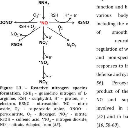
include species derived from nitric oxide (Figure I.3), that arise in

physiological environments, and include NO

2

-

(nitrite), S-nitrosothiols,

peroxynitrite (ONOO

), dinitrosyl-iron complexes (DNIC, chapter II), among

others (Figure I.3) (55). NO and related nitrogen oxides are endogenous

regulators of cell and tissue

function and have a role in

various

body

functions,

including the vasodilatation

of

smooth

muscle,

neurotransmission,

regulation of wound healing

and non-specific immune

responses to infection, host

defense and cytotoxicity (52,

56).

Peroxynitrite,

the

product of the reaction of

NO

and

superoxide,

is

involved in inflammation

(57) and in bacteria killing

(10, 58-60).

Figure I.3

Reactive

nitrogen

species

formation.

RNH

2

guanidi no nitrogen of

L-arginine, RSH - sulphydril, H

+

- proton, e

-

-electron, RSNO - nitrosothiol,

NO

nitric

oxide,

O

2

-

- superoxide anion,

ONOO

-

-peroxinitrite, O

2

- dioxygen, NO

2

-

- nitrite,

RSOH

sulfenic acid,

NO

2

nitrogen dioxide,

NO

3

-

- nitrate. Adapted from (

55

).

RNH

2

NO

NO

2

-•

NO

2

NO

3

-RSNO

RSOH

OONO

-O

2

-RSH + O

2

NO

2

-RSH

H

+

+ e

-N

2

O

3

O

2

e

-e

(45)

-17

In mammalian cells, nitric oxide is produced by NOS (Nitric

Oxide

Synthase). This class of enzymes is widely distributed throughout the

mammalian tissues. Endothelial (eNOS, constitutive), neuronal (nNOS,

constitutive) and inducible (iNOS) isoforms are prompted to convert

arginine into nitric oxide (L-arginine + O

2

+ NADPH

NO + L-citrulline +

NADP

+

) (57). The NOS isoforms are soluble and contain flavin adenine

dinucleotide (FAD), flavin mononucleotide (FMN) and haem iron as

prosthetic groups, requiring also the cofactor tetrahydrobiopterin (BH

4

)

(61). The constitutive forms are low-activity enzymes that produce small

amounts of NO as a signaling molecule (62). The inducible form is an

enzyme produced by phagocytes when they are stimulated (63).

When macrophages are activated with bacterial cell-wall

components, such as lipopolysacharide (LPS), together with a

T-cell-derived cytokine (IFN-

γ)

, they express high levels of inducible nitric oxide

synthase (64-68).

iNOS is present in other phagocytic cells of the immune

system, such as dendritic cells and natural killer cells (69). The activity of

iNOS affords effective protection against infection (69). The NO produced

diffuses across membranes and can kill or inhibit a broad range of

organisms, such as bacteria, fungi, parasitic worms and protozoa, persisting

for a longer period of time, when compared to the short-lived oxidative

burst (31, 55, 70). The most important targets of nitric oxide and RNS are

protein thiols, aromatic amino acid residues, metal centres, lipids and

nucleotide bases (DNA), resulting in the blockage of essential microbial

physiological processes such as respiration and DNA replication (71-76).

Mice deficient in iNOS are more susceptible to infection by many

intracellular

pathogens

like

Mycobacterium

tuberculosis,

Listeria

(46)

18

Porphyromonas gingivalis,

Trypanosoma cruzi and

Plasmodium falciparum,

when compared to the wild type (10, 55, 77-85).

Interestingly, nitric oxide synthase enzymes were also found to be

produced by bacteria. As occurs for their homologues in mammals, all the

bacterial NOS possess a haem ring and are able to convert L-arginine to

nitric oxide, but in contrast they lack the associated NOS

red

(C-terminal

flavoprotein reductase domain) (86-91). Moreover, the NO release rates are

lower when compared to mammalian NOS (86). Homologues were found

mostly in Gram-positive bacteria (e.g.

Streptomyces

sp.,

Bacillus

sp. and

Sthapylococcus sp.) and so far in one Gram-negative bacterium (Sorangium

cellulosum

) (87). In bacteria, this enzyme is able to provide protection

against oxidative and nitrosative stress, as expression of bacterial NOS will

result in production of small concentrations of NO, which leads to the

activation of sensors that respond to nitric oxide (see I.2). The result is the

activation of regulons, which include genes encoding enzymes that

participate in protection against oxidative and nitrosative stress (87, 90,

91).

I.2

Bacterial response to nitrosative stress

The ability to scavenge or detoxify NO produced by phagocytic cells

is a survival characteristic in several microorganisms that depends on

genetic, biochemical and physiological factors. Bacteria possess many

mechanisms of defence against nitrosative stress. One of these strategies

involves bacterial regulators/sensors that will activate large-scale changes

(47)

19

(92). Till now several transcriptional regulators that contain different

transition metal cofactors had been identified, from Fe-S clusters to haem

centres, which control gene expression towards the stimuli nitric oxide.

Fe-S clusters containing-regulators that respond to nitric oxide are

SoxR, FNR (Fumarate and

Nitrate reduction

Regulator) and IscR

(Iron-Sulphur

Cluster assembly system

Regulator). SoxR belongs to the MerR

(Mercury

Resistance) family, regulates the stress response to superoxide

and nitric oxide and contains a [2Fe-2S] cluster (93, 94). SoxR switches on

the transcription of the gene encoding the regulator SoxS, which

consequently activates the expression of SoxRS regulon members. The

SoxRS switches on in response to NO, inducing the expression of genes

encoding a superoxide dismutase, a DNA repair enzyme (endonuclease IV),

and a nitroreductase, among others (95, 96).

The FNR subfamily members belong to a large family of widely

distributed regulators that control gene expression in response to nitric

oxide (and other ligands such as oxygen) activating the transcription of

NO-related genes (94). The cofactor in this regulator differs between

organisms, as a Fe-S cluster was identified in

E. coli, whereas in other

bacteria this regulator has a haem group (e.g.

Pseudomonas sp. DNR) (93,

94). The most studied regulator of this family is FNR that contains a

[4Fe-4S] cluster that forms DNICs (DiNitrosyl

Iron

Complexes, see Chapter II)

upon reaction with NO (93, 94). FNR controls gene expression during

anaerobic growth and one of the genes from the E. coli FNR regulon is the

important NO-detoxifying protein flavohaemoglobin, whose characteristics

will be further discussed (II.3.1/2). Moreover,

Neisseria gonorhoeae FNR

regulates

aniA, a gene coding a nitrite reductase (93, 94). Also, in

Campylobacter jejuni, the NssR (Nitrosative stress Regulator, an FNR family

(48)

20

are important for nitric oxide detoxification (97). Another important Fe-S

cluster-containing regulator that senses NO is IscR whose role and

characteristics will be further discussed in Chapter II.

NsrR (Nitrite

sensitive

repressor

Regulator) is a regulator of NO

metabolism in gamma- and beta-proteobacteria but also in Gram-positive

bacteria like

Bacillus and

Streptomyces species (98). This transcription

factor binds a Fe-S centre through a sequence motif that contains three

conserved cysteine residues (99-101). NsrR contains in its regulon genes

encoding flavohaemoglobin and nitrite reductase proteins (98, 102).

The major sensor of nitric oxide in eukaryotes is the soluble

guanylate cyclase which contains a haem cofactor that binds NO, resulting

in cyclization of GTP to cyclic GMP, which leads to the activation of several

pathways. The haem is located in a domain that displays a high sequence

identity to a family of sensors designated as H-NOX (Haem

NO/OXygen

binding) which are widespread in Bacteria, like Vibrio chloraea, Clostridium

botulinum,

Shewanella oneidensis and

Thermoanaerobacter tencongensis

(93, 94). The physiological role of these regulators is poorly understood in

bacteria; nevertheless, two studies carried out in

Legionella pneumophila

and Vibrio fischeri showed that H-NOX regulates biofilm formation and iron

metabolism, respectively (103, 104).

Mycobacterium tuberculosis contains

the DosRST (Dormancy

survival) system. Most DosR-regulated genes are

induced in activated macrophages in a NO synthase-dependent manner,

and the DosRST regulon includes

glbN encoding a truncated haemoglobin

which has a role in NO detoxification (93, 94).

Chapter II will describe the role of the regulatory protein Fur that

(49)

21

bacteria. This regulator is inactivated upon exposure to NO due to the

formation of DNIC species leading to derepression of the Fur regulon (105).

Furthermore, the regulon of PerR (a Fur homologue) is also derepressed

upon exposure to nitric oxide in vivo (106).

The regulatory and biochemical properties of the specific NO

regulator, NorR, will be discussed in the flavodiiron proteins subchapter.

I.3

NO detoxification systems

Among several mechanisms used by bacteria to resist nitrosative

stress are the nitric oxide detoxification systems. In enteric bacteria, three

proteins were identified as NO detoxificants: the periplasmic cytochrome c

nitrite

reductase

formate-dependent, NrfA; the flavorubredoxin, FlRd and

its associated oxidoreductase, NorW; and the flavohaemoglobin

protein,

Hmp.

I.3.1

Flavodiiron family of proteins

Flavodiiron proteins (FDP) represent a large family of enzymes,

widespread among Archaea, Bacteria and Protozoa, which contain a

conserved two-domain structural core, built by a metallo-

β

-lactamase-like-domain at the N-terminal region harbouring a non-haem diiron site, and a

flavodoxin-like domain, containing a FMN moiety (107, 108). FDPs possess

NO reductase activity and are involved in microbial resistance to nitric

oxide (107, 108). Although, the two-domain structural core is conserved in

this family of proteins, several members have extra domains fused at the

(50)

22

proteins were divided into four classes, from A to D: class A in which

proteins contain only the two domain core; class B with enzymes that have

an extra rubredoxin-like domain containing a FeCys

4

binding motif; class C

where proteins have an additional module that contains significant

similarities to NAD(P)H:flavin oxidoreductases; and class D with proteins

containing NAD(P)H:rubredoxin oxidoreductase and rubredoxin modules

fused to flavodiiron core (107-109).

The first reports of a protein from FDP class A arose with work on

Desulfovibrio gigas. The protein was named ROO (Rubredoxin:oxygen

oxidoreductase) and was shown to accept electrons directly from the

rubredoxin partner (107, 108). ROO has NO reductase activity, its deletion

results in sensitivity to NO and the expression of this enzyme is able to

complement the

norV

(encoding a FDP class B enzyme) mutant of

E. coli

under anaerobic conditions (110).

The class C enzymes were so far found in cyanobacteria and some

eukaryotic oxygenic phototrophic organisms (107, 111). In this class of

enzymes, the long electron transfer chains are not required, as the fusion of

the NAD(P)H:flavin oxidoreductases module to the flavodiiron core allows

the protein to accept electrons directly from NAD(P)H and perform several

intra-molecular electron transfer steps onto the diiron centre, which

reduces the diatomic substrate. This enzymes are proposed to reduce

dioxygen to water, avoiding the formation of ROS (112). A recent study in

Synechocystis sp.

showed that FDP proteins of class C are induced upon

nitrosative stress (113).

The class D enzymes were found so far encoded in the genomes of

Clostridiales and of the pathogen Trichomonas vaginalis; further studies are

Imagem

Figure I.3 – Reactive nitrogen species formation. RNH 2 – guanidi no nitrogen of  L-arginine, RSH - sulphydril, H + - proton, e -  -electron, RSNO - nitrosothiol, • NO – nitric oxide, O 2 - - superoxide anion, ONOO -  -peroxinitrite, O 2 - dioxygen, NO 2 -
Figure I.4 – Model of NorR-dependent activation of norVW . (A) NorR contains the regulatory GAF domai n (blue), the AAA + dom ain (green) and DNA-bindi ng domai n (light red)
Figure I.5 – The regulation of Hmp by NsrR. The hmp transcription start site and consensus NsrR-binding sites are shown i n blue and red, respec tively
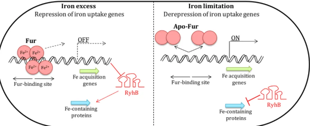
Figure II.1 – Fur regulation mechanisms . When iron is abundant, the Fur- Fur-Fe 2+ dimer blocks the transcription of downs tream genes (Fe ac quisition genes)
+7

Referências

Documentos relacionados

redemocratização no Brasil, expressada pela Constituição de 1988 (BRASIL, 1988), estabeleceu um novo paradigma para as liberdades individuais, diferenciando-se do período de

67 Ora, é perante este conjunto de problemas, que dizem respeito aos domínios da história do Egipto, da ciência, do colonialismo, do Estado, da economia e da política

A teoria Kohlbergiana tem em linha de conta o problema das relações entre o pensamento moral e acção moral, que procura saber quando, como e em que circunstâncias é que

Relativamente à forma como homens e mulheres que passam por um ou mais tratamentos e homens e mulheres que não passaram por nenhum tratamento, caraterizam a relação

De acordo com os normativos vigentes, a avaliação é considerada como parte constitutiva da aprendizagem, um elemento integrante e regulador de toda a prática educativa,

O Estado na prossecução do interesse público e numa articulação entre contenção orçamental e necessidade de realização de grandes empreendimentos, tem optado

mulheres e os homens de forma diferente de acordo com o seu ESE - e um efeito prevalecente de sexo nos participantes do sexo masculino – avaliam mulheres e homens do mesmo ESE de

In a study performed by Barakat, Moustafa and Bikhazi (2012), it was found that 4 weeks of treatment with sodium selenite (5 ppm in drinking water) increased mRNA and protein