Vanessa Andreia da Costa Rocha
Characterization of cells that survived
to disinfection in food processing areas
of retail facilities
Universidade do Minho
Escola de Engenharia
October, 2016 V anessa Andr eia da Cost a R oc ha C h a ra c te ri za ti o n o f c e lls t h a t s u rv iv e d t o d is in fe c ti o n i n f o o d p ro c e ss in g a re a s o f r e ta il f a c ili ti e s Minho | 20 1 6 UVanessa Andreia da Costa Rocha
Characterization of cells that survived
to disinfection in food processing areas
of retail facilities
Master dissertation
Master in Bioengineering
Supervisor: Doctor Maria do Pilar de Araújo Teixeira
Co-supervisor: Doctor Diana Alexandra Ferreira
Rodrigues
|iii
Vanessa Andreia da Costa Rocha
Characterization of cells that survived
to disinfection in food processing areas
of retail facilities
Master dissertation
Master in Bioengineering
Supervisor: Doctor Maria do Pilar de Araújo Teixeira
Co-supervisor: Doctor Diana Alexandra Ferreira
Rodrigues
É autorizada a reprodução integral desta dissertação apenas para efeitos de investigação, mediante a declaração escrita do interessado, que a tal se compromete.
Universidade do Minho, _____/_____/_________
|v
Agradecimentos
A vida é uma longa caminhada, constituída por vários capítulos, e mais um desses capítulos termina agora, com a passagem de várias pessoas.
Em primeiro lugar, queria agradecer à minha orientadora, Doutora Pilar Teixeira e co-orientadora, Doutora Diana Rodrigues, pela orientação, motivação e ajuda na realização deste trabalho. A cada uma queria agradecer, à Doutora Pilar Teixeira pelas suas sugestões, conselhos, amizade, força e preocupação. À Doutora Diana Rodrigues pela partilha de conhecimentos, disponibilidade demonstrada, pela ajuda laboratorial e, especialmente pela amizade e por vezes pela confidencialidade. Muito obrigada pela aprendizagem, por tudo o que me conseguiram passar!
A todos os colegas do LIBRO também queria agradecer, pelo bom ambiente e bons momentos passados, bem como a boa disposição.
Especialmente à Diana Alves, Andreia Magalhães, Carla Faria e Sílvia Vilaça, por fazer dos longos dias passados no laboratório, muito mais leves e divertidos.
Um agradecimento especial vai para a minha amiga Tânia Grainha, pela amizade durante estes anos, confidencialidade e que sempre me acompanhou nos bons e maus momentos.
Queria agradecer à Vânia Gaio pela ajuda, tempo disponibilizado e amizade e à Ritinha pela ajuda na realização do PCR.
Queria muito agradecer à minha família, mãe e irmãos que sem dúvida é a família “união”. Agradecer pela força e apoio, preocupação e confiança transmitida, sempre acreditaram em mim e deram-me força nos momentos mais difíceis. Um obrigado à minha irmã que é como uma segunda mãe. Obrigado pelo exemplo que tens sido e serás sempre para mim!
|vii
Abstract
Microbial contamination is a serious concern in areas of food processing because of its negative impact on public health. Numerous foodborne outbreaks occur every year due to various types of pathogenic microorganisms. The most common procedures to control these undesirable microorganisms include the use of disinfectants, however the disinfection procedures often demonstrate some inefficiency in the inactivation and removal of the same. Thus, the main objective of this study was the characterization of cells that survived to cleaning and disinfection in food processing areas of one meat retail facility. For that, samples were collected from meat processing surfaces of one meat retail facility in the center of Braga (Portugal), before and after the cleaning and disinfection procedures. Then the isolation of bacteria by selective media and the identification by 16S sequencing were performed. Finally, the isolates were phenotypically characterized in terms of biofilm formation ability, susceptibility of planktonic cells and biofilms to two disinfectants commonly used in the food industry (sodium hypochlorite (SH) and hydrogen peroxide (HP)), and susceptibility of planktonic cells to broad spectrum antibiotics (ampicillin (AMP) and rifampin (RIF)).
Listeria innocua, Serratia spp. and Hafnia alvei were the microorganisms identified in the samples collected before cleaning and disinfection procedures, while Cellulosimicrobium spp., Serratia spp., and Enterobacter spp. were identified in the samples collected after cleaning and disinfection procedures. These microorganisms can represent a risk for public health, since some species are pathogenic. The results demonstrated that planktonic cells of all isolates tested were more susceptible to HP compared to SH. Moreover, these isolates showed good biofilm formation capacity, with an increase of total biomass along the time and, as expected, less susceptibility to disinfectants compared to planktonic cells. After exposure of biofilms to HP and SH at concentrations higher than the recommended ones, biofilms were still able to survive which may contribute to bacterial resistance to these compounds, as well as cross-resistance to antibiotics.
In view of the results obtained in this study, it was concluded that cells that survive to cleaning and disinfection procedures represents a risk in terms of contamination due to biofilm formation ability, which can contribute to bacterial resistance to these compounds as well as cross-resistance to antibiotics.
|ix
Resumo
A contaminação microbiana constitui um grave problema em áreas de processamento de alimentos devido ao impacto negativo na saúde pública. Diversos surtos alimentares ocorrem todos os anos devido aos vários tipos de microrganismos patogénicos presentes. Os procedimentos mais comuns de controlo destes microrganismos indesejáveis incluem a aplicação de desinfetantes, contudo esses procedimentos de desinfeção demonstram, frequentemente, alguma ineficiência na inativação e eliminação dos mesmos. Assim, o principal objetivo deste estudo foi a caracterização das células que sobrevivem à limpeza e desinfeção em áreas de processamento de alimentos num talho. Para isso, recolheram-se amostras antes e após a limpeza e desinfeção das superfícies de processamento das carnes num estabelecimento no centro de Braga (Portugal) e, de seguida, foi realizado o isolamento de microrganismos por meios seletivos e identificados por sequenciação 16S. Finalmente, os isolados foram caracterizados fenotipicamente em termos da sua capacidade de formação de biofilme, suscetibilidade das células planctónicas e do biofilme a dois dos desinfetantes comummente utilizados na indústria alimentar (hipoclorito de sódio (HS) e peróxido de hidrogénio (PH)), a susceptibilidade de células planctónicas a dois antibióticos de largo espectro (rifampicina (RIF) e ampicilina (AMP)).
Listeria innocua, Serratia spp. and Hafnia alvei foram os microrganismos identificados nas amostras recolhidas antes dos processos de limpeza e desinfeção, enquanto que Cellulosimicrobium
spp., Serratia spp., and Enterobacter spp. foram identificados nas amostras recolhidas após os processos de limpeza e desinfeção. Estes microrganismospodem representar um risco para a saúde pública devido a algumas espécies serem patogénicas. Os resultados obtidos mostraram que as células planctónicas foram mais suscetíveis ao peróxido de hidrogénio em comparação com o hipoclorito de sódio. Os isolados apresentaram boa capacidade de formação de biofilme, com um aumento de biomassa total ao longo do tempo e, como esperado, menor suscetibilidade aos desinfetantes em comparação com as células planctónicas. Após a exposição dos biofilmes ao peróxido de hidrogénio e ao hipoclorito de sódio, em concentrações mais elevadas do que as recomendadas, os biofilmes foram ainda capazes de sobreviver e podem contribuir para a resistência bacteriana a esses compostos, bem como uma resistência cruzada aos antibióticos.
Face aos resultados obtidos é possível concluir que as células que sobrevivem aos processos de limpeza e desinfeção representam um risco em termos de contaminação devido à capacidade de formação de biofilme, contribuindo para a resistência bacteriana a esses compostos bem como para a resistência cruzada aos antibióticos.
|xi
Content
Chapter 1 Introduction ... 21
1.1. Context ... 23
1.2. Objectives ... 23
1.3. Outline of the dissertation ... 24
Chapter 2 State of the art... 25
2.1. Microbial food contamination and cross-contamination ... 27
2.2. Foodborne diseases and associated pathogenic microorganisms ... 27
2.3. Microbial contamination of food contact surfaces ... 30
2.3.1. Bacterial adhesion ... 31
2.3.2. Biofilm formation ... 32
2.4. Control of foodborne pathogens ... 34
2.4.1. Cleaning and disinfection ... 35
2.4.2. Disinfectants in the industry ... 35
2.4.3. Cellular and biofilm resistance ... 38
2.4.4. Cross-resistance to antibiotics ... 39
Chapter 3 Considerations of the methodology ... 41
3.1. Surfaces sampling methods ... 43
3.2. Isolation Methods ... 43
3.3. Rapid methods for identification of isolates ... 45
3.4. Biofilm formation ... 46
3.5. Quantification of biofilm biomass ... 47
3.6. Microbial susceptibility testing ... 48
Chapter 4 Materials and methods ... 51
4.1. Sampling ... 53
4.2. Isolation of microorganisms ... 53
4.3. Identification by 16S sequencing ... 54
4.4. Microorganisms and growth conditions ... 55
4.5.1. Chemical disinfectants and antibiotics preparation ... 55
4.5.2. Determination of Minimum Inhibitory Concentration (MIC) ... 56
4.6. Biofilm formation ... 56
4.7. Evaluation of biofilm formation ability by Cristal Violet assay ... 57
4.8. Evaluation of biofilm susceptibility to chemical disinfectants ... 57
4.8.1. Disinfectants and Neutralizer Preparation... 57
4.8.2. Minimum Biofilm Eradication Concentration assay ... 58
4.9. Statistical Analysis ... 58
Chapter 5 Results... 59
5.1. Isolates collected before cleaning and disinfection ... 61
5.1.1. Isolation of bacteria by liquid enrichment and solid selective media ... 61
5.1.2. Identification of isolates by 16S sequencing ... 62
5.1.3. Evaluation of susceptibility to disinfectants and antibiotics ... 62
5.1.4. Evaluation of biofilm formation ability... 64
5.1.5. Minimum Biofilm Eradication Concentration ... 65
5.2. Isolates collected after cleaning and disinfection ... 65
5.2.1. Isolation of bacteria by liquid enrichment and solid selective media ... 65
5.2.2. Identification of isolates by 16S sequencing ... 67
5.2.3. Evaluation of susceptibility to disinfectants and antibiotics ... 67
5.2.4. Evaluation of biofilm formation ability... 69
5.2.5. Minimum Biofilm Eradication Concentration ... 70
Chapter 6 Discussion ... 71
Chapter 7 Conclusions and future work ... 79
7.1. Conclusions ... 81
7.2. Future Work ... 81
|xiii
List of Figures
Chapter 2
Figure 2.1. Foodborne outbreaks in Europe Union, 2013. Adapted from: EFSA, 2015.
Figure 2.2. Mechanisms of bacterial adhesion. Initially, the surface is conditioned by presence of food waste. Then transport of planktonic cells from the bulk liquid to the surface, where adsorption of cells at the surface is initiate and starting of EPS formation and production of cell-cell signaling molecules. Last step is irreversible adsorption of cell-cells. Adapted from: Shi and Zhu, 2009.
Figure 2.3. Representation of the five main stages of biofilm formation. Stage 1 - Initial and reversible attachment; Stage 2 - Irreversible attachment; Stage 3 - Development of biofilm architecture; Stage 4 - Biofilm maturation; Stage 5 - Dispersion of biofilm cells.
Chapter 3
Figure 3.1. Isolation procedure: enrichment and plating in selective media.
Chapter 4
Figure 4.1. Sampling scheme used in the present work (cutting boards and mincer).
Chapter 5
Figure 5.1. Aspect of the colonies grown (samples collected before cleaning and disinfection) using Oxford Listeria Agar Base and CHROMagarTM E.coli media.
Figure 5.2. Evaluation of biofilm formation ability of the isolates (collected before cleaning and disinfection) by violet crystal staining. Each bar represents average CV–OD570 values and standard
errors. Symbols indicate statistically different values (p < 0.05) between different growth periods for the same bacteria (*), and between different bacteria considering the same growth period (ƒ). Figure 5.3. Aspect of the colonies grown (samples collected after cleaning and disinfection) using Oxford Listeria Agar Base and CHROMagarTM E.coli media.
Figure 5.4. Evaluation of biofilm formation ability of the isolates (collected after cleaning and disinfection) by violet crystal staining. Each bar represents average CV–OD570 values and standard errors. Symbols indicate statistically different values (p < 0.05) between different growth periods for the same bacteria (*), and between bacteria considering the same growth period (ƒ).
|xv
List of Tables
Chapter 2
Table 2.1. Microorganisms responsible for common foodborne disease. Adapted from: FDA, 2014 and Varnam and Evans, 1996.
Table 2.2. Mechanisms of antibacterial action of some disinfectants used in the food industry. Adapted from: Sheldon, 2005.
Chapter 5
Table 5.1. Microbial growth of samples collected before disinfection on liquid enrichment media (Listeria Enrichment Broth Base, or E.E Broth), and on solid selective media (Oxford Listeria Agar Base, or CHROMagarTM E.coli).
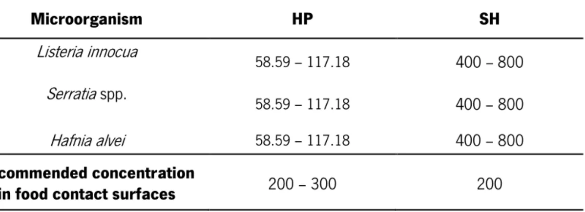
Table 5.2. MIC values obtained with the isolates collected before disinfection, and recommended concentrations of each disinfectant (µg/ml).
Table 5.3. MIC values of each antibiotic obtained with the isolates collected before disinfection (µg/ml).
Table 5.4. MBEC value obtained with the isolates collected before disinfection, and recommended concentration of each disinfectant (µg/ml).
Table 5.5. Microbial growth of samples collected after disinfection on liquid enrichment media (Listeria Enrichment Broth Base, or E.E Broth), and on solid selective media (Oxford Listeria Agar Base, or CHROMagarTM E.coli).
Table 5.6. MIC values obtained with the isolates collected after disinfection, and recommended concentrations of each disinfectant (µg/ml).
Table 5.7. MIC values of each antibiotic obtained with the isolates collected after disinfection of (µg/ml).
Table5.8. MBEC value obtained with the isolates collected after disinfection, and recommended concentration of each disinfectant (µg/ml).
|xvii
Glossary of abbreviations
AMP Ampicillin
CLSI Clinical and Laboratory Standards Institute
CV Crystal Violet
EU European Union
EUCAST European Committee on Antimicrobial Susceptibility Testing EPS Extracellular Polymeric Substance
HACCP Hazard Analysis Critical Control Point
HP Hydrogen Peroxide
ISO International Organization for Standardization LPS Lipopolysaccharide
MIC Minimum Inhibitory Concentration
MBEC Minimum Biofilm Eradication Concentration PBS Phosphate-Buffered-Saline
PS Peak Serum
RIF Rifampicin
SH Sodium Hypochlorite
SPSS Statistical Package for the Social Sciences
TAE Tris-acetate-EDTA
TSA Tryptic Soy Agar
TSB Tryptic Soy Broth
|xix
Scientific Output
Abstract in a conference:
Rocha V., Rodrigues D., Teixeira P. Characterization of biofilms of isolates from meat retail facilities. Biofilms7 International Conference, 26-28 Jun, 2016 Porto, Portugal.
|21
Chapter 1
Introduction
This chapter provides a general framing of this thesis, as well as a description of the objectives of the work. Finally, it is presented the outline of the dissertation, working as a guide line to the overall works presented in the further chapters.
|23
1.1. Context
Develop products with quality and that do not harm the health of consumers is a challenge for industries, and the food industry has a big importance in relation to others mainly due to its impact in global public health. In this sector,apart the floor and windows, there are equipments that require an efficient cleaning due to the existence of microorganisms that survive to disinfection programs leading to biofilm formation, which constitutes a higher risk of food contamination. Throughout of the food chain, maintenance of hygienic conditions is a fundamental factor in the control of foodborne diseases. Microorganisms such as Listeria monocytogenes, Escherichia coli and Salmonella spp. represent the most common foodborne pathogens that cause diseases in humans. Products provided from retail facilities are fractionated and can be subjected to the risks of contamination. Various factors such as cross-contamination, poor hygiene in food preparation and inappropriate processing may allow the multiplication of microorganisms until they reach infectious doses. When this happens, the products that do not undergo heat treatment can cause foodborne diseases, with unpredictable implications for consumers. Inadequate cleaning and disinfection of food processing environments causes economic losses and represents a serious risk to public health. Foodborne pathogens present on food contact surfaces have ability to adhere and form biofilms on different surfaces, as shown by numerous studies. Also, it is very well documented the difficulty in eliminating adhered and biofilm forms of these microorganisms compared to planktonic cells.
Disinfection of surfaces is an important issue related with the acquisition of bacterial resistance to disinfectant agents and the possible relation between disinfectants and the resistance to antibiotics. Disinfectants and antibiotics can have similar modes of action and, thus, induce in similar ways the development of bacterial resistance.
1.2. Objectives
Given the context described above, the main aim of this work was the characterization of cells that survived to cleaning and disinfection in food processing areas of one meat retail facility in the center of Braga, Portugal. More specifically, the work was focused on the following goals:
|Introduction
Phenotypical characterization of the isolates in terms of biofilm formation ability, susceptibility to disinfectants in planktonic cells and biofilm, and eventual cross-resistance to antibiotics.
1.3. Outline of the dissertation
This dissertation is divided in eight chapters. This first chapter consists on a brief introduction to the theme of this dissertation and defines the objectives proposed with this work.
On the second chapter it is presented the state of the art regarding microbial food contamination and cross-contamination, foodborne diseases, as well as a description of the related microorganisms. Several aspects related with microbial contamination of food contact surfaces and control of foodborne pathogens is also presented.
The third chapter describes some considerations of the methodology. The fourth chapter presents the methods used for realization this thesis.
The fifth chapter shows the results regarding the identification of isolates collected before and after disinfection, biofilm formation ability of the bacterial isolates, the susceptibility of planktonic cells and biofilm to different disinfectants, and the eventual acquisition of cross-resistance to antibiotics.
The sixth chapter presents a discussion about the results.
The seventh chapter contains the conclusions of the work done and the proposals for future work.
|25
Chapter 2
State of the art
This chapter encloses the literature review, presenting in the first sections a brief introduction to microbial food contamination and cross-contamination, foodborne diseases and pathogenic microorganisms associated. Also, adhesion and biofilm formation, different approaches to control foodborne microorganisms, and cross-resistance to antimicrobials are described.
|27
2.1. Microbial food contamination and cross-contamination
Food contamination is a constant public concern. There are three main types of food contaminants: microbiological, chemical, and physical (Scott, 1996) but the biggest problem related with food is due to pathogenic microorganisms rather than chemical or physical contaminants. The consumption of food contaminated with particular microorganisms or microbial products can cause diseases, as food infections and food poisoning (Reij and Den Aantrekker, 2004). There is a relationship between certain types of food and certain pathogens, which results from the natural contamination of the different foods and the processing or cooking customarily applied (Varman and Evans, 1996). There are several factors responsible for the occurrence of foodborne diseases. Cross-contamination of food is one of the most important factors contributing to the increasing number of foodborne diseases. This term is frequently used to refer to, in a general way, direct or indirect transfer of bacteria/virus from a contaminated product to a product not contaminated. In addition, other terms have been used to refer to bacterial transfer, as recontamination (contaminating food after an inactivation process), poor hygiene of food handlers, and contaminated equipment (Rodríguez et al., 2008). The risk of cross-contamination is associated with various stages of food preparation (Greig and Ravel, 2009). It has been observed that among the most common causes associated with cross-contamination of food are the inadequate cleaning of equipment and utensils, as well as poor personal hygiene (Rodríguez et al., 2008). Food industries has taken measures to ensure food safety principles, as the use of effective programs of quality control, implementation of Hazard Analysis Critical Control Point (HACCP) programs, and the increasingly use of safe methods during processing, transportation, storage and distribution of food. However, it is very important the training of food handlers and education of consumers in order to avoid foodborne diseases (Havelaar et al., 2010).
2.2. Foodborne diseases and associated pathogenic microorganisms
A diverse range of microorganisms can be present in food and food ingredients, which can include spoilage organisms as Pseudomonasaeruginosa, and also important pathogens as E. coli, L. monocytogenes and Salmonella enterica. These organisms are able to grow due to intrinsic (water activity and pH) and extrinsic (temperature, processing conditions and gaseous
|State of the art
atmosphere) properties of the food (Eviras, 2001). Table 2.1 shows the major foodborne pathogens and the main characteristics of the diseases they cause.
Table 2.1. Microorganisms responsible for common foodborne disease. Adapted from: FDA, 2014 and Varnam and Evans, 1996.
Microorganism Symptoms Disease Food sources
Salmonella spp.
Diarrhea, vomiting, fever and malaise, usually 12 to 16 hours after ingestion.
Infection
Contaminated eggs, poultry, unpasteurized milk or juice, cheese, contaminated raw fruits, vegetables.
Shigella spp.
Diarrhea with mucoid, bloody stools, 12 to 50
hours after ingestion. Infection
Food or water contaminated with human fecal material, ready-to-eat foods touched by infected food workers (raw vegetables, salads, sandwiches).
Campylobacter jejuni
Prodromal fever and malaise, 2 to 11 days after ingestion followed by abdominal pain and profuse diarrhea.
Infection
Raw and undercooked poultry, unpasteurized milk, contaminated water.
Escherichia coli Symptoms vary according
to type of E.coli infection. Infection
Water or food contaminated with human feces, undercooked beef, unpasteurized milk and juice, raw fruits and vegetables.
Listeria monocytogenes
Meningitis in neonates, abortion in pregnant females, septicaemia. Usually extended period between ingestion and appearance of symptomas.
Infection
Fresh cheeses, unpasteurized or inadequately pasteurized milk, ready-to-eat deli meats.
Staphylococcus aureus
Vomiting, abdominal pain and diarrhoea on some accasions, 2 to 6 hours after ingestion. Severe dehydration may result in collapse.
Intoxication
Unrefrigerated or improperly refrigerated meats, potato and egg salads, cream pastries.
|29
Table 2.1. (Cont.) Microorganisms responsible for common foodborne disease. Adapted from: FDA, 2014 and Varnam and Evans, 1996.
Microorganism Symptoms Disease Food sources
Bacillus cereus Two distinct syndromes:
diarrhea and emetic. Intoxication
Meats, stews, gravies, vanilla sauce.
Clostridium botulinum
Fatigue, lassitude, dizziness and effects on the central nervous system including speech difficulties and visual disturbances. Onset is 24 to 72 hours after ingestion.
Intoxication
Inadequately processed, home-canned foods, sausages, seafood
products, chopped bottled garlic, honey.
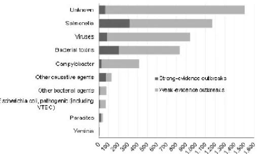
Foodborne diseases represent a global threat to human health. Most of these diseases are associated with pathogenic microorganisms and/or their toxins, while other causes, such as parasites and chemical substances naturally present in some foods, are also responsible (Newell et al., 2010). According to the World Health Organization (WHO) a foodborne disease is often of toxic or infectious nature, and is caused by pathogenic microorganisms that come in contact with human body through ingestion of contaminated food or water (Balbani and Butugan 2001). In the year 2013, most of the reported outbreaks in European Union (EU) remain to be caused by Salmonella, followed by viruses, bacterial toxins, and Campylobacter (Figure 2.1).
In 2013, 5196 foodborne outbreaks were reported in EU causing a high number of human cases and hospitalizations. According to European Food Safety Authority (EFSA) and European Centre for Disease Prevention and Control (ECDC), the three main food vehicles in the reported foodborne outbreaks were eggs and egg products (18.5%), followed by others foodstuffs (15.4%) and finally mixed food (10.7%). The category ‘Household/domestic kitchen’ (38.5 %) was the most commonly reported setting, followed by ‘Restaurant, cafe, pub, bar, hotel’ (22.2 %). Apart from restaurants and households, the next most common settings in strong-evidence outbreaks were ‘Other settings’ (8.6 %) and ‘School, kindergarten’ (8.3 %) (EFSA, 2015).
|State of the art
Figure 2.1. Foodborne outbreaks in Europe Union, 2013. Bacterial toxins include toxins produced by Bacillus, Clostridium and Staphylococcus. Foodborne viruses include calicivirus, hepatitis A virus, Flavivirus, Rotavirus and other unspecified viruses. Other causative agents include mushroom toxins, marine biotoxins, histamine, mycotoxins and escolar fish (wax esters). Parasites include primarily Trichinella, but also Cryptosporidium, Giardia and other unspecified parasites. Other bacterial agents include Listeria, Brucella, Shigella, Vibrio and other unspecified bacterial agents. In this figure, the category ‘Escherichia coli, pathogenic (including VTEC)’ also includes one strong-evidence outbreak due to pathogenic E. coli other than VTEC. Adapted from: EFSA, 2015.
2.3. Microbial contamination of food contact surfaces
Adhered cells and biofilm show increased resistance against stress factors commonly used in the decontamination of food contact surfaces and, due to this fact, the adherence and biofilm formation of bacteria on these surfaces have implications on hygiene (Aarnisalo et al., 2000). The existence of pathogenic bacteria on food and food contact surfaces increases the food safety risk (Shi and Zhu, 2009). Pathogenic microorganisms that cause diseases associated with biofilms have been related with the presence of L. monocytogenes, Yersinia enterocolitica, Campylobacter jejuni, Salmonella spp., Staphylococcus spp. and E. coli O157:H7 (Aarnela et al.,
|31
2007; Dykes et al., 2003; Sharma and Anand, 2002; Waak et al., 2002) thus, it is important to know the mechanisms involved in bacterial adhesion and biofilm formation.
2.3.1. Bacterial adhesion
Biofilm development is subsequent to adhesion of microorganisms to surfaces. Both processes are very complex and are affected by various factors. To explain the adherence process and biofilm formation on food contact surfaces several mechanisms have been suggested. Microbial adhesion corresponds to the first stages of biofilm formation and can be divided into reversible and irreversible adhesion. In the initial phase, the surface is conditioned by the presence of food residues and, then, planktonic cells are transported from the bulk liquid to the surface, reversibly attaching to the surface. At this stage, the interactions involved in bacterial adhesion to materials’ surface can be classified as nonspecific or specific (Busscher, 1987). The nonspecific interactions comprehend physicochemical interactions between bacterial cell wall and materials’ surface. These interactions involve Van der Waals forces, electrostatic interactions and hydrophobic effects. The specific interactions are those in which the adhesion becomes irreversible. In irreversible adhesion, chemical reactions between the cells that remain immobilized and the surface may occur, determining firmer adhesion of bacteria to the surface by the bridging function of microbial surface polymeric structures (Figure 2.2) (Dunne, 2002).
Figure 2.2. Mechanisms of bacterial adhesion. Initially, the surface is conditioned by presence of food waste. Then transport of planktonic cells from the bulk liquid to the surface, where adsorption of cells at the surface is initiate and starting of EPS formation and production of cell-cell signaling molecules. Last step is irreversible adsorption of cell-cells. Adapted from: Shi and Zhu, 2009.
|State of the art
Microbial adhesion can be influenced by several factors such as properties of the bulk media, properties of the surface, and properties of the microorganisms (Schryver et al., 2009). The properties of the bulk media that mostly influence the adhesion process include the presence of conditioning substances or antimicrobial compounds, pH, temperature, flow velocity, exposure time, microorganism’s concentration, surface tension and ionic strength. For example, it was showed that maximum adhesion to stainless steel surfaces at 30 ⁰C occurred at pH 7 for L. monocytogenes and pH 8-9 for Y. enterocolitia (Herald and Zoottola, 1988a, 1988b). In a laboratory study it was observed that an increase in nutrient concentration correlated with an increase in the number of attached bacterial cells (Cowan et al., 1991). The physicochemical and morphological properties of the surface also contribute to the effectiveness of microbial adhesion as, for example, the van der Waals forces or the repulsive electrostatic forces, which strongly depend on surface charge and hydrophobicity (Kokare et al., 2009). Also, chemical composition, porosity and roughness of the surface determine the higher or lower affinity of microorganisms to the substratum. Physicochemical properties of microorganisms’ surface are also responsible for the adhesion process. Other characteristics of microorganisms that are also known for their important role in the adhesion process include their ability to produce extracellular polymeric substances (EPS) and the presence of extracellular appendages (Watnick and Kolter, 2000). The surfaces of several bacterial cells have negative charge, and this varies with growth environments. Due to electrostatic repulsive forces, the negative charge of the cell surface is adverse to bacterial adhesion, which maintains the cells at a short distance from the surface. Hydrophobicity plays an important role in the reduction of the repulsive forces of interaction between two surfaces and, for this reason, the existence of fimbriae, flagella and LPS, with an hydrophobic behavior, leads to an easier attachment of the cells to the surfaces (Shi and Zhu, 2009).
2.3.2. Biofilm formation
Several areas are affected by biofilm formation, and food environment is of special importance since biofilms act as a source of contamination that may lead to food contamination (Maukonen et al., 2003). Biofilm is a well-organized community of microorganisms capable of grow on biotic (living tissue or cells) as well as abiotic surfaces (metal, concrete, biomedical implants, etc.). Genetic studies showed that biofilms are formed through multiple steps. These
|33
steps require intracellular signaling, involving cell–surface and cell–cell interactions which determine structure, function and composition of biofilm. Moreover, a different set of genes is transcribed compared with planktonic cells and, thus, biofilm formation can be considered a developmental process, since it shares some of the features of other bacterial developmental processes (Watnick and Kolter, 2000; Wong and O’Toole, 2011). Biofilm formation occurs step by step, such as: (1) initial attachment, that may be mediated by fimbriae, pili, flagella, and EPS that act to form a bridge between bacteria and the conditioning film; (2) irreversible attachment; (3) early development of biofilm architecture; (4) maturation; and (5) dispersion (Figure 2.3) (Srey et al., 2013).
Figure 2.3. Representation of the five main stages of biofilm formation. Stage 1 - Initial and reversible attachment; Stage 2 - Irreversible attachment; Stage 3 - Development of biofilm architecture; Stage 4 - Biofilm maturation; Stage 5 - Dispersion of biofilm cells.
Initial (stage 1) and irreversible attachment (stage 2) occur between cell-surface (explain in section 2.3.1.). Division and growth of microorganisms as well as greater production and excretion of EPS, leads to the development of biofilm architecture (stage 3) (Srey et al., 2013). Then, the development of an organized and defined structure, with quorum sensing playing an important role, contributes to biofilm maturation (stage 4). Quorum sensing is a process by which bacteria sense and respond to their own population density or changes in their environment and is related with cell-to-cell communication, being an important factor in biofilm regulation. In addition, quorum sensing is related with the expression of exopolysaccharide biosynthesis genes (Davey and O’Toole, 2000) and it regulates colonization and virulence (Walters and Sperandio, 2006). Finally (stage 5), release of the cells occurs due to endogenous enzymatic degradation, release of EPS, movement of fluid or mechanical shock (Srey et al., 2013), and thus allows motile cells to colonize others surfaces. Shedding of planktonic cells is part of the biofilm cycle,
|State of the art
being important in the dissemination of the infection in the host or contamination in the food processing plant (Stoodley et al., 2001).
Biofilms in nature may exist in single or multiple species communities. By forming biofilms on surfaces, the microorganisms are protected against dehydration, biocides and other environmental threats, and due to this fact biofilms can be seen as a survival strategy for microorganisms (Costerton et al., 1995). A Biofilm consists of microcolonies of distinct or of the same species of microbial cells, embed themselves in a slimy matrix composed of extracellular polymeric substances (EPS) (Donlan and Costerton, 2002). These substances are considered compounds to determinate the physicochemical properties of biofilms, and consist on polysaccharides, proteins, nucleic acids, and lipids. EPS promote a matrix that allows the cells to stand firm compared with planktonic cells (Kokare et al., 2009). Moreover, the spatial localization of cells within biofilm matrix may be responsible for different behaviors and expression patterns (Bridier et al., 2011), and even a development of a dormant state (Monds and O'Toole, 2009). EPS are responsible for the morphology and internal structure of biofilms, allowing the functional and structural integrity of the biofilm (Flemming et al., 2003).
2.4. Control of foodborne pathogens
The objective of microbial control includes biofilm and adhered cells removal to prevent spoilage of products and to ensure that quality specifications of the products are met. The most important means to maintain an efficient microbial control include: reducing the microbial load from outside sources to the process; efficient control of growth at microbiologically vulnerable area (area with bigger risk of bacterial growth), as for example, cracks in table; and an adequate cleaning and disinfection of the process lines (Wirtanen et al., 2000). There are evidences that the biofilm mode of life leads to increased resistance to antimicrobial products. There are researches that showed that biofilms are more resistant to antimicrobials compared to planktonic cells, and this makes their elimination from food processing facilities a great and very important challenge (Simões et al., 2009).
|35
2.4.1. Cleaning and disinfection
Disinfection means the use of disinfectants to kill microorganisms, aiming to reduce the population of viable cells after cleaning, and prevent an eventual microbial growth and biofilm formation on surfaces before production restart (Bremer et al., 2002). In food industry, disinfection is influenced by biological factors, pH, exposure concentration, surface characteristics, contact time, temperature, and chemical and physical properties of contaminating substances that may be present (Schmidt, 2003). Also, the design and type of surfaces used can lead to the efficacy, or not, of the cleaning and disinfection processes. The effect of these processes is limited by the ability of bacteria to attach to surfaces and, eventually, to form biofilms (Carballo and Araújo, 2012). Cleaning does not allow the total removal of bacteria from the surfaces, leading to possible re-attach and formation of biofilm (Srey et al., 2013). Thus, the application of cleaning and disinfection procedures is essential for maintaining health and safety in food industries. There are three types of cleaning (physical, chemical, and microbiological) essential in food processing plants. Physical cleaning means that there is no visible foreign matter, waste on the equipment surfaces. Chemical cleaning surfaces are those on which there are no undesirable chemical residues, while microbiological cleaning surfaces imply the elimination of spoilage microbes and pathogens (Gould, 1994).
2.4.2. Disinfectants in the industry
Biocides are used to control unwanted organisms that are harmful to human or animal health, or that cause damage to human activities. Biocides include disinfectants which are products used for a thorough combined cleaning and disinfection, being essential in the food industry hygiene to control pathogenic and spoilage microorganisms (Holah, 2000). In this way, the use of disinfectants can also enhance the shelf life of the products, and reduce the risks of foodborne diseases (Wirtanen, 2003). The mode of action and disinfectant efficacy against different microorganisms depends on the chemical nature, thus disinfectants can be classified according to their chemical nature and activity (Morello et al., 1998). In general, the disinfectants initially binds to targets within the cell wall to disrupt the latter’s integrity and then penetrates the cell wall and interacts with cytoplasmic constituents (Maillard, 2002). A summary of
|State of the art
disinfectants used in the industry, as well as their targets and mechanism of action, is presented in Table 2.2.
Table 2.2. Mechanisms of antibacterial action of some disinfectants used in food industry. Adapted from: Sheldon, 2005.
Class Antimicrobial Agent
Antimicrobial
target Mechanism(s) of action Alcohols Ethanol
Isopropanol Bacterial membrane
Denaturation of proteins; inhibition of DNA, RNA, protein and peptidoglycan synthesis.
Aldehydes Glutaraldeyde Formaldehyde
Cell envelope (cell wall, outer membrane) and
cross-linking of macromolecules
Cross-linking of proteins, RNA and DNA; inhibition of cellular metabolism and replication.
Bisphenols Triclosan Essential enzymes and cell wall
Binding to enoyl-acyl carrier protein reductase, causing inhibition of fatty acid biosynthesis and precipitating cell wall proteins.
Halogen releasing
agents Iodine and chlorine compounds groups in proteins DNA and amino
Inhibition of DNA synthesis; disrupt; oxidative phosphorylation and membrane-associated activities.
Peroxygens Hydrogen peroxide Peracetic acid (PAA)
DNA and protein thiol groups
Hydrogen peroxide produces hydroxyl free radicals that function as oxidants, which react with lipids, proteins, and DNA, thus increasing cell permeability; PAA causes disruption of thiol groups in proteins and enzymes.
Phenols Lysol Staphene Amphyl
Cytoplasmic membrane
Rupture of cell membranes and denaturation of cellular constituent.
Quaternary ammonium compounds Cetrimide Benzalkonium chloride Cytoplasmic membrane
Damage of cell wall and
cytoplasmic membrane
mediated by binding to phospholipids, resulting in loss of structural integrity of the cytoplasmic membrane, leakage of intracellular components and cell lysis.
|37
Chlorine compounds, peroxygen compounds, quaternary ammonium compounds (QACs) and bis-phenols are among the types of disinfectant more commonly used in food processing areas and, as the two first were used in this study, these chemicals agents will be addressed here.
Chlorine Compounds – Sodium Hypochlorite
Chlorine is one of the most commonly used sanitizer in the food industry, on processing and handling applications (Schmidt, 2003). Chlorine compounds are available in various forms that include: liquid chlorine, hypochlorites, inorganic chloramines, and organic chloramines. Chlorine compounds are broad spectrum germicides that have as target the microbial membrane, inhibiting cellular enzymes involved in glucose metabolism; have a lethal effect on DNA, and oxidize cellular protein (Dychdala, 2001). This compound has activity at low temperature, is relatively cheap, and leaves minimal residue or film on surfaces. The activity of chlorine is affected by factors such as pH, temperature, and organic load (Huss, 2003). However, chlorine is less affected by water hardness when compared to other sanitizers (especially the quaternary ammonium compounds). Chlorine is a disinfectant that presents disadvantages as be corrosive to many metal surfaces (especially at higher temperatures) (Schmidt, 2003) and deteriorate fabrics; in high concentrations irritates the mucus membranes, eyes and skin (Fukuzaki, 2006). However, also presents advantages as the efficiency at low concentrations for disinfecting objects, and its low cost, which are advantageous characteristics to be used on a wide scale in food industries. Finally, chlorine is also effective against fungi, bacteria, and algae but is not effective against spores (Kennedy et al., 2000).
Peroxides – Hydrogen Peroxide
Peroxides contain at least one pair of covalently bonded oxygen atoms (-O-O-) and one of the oxygen atoms is loosely bound in the molecule and is readily detached as freely active oxygen. Peroxides can be divided into two groups: the inorganic group (includes hydrogen peroxide), and the organic group (includes peroxyacetic acid). HP has been widely used in the medical field and has become commonly used as a sanitizer in food industry for disinfection, sterilization, and antisepsis (Schmidt, 2003). The HP agent has as primary mode of action that
|State of the art
creates an oxidizing environment and generates a single or superoxide oxygen (O2 •). HP is fairly broad spectrum with a slightly higher activity against gram-negative than gram-positive organisms (Schmidt, 2003). HP, which chemical formula is H2O2, acts as an oxidant by producing hydroxyl free radicals (• OH) that attack essential cell components, including lipids, proteins, and DNA (McDonnell and Russell, 1999). This disinfectant is a clear, colorless liquid, and is commercially available in a variety of concentrations, ranging from 3 to 90%. It is also considered to be environmentally friendly, since it can rapidly degrade into the innocuous products water and oxygen (McDonnell and Russell, 1999). The consequences of using high concentrations of HP (5% and above) include eye and skin irritation, thus high concentrations should be handled with care (Schmidt, 2003).
2.4.3. Cellular and biofilm resistance
Resistance is defined as the insusceptibility of a microorganism to a particular treatment under a particular set of conditions (Gilbert and McBain 2003). Resistance is an important term regarding disinfection in food industry. If a microorganism (or species) survives or grows in a higher concentration of disinfectant than another microorganism (or species), it is said to have higher resistance. Within a species, strains that survive to (or are not inhibited by) a concentration of disinfectant that kills (or inhibits) the majority of the strains of that species will be termed resistant. Strains with intermediate resistance will be termed tolerant (Langsrud et al., 2003). Among several studies, industrial disinfectants including quaternary ammonium compounds, sodium hypochlorite, alcohols, chlorinated compounds, and other oxidizing agents such as peracetic acid, ozone and peroxide derivatives have been tested in several microorganisms (Ibusqyuiza et al., 2011; Yan et al., 2013). Typically there are three types of bacterial resistance described: natural or intrinsic resistance, acquired resistance, and resistance by adaptation (McDonnell and Russell, 1999). The intrinsic microbial resistance is a natural property of a microorganism and is frequently associated to cellular impermeability imparted by the outer layers of a bacterial cell that limit the uptake of antimicrobial agents, although active efflux pumps are also an important transposon process. Efflux pumps are common membrane components in all cell types, from prokaryotes to eukaryotes. These components aim at conferring to bacteria a common and basic mechanism of resistance by extruding toxic molecules (Bambeke et al., 2003). Moreover, reduced susceptibility of microorganisms to
|39
antimicrobial agents may be acquired through mutation, or by the acquisition of a plasmid or transposon (Gilbert and McBain, 2003).
It has been demonstrated that the survival and persistence of microorganisms in food matrices and food contact surfaces is problematic, since microorganisms have resisted to disinfectants (Burgess et al., 2014). In particular, biofilms exhibit a pattern of high resistance towards biocidal agents when compared with the planktonic counterparts (Wong et al., 2010). Reports showed that bacteria in biofilms can respond to antibiotic treatment by increasing the synthesis of EPS, which contribute to the matrix of the biofilm (Sailer et al., 2003; Bagge et al., 2004). When biofilms are exposed to disinfectants, reaction-diffusion limited penetration might result in only low levels of the disinfectant reaching the deeper regions of biofilms (Szomolay et al., 2005). Thus, the sheltered cells are then able to enter in an adapted resistant state if the local time scale for adaptation is faster than that of disinfection, and this mechanism is not available to a planktonic population (Szomolay et al., 2005). Other defenses, between the most common in biofilms, are based on a high population density, as well as less defined phenotypic changes. These phenotypic changes can be caused by nutrient gradients and toxins within the biofilm (Champman, 2003). Persister cells also are resistant to disinfectants and antibiotics. These cells are small subpopulations of bacteria that become tolerant to lethal concentrations of disinfectants and antibiotics without any specific resistance mechanisms. Usually, these cells comprise about 1% in the stationary state and in biofilms, due to a state of dormancy (Lewis, 2008). Biofilms that contain them, exhibit tolerance and, thus, cells do not grow in the presence of disinfectants and antibiotics but also do not die.
2.4.4. Cross-resistance to antibiotics
Cross-resistance is another problem related with bacterial resistance. This term means that a microorganism that is resistant to a biocidal agent may also acquire resistance to other antimicrobials. This fact can be due to a pre-exposure or adaptation to a biocidal agent that can affect the bacterial susceptibility to other different desinfectants or to antibiotic, leading to similar resistance responses by bacteria. Cross-resistance may occur if two antibacterial agents (1) use the same pathway to reach the target (e.g. porins), (2) have a similar mechanism of action (e.g. inhibition of protein synthesis), or (3) are affected by the same resistance mechanisms (e.g. reduction in permeability). Alternation between two disinfectants is commonly used to avoid
|State of the art
resistant strains in food production environments (Gilbert and McBain, 2003). Although disinfectants and antibiotics present several distinct aspects, there are also similarities in the mode of action of these two kinds of biocidal compounds. Thus, between the similarities, these may be: the uptake through bacterial envelope by passive diffusion; the effect on the membrane integrity and morphology; and the effect on diverse key steps of bacterial metabolism (SCENIHR, 2009). Concerning antibiotics, the main factors related with the increase of resistance are related with the misuse and overuse of these compounds, misdiagnoses, bacteria lacking the target structure of a given antibiotic, and the widespread use and abuse of poorly controlled antibiotics given to cattle as prophylaxis, growth promoters or treatment, as well as in the meat and aquaculture industries (Adetunji and Isola, 2011). Other hypothesis may be related with a possible sub-lethal exposure to disinfectants that are common in food industry, which can induce an increased tolerance to multiple antibiotics (Condell et al., 2012).
|41
Chapter 3
Considerations of the methodology
This chapter describes the theoretical foundations of the methodology used to perform the work presented and others methods that will be possible of to apply.
| Considerations of the methodology
|43
3.1. Surfaces sampling methods
Microbiological sampling methods including swabs, agar contact plates, wipes, tapes, hygiene monitors, dust and bulk sampling, as well as microscopy of the surface are employed to assess the cleanliness of surfaces. Traditional swabs can be made from a wooden or plastic shaft with cotton, rayon, Dacron®, or alginate fibers, spun to form a bud at one end. In this work was used the swab made from plastic shaft with cotton. The sterile swab is put over the surface to be tested. Bacteria are removed and then transferred directly to a transport media that permits the transfer of swabbed organisms to the laboratory without loss in viability. However with the type of swab used, there is a wide variation in the choice of wetting solution or diluent. Some formulations, for example, aid the recovery of stressed bacteria, whilst others include agents capable of neutralizing the effects of any residual detergents and/or disinfectants that may be picked up by the swab during sampling. It is imperative that the characteristics of the wetting solution do not alter, either qualitatively or quantitatively, the microbial population between sampling and enumeration (Moore and Griffith, 2007).
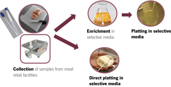
3.2. Isolation Methods
Enrichment
The enrichement step is applied when pathogenic microorganisms are present in small numbers. Enriched media is a media with specific and known qualities that favors the growth of a particular microorganism while inhibiting the growth of others (Varman and Evans, 1996). The selective agents frequently contained in enrichment broths are acriflavine, nalidixic acid, potassium tellurite and antibiotics, as novobiocin (Gasanov et al., 2005), depending on each media for each microorganism. There are some variety in terms of enrichment media for Listeria spp. such as Listeria Enrichment Broth Base, Listeria Fraser Broth, and Listeria Enrichment Broth, Modified and for E. coli as E.E. Broth (Enterobacteriaceae Enrichment Broth), and TSB Broth with Novobiocincin (Modified Tryptic Soy Broth with Novobiocin). For identification of L. monocytogenes was used the Listeria Enrichment Broth Base media and for E. coli was utilized the E. E. Broth media. The enrichment procedure includes a period of incubation in a selective
| Considerations of the methodology
media to suppress the competitive flora and enable the multiplication of the target organism for subsequent isolation or detection by a variety of techniques (Jasson et al., 2010).
Selective plating
Enrichment methods are followed by the isolation of the target microorganism on specific plate media (Figure 3.1). Selective media allows the growth of certain type of organisms, while inhibiting the growth of other organisms (Gasanov et al., 2005). Examples of these type of media are Listeria PALCAM Agar and Listeria Oxford Agar Base for Listeria spp. For E. coli there is E. coli Direct Agar. For isolation of L. monocytogenes was used Listeria Oxford Agar Base and for E. coli was CHROMagarTM E.coli. Nowadays, this task has been facilitated by the introduction of chromogenic and fluorogenic media for the detection. A wide range of chromogenic (colour reaction) and fluorogenic (fluorescent reaction) substrates are available, and these compounds are applied in several commercial systems and media. The target population is characterized by enzyme systems that metabolize the substrate (sugar or amino acid) that leads to the release of chromogen/fluorogen, resulting in a colour change in the media and/or fluorescence under long wave UV light (Perry and Freydiere, 2007). As example, it is known that L. monocytogenes and L. ivanovii produce the enzyme phosphatidylinositol-specific phospholipase C (PIPL-C) (Coffey et al., 1996), which activity is measured on chromogenic media. There are however chromogenic agars used in others bacteria, as is the case of some STEC (Shiga toxin-producing Escherichia coli) strains that are sensitive for sorbitol-fermenting (Boer, 2000).
Figure 3.1. Isolation procedure: enrichment and plating in selective media.
Plating in selective media
Oxford Listeria Agar Base (37 ºC, 96 h) CHROMagarTM
E.coli (37 ºC, 24 h)
Enrichmentin selective media
Listeria Enrichment Broth Base (30 ºC, 48 h) E.E. Broth (37 ºC, 24 h)
|45
3.3. Rapid methods for identification of isolates
Rapid methods are used for the detection and identification of microbial pathogenic agents, and include nucleic acid-based methods, immunological-based methods, oligonucleotide DNA microarray, and biossensors-based methods (Law et al., 2015).
Polymerase chain reaction (PCR)
PCR is a method which operates by amplifying a specific target DNA sequence, and involves a cyclic three steps process. The choice of specific primers is made in order to amply the interest region. Primers are short oligonucleotides, usually 20-30 nucleotides in length, whose sequence matches the end of the region of interest. For this work were chosen universal primers (27F and 338R).Universal primers are complementary to nucleotide sequences that are very common in a particular set of DNA molecules and cloning vectors. Thus, they are able to bind to a wide variety of DNA templates (Jill and Claridge, 2004). During each cycle, the double-stranded DNA is denatured into double-stranded DNA at high temperature (step 1). Two single-stranded synthetic oligonucleotides or specific primers (forward and reverse primer) will anneal to the DNA strands (step 2). Then, the primers complementary to the single-stranded DNA are extended due to the presence of deoxyribonucleotides and a thermostable DNA polymerase, whose process is polymerization (step 3). In subsequent cycles, primers will bind to both the original DNA and the newly synthesize DNA resulting in an exponential increase in the number of copies. The size of the PCR product is usually detected by electrophoresis gel (Ikeda et al., 2007).
This method has advantages over culture and other standard methods including specificity, sensitivity, rapidity, accuracy and capacity to detect small amounts of target nucleic acid in a sample (Ikeda et al., 2007). It has been used in the detection of distinct foodborne pathogens like L. monocytogenes, E. coli O157:H7, S. aureus, C. jejuni, Salmonella spp. and Shigella spp. (Lee et al., 2008; Alves et al., 2012; Chiang et al., 2012; Zhou et al., 2013). PCR is also used for toxins detection by amplification of specific genes that encode bacterial toxins. This has been performed with several bacterial species, as Vibrio cholera, B. cereus, E. coli, and S. aureus (Planche et al., 2008).
| Considerations of the methodology
16S rRNA Gene Sequencing
16S rRNA gene sequencing involves the amplification of a phylogenetically informative target as the 16S rRNA gene (Bosshard et al., 2003). Several primers that recognize 16S ribosomal (rDNA) sequences (conserved in a wide variety of bacteria) are used to amplify regions of interest (Kolbert and Persing, 1999). From the sequence determination and from the comparative database searches, it can be assigned a group of bacteria to the unknown isolate (Bosshard et al., 2003). This technique traces phylogenetic relationships between bacteria and identifies bacteria from various sources, such as environmental or clinical specimens. Identification based on the 16S rRNA sequence is of interest because ribosomal SSU (small subunit) exists universally among bacteria and includes regions with species specific variability (Mignard and Flandrois, 2006). This technology is used today in clinical laboratories to provide genus and species identification for isolates that do not fit any recognized biochemical profiles, for strains generating only a “low likelihood” or “acceptable” identification according to commercial systems, or for taxa that are rarely associated with human infectious diseases (Janda and Abbott, 2007).
3.4. Biofilm formation
Currently there are a variety of systems for examining biofilm formation and several devices available for the formation of bacterial biofilms. These in vitro systems are usually divided into flow (Modified Robbins Device, and flow cell biofilm system) and static models (microtiter plate, Calgary Biofilm Device, perfused biofilm fermentor, and Constant Depth Film Fermentors). Static models are preferable due to the facility to handle and versatility, allowing to study the effect of different conditions of biofilm formation as well as different behaviors of these bacterial communities (Abdallah et al., 2014).
The microtiter plate assay was developed by Christensen, and is the most widely used method to assess the early stages of biofilm formation in abiotic surfaces (Christensen et al., 1985), providing a large number of parallel and miniaturized reactors in small scale with the same conditions of space and fluid dynamics (Kumar et al., 2004). A microtiter plate may have 6, 24, 96, 384 or even 1536 sample wells arranged in a rectangular matrix, but in laboratory the most used is the 96-well format (Coenye and Nelis, 2010; Kumar et al., 2004). The use of this
|47
method has the advantage of being easily combine with compatible side equipment, such as micropipettes, pipetting robots and microplate’s readers (Duetz, 2007). Also, the advantage of a regular renewal of liquid phase allows avoiding the limitation of nutrients and accumulation of potentially toxic metabolites, and due to its low cost, flexibility and speed, this technique allows the processing of multiple samples simultaneously with simplicity. Furthermore, it is in agreement with the requirement of common laboratory equipment to use smaller quantities of reagents and culture media (Machado et al., 2012). Microtiter plates can be used for a wide variety of biofilm analysis, including assays for biomass quantification (Lopes et al., 2010), assessment of biofilm metabolic activity (Lopes et al., 2010), enumeration of biofilm cells (Lopes et al., 2010), quantification of biofilm matrix (Peeters et al., 2008), and susceptibility testing (Sandberg et al., 2008). However, this system does have some drawbacks such as: its static nature, that results in possible nutrient limitation and the inability to easily generate mature biofilms (Merritt et al., 2005); the operator-dependent nature of its procedures, for example washing steps, that can cause a poor reproducibility even in intra-laboratory experiments; and its configuration that hinders the observation of the biofilm structure by direct microscopy (Azevedo et al., 2009).
3.5. Quantification of biofilm biomass
Biofilm biomass can be measured by distinct methods, such as microscopy, molecular probes, biochemical analysis of biomass components, and, the most usual, staining of biofilms with specific compounds and subsequent determination of optical density (Stepanovic et al., 2000; Li et al., 2003; Peeters et al., 2008; Azevedo et al., 2009).
Crystal violet (CV) staining was first described by Christensen and has been one of the most expedites methods to determinate biofilm biomass (Christensen et al., 1985). In this staining assay, the dye binds to negatively charged surface molecules and polysaccharides located in the extracellular matrix staining also both living and dead cells (Li et al., 2003), allowing the determination of biofilm biomass without disrupting the biofilm. Once total biomass (cells and matrix) is stained in purple, the dye can be easily dissolved in acetic acid (Stepanovic et al., 2000) and, finally, the absorbance is read at 570 nm. However, problems are frequent when using this method, due to the existence of clear fails in the reproducibility of results, and the requirement of successive washing steps, which may lead to loss of part of the biomass
| Considerations of the methodology
present (Peeters et al., 2008). Although of the disadvantages of this technique, the CV method is a straightforward, quick, and low cost technique to indirect quantification of microbial adhesion and amount of biofilm formed on inert surfaces by a broad range of microorganisms (Stepanovic et al., 2000; O’Toole, 2011),
3.6. Microbial susceptibility testing
For the assessment of microbial susceptibility to disinfectants and antibiotics, different methods may be used, as disk diffusion and broth dilution. These assays are standard procedures to quantify planktonic cells’ susceptibility to biocidal or antimicrobial agents, but they are not directly applicable to biofilms. With the broth dilution method it is evaluated the ability of bacteria to grow in a range of concentrations of a given biocidal/antimicrobial agent. Briefly, dilutions of biocidal/antimicrobial compounds solutions are prepared in a liquid bacterial growth media, which is then inoculated with the standardized cell suspension. Microtiter plates are then incubated overnight, at the incubation conditions recommended for the bacteria under study, and then the plates are examined for visible evidence of bacterial growth in the form of turbidity. The biocidal activity of a compound can be quantified by determining the minimum concentration of the compound capable of inhibiting the visible growth of a microorganism, a value called Minimum Inhibitory Concentration (MIC) (mg/l or µg/ml) (Andrews, 2001). Posteriorly, MICs values may be translated into clinical categories, namely sensitive (S), intermediary (I) or resistant (R). Concerning antibiotics, these clinical categories and correlated MICs are provided by several committees, including the European Committee on Antimicrobial Susceptibility Testing (EUCAST) (EUCAST, 2014).
Several techniques are also available to evaluate susceptibility of biofilms, such as Biofilm Eradication Surface Test (BEST) Assay™ (Harding et al., 2011) and BioTimer Assay (De Giusti et al., 2011). Calgary Biofilm Device is usually used to determinate Minimum Biofilm Eradication Concentration (MBEC) which is the lowest biocidal concentration that eradicates the biofilm (Ceri et al., 1999). However, susceptibility assay of biofilms on microtiter plates is also a method used. This technique consists in, after biocidal exposure, the biocidal compound is removed from the wells and biofilms are then scraped thoroughly. Posteriorly, samples correspondent to each concentration of biocidal agent are plated on solid media and incubated from a specific period of time. In order to assess MBEC, the presence of colonies is evaluated
|49
and MBEC value is determined, as the lowest concentration of antibiotic that prevented bacterial growth (Mataraci and Dosler, 2012).
|51
Chapter 4
Materials and methods
|53
4.1. Sampling
Samples were collected from one meat retail facility in center of Braga, Portugal. The sampling procedure followed the standard ISO method 18593:2004. Contact surfaces swabbed were cutting boards and the mincer (Figure 4.1) due to a higher probability of contamination. For each one of these surfaces, triplicate samples were collected before and after cleaning and disinfection procedure. After collecting samples, these were transported in a refrigerated box to the laboratory, where they were processed.
Figure 4.1. Sampling scheme used in the present work (cutting boards and mincer).
4.2. Isolation of microorganisms
Listeria monocytogenes: Some swabs were directly placed into plates containing solid selective media (Oxford Listeria Agar Base, Liofilchem) with supplement (Listeria Oxford Supplement, Liofilchem) and incubated at 37 ⁰C for 96 h. The other swabs were placed into 40 ml containing selective enrichment broth media (Listeria Enrichment Broth Base, Liofilchem) with supplement (Listeria for Enrichment Supplement, Liofilchem) and incubated at 30 ⁰C for 48 h. After growth in selective enrichment broth media, 100 µL of broth was inoculated into plates containing selective solid media and incubated at 37 ⁰C for 96 h. These steps were performed for samples collected before and after disinfection in both surfaces analyzed. For the selective media it would be expected to have brown-gray colonies with black center and black halo correspondents to L. monocytogenes.
Collection of samples from meat retail facilities Enrichment in selective media Platting in selective media Direct platting in selective media