UNIVERSIDADE DO ALGARVE
Departamento de Ciências Biomédicas e Medicina
Gluconeogenesis and amino acids
metabolism in ovarian clear cell
carcinoma
Filipa Lopes Coelho
Dissertação
Mestrado em Ciências Biomédicas
Trabalho efetuado sob orientação de: Profª. Doutora Jacinta Serpa (orientação externa)
Prof. Doutor Álvaro Tavares (orientação interna)
UNIVERSIDADE DO ALGARVE
Departamento de Ciências Biomédicas e Medicina
Gluconeogenesis and amino acids
metabolism in ovarian clear cell
carcinoma
Filipa Lopes Coelho
Dissertação
Mestrado em Ciências Biomédicas
Trabalho efetuado sob orientação de:
Profª. Doutora Jacinta Serpa (orientação externa)
UIPM (IPO Lisboa)/CEDOC – Faculdade de Ciência Médicas da Universidade Nova de Lisboa Prof. Doutor Álvaro Tavares (orientação interna)
CBME – Universidade do Algarve
Título do trabalho: “Gluconeogenesis and amino acids metabolism in ovarian clear cell carcinoma”
Declaro ser a autora deste trabalho, que é original e inédito. Autores e trabalhos consultados estão devidamente citados no texto e constam da listagem de referências incluída.
Copyright Filipa Lopes Coelho
A Universidade do Algarve tem o direito, perpétuo e sem limites geográficos, de arquivar e publicitar este trabalho através de exemplares impressos reproduzidos em papel ou de forma digital, ou por qualquer outro meio conhecido ou que venha a ser inventado, de o divulgar através de repositórios científicos e de admitir a sua cópia e distribuição com objetivos educacionais ou de investigação, não comerciais, desde que seja dado crédito ao autor e editor.
i
Agradecimentos
Gostaria de dar o meu especial agradecimento à Doutora Jacinta Serpa, minha orientadora, que tornou possível este trabalho e me ajudou a superar e ultrapassar todos os obstáculos com que me deparei, ao longo deste ano. Obrigada por toda a disponibilidade, ajuda e conhecimentos transmitidos. Agradeço ainda toda a paciência, confiança, incentivo ao pensamento crítico e autonomia. Um sincero muito obrigada!
O meu agradecimento ao Doutor Álvaro Tavares pela disponibilidade de aceitar co-orientar a minha tese.
Gostaria de agradecer ao Doutor Luís Gafeira por me ter apresentado o enorme mundo do NMR e por toda a simpatia e disponibilidade.
Agradeço a todas as minhas colegas do grupo. À Fernanda por toda a simpatia e alegria que sempre transmitiu. Um especial agradecimento à Lídia e à Sofia por todos os ensinamentos e palavras de incentivo.
Um obrigada a todos os meus colegas do UIPM, que direta ou indiretamente tornaram este trabalho possível.
Agradeço aos meus amigos, que apesar de alguns deles estarem longe ouviram incondicionalmente os meus desabafos e as minhas irritações.
E por último, mas não menos importantes, agradeço à minha família por me ter dado um apoio incondicional, aturado as minhas frustrações e irritações e por todas as palavras de incentivo e força. Um especial obrigada aos meus pais e à minha irmã, sem eles não teria sido possível!
Obrigado a todos os que estiveram presentes nesta fase da minha vida que, aliás, foi das mais estimulantes nesta minha breve vida, no mundo da “ciência”.
ii
Resumo
O cancro é considerado como um dos maiores problemas de saúde pública (Siegel et al., 2013). De acordo com a OMS (Organização Mundial de Saúde) o cancro é caracterizado como sendo um crescimento anormal de células, podendo desenvolver-se em qualquer parte do corpo e alastrar-se para outros órgãos (metástases). Sabe-se ainda que as metástases são maioritariamente responsáveis pela morbilidade e mortalidade em doentes com cancro (Seyfried and Shelton, 2010). Biologicamente, as células normais podem sofrer transformação maligna, sendo em parte esta transformação influenciada pelo microambiente envolvente, onde o tumor e as células circundantes estabelecem uma rede funcional (Hanahan and Weinberg, 2011; Serpa and Dias, 2011).
O carcinoma do ovário, é uma grande causa de mortalidade em mulheres, constitui cerca de 90% de todas neoplasias malignas do ovário, sendo 3-10% dos casos classificados como carcinoma de células claras (OCCC). Cerca de 57-81% de OCCC são diagnosticados nos estádios I/II, apresentando-se usualmente como uma massa pélvica. Doentes com CCC apresentam um comportamento clínico distinto e com pior prognóstico do que outras neoplasias de ovário (Feeley et al., 2001, Bjorkholm et al., 1982; Sorbe et al., 1982; Hogberg et al.,1993). Um dos motivos que contribuem para o mau prognóstico em CCC está relacionado com a baixa resposta à quimioterapia convencional baseada em cis-platina (Goff et al., 1996). Este tipo histológico de carcinoma do ovário deve o nome de células claras a algumas alterações morfológicas devidas à acumulação de glicogénio, cuja síntese é regulada pelo HNF1 (hepatocyte nuclear factor 1 ), o gene central na tumorigénese do CCC (Anglesio et al., 2011).
Atualmente sabe-se que as células tumorais poderão apresentar perfis metabólicos distintos das células normais (Fuchs and Bode, 2005). Em 1956, Otto Warburg estabeleceu o primeiro elo de ligação entre o metabolismo e o cancro através das observações em que as células tumorais, mesmo na presença de oxigénio, apresentam um aumento da taxa da glicólise, tendo lactato como produto final (Warburg, 1956). Este fenómeno, conhecido como efeito de Warburg, coloca a hipótese das células tumorais apresentarem defeitos na mitocôndria, responsável pela anulação do ciclo TCA (ou ciclo de Krebs) e consequentemente aumento da taxa de glicólise (Vander Heiden et al., 2009), suportando a proliferação celular. Em vários tipos de cancro, existe uma associação entre o desregulação e aumento da proliferação /sobrevivência celular com um aumento da glicólise aeróbica (Wise and Thompson, 2010), onde as fontes de carbono, NADPH e ATP provêm da glucose (Dang, 2012).
A glucose e a glutamina são os principais substratos a serem utilizados como fontes de ATP e carbono, essenciais à síntese de macromoléculas e ao suporte da proliferação celular. A
iii
glucose é responsável pela produção de ribose (fundamental para a síntese de ácidos nucleicos) e ATP, através da via das pentoses-fosfato (PPP). A glutamina, o aminoácido mais abundante na corrente sanguínea, é considerado um substrato bioenergético e dador de nitrogénio, sendo que este aminoácido se revela essencial para a produção de biomassa e energia (Dang, 2012).
A neoglucogénese é responsável por cerca de 35-50% da produção total de glucose, sendo uma via que requer ATP e NADPH para a conversão de piruvato, lactato e aminoácidos (como alanina e glutamina) em glucose de novo. Nos tecidos normais, a neoglucogénese é ativada por modificações pós-translacionais ou pela ativação alostérica de enzimas chave limitantes, responsáveis pelos eventos moleculares desta via. Geralmente, e de acordo com os requisitos metabólicos, os órgãos adaptam a importação, armazenamento, produção e libertação de glucose (Oh et al., 2013; Telang et al., 2012).
A conversão de piruvato em glucose é considerada como o maior passo da neoglucogénese, sendo catalisado por várias enzimas citoplasmáticas e mitocôndriais. Parte dos passos da neoglucogénese são catalisadas, no sentido inverso, por enzimas glicolíticas (Quintas et al., 2008a).
A nível molecular, o piruvato é transportado diretamente do citoplasma para a mitocôndria ou, tendo a alanina como percursor, a conversão para piruvato ocorre na mitocôndria por transaminação. O piruvato promove a formação de oxoloacetato (OAA) por ação da piruvato carboxilase (PC). O transportador carnitina permite o influxo de acetil-CoA na mitocôndria, que servirá como um activador essencial de PC. OAA é então reduzido a malato e transportado para o citoplasma via transportador malato- -cetoglutarato. No citoplasma, o malato é oxidado a OAA e posteriormente a fosfoenolpiruvato (PEP), através de fosfoenolpiruvato carboxicinase citosólica (PCK1). Caso o OAA seja convertido diretamente no citoplasma, a conversão a PEP é catalisada por fosfoenolpiruvato carboxicinase mitocondrial (PCK2). A conversão de fructose-1,6-bifosfato (F1,6BP) em fructose-6-fosfato (F6P) pela fructose-1,6-bifosfatase (FBP1) é outra reação irreversível na neoglucogénese (Quintas et al., 2008b). Posteriormente, glucose-6-fosfatase (G6Pase) é então responsável pela desfosforilação de F6P em glucose de novo (Weickert and Pfeiffer, 2006). Por sua vez, a função da enzima 6-fosfofructo-2-cinase/ fructose-2,6-bifosfatase (PFKFB1) está associada à estimulação da glicólise e inibição da neoglucogénese, através da modulação da sua atividade cinase.
A glutamina pode ser considerada tão importante como a glucose a nível da produção de energia, estando provado que restrições de glutamina ou inibição de enzimas glutaminoliticas diminuem a proliferação celular, tanto in vitro como in vivo (Reynolds et al., 2013).
iv
Os passos fundamentais da glutaminólise são a hidrólise de glutamina a glutamato, por ação da glutaminase (GA), e a subsequente conversão na mitocôndria do glutamato em -cetoglutarato ( -KG). -KG pode integrar o ciclo TCA e fornecer os intermediários metabólicos, como NADH (Piao et al., 2013). Porém, um metabolismo oxidativo incompleto pode conduzir à produção de lactato como produto final. O glutamato pode ainda ser convertido em piruvato pela transaminase glutamato-piruvato (GPT) e ser usado como substrato para a neoglucogénese (Matés et al., 2013).
O glutatião (GSH) é um tripeptido γ-glutamil-cisteinil-glicina, sendo considerado o antioxidante mais importante do organismo. GSH, um tiol antioxidante intracelular, atua como tampão redox contra o stress oxidativo. Alterações moleculares no sistema GSH em alguns tumores pode ser responsável por um aumento da proliferação celular, bem como da resistência a drogas (Traverso et al., 2013).
A cisteína, um aminoácido utilizado na síntese de GSH, é obtido através da dieta ou da transulfuração de outros amino acidos. A transulfuração desempenha um papel importante na homeostase de cisteína, sendo mediada em parte pela cis-tationa-gama-ligase (CTH) e 3- sulfotransferase de mercaptopiruvato (MPST). CTH é uma enzima citoplasmática e a última enzima-chave na via de transulfaração, catalisando a conversão de cistationina (derivada da metionina) em cisteína, amónia, 2-oxobutirato e é ainda responsável pela produção de H2S
(Wang and Hegele, 2003). A CTH tem ainda a capacidade de degradar cisteína, o que conduz à produção de lactato (Steegborn, 1999). Por sua vez, MPST é uma enzima envolvida da degradação da cisteína, catalisando a transferência de ião enxofre do 3-mercaptopiruvato em cianina ou outro tiol (Jurkowska et al., 2011).
Com base no trabalho científico que vem sendo dedicado na área do metabolismo tumoral nesta tese foram definidos dois objectivos principais que foram abordados, utilizando um modelo in vitro de OCCC (ES2). O primeiro objectivo é verificar se a neoglucogénese é a fonte de glucose, de modo a suportar as necessidades celulares. Investigámos a glutamina, lactato e butirato como substrato da neoglucogénese, bem como o perfil de expressão de enzimas chave da neoglucogénese (PCK1, PCK2, FBP1 e PFKFB1) e a modulação de características celulares como a migração e ciclo celular. O segundo objetivo, que partiu dos resultados obtidos no primeiro objetivo, e é clarificar a importância do metabolismo da glutamina e da cisteína e a sua influência na síntese de GSH. Pretendemos ainda determinar a relevância da glutamina e cisteína na via de síntese de GSH, bem como na progressão tumoral.
v
O perfil metabólico de ES2 por NMR revelou a presença de glicogénio não marcado, assim como também não foi detetada glucose 13C marcada, o que é indicativo de que as células
não estão a recorrer aos substratos testados como fonte para neoglucogénese.
Ao nível das enzimas neoglucogénicas, a acetilação de PCK1 pode ser responsável pelos resultados obtidos, onde maiores níveis de expressão de mRNA PCK1 nas condições de glucose e butirato não se refletiram num aumento dos níveis proteicos de PCK1, sendo por sua vez verificado um aumento dos níveis proteicos de PCK1 na condição lactato e glutamina. Em relação ao PCK2, os resultados suportam o descrito na literatura, em que a expressão de PCK2 permanece praticamente inalterada, sendo porém a sua expressão mais acentuada quando a neoglucogénese recorre ao lactato como substrato.
Os nossos resultados também sugerem que quando as células estão num meio privado de glucose os níveis proteicos de FBP1 encontram-se sobre expressos, enquanto que os níveis de PFKFB1 permanecem baixos, estimulando a neoglucogénese. No geral, a nível da modulação de enzimas envolvidas na neoglucogénese o lactato parece ser o principal substrato.
Como referido anteriormente, a alanina também pode ser usada como substrato da neoglucogénese, através da conversão de alanina em piruvato, via transaminação. Observámos um aumento nos níveis de mRNA de transaminase de alanina (ALT), nas condições glucose, lactato e glutamina, o que sugere que a alanina também desempenha um papel importante como precursor da neoglucogénese.
A análise de NMR permitiu ainda verificar que ES2 são capazes a metabolizar (pelo menos parte) o butirato, favorecendo o ciclo celular e a migração.
Por sua vez, e de acordo com os resultados obtidos por NMR, a glutamina parece ser o substrato mais usado. A análise metabólica revelou que a glutamina foi metabolizada em intermediários do ciclo TCA 13C marcados, bem como intermediários 13C marcados para a
síntese de macromoléculas, o que sugere que a glutamina é metabolizada pela glutaminólise em vez de ser utilizada como substrato para a neoglucogénese. Os resultados obtidos tambem confirmam que a glutaminólise favorece a migração celular e a progressão do ciclo celular.
O perfil metabólico de ES2 revelou ainda a presença intracelular de GSH 13C marcado nos
resíduos de glicina e de glutamato, bem como glicina 13C marcada e glutamato13C livres.
Observámos que a suplementação de cisteína conduziu a um aumento dos níveis de GSH, que se encontra associado à proliferação e ciclo celular. Detetámos ainda que na presença de cisteína, ocorre um aumento da síntese GSH, que especulativamente estará associado ao decréscimo dos níveis de metionina no interior das células. Assim, o modelo in vitro de OCCC revelou que a glutaminólise parece ser relevante, não só ao nível da produção de energia e de
vi
manter o potencial redox favorável, mas também está associada à síntese de GSH, sendo por sua vez, a síntese de GSH favorecida pela suplementação de cisteína.
Resumindo, o metabolismo de aminoácidos revelou-se crucial na tumorigénese de OCCC, bem como na resistência a drogas anti tumorais.
Palavras-chave: carcinoma de células claras do ovário (OCCC), neoglucogénese,
vii
Abstract
Tumor cells may exhibit different metabolic profiles compared to normal tissues from which they are derived. Those observations gave rise to the new concept that tumorigenesis requires metabolic alterations to sustain cell proliferation. Several studies reveal that increased cell proliferation is accompanied by increased glucose consumption. In OCCC, a typical morphological feature is the accumulation of glycogen in cancer cells cytoplasm this fact allows us to raise the stepwise questions: 1) Is glucose the main energy and carbon source used by OCCC?; 2) Being glucose a relevant energy and carbon source, can gluconeogenesis be working on ovarian clear cell carcinoma?, and 3) If gluconeogenesis is working on, which are the gluconeogenic substrates used by ovarian clear cell carcinoma?
In the present thesis we try to clarify, in an OCCC in vitro model (ES2) and in a glucose privation microenvironment, if gluconeogenesis pathway is working on, in order to support cells energy demands. Glutamine, lactate and butyrate were testes as putative gluconeogenesis substrates.
Metabolic analysis by NMR of ES2 cell line showed the presence of glycogen, characteristic of this cell line, though 13C unlabelled, meaning that it was not synthesized from the
tested substrates. At the same time the tested time points were not enough to clarify if glucose is de novo synthesized through gluconeogenesis. Nevertheless, NMR results showed that glutaminolysis was the major pathway used to sustain ES2 cell proliferation, with the production of TCA intermediates, as well as intermediates for macromolecular synthesis.
These findings prompted us to investigate the role of glutaminolysis in OCCC. In NMR analysis, we observed the synthesis of glycine and glutamate as well as the synthesis of GSH. Gathering cysteine to cells exposed to glutamine, we verified an increase in the synthesis of GSH. In cancer, GSH high levels is related to drug resistance which is in agreement with the fact that OCCC poor prognosis is also due to resistance to therapy.
Summing up, amino acids can play a central role in OCCC concerning energy and carbon metabolism as well as drug resistance.
Keywords: gluconeogenesis, glutaminolysis, glutathione (GSH), ovary clear cell
viii
Table of contents
Agradecimentos ... i Resumo ... ii Abstract ... vii Index of figures ... xiIndex of table ... xii
List of abbreviations ... xiii
1. Introduction ... 1
1.1 Cancer ... 1
1.2 Ovarian clear cell carcinoma (OCCC) ... 1
1.3 Cancer Metabolism ... 3
1.3.1 Oncogenic alterations ... 4
1.3.2 Nutrients uptake ... 5
1.4 Glucose and Glutamine ... 6
1.4.1 Gluconeogenesis and Glycolysis ... 8
1.4.1.1 Gluconeogenesis ... 9 1.4.1.1.1 Molecular regulators ... 10 1.4.1.1.2 Molecular pathway ... 10 1.4.1.1.3 Key enzymes ... 12 1.4.2 Glutaminolysis ... 13 1.4.2.1 Molecular pathway ... 14 1.4.2.2 Key enzymes ... 14 1.3 Glutathione (GSH) ... 15 1.4 Cysteine homeostasis ... 17 2. Aims ... 18
ix
3.1 Role of glucose, glutamine, lactate and butyrate as gluconeogenic precursor and
cysteine associated with glutaminolysis ... 19
3.1.1 Cell culture ... 19
3.1.2 Nuclear magnetic resonance (NMR) for metabolomic detection ... 20
3.1.3 Gene expression ... 21
3.1.3.1 Reverse transcription polymerase chain reaction (RT-PCR) ... 21
3.1.3.2 Relative quantifying PCR (RQ-PCR) ... 22
3.1.4 Protein levels ... 23
3.1.4.1 Western Blotting ... 23
3.1.5 Promoter activity using Luciferase reporter gene assay ... 24
3.1.5.1 Amplification of promoter sequences ... 24
3.1.5.2 DNA automate sequencing of promoter sequences ... 25
3.1.5.3 Plasmid generation and cloning ... 26
3.1.5.4 Plasmid isolation ... 27
3.1.5.5 Transient transfection of promoters constructs into ES2 cell line ... 29
3.1.5.6 Luciferase activity ... 30
3.2. Role of glucose, glutamime, lactate, butyrate and cysteine in cell migration, cell cycle and apoptosis regulation ... 30
3.2.1 Cell migration - In vitro wound healing assay ... 30
3.2.2 Cell cycle analysis by Flow Cytometry ... 31
3.2.3 Apoptosis analysis by Flow Cytometry ... 31
3.3 Role of cysteine in aminothiols production ... 32
3.3.1 High-performance Liquid Chromatography (HPLC) for aminothiols detection ... 32
3.4 Statistical analysis ... 34
4. Results ... 36
4.1 Role of glucose, glutamine, lactate and butyrate as gluconeogenic precursor ... 36
x
4.1.2 PCK1, PCK2, FBP1, PFKFB1 and ALT gene expression and protein levels in ES2
cell line ……….………...38
4.1.3 Activity of PCK1, PCK2, FBP1 and PFKFB1 promoters in ES2 cell line ... 41
4.2 Role of glucose, glutamine, lactate, butyrate in cell migration and cell cycle in ES2 cell line ……… ... 42
4.2.1 Cell migration - In vitro wound healing assay ... 42
4.2.2 Cell cycle analysis by Flow Cytometry ... 44
4.3 Role of cysteine associated with glutamine and endogenous antioxidant synthesis .... 45
4.3.1 CTH, MPST and mTOR gene expression in ES2 cell line ... 45
4.3.2 Activity of CTH and MPST promoters in ES2 cell line ... 47
4.4 Role of cysteine in cell migration, cell cycle and apoptosis in ES 2 cell line ... 48
4.4.1 Cell migration - In vitro wound healing assay ... 48
4.4.2 Cell cycle analysis by Flow Cytometry ... 48
4.4.3 Cell death analysis by Flow Cytometry ... 49
4.5 Role of cysteine in aminothiols production ... 50
4.5.1 High-performance Liquid Chromatography (HPLC) for aminothiols detection in ES2 cell line . ... 50 5. Discussion ... 53 6. Conclusions ... 63 7. Future Perspectives ... 64 Bibliography references ... 65 Appendices ... 73 Appendix I ... 74 Appendix II ... 78
xi
Index of figures
Figure 1 ... 4
Figure 2: mTOR pathway ... 6
Figure 3: ... 7
Figure 4: ... 8
Figure 5: Gluconeogenesis Pathway ... 11
Figure 6: Glutaminolysis . ... 15
Figure 7: Gluthatione formation . ... 16
Figure 8: a) pGL3-Basic Vector. b) pGL3-Control Vector. Adapted from Technical Manual - pGL3 Luciferase ... 27
Figure 9: NOESY 1D (1H) spectra for ES2 cell line. ... 36
Figure 10:13C-1H-HSQC spectra of ES2 supernant (a) and cell extracts (b) incubated with 13C -glutamine ... 37
Figure 11- Relative gene expression of PCK1, PCK2 and PFKFB1 ... 39
Figure 12– PCK1 (a),FBP1 (b) and PFKFB1 (c) protein levels in ES2cells assessed by western-blotting. ... 40
Figure 13- Relative gene expression of ALT. ... 41
Figure 14 - Luciferase activity of PCK1, PCK2, FBP1 and PFKFB1 constructs in transfected ES2 cells. ... 42
Figure 15- In vitro Wound Healing assay in ES2 cells... 43
Figure 16- Quantification of wound closure in ES2 cells ... 44
Figure 17– Cell cycle analyzes by flow cytometric (PI staining) in ES2 cells ... 45
Figure 18- Relative gene expression of CTH (a) and MPST (b). ... 46
Figure 19- Relative gene expression of mTOR.. ... 47
Figure 20- Luciferase activity of CTH and MPST constructs in transfected ES2 cells. ... 47
Figure 21 - In vitro Wound Healing assay in ES2 cells... 48
Figure 22- Quantification of wound closure in ES2 cells. ... 48
Figure 23– Cell cycle analyzes by flow cytometric (PI staining) in ES2 cells.. ... 49
Figure 24– Apoptosis and necrosis analyzes by flow cytometric (Annexin V and PI staining) ... 50
Figure 25 –Thiols quantification in crude culture medium and supernatant in ES2 cell line. ... 51
xii
Figure A1: HPLC spectrum in control crude medium and in cysteine crude medium. ... 78
Figure A2 - HPLC spectrum in control crude medium and supernatant.. ... 78
Figure A3– HPLC spectrum in cysteine crude medium and supernatant. ... 79
Figure A4- HPLC spectrum of intracellular contents in control and cysteine condition... 79
Index of tables
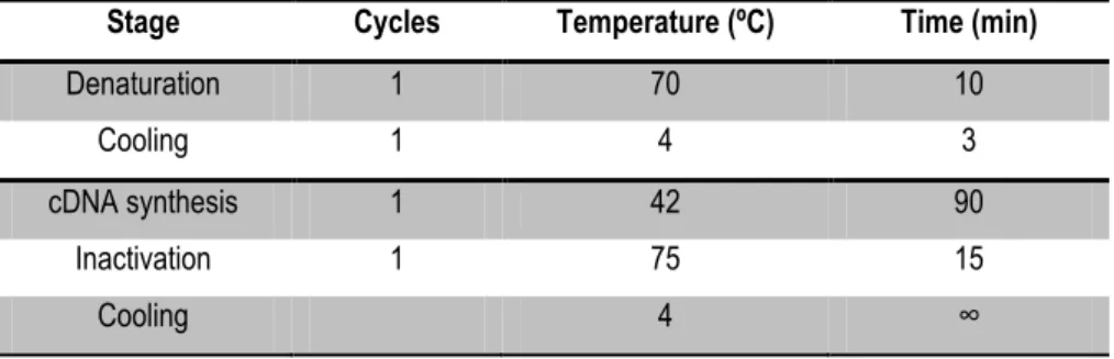
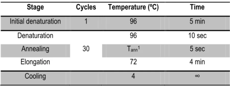
Table 1- Program used for cDNA synthesis ... 21Table 2- Relative quantifying PCR program ... 22
Table 3- PCR program used for amplification of promoters sequences ... 25
Table 4- Automate sequencing program ... 26
Table 5- PCR program for plasmid constructs amplification ... 28
Table 6- Automate sequencing program ... 28
Table 7- Primers used during the experimental work ………..……….37 22
xiii
List of abbreviations
KG – -Ketoglutarate AA – Antibiotic-Antimycotic ADP – Adenosine diphosphate AKT – Protein kinase B ALT- Alanine transaminase
AMPK – AMP-activated protein kinase ATP – Adenosine Triphosphate BSA – Bovine serum albumin
cAMP – Cyclic adenosine monophosphate cDNA – DNA copy
ChIP – Chromatin immunoprecipitation CREB – Responsive element binding protein CS – Citrate synthase
CTH – Cystathionine gamma-lyase CysGly – Cysteinylglycine
ddH2O – Bidestilled water
DMEM – Dulbecco’s modified essential medium DNA – Deoxyribonucleic acid
DTT – Dithiothreitol
EMSA – Electrophoretic mobility shift assay ERR - Estrogen receptor related gamma ETC – Electron transport chain
F1,6BP – Fructose-1, 6-biphosphate F2,6BP – Fructose-2,6-biphosphatase F6P – Fructose-6-phosphate
FBP1 – Fructose-1,6-bisphosphatase FBS – Fetal bovine serum
For – Forward
FoxOs – Forkhead box class O G6Pase – Glucose-6-phosphatase GA – Glutaminase
GCL – γ-glutamylcysteine ligase gDNA – Genomic DNA
GLS1 – Glutaminase isoform 1 GLS2 – Glutaminase isoform 2 GLUD1 – Glutamate dehydrogenase GLUL – Glutamate- ammonia ligase GPT – Glutamate pyruvate transaminase GR – Glucocorticoids receptors
GSH – Glutathione GSH – Glutathione
GSH-R – Glutathione reductase GSS – Glutathione synthetase
xiv
GSSG – Glutathione disulfide (or in oxidized form) h – Hours
H2S – Hydrogen sulfide
HDAC – Histone deacetylase HIF1 - Hypoxia inducible factor HK2 – Hexokinase 2
HMDB – Human Metabolome database
HPLC – High-performance Liquid Chromatography HPRT – Hypoxanthine phosphoribosyltransferase HRP – Horse raddish peroxidase
LB – Luria-Bertani medium
LDH-A – Lactate dehydrogenase subunit A LDH-B – Lactate dehydrogenase subunit B MgCl2 – Magnesium chloride
Mg2+ - Magnesium min – Minutes
MPC – Mitochondrial pyruvate transporter MPST – 3-mercaptopyruvate sulfurtransferase mRNA – messenger ribonucleic acid
mTOR – Mammalian target of rapamycin
mTOR C1 – Mammalian target of rapamycin complex 1 mTOR C2 – Mammalian target of rapamycin complex 1 NADH – Nicotinamide adenine dinucleotide reduced NaOH – Sodium hydroxide
NMR – Nuclear magnetic resonance OAA – Oxoloacetate
OCCC – Ovarian clear cell carcinoma ON – Overnight
OXPHOS – Oxidative phosphorylation PBS – Phosphate buffered saline PC – Pyruvate carboxylase
PCK1 – Phosphoenolpyruvate carboxykinase cytosolic PCK2 – Phosphoenolpyruvate carboxykinase mitochondrial PCR - Polymerase chain reaction
PDH – Pyruvate dehydrogenase
PDK1 – Pyruvate dehydrogenase kinase PEP – Phosphoenolpyruvate
PFK1 – Phosphofructokinase-1
PFKFB1 – Fructose-2, 6-bisphosphatase PI – Propidium iodite
PK – Pyruvate kinase
PKM2 – Pyruvate kinase isozymes M2 PKM2 – Pyruvate kinase-2
PPP – Pentose phosphate pathway PS – Phosphatidyl serine
xv
R5P – Ribose-5-phosphate Rb – Retinoblastoma Rev – Reverse
RIPA – Radio-Immunoprecipitation Assay RNA – Ribonucleic acid
ROS – Reactive oxygen species
RQ-PCR – Relative quantifying polymerase chain reaction RT – Room temperature
RTK – Tyrosine kinase receptor
RT-PCR – Reverse transcription polymerase chain reaction SBD-F – Ammonium 7-Fluoro-2,1,3-benzoxadiazole-4-sulfonate T75 – 75 cm2 tissue culture flasks
TCA – Trichloroacetic acid
TCA cycle – Tricarboxylic acid cycle, also known as citric acid cycle or Krebs cycle TCEP – Tris (2-carboxyethyl)phosphine
V – Volume W – Weig
1
1. Introduction
1.1 Cancer
Cancer is considered as one of the major public health problem worldwide (Siegel et al., 2013). According to WHO (World health organization) cancer is characterized by an abnormal growth of cells and it can develop in any part of the body and spread to other organs, process known as metastasis. Metastasis is the main responsible for morbidity and mortality in cancer patients (Seyfried and Shelton, 2010).
Nowadays because tumor progression is considered a stepwise process several studies have been developed to clarify cancer initiation and progression. Biologically, normal cells can suffer changes in cell physiology that can lead to malignant tumors. Due to the complexities of neoplastic pathways, tumor progression is characterized by hallmarks. The main alterations in cell physiology sustain proliferative signaling, insensitivity to growth inhibition, resistance to cell death, replicative immortality, angiogenesis and tissue invasion (metastasis) (Hanahan and Weinberg, 2011; Seyfried and Shelton, 2010). Additionally, two other important emerging hallmarks in cancer were recently raised, reprogramming of energy metabolism and evading immune system (Hanahan and Weinberg, 2011). Malignant transformation is always conditioned by tumor microenvironment, where tumor and surrounding cells act as a functional network (Serpa and Dias, 2011).
1.2 Ovarian clear cell carcinoma (OCCC)
Epithelial ovarian cancer make up 90% of all ovarian malignancies (Feeley et al., 2001) and is the leading cause of death in women with subtypes: serous (30%–70% of cases), endometrioid (10%–20% of cases), mucinous (5%–20% of cases), clear cell (3%–10% of cases), and undifferentiated (<1% of cases) carcinomas (Bjorkholm et al., 1982; Sorbe et al., 1982; Hogberg et al.,1993). The majority of patients present with stage III and IV disease, for which the 5-year survival rates are less than 20%, however recent reports showed that 57-81% of clear cell carcinoma (CCC) were diagnosed at stage I/II being usually presents as a pelvic mass (Anglesio et al., 2011). In Portugal the incidence of ovarian cancer is 10.61/100.000 and 5-year survival rate is 40% (Registo Oncológico Regional Sul; ISSN: 1646-6675). Most of the patients with advanced disease is not cured by surgery (Kobayashi et al, 2009).
2
CCC has been recognized by WHO, since 1973 as a distinct histologic type in the classification of ovarian tumors (Sugiyama et al., 2000) and despite the low incidence among epithelial ovarian cancers, patients with CCC have a distinctly different clinical behaviour with significantly worse prognosis than patients with serous carcinoma, the most prevalent type (O’Brien et al., 1993; Tammela et al.,1998). One of the reasons why CCC has such a poor prognosis is its low response to standard platinum-based chemotherapy (Goff et al., 1996). CCC are characterized, in part, by their glycogen containing clear (Anglesio et al., 2011).
Substantial histopathology data, that is also supported by epidemiological studies (Prowse et al., 2006), provide evidence that endometriosis might be viewed as a pre-neoplastic process of CCC and endometrioid carcinomas, possibly via intermediary atypical borderline lesions. An atypical glandular change in endometriosis, the so called atypical endometriosis, is often present associated with CCC, while it is rare in endometriosis without a neoplasm (Czernobilsky e al.,1979; Fukunaga et al., 1997; LaGrenade et al., 1998). Thus, many clinicopathological studies have strongly suggested a malignant transformation of endometriosis to CCC, but little molecular evidence exists to support the notion that endometriosis is the precursor of CCC.
In ovarian endometriosis, especially in that associated with a neoplasm, metaplastic changes are often observed: eosinophilic metaplasia is the most common, followed by ciliated cell, hobnailand mucinous metaplasias. Papillary, squamous and clear cell metaplasias are rare (Fukunaga et al., 1998).
Molecular genetic alterations play a key role in carcinogenesis and in CCC are specifically found mutations of K-RAS and the signaling pathways of HNF-1 (hepatocyte nuclear factor , Emi1 (early mitotic inhibitor-1) and mTOR (mammalian target of rapamycin) are upregulated (Kobayashi et al, 2009). CCC is also associated with mutations in PIK3CA encoding PI3K, being suggested that the PI3K–AKT–mTOR–HIF1 (phosphoinositide 3-kinase, v-akt murine thymoma viral oncogene homolog, mammalian target of rapamycin, and hypoxia induced factor 1 ) could be useful as therapeutically target. Most recently, it was described that in almost half of CCC cases the tumor suppressor gene ARID1 was mutated. The ARID1A gene product is part of chromatin remodelling complex, which interacts with various hypoxia and cytokine related transcription factors, including HIF1 (Anglesio et al., 2011).
Understanding metabolic features and molecular alterations could help to explain many of the distinctive prognostic features of the ovarian CCC type.
3
1.3 Cancer Metabolism
Metabolism is one of the emerging hallmarks in cancer (Hanahan and Weinberg, 2011) and this area has gained more attention from many research groups in the whole world.
Nowadays, it is known that tumor cells may have different metabolic profiles (Fuchs and Bode, 2005) compared to normal cells.
In 1956, Otto Warburg established the first bond between metabolism and cancer, since he observed that tumor cells exhibit an increased rate of glycolysis with lactate production even in the presence of oxygen (Warburg, 1956), so called aerobic glycolysis. In these observations known as Warburg effect, Warburg hypothesized that cancer cells have a defect in mitochondria that was responsible for the abrogation of tricarboxylic acid (TCA) cycle and consequent increased rate of glycolysis (Vander Heiden et al., 2009) to sustain metabolic requirements for cell proliferation. Warburg effect gave rise to a new concept that tumorigenesis require metabolic adaptation (Deberardinis et al., 2008).
Energy metabolism in normal cells, in the presence of oxygen, is characterized by cytoplasmic glycolysis (the precursors result from glucose oxidation), TCA acid cycle and electron transport chain (ETC)/oxidative phosphorylation (OXPHOS). OXPHOS pathway is an efficient process to generate 32 moles of ddenosine triphosphate (ATP)/mole from 1 mole of glucose (Ganapathy et al., 2009a).
When cells are under hypoxic conditions the mitochondrial function is suppressed. Instead, cells have to use an alternative pathway to produce energy, that is the aerobic glycolysis with lactate as final product (Ganapathy et al., 2009). In many cancers there is an association between the deregulated and enhanced cell proliferation and survival with an increase in aerobic glycolysis (Wise and Thompson, 2010) where carbon skeletons, NADPH and ATP are produced by glycolysis (Dang, 2012). Cells using aerobic glycolysis have higher levels of ATP/ adenosine diphosphate (ADP) and NADH/NAD+. In normal cells alterations on those levels can impair growth
and apoptosis (Vander Heiden et al., 2009).
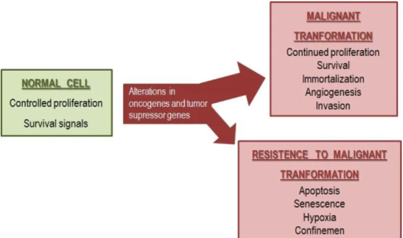
Many authors thought and defend that genomic mutability acquired during tumor progression lead to metabolic modifications and in some cases to the Warburg effect (Kim and Dang, 2006; Seyfried and Shelton, 2010). Studies in energy metabolism of cancer cells have demonstrated that there was a link between alteration in several oncogenes/ tumor suppressor genes and the regulation of metabolic processes (Wise and Thompson, 2010). These alterations could lead to genomic instability and DNA damage, having the potential to increased mutability of cells. Those pre-malignant cells start to display hallmarks of cancer (Figure 1) (Seyfried and
4
Shelton, 2010). In most cases of malignant transformation the bioenergetic demands are supported, in part, by some oncogenic mutations responsible for an increase in nutrients uptake (Vander Heiden et al., 2009).
Cell metabolism can trigger the production of toxic products like reactive oxygen species (ROS). Increase in ROS production or abolishment/decrease in detoxification can lead to membrane damage and mutagenesis which can result in malignant transformation (Dang, 2012). Increased ROS levels induce alterations in pathways, like pentose phosphate pathway (PPP). ROS oxidizes and consequently inactivates pyruvate kinase isozymes M2 (PKM2) responsible for the last step in glycolysis by modifying a critical sulphydryl group of PKM2 (Dang, 2012). Inactive PKM2 decreases the rate of PPP, reflecting in a NADPH reduction and hence in a decrease of glutathione reductase (GSH-R) responsible for glutathione (GSH) reduction (antioxidant compound) (Dhivya, 2012).
1.3.1 Oncogenic alterations
During malignant transformation, mutations/overexpression of oncogenes and loss of tumor suppressor genes can lead to alterations in metabolism, namely a switch from aerobic respiration to aerobic glycolysis (Warburg effect) (Dang, 2012).
In most cases, oncogenes control mitosis and cell survival by affecting steps on signaling mechanism namely by increasing signals in a cascade similar to “tyrosine kinase receptor” (RTK) (Israël and Schwartz, 2011a). One of the influences of oncogenes and tumor suppressor genes mutations is alterations in the production of ribose-5-phosphate (R5P), essential for nucleotide biosynthesis (Deberardinis et al., 2008).
Figure 1 – Cell proliferation and survival is
regulated by some pathways that can be abrogated or altered leading to malignant transformation. Cancer cells have the potential to proliferate beyond the limitations of normal cells. Some cells can resist to this alterations by blocking clonal cancer expansion and neoplastic evolution.
5
Tumor suppressors are associated with DNA repair. Tumor suppressor p53 target TP53-induced glycolysis and apoptosis regulator (TIGAR) that suppresses glycolysis by inhibiting activation of phosphofructokinase-1 (PFK1). This enzyme increase substrate availability to oxidative pathway as NADPH and R5P generation, via PPP. In tumors p53 deficiency is frequent, leading to glycolysis triggering and PPP impair.
Cancer cells mostly choose an alternative mechanism to sustain cell R5P requirements. Tumors may express pyruvate kinase-2 (PKM2), a glycolytic enzyme that stimulates PPP, inducing R5P production through a non-oxidative pathway (Deberardinis et al., 2008).
Protein kinase B (AKT) is another oncogene associated with tumorigenesis. AKT encodes serine-threonine kinase protein responsible for the regulation of glucose uptake and aerobic glycolysis. This oncogene mobilizes glucose transporters to the cell surface, increasing the levels of glucose import and activating hexokinase 2 (HK2) which is responsible for glucose phosphorylation and intracellular trap. Additionally, the oncogene MYC is also associated to tumorigenesis triggering direct activation on glycolysis (Kim and Dang, 2006).
1.3.2 Nutrients uptake
Cancer cells can supply metabolites requirements due to the expression of nutrient transporters (Fuchs and Bode, 2005). In normal cells the uptake of nutrients from their environment is stimulated by growth factors avoiding uncontrolled proliferation (Vander Heiden et al., 2009).
Glucose is one of the most important nutrient for cell mass production and cell proliferation. This compound is responsible for carbon and hydrogen supply but not nitrogen, phosphorus and sulfur that are also need for cell development. Those elements essentials to build new cells are supplied by other nutrients, namely amino acids as glutamine (Dang, 2012).
Glucose, amino acids, fatty acids and vitamins are essential nutrients required for cell survival and proliferation. In cancer cells, nutrient transporters are up-regulated or altered in plasma membrane (Ganapathy et al., 2009). Amino acid transporters support metabolic needs and are essential requirements for production of nucleotides, cellular nitrogen and proteins (Ganapathy et al., 2009; Mazurek and Eigebbrodt, 2003).
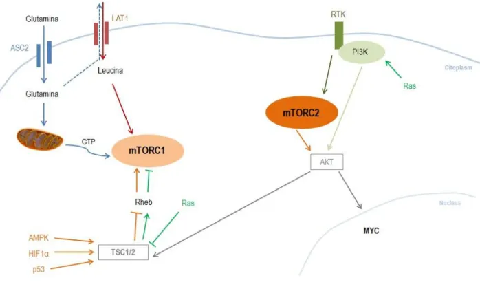
Cell growth is regulated in part by mammalian target of rapamycin (mTOR) mTORC1/C2 complexes being activated by amino acids and growth factors. mTOR is a regulatory-associated protein which is located downstream of PI3K and AKT and that can also be activated by insulin-like growth factor, oxidative stress and nutrients, essentially leucine that is imported in exchange
6
by glutamine. (Fuchs and Bode, 2005b; Laplante et al., 2010; Wise and Thompson, 2010). Leucine mobilizes mTORC1 to lysosomal membrane by G proteins turning it active to phosphorylate substrates that will be responsible for stimulating translation, ribosome biogenesis, cell growth and autophagy inhibition. Growth signals are transmitted by RTK to activate mTORC2 and PI3K. mTORC2 and PI3K stimulate AKT initiating the pathway responsible for an increase in metabolism, survival and proliferation. This process is also responsible for a transcriptional program, namely in genes such as MYC. AKT activated by mTORC2 can also influence mTORC1 (Figure 2) (Dang, 2012; Sabatini, 2006).
Figure 2: mTOR pathway - Glutamine (Gln) enters in the cell by neutral amino acid transporter (ASC2) membrane
transporter, allowing leucine (Leu) influx by human L-type amino acid transporter 1 (LAT1). Leu actives mTORC1 (red) and in mitochondria gln realize TCA cycle with GTP production (blue). Hypoxic genes (HIF1α), DNA damage genes (p53) and energy depletion genes (AMPK) phosphorylated a negative regulator of mTORC1 (tuberous sclerosis complex - TSC1/2), overturning Rheb (orange arrows) and leading to mTORC1 activation. Ras inhibits mTORC1 by suppressing TSC1/2 (green arrows). RTK receive growth factor signaling activating PI3K and mTORC2. PI3K (also stimulated by Ras) and AKT induce mTORC2 (grey and dark orange arrows) resulting in myc activation that stimulate glycolysis and glutaminolysis. mTORC1 can also be activated by AKT expression.
1.4 Glucose and Glutamine
Glucose and glutamine are two major substrates for cell proliferation essentially because there are the two main sources of ATP and carbon skeletons required for macromolecules
7
synthesis. Glucose is responsible for ribose (fundamental for nucleic acid synthesis) and ATP production through pentose phosphate pathway (PPP). Glutamine that is the most abundant amino acid in bloodstream and the main bioenergetic substrate and nitrogen donnor (Dang, 2012).
In normal conditions glucose enters the cells through facilitative transport where is converted by glucose phosphorylation in hexose phosphate. This compound is phosphorylated and converted into glycerol, essential for lipid synthesis or transformed into pyruvate (Dang, 2012).
Proliferating cells in hypoxia and under glucose limitations reprogramme glutamine catabolism by TCA cycle to increase lipid synthesis. In hypoxia and in unrestricted glucose conditions the conversion of pyruvate to acetyl- CoA is switch to the conversion of pyruvate into lactate (Figure 3). This process is mediated by the inhibition of pyruvate dehydrogenase due the activation of pyruvate dehydrogenase kinase (PDK1) induced by hypoxia inducible factor (HIF1 ) (Dang, 2012). HIF1 actives target genes like PDK1, consequently pyruvate is not used in mitochondrial oxidation, activating cellular responses to metabolic stresses.
Glutaminolysis, that is also increased in some tumors, is considered as a glutamate store. Glutamine is converted into glutamate by glutaminase, generating NAD+ that is essential for glycolysis (Israël and Schwartz, 2011b; Dang, 2012).
Figure 3: Pyruvate resulting from glycolysis can
converted into acetyl-CoA in TCA cycle, increasing the levels of OAA and citrate which consequently permits an energy gain. Pyruvate can also be converted in lactate. Glutamine enters in TCA through conversion into glutamate. TCA can be independent of glucose or responsible for lipid synthesis via reductive carboxylation.
8
Decrease in glucose and glutamine levels lead to a reduction in ATP levels and an increase in AMP/ATP ratio. AMP-activated protein kinase (AMPK) is sensitive to AMP/ATP and have the potential to phosphorylate substrates to increase energy production and to decrease the activity of pathways that consume energy. On the other hand, AMPK that recycles cellular components for energy production, inhibits Acetyl-CoA carboxylase by phosphorylation and also induces mTOR pathway (Dang, 2012).
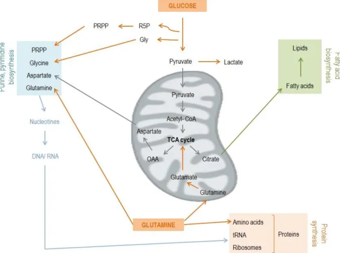
Figure 4: Cells have to duplicate genomic material, proteins and lipids to support essential requirements for
replicative division, enhanced in tumors. Increase in glucose and glutamine influx is common in tumorigenesis. Glucose is converted into pyruvate with lactate production. Part of the pyruvate goes to TCA cycle in mitochondria resulting in citrate and aspartate production (TCA intermediates). Citrate is involved in fatty acids biosynthesis and asp in nucleotides production. Glucose is also responsible for amino acid precursors such as glycine and R5P. Glutamine also enters the mitochondria where is converted into glutamate that origin KG, a TCA precursor. PRPP: phosphoribosyl pyrophosphate. Adapted from DeBerardinis et al., 2008
1.4.1 Gluconeogenesis and Glycolysis
Some enzymes are common to gluconeogenesis and glycolysis however working on different directions. Others are exclusive to gluconeogenesis or glycolysis, such as pyruvate
9
kinase (PK), in glycolysis, which is responsible for the conversion of phosphoenolpyruvate (PEP) into pyruvate, being transfered to mitochondria as acetyl- CoA and entering in TCA cycle and oxidative metabolism (Israël and Schwartz, 2011b).
Cells have an adaptative response to their metabolic needs, for example in starvation glycolytic enzymes, such as PK and pyruvate dehydrogenase (PDH) are phosphorylated and inactivated due to cAMP- glucagon- adrenergic signals. The inactivation of these enzymes leads to the interconnection break between glycolysis and TCA cycle and stops the oxidative metabolism (Israël and Schwartz, 2011b).
In normal cells, citrate synthase (CS) is inhibited by an increase in NADPH levels whereas in cancer cells this enzyme is active and overexpressed. Overexpression of CS increases levels of acetyl-CoA, oxoloacetate (OAA) and citrate and decrease levels of ketone bodies. On the other hand, low levels of ketone bodies are responsible for the decrease in the stimulation of pyruvate carboxylase (PC). Decreases in PC activity leads to more pyruvate to be “processed” by lactate dehydrogenase subunit A (LDHA). Overexpression of LDHA increase the levels of lactate and NAD+, essential and required for glycolysis (Israël and Schwartz, 2011a,
2011b). This alternative pathway in tumors can explain, at least in part, the previously referred Warburg effect.
1.4.1.1 Gluconeogenesis
Gluconeogenesis accounts for 35-50% of total basal glucose production, being regulated in part by the interaction of different regulatory mechanisms discussed later on. Glucose is used by the majority of the tissues as energy source, being its production, in physiological conditions, restricted to the liver, kidneys and some cells in small intestine. Glucose-6-phosphatase (G6Pase) is an essential gluconeogenic enzyme and its expression is limited to these organs (Corssmit et al., 2001).
Glucose biosynthesis via gluconeogenesis requires ATP and NADPH for the conversion of pyruvate, lactate and amino acids, as glutamine and alanine, into the final product, de novo glucose. Lactate is a common substrate for kidney and liver whereas alanine conversion to glucose takes place almost exclusively in liver. In healthy humans is estimated that the contribution of lactate for total glucose production is approximately 15%(Corssmit et al., 2001).
10
1.4.1.1.1 Molecular regulators
In normal tissues, gluconeogenesis is activated by pos-translational modifications or by allosteric activation of key-rate limiting enzymes, responsible for the molecular events of this pathway. Liver is the most active organ for gluconeogenesis and in physiological conditions this pathway contributes to the maintenance of glucose homeostasis, controlling glucose levels between bloodstream and hepatocytes. In general and according to the metabolic requirements, this organ adjusts the regulation of glucose uptake and storage, as well as its production and release (Oh et al., 2013; Telang et al., 2012). Hormones have an important physiological role in the regulation of metabolism. Insulin inhibits gluconeogenesis while other hormones like glucagon and glucocorticoids induce the synthesis of de novo glucose by regulating the expression of glycolytic enzymes. Glucose concentration have also been reported to directly suppress the expression of the gene phosphoenolpyruvate carboxykinase cytosolic (PCK1) and gluconeogenesis (Weickert and Pfeiffer, 2006).
Additionally stress hormone cortisol is responsible for the initiation of signaling cascades that activate transcriptional regulators like cyclic adenosine monophosphate (cAMP), responsive
element binding protein (CREB), nuclear glucocorticoids receptors (GR), estrogen receptor related gamma (ERR and forkhead box class O (FoxOs) transcription factors. These transcriptional regulators promote the expression of glucogenic enzymes, namely PCK1 and G6Pase (Oh et al., 2013; Weickert and Pfeiffer, 2006).
1.4.1.1.2 Molecular pathway
The conversion of pyruvate into glucose is the major step of gluconeogenesis that is catalyzed by the action of diverse cytoplasmic and mitochondrial enzymes. Some glycogenic enzymes catalyze steps of gluconeogenesis (seven reactions in ten), in a reversible way (Figure 5).
Pyruvate is transported directly from cytoplasm to mitochondria or having alanine as a precursor, which conversion occurs in the mitochondria by transamination. Pyruvate promotes the OAA formation by the action of PC. Carnitine transporter permits the influx of Acytil-CoA in mitochondria, being an essential activator of PC due to the carboxylation of a biotin residue that occurs exclusively in the presence of acytil-CoA. OAA is then reduced to malate being transported to cytoplasm by malate- -ketoglutarate transporter. In cytoplasm, malate is oxidized to OAA and PEP through PCK1. Nevertheless, if OAA is exported directly from the cytoplasm this reaction is catalyzed by phosphoenolpyruvate carboxykinase mitochondrial (PCK2), as it will
11
be depicted latter on. Another irreversible reaction in gluconeogenesis is the conversion of 1, 6-biphosphate (F1,6BP) in 6-phosphate (F6P), that is catalyze by fructose-1,6-bisphosphatase (FBP1) (Quintas et al., 2008a). G6Pase is then responsible for the dephosphorylation of glucose 6-phosphate (Glc-6-P) to de novo glucose (Figure 5) (Weickert and Pfeiffer, 2006).
Figure 5: Gluconeogenesis Pathway - Pyruvate provided by alanine transamination or lactate dehydrogenation is
converted to OAA by PC envolving the hydrolysis of one ATP molecule. Pyruvate from lactate, enters mitochondria by mitochondrial pyruvate transporter (MPC) (orange arrows). Acytil-CoA gets in mitochondria by carnitine transporter (blue arrows). OAA is reduced into malate and exported from mitochondrial by malate -ketoglutarate transporter (green arrows) while OAA from lactate is directly converted into PEP by PCK2 and exported for cytoplasm. In cytoplasm, malate is oxidized to OAA and then converted to PEP by the action of PCK1. The conversion of PEP in 2- phosphoglycerate , 3- phosphoglycerate and 1, 3 - bisphosphoglycerate are reversible reactions with glycolysis. 1, 3 – bisphosphoglycerate are then converted to frt-1,6-BP by FBP1. Frt-1,6-BP generates Frt-6-P in a reversible way. G-6-P by the action of G6Pase origin glucose, the final product. Adapted from Quintas et al., 2008.
12
1.4.1.1.3 Key enzymes
The enzyme 6-phosphofructo-2-kinase/ fructose-2, 6-bisphosphatase (PFKFB1) has its function associated with the promotion of glycolysis and the inhibition of gluconeogenesis through the modulation of their kinase activity. Although, gluconeogenesis is also regulated by others rate-limiting enzymes, the PCK1 and G6Pase (Noguchi et al., 2013). The expression of PCK1 and G6Pase genes is regulated at the transcriptional level by a complex network of transcription factors and cofactors including CREB, HNF-4α and FoxO1 (Kim et al., 2011). Also the activation of AKT-signaling pathway modulates gluconeogenesis, leading to the downregulation of PCK1 and G6Pase proteins and hence the suppression of gluconeogenesis (Noguchi et al., 2013). Those enzymes could also be activated by allosteric alterations, due to the relative proportion of AMP and ATP (Oh et al., 2013).
Alanine aminotransferase (ALT) plays an important role in gluconeogenesis from alanine, being an increase in ALT activity associated with higher rates of gluconeogenic pathway (Moraes-Silva et al., 2012). Previous studies had reported that gluconeogenesis from alanine was increased in cancer patients (Moreira et al., 2013). ALT catalyses the conversion of alanine into pyruvate and then converted into OAA by the action of PC, involving the hydrolysis of one ATP molecule (Quintas et al., 2008a).
PCK1 includes two enzymes with different intracellular locations, the cytosolic and
mitochondrial forms that are encoded by two nuclear genes (Greenfield et al., 2000). In human 60% of PEPCK is confined to mitochondria while 40% to cytosol (Hanson and Garber, 1972). PCK1, also known as PEPCK, is regulated by nutritional and hormonal stimuli at the transcriptional level, whereas mitochondrial form, also referred as PCK2, remains relatively unaltered appearing to be constitutive. Shifts in the ratio of PCK1 to PCK2 during feed restrictions or diabetes has been reported. PCK1 is required for gluconeogenesis from amino acids and PCK2 is more suited to gluconeogenesis from lactate. Due to the stoichiometry of gluconeogenesis, PEP formed from pyruvate and some amino acids requires the independent synthesis of NADH in cytosol (Agca et al., 2002). The reaction of conversion of lactate into pyruvate, in cytoplasm, is promoted by the enzyme lactate dehydrogenase B chain (LDH-B) with NADH production. Thereby the production of NADH by malate oxidation is not necessary and the pyruvate is directly converted into PEP by PCK2, inside mitochondria. PEP is exported to cytoplasm to sustain gluconeogenesis (Quintas et al., 2008a).
13
FBP1 is a magnesium (Mg2+) dependent enzyme that promote the irreversible hydrolysis of carbon 1 (C1) from fructose-1, 6-biphosphate. This reaction results in the release of an inorganic phosphate and F6P (Quintas et al., 2008a).
The molecular function of PFKFB1 is to synthesize fructose-2, 6- bisphosphate (F 2, 6 BP) from F6P. PFKFB1 activates PFK1, an essential and irreversible enzyme on controlling the glycolytic pathway. PFKFB1 expression could be stimulate by a decrease in ATP levels and by the abundance of energy stores leading to an enhancement in glycolytic flux and cell growth. When ATP levels and energy are abundant to supply cell requirements, ATP directly activates PFK1 by negative feedback. In cancer, an increase in PFKFB1 levels is commonly caused by an overexpression of HIF-1α, myc and activation of ras or loss of p53 (Yalcin et al., 2009).
G6Pase is responsible for the last step of gluconeogenesis, with glucose as final product.
This enzyme promotes phosphate hydrolysis, in a Mg2+ dependent reaction. Glucose is then
transported to bloodstream until target organs (Quintas et al., 2008a).
1.4.2 Glutaminolysis
Glutamine is a non-essential amino acid, although is a required nutrient source to maintain rapid cell division of cancer cells, being an increase of glutamine uptake and glutaminolysis associated with carcinogenesis (Matés et al., 2013). This non essential amino acid provides the anaplerotic carbon in mitochondrial TCA cycle as well as nitrogen and carbon skeletons. Glutamine is also used for glutathione (GSH) synthesis, fatty acids production, protein and nucleotide biosynthesis (Dang, 2010; Reynolds et al., 2013).
Since 1990 it has been reported that in cancer cells the activity of glutaminase is similar or higher than hexokinase from glycolysis. This result suggested that glutamine may be as important as glucose for energy generation (Board et al., 1990). More recently, it had been proved that glutamine restrictions or inhibitors of glutaminolytic enzymes decrease cell proliferation in vitro and in vivo (Reynolds et al., 2013).
MYC oncogene and tumor suppressor gene TP53 contribute to the glutamine uptake and glutaminolysis. MYC is responsible for inducing glutaminolysis through the activation of -catenin being glutamine synthetase a direct target of activated -catenin. Recently, dysfunctional retinoblastoma (Rb) cascate, besides promoting loss of proliferative control, it had been proposed to directly regulate the uptake and conversion of glutamine into anabolic precursors, required for neoplastic cell growth and survival (Dang, 2010; Reynolds et al., 2013).
14
1.4.2.1 Molecular pathway
The fundamental steps in glutaminolysis is the hydrolysis of glutamine into glutamate by glutaminase (GA) and subsequently conversion into -KG in mitochondria. -KG can enter TCA cycle and replenish the metabolic intermediates such as NADH (Piao et al., 2013).
GA localizes in the inner mitochondrial membrane, being responsible for the hydrolysis of the -amino group of glutamine, forming glutamate and ammonia. Ammonia could be used to form carbamoyl phosphate or may diffuse from the mitochondria and the cell. Glutamate can suffer deamination by glutamate dehydrogenase (GLUD1) forming -ketoglutarate ( -KG) entering in TCA cycle for energy production. Glutamate can also be converted into pyruvate by glutamate pyruvate transaminase (GPT) (Figure 5). Hence, glutamate catabolism in TCA cycle produces reducing equivalents for the generation of ROS by OXPHOS (Matés et al., 2013).
As mentioned above glutaminolysis enables NADH production through TCA cycle; however an incomplete oxidative metabolism can lead to lactate as end product. Zagari et al. (2013) reported that glutamine has an important role in modulating the initial lactate production being this phenomenon observed in vitro. In this process, lactate production involves the efflux of malate from mitochondria and its conversion into pyruvate and finally into lactate, with NADH production. However, when glutamine is depleted, lactate is consumed to “refuel” the TCA cycle (Board et al., 1990; Zagari et al., 2013). The highest percentage of consumed glutamine is metabolized into molecules such as lactate (Matés et al., 2013).
1.4.2.2 Key enzymes
Glutamine could be lysed by mitochondrial GA into glutamate and ammonia and some cancer cells can survive without glutamine when provided with ammonia as nitrogen source. Cancer cells can also have an increase in glutamate-ammonia ligase (GLUL), that is responsible for the conversion of glutamate and ammonia into glutamine (Dang, 2010).
GA is a central enzyme is glutamine catabolism. Glutamate, from glutamine catabolism, is involved in mitochondrial bioenergetic via TCA cycle responsible for ATP production. GA family is composed by two isoforms, GLS and GLS2, encoded by separate genes. Only GLS enzyme is associated and increased in cancer cells whereas GLS2 plays a critical role in protection against oxidative stress by up-regulating glutathione levels. Therefore GLS up-regulation is linked with increased rates of proliferation while GLS2 is associated with resting, non-proliferative or quiescent cell states, working as a tumor suppressor (Matés et al., 2013).
15
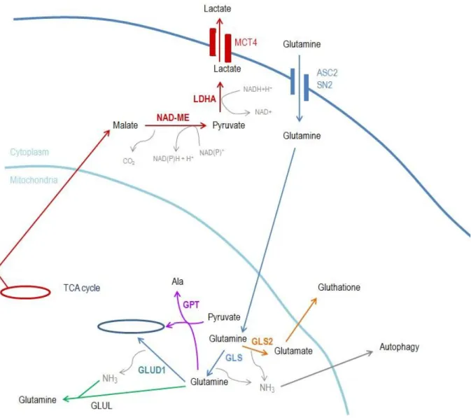
Figure 6: Glutaminolysis - Glutamine enters in the cell by cytoplasmic transporter such ASCT2 or solute carrier
family 38 (SN2). Glutamine is transferred to mitochondria where is hydrolyzed by GLS (an isoform of GA) forming glutamate and ammonia (NH3) (blue arrows). If glutamine is hydrolyzed by GLS2 (another GA isoform), in similarity to GLS reaction originates glutamate and also NH3 but glutamate is responsible for glutathione production, a potent antioxidant agent (orange arrows). NH3 and glutamate can be used to novo glutamine by the action of glutamine synthetase (green arrows). Glutamate is converted into -KG (alanine production by glutamate-pyruvate transaminase – GPT is also possible), entering in TCA cycle (blue arrows). If an incomplete oxidative metabolism occurs, malate (a TCA cycle intermediate) is exported from mitochondria where originates pyruvate (by the action of malate dehydrogenase –NAD ME) and then lac (by LDHA). Lac is exported from cell through monocarboxylate transporter 4 (MCT4) (red arrows).
1.5 Glutathione (GSH)
GSH which is a tripeptide gamma-glutamyl-cysteinyl-glycine is a major free radical scavenger, immune booster and detoxifier of the body. Can be found in reduced (GSH) and oxidized forms (GSSG) (Figure 6) being the cysteine residue responsible for the readily oxidation of GSH to GSSH by electrophilic substances as free radicals and ROS. GSH is the major
thiol-16
containing endogenous antioxidant and acts as a redox buffer against several sources of oxidative stress (Dhivya, 2012; Matés et al., 2013). Quotient [GSH]: [GSSH] is used as indicator of the redox state of cells being values >10 verified under normal physiological conditions (Quintas et al., 2008b) where glutathione disulfide (GSSH) levels accounts for less than 1% of the intracellular content. In the cytosol, 85-90% of the GSH is freely distributed, although it can also be compartmentalized in different organelles, like mitochondria, peroxisomes and nuclear matrix (Singh et al., 2012).
GSH provides greater antioxidant protection participating directly in ROS destruction and plays a critical role in carcinogenesis inhibition (Dhivya, 2012). Aerobic glycolysis, which is a metabolic characteristic of some tumors, is responsible for the production of ROS where their increase (higher than physiological conditions) is associated with higher levels of oxidative stress that lead to cell injury and death. Increase in ROS levels have also been implicated in cancer development and progression and molecular alterations in the components of the GSH system and in various tumors can lead to an increased survival and tumor drug resistance (Traverso et al., 2013). ROS formation and GSH depletion may also cause mitochondrial dysfunction and subsequent cytochrome c release triggering cell viability (Han et al., 2010).
GSH, beyond detoxification and cellular protection from damage by free radicals, peroxides and toxins, has also other essentials functions in metabolic and biochemical reactions, such as DNA synthesis and repair, protein and prostaglandins synthesis, amino acid transporters and enzymes activation (Dhivya, 2012). GSH is also implicated in essential functions like signal transduction, molecular regulation of cell physiology and regulation of apoptosis (Estrela et al., 2006; Singh et al., 2012).
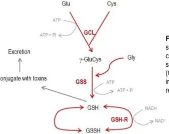
Figure 7: Gluthatione formation - -glutamyl cysteine
synthease (GCL) that catalyzes the ligation of glu with cysteine (cys) through ATP activation. Glutathione synthetase (GSS) allowing gly ligation and glutathione (GSH) production. GSH is easily oxidized into GSSH, an inactive form. GSSH can be regenerated by glutathione reductase. Adapted from Quintas et al., 2008.
17
1.6 Cysteine homeostasis
Cysteine is a semi-essential amino acid, provided through diet or trans-sulfuration pathway. High cysteine concentrations in human plasma could be cytotoxic, leading to pathologies like pre-eclampsia, premature delivery, low birth weight and cardiovascular diseases. Cysteine is used to GSH, taurine or inorganic sulphate synthesis and in similarity to GSH, taurine a most abundant free amino acid in humans, also acts as an endogenous antioxidant (Ishii et al., 2004).
Trans-sulfuration pathway is mediated, in part, by cystathionine gamma-lyase (CTH) and 3-mercaptopyruvate sulfurtransferase (MPST). CTH, a cytoplasmic enzyme and the last key enzyme in trans-sulfuration pathway, catalyze the conversion of cystathionine (derived from methionine) to cysteine, ammonia and 2-oxobutyrate. CTH protein can also accept homocysteine (HCYS) as substrate and could be responsible for hydrogen sulfide (H2S) production (Wang and
Hegele, 2003). In literature, CTH was also characterized as being able to degradate cysteine, leading to pyruvate production (Steegborn, 1999).
Under physiological conditions, oxidative stress induces an increase of HCYS flux through the trans-sulfuration pathway. As results cystathionine levels, a direct substrate of CTH, increase leading to an increase of downstream metabolites of CTH, cysteine and GSH. Then, GSH permits the maintenance of cellular redox status (Jurkowska et al., 2011).
MPST is an enzyme involved in cysteine degradation, catalyzing the transfer of sulfur ion from 3-mercaptopyruvate to cyanine or other thiol compound, yielding sulfane sulfur. In cancer had been reported a decrease in MPST activity leading to a reduction of non-oxidative metabolism of cysteine and consequently to a sulfane sulfur deficiency. Insufficient levels of sulfane sulfur induce an uncontrollable action of enzymes inactivated by sulfane sulfur (Jurkowska et al., 2011).
As referred, CTH and MPST, plays an important role in cysteine homeostasis, GSH-taurine synthesis and H2S production. In humans, CTH mutations and deficiency levels causes
cystathionimaemia, a disease characterized by accumulation of cystathionine in blood, tissue and urine with no consistent clinical consequences (Ishii et al., 2004; Jurkowska et al., 2011; Kraus et al., 2009).
18
2. Aims
The first main objective of this thesis is to understand if gluconeogenesis is a source of glucose supply in ovarian clear cell carcinoma (OCCC). This main objective is subdivided in 3 specific aims: 1) To verify if gluconeogenesis is working on OCCC by testing glutamine, lactate and butyrate as gluconeogenic substrates; 2); To evaluate the expression profile of key gluconeogenic enzymes (PCK1, PKC2, FBP1 and PFKF1) under these gluconeogenic substrates exposure; 3) To evaluate the modulation of cell features such as migration, cell viability and cell cycle in the presence of these gluconeogenic substrates.
The second main objective of this thesis came up from the evaluation of the results obtained from the first main objective accomplishment; and it is to clarify the role of glutamine and cysteine metabolism in OCCC. This objective is accomplished through 4 specific aims: 1) To verify if glutaminolysis is working on OCCC; 2) To evaluated cysteine degradation and synthesis as well as to determine the expression profile of genes involved in cysteine metabolism; 3) To evaluate the modulation of cell features such as migration, cell viability and cell cycle by cysteine in cells grown in glutamine, and 4) To determine the relevance of glutamine and cysteine metabolism in GSH synthesis.