UNIVERSIDADE DA BEIRA INTERIOR
Ciências
Hepatocellular carcinoma therapy based on
human amniotic membrane
Andreia Pereira Alves
Dissertação para obtenção do Grau de Mestre em
Bioquímica
(2
ºciclo de estudos)
Orientador: Prof. Doutor Cláudio Jorge Maia Batista
Co-orientador: Prof. Doutor Maria Filomena Botelho
Ao Samuel, pais e Arminda
Agradecimentos
Este trabalho foi feito com muito esforço e trabalho árduo, que me fez crescer não só em conhecimento, mas também como mulher. Ao longo deste caminho pude contar com algumas pessoas, a quem gostaria de agradecer:
Em primeiro lugar, ao professor Doutor Cláudio Maia Jorge, orientador desta dissertação, obrigado pela disponibilidade, orientação e pela partilha de conhecimento e experiência científica, sempre patentes durante todo o desenvolvimento deste trabalho.
À professora Doutora Maria Filomena Botelho, diretora Unidade de Biofísica da Faculdade de Medicina da Universidade de Coimbra, co-orientadora desta dissertação, obrigada pela hospitalidade, disponibilidade prestadas durante o desenvolvimento deste trabalho e especialmente pelos conhecimentos adquiridos.
À Mestre Catarina Mamede, o meu maior obrigada, pela paciência, ajuda, dedicação, carinho e amizade, ao longo destes dois anos. Obrigada pelo apoio incondicional que me ajudou a finalizar esta dissertação.
Agradeço a todos os colegas de trabalho do CICS e da Unidade de Biofísica, a criação de um ambiente de trabalho saudável, e por me oferecer apoio e carinho nos momentos mais difíceis.
Obrigado pelo apoio incondicional e incansável dos meus amigos de sempre, Rita, Filipa, Petra, Luís e Válter, gostaria de expressar a minha mais profunda gratidão, por estarem ao meu lado nos momentos mais sombrios, assim como nos momentos mais felizes e dos meus colegas e amigos de Bioquímica, que fizeram parte desta vida académica, que agora chega ao fim.
Por último, mas não menos importante, à minha família, especialmente ao meu irmão e aos meus pais, que foram sem dúvida, o maior pilar que tive nesta fase e em toda a minha vida. Samuel obrigado por todo o apoio, sei que sou a tua heroína e isso traz-me muita responsabilidade, espero deixar-te mais uma vez orgulhoso. Mãe e pai, vocês foram e são o meu maior apoio, obrigado por todas as palavras e sermões, e especialmente pela confiança depositada, que fizeram de mim o que hoje sou.
Acknowledgments
This dissertation was done intense effort and hard work, which made me grow up not only in knowledge but also as a woman. Along this path I could count on some people, whom I would like to thank:
Firstly, to my supervisor Professor Cláudio Maia Jorge, thank you for availability, guidance and sharing of scientific knowledge and experience, always patents during this Master thesis. I would also like to show my appreciation to Professor Maria Filomena Botelho from Unit of Biophysics of Faculty of Medicine of University of Coimbra, co-supervisor of this dissertation, for the hospitality, availability provided during the development of this work and especially the acquired knowledge.
To Master Catarina Mamede, my greatest thanks for their patience, support, dedication, affection and friendship, throughout those two years. Thank you for the unconditional support that helped me complete this dissertation.
I thank to work group of CICS and of Biophysics Unit, the creating a healthy working environment, and for offering me support and kindness in the toughest moments.
Thank you for the unconditional and tireless support, to my dear friends, Rita, Filipa, Petra, Luís e Válter, I would like to express my deepest gratitude, for being at my side in the darkest times, but as well as in the happiest and to the rest of the friends and colleagues of the Biochemistry, that were part of this academic life, which now comes to an end.
Last but not least, to my family, especially to my brother and my parents, which were undoubtedly the greatest pillar that have had at this stage and in all my life. Samuel thanks for all the support, I know I'm your hero and that brings me a lot of responsibility, I hope to make you proud one more time. Mother and father, were and are my biggest support, thank you for all the words and sermons and especially for their belief, that made me who I am today.
Resumo
O carcinoma hepatocelular é dos tumores primários do fígado mais frequentemente detetado e representa um problema de saúde pública em todo o mundo, devido à sua elevada taxa de morbidade e mortalidade, que em parte se deve ao estado avançado em que é comumente diagnosticado e à elevada resistência às terapias convencionais, nomeadamente à radioterapia e à quimioterapia. Por esta razão, é muito importante encontrar novas e eficazes terapias para o carcinoma hepatocelular.
A membrana amniótica humana (hAM, do inglês, human amniotic membrane) foi referenciada em várias publicações como uma potencial opção terapêutica para o cancro devido às suas propriedades pro-apoptóticas, anti-angiogénicas e imunorreguladoras. As células derivadas da hAM e o seu meio condicionado já foram testados, in vitro e in vivo, em vários tipos de cancro. De forma inovadora, o nosso grupo de investigação avaliou o efeito anticancerígeno de extratos proteicos da hAM (hAMPE, do inglês, human amniotic membrane protein extracts) na terapia do cancro. Este extrato é obtido a partir de células epiteliais e mesenquimais, assim como da matriz extracelular. Foi demonstrado que o hAMPE pode inibir a atividade metabólica das três linhas celulares de carcinoma hepatocelular humano: Hep3B2.1 -7, HepG2 e HuH7.
Tendo em conta os resultados obtidos até agora pelo nosso grupo de investigação, este trabalho experimental teve como objetivo estudar o efeito do tratamento com o hAMPE no metabolismo da glicolítico no carcinoma hepatocelular.
Verificou-se através da técnica de ressonância magnética nuclear que o tratamento com o hAMPE aumentou o consumo de glicose nas linhas celulares Hep3B2.1-7, HepG2 e HuH7, no entanto essa glicose não é convertida em lactato. O tratamento com o hAMPE induziu efeitos semelhantes nas Hep3B2.1-7 e HepG2, aumentando a expressão de lactato desidrogenase (LDH, do inglês, lactate dehydrogenase) e do transportador de monocarboxilato isoformas 4 (MCT4, do inglês monocarboxylate transporter isoform 4), não se tendo verificado alterações significativas nos níveis de lactato. No que diz respeito às células HuH7, os níveis de lactato, assim como a expressão de LDH e MCT4, diminuíram em resposta ao tratamento com o hAMPE, sugerindo que níveis reduzidos de lactato podem indicar um decréscimo na agressividade tumoral.
Este trabalho revela que o hAMPE é capaz de alterar o metabolismo glicolítico das células de carcinoma hepatocelular humano. No entanto, mais estudos são necessários de forma a esclarecer a relação entre o tratamento com o hAMPE e o metabolismo glicolítico das linhas celulares do carcinoma hepatocelular.
Palavras-chave
Placenta, Membrana amniótica humana, Carcinoma hepatocelular, Terapia, Metabolismo, Glicólise.
Resumo Alargado
O carcinoma hepatocelular é um tumor primário do fígado que representa um problema de saúde pública, pois é responsável por elevadas taxas de incidência e mortalidade. A elevada mortalidade deve-se em parte ao estado avançado em que o hepatocarcinoma é diagnosticado e à elevada resistência às terapias convencionais, nomeadamente à radioterapia e/ou à quimioterapia.
O processo de hepatocarcinogénese é multifatorial e ainda está pouco caraterizado. No entanto sabe-se que as principais alterações genéticas são ao nível dos genes que codificam a proteína 53 (p53) e da β-catenina. Para além destas alterações genéticas, as células tumorais caraterizam-se por alterações metabólicas ao nível da glicólise, apresentando uma elevada taxa de glicólise, mesmo em presença de níveis normais de oxigénio. Nos tumores em ambiente hipóxico, como o carcinoma hepatocelular, ocorre um aumento acentuado na dependência da glicólise, visto que o tumor depende quase exclusivamente da glicólise para a obtenção de energia.
Devido à falta de terapias eficazes para o tratamento do carcinoma hepatocelular, é imperativo encontrar novas terapias que permitam reduzir a mortalidade deste tumor. Neste contexto, surge a membrana amniótica humana (hAM, do inglês, human amniotic membrane) como possível terapia para o hepatocarcinoma.
A hAM tem sido referenciada em várias publicações como uma potencial opção para o tratamento oncológico, devido às suas propriedades anti-angiogénicas, pro-apoptóticas e imunorreguladoras. Vários derivados da hAM, como as suas células e o seu meio condicionado, foram testadas, in vitro e in vivo, em vários tipos de cancro. De forma inovadora, o nosso grupo de investigação estudou o efeito anticancerígeno de extratos proteicos da hAM (hAMPE, do inglês human amniotic membrane protein extracts), obtidos a partir de células epiteliais e mesenquimais, assim como da matriz extracelular.
Foi demonstrado que o hAMPE pode inibir a atividade metabólica das três linhas celulares humanas de carcinoma hepatocelular: Hep3B2.1 -7, HepG2 e HuH7. Tendo em conta os resultados obtidos até agora pelo nosso grupo de investigação e ao aumento do metabolismo glicolítico que ocorre nas células cancerosas, colocou-se a hipótese de que o tratamento com o hAMPE pode inibir a glicólise. Por este motivo, este trabalho experimental teve como objetivo estudar o efeito do tratamento com hAMPE no metabolismo glicolítico do carcinoma hepatocelular.
Verificou-se através da técnica de ressonância magnética nuclear (1NMR, do inglês proton nuclear magnetic resonance) que o tratamento com o hAMPE aumentou o consumo de glicose
Ao nível da expressão dos transportadores de glucose (GLUT, do inglês glucose transportes) 1 e 3, apenas na linha Hep3B2.1-7 há um aumento da expressão do GLUT-1 que justifique o aumento do consumo de glucose, no entanto nas restantes linhas celulares não se verificaram alterações que justifiquem o aumento do consumo de glucose. Tal aumento pode dever-se ao aumento da expressão de outras isoformas de GLUT.
De forma geral, o tratamento com o hAMPE induziu efeitos semelhantes nas linhas celulares Hep3B2.1-7 e HepG2, aumentando a expressão de lactato desidrogenase (LDH, do inglês,
lactate dehydrogenase) e do transportador de monocarboxilato isoformas 4 (MCT4, do inglês monocarboxylate transporter isoform 4), embora não tivessem sido registadas alterações nos
níveis de lactato. Por outro lado, nas células HuH7 os níveis de lactato, assim como a expressão de LDH e MCT4, diminuíram em resposta ao tratamento com o hAMPE, sugerindo que níveis reduzidos de lactato podem indicar um decréscimo na agressividade deste tipo de tumor.
Este trabalho revelou que o hAMPE altera o perfil glicolítico das linhas celulares de carcinoma hepatocelular. No entanto, são necessários mais estudos para esclarecer a relação entre o tratamento com o hAMPE e o metabolismo glicolítico.
Abstract
The hepatocellular carcinoma is the most frequently detected primary malignant liver tumor, representing a worldwide public health problem due to its morbidity and mortality rates. The high mortality rate is in part due to the late stage at which is diagnosed and its high resistance to conventional therapies, such as radiotherapy and chemotherapy. For this reason, it is very important to find new and effective therapies for hepatocellular carcinoma. The human amniotic membrane (hAM) has been referenced in several publications as a potential therapeutic option in cancer due to its pro-apoptotic, anti-angiogenic and immunoregulatory properties. The hAM-derived cells and their supernatant were already evaluated, in vitro and in vivo, in cancer therapy. In an innovative way, our research group evaluated the protein extracts of human amniotic membrane (hAMPE) in cancer therapy. The extracts were obtained from epithelial and mesenchymal cells, as well as from the extracellular matrix. It was demonstrated that the hAMPE may inhibit the metabolic activity of three human hepatocellular carcinoma cell lines: Hep3B2.1 -7, HepG2 and HuH7.
Taking into account the results obtained so far by our research group, associated with the increased glycolytic metabolism in cancer cells, this experimental work aimed to study the effect of hAMPE treatment in the glucose metabolism of hepatocellular carcinoma.
It was verified by proton nuclear magnetic resonance (1NMR) that hAMPE treatment increased
glucose consumption on Hep3B2.1-7, HepG2, and HuH7 cell lines. However, it was verified that glucose is not converted to lactate. The hAMPE treatment induced similar effects in Hep3B2.1-7 and HepG2, increasing the expression of lactate dehydrogenase (LDH) and monocarboxylate transporter isoform 4 (MCT4), but no changes in lactate levels. Regarding HuH7 cells, the levels of lactate, as well as the expression of LDH and MCT4, decreased in response to hAMPE treatment, suggesting that reduced levels of lactate may indicate a decrease in tumor aggressiveness.
This work reveals the anticancer potential of hAMPE, modulating the glycolytic profile of the human hepatocellular carcinoma cells under study. However, further studies are required in order to clarify the relationship between hAMPE treatment and the glycolytic metabolism.
Keywords
Placenta, Human amniotic membrane, Hepatocellular carcinoma, Therapy, Metabolism, Glycolysis
Index
Chapter 1 - Introduction ... 1
1.1. Hepatocellular carcinoma... 1
1.1.1. Epidemiology and etiology ... 1
1.1.2. Pathogenesis ... 6
1.1.3. Diagnosis and therapy ... 9
1.1.4 Glycolytic metabolism ... 11
1.2. Human amniotic membrane ... 14
1.2.1. Structure and function ... 14
1.2.2. Properties of human amniotic membrane ... 16
1.3. Human amniotic membrane and cancer ... 17
Chapter 2 - Aims ... 23
Chapter 3 - Material and methods ... 25
3.1. Collection of the human amniotic membrane ... 25
3.2. Reception of the human amniotic membrane ... 25
3.3. Extraction and quantification of proteins ... 25
3.4. Cell culture ... 26
3.5. Experimental design ... 26
3.6. Glucose consumption and lactate production by 1H NMR ... 27
3.7. Protein quantification by Western blot ... 28
3.8. GLUT-1 and -3 expression evaluation by fluorescence ... 29
3.9. Statistical analysis ... 29
4.1. Glucose consumption evaluation by 1H NMR ... 31
4.2. Lactate production evaluation by 1H NMR ... 32
4.3. Expression of GLUT-1 and GLUT-3 by fluorescence ... 33
4.3.1. Expression of GLUT-1 ... 33
4.3.2. Expression of GLUT-3 ... 34
4.5. Expression of LDH by Western Blot ... 34
4.6. Expression of MCT4 by Western Blot ... 35
Chapter 5 - Discussion ... 37
Chapter 6 - Conclusions and Future Perspectives ... 43
Figure list
Figure 1. Estimated new cancer cases and deaths worldwide by sex ... 1
Figure 2. Liver Cancer Incidence Rates by Sex and World Area, in all ages for males and females. ... 2
Figure 3. Predictions of the number of new cases (A) and of the number of cancer deaths (B) by hepatocellular carcinoma ... 3
Figure 4. The function of p53 ... 8
Figure 5. WNT/β-catenin pathway in hepatocellular carcinoma ... 9
Figure 6. Schematic diagram of cancer cells glycolytic metabolism ... 11
Figure 7. The placenta structure... 14
Figure 8 . Histologic section of hAM, obtained through hematoxylin–eosin staining ... 15
Figure 9. Experimental design. ... 27
Figure 10. Evaluation of glucose consumption by 1H NMR in Hep3B2.1-7, HepG2, and HuH7 cell lines.. ... 31
Figure 11. Lactate production evaluation by 1H NMR in Hep3B2.1-7, HepG2, and HuH7 cell lines, respectively ... 32
Figure 12. Lactate production in Hep3B2.1-7, HepG2, and HuH7 cell lines, respectively. ... 33
Figure 13 . GLUT-1 expression evaluation by fluorescence in Hep3B2.1-7, HepG2 and HuH7 cell lines after incubation with 1µg/µL of hAMPE ... 34
Figure 14 . GLUT-3 expression evaluation by fluorescence in Hep3B2.1-7, HepG2 and HuH7 cell lines after incubation with 1µg/µL of hAMPE. ... 34
Figure 15. LDH expression in Hep3B2.1-7, HepG2 and HuH7 determined by Western Blot. .... 35
Table list
Table 1. Risk factors for hepatocellular carcinoma by geographical area ... 4 Table 2. Mutations responsible for hepatocellular carcinoma development ... 6 Table 3. Metabolic activity of several human cancer cell lines evaluated after incubation of hAMPE ... 19
Abbreviations
1H NMR Proton nuclear magnetic resonance
5-FU 5-fluorouracil
AFB1 Aflatoxin B1
APC Adenomatous polyposis coli
ATCC American Type Culture Collection
ATP Adenosine triphosphate
BAX BCL-2 associated X protein
BCA Bicinchoninic acid assay
BCL-2 B-cell lymphoma 2
BRCA2 Breast cancer type 2 susceptibility protein CHUC Coimbra Hospital and University Centre
CK1 Casein kinase
DLC1 Deleted in liver cancer 1 protein DMEM Dulbecco’s Modified Eagle’s Medium
DNA Deoxyribonucleic acid
EGF Epidermal growth factor
ECL Enhanced chemiluminescent
epCAM Epithelial cell adhesion molecule
FBS Fetal bovine serum
FZD Frizzled receptors
GLUT Glucose transporter
HAECs Human amniotic membrane epithelial cells
hAM Human amniotic membrane
HAMCs Human amniotic membrane mesenchymal cells hAMPE Human amniotic membrane protein extract
HBV Hepatitis B virus
HCV Hepatitis C virus
HIF-1 Hypoxia-inducible factor 1
HIV Human immunodeficiency virus
HK Hexokinase
HLA Human leukocyte antigen
IFN-γ Interferon gamma
IGF Insulin-like growth
IL Interleukin
JCRB Japanese Collection of Research Bioresources
LDH Lactate dehydrogenase
LRP Low-density lipoprotein receptor-related protein M2-PK Pyruvate kinase isoform M2
M-CSF Macrophage colony-stimulating factor MCT4 Monocarboxylate transporter 4
NAFLD non-alcoholic fatty liver disease NASH Nonalcoholic steatohepatitis
p53 Protein 53
PE Phycoerythrin
PEDF Pigment epithelium-derived factor
PFK Phosphofructokinase
PTEN Phosphatase and tensin homolog PVDF Polyvinylidene difluoride
RB Retinoblastoma
RF/PEI Radiofrequency ablation/ Conventional percutaneous ethanol injection
RNA Ribonucleic acid
ROS Reactive oxygen species
SDS-PAGE Sodium dodecyl sulfate/ polyacrylamide gel
siRNA Small interfering RNA
SOCS-1 Suppressor of cytokine signaling 1 TACE Transarterial chemoembolization
TCF/LEF T-cell factor/ lymphoid enhancer-binding factor TGF-β Transforming growth factor beta
TNF Tumor necrosis factor
UV Ultraviolet
Chapter 1 - Introduction
1.1. Hepatocellular carcinoma
1.1.1. Epidemiology and etiology
Liver tumors can be classified as primary or secondary, according to their origin (Goodman, 2007). Among the primary liver tumors, whose the origin is the liver itself, the hepatocellular carcinoma is the most common epithelial tumor and accounts for about 80% of the primary liver tumors (Befeler and Bisceglie, 2002; Gomes et al., 2013; Jemal and Ferlay, 1999; Reid, 2001).
Hepatocellular carcinoma represents a worldwide health problem (Llovet et al., 2008)due to a high morbidity and mortality (El-Serag and Rudolph, 2007; Ferlay et al., 2010; Yang and Roberts, 2010). Hepatocellular carcinoma is the second cause of cancer death in the world. According to GLOBOCAN, in terms of incidence, hepatocellular carcinoma is the sixth most incident in the world (Llovet and Bruix, 2008). The high rate of mortality is mainly due to the asymptomatic nature of the disease at early stages, and consequently, the majority of hepatocellular carcinoma cases are detected in advanced stages (Ferlay et al., 2010; reviewed by Gomes et al., 2013; Jemal et al., 2011). However, the incidence varies according to age, ethnicity, gender and geographic distribution (Yang and Roberts, 2010). In relation to gender, hepatocellular carcinoma is the fifth incident cancer in male and the ninth in females. In terms of mortality, hepatocellular carcinoma is the second cause of cancer death in male and sixth in females (Torre et al. 2015), as shown in Figure 1.
Figure 1. Estimated new cancer cases and deaths worldwide by sex. Source: GLOBOCAN 2012. Adapted from (Torre et al., 2015).
Hepatocellular carcinoma rarely surges before 40 years old, except in the case of infection by the hepatitis virus. The mean ages of hepatocellular carcinoma diagnosis are between 55 and 59 years old in China and between 63 and 65 years in Europe and North America. In low-risk populations, the highest incidence of hepatocellular carcinoma is among individuals aged 75 or older (El-Serag, 2012).
As mentioned above, the incidence varies due to geographic distribution. In fact, hepatocellular carcinoma is more common in less developed countries, representing 82,9% of the cases of hepatocellular carcinoma, while that in more developed countries the incidence of hepatocellular is lower (Figure 2) (Torre et al., 2015; Yang and Roberts 2010). More than half a million cases are annually diagnosed, and in the last years, the incidence has been increasing in more developed countries, such as the United States, Canada and Europe (Llovet et al., 2008).
Figure 2. Liver Cancer Incidence Rates by Sex and World Area, in all ages for males and females. Source: GLOBOCAN 2012. Adapted from (Torre et al., 2015).
In Portugal, according to data from GLOBOCAN 2012, the incidence rate of this cancer is lower when compared with other types of cancer, accounting only 1.1%. Regarding the mortality rate, it accounts approximately 2% of cancer deaths in Portugal (GLOBOCAN, 2012). According to predictions from GLOBOCAN, 1014 new cases and over 1110 deaths by hepatocellular carcinoma will occur in 2020 (see Figure 3 A and B).
Figure 3. Predictions of the number of new cases (A) and of the number of cancer deaths (B) by hepatocellular carcinoma. Retrieved from GLOBOCAN, predictions 2020.
Etiologically, hepatocellular carcinoma may arise from gene mutations, which in turn change the metabolic pathways of the cell. Genetic mutations may be a consequence of several external factors, such as viral infection by hepatitis B (HBV) and C (HCV), or proliferation associated with chronic hepatitis. This proliferation caused by hepatitis infection may increase the risk of genetic alterations, such as gene duplication (Ferlay et al., 2010; Gomes et al., 2013; Jemal et al., 2011). This can explain the relationship between hepatocellular carcinoma and hepatitis since the majority of the cases occur in less developed countries. In addition, the highest incidence rates of hepatocellular carcinoma are described in regions where HBV is endemic, which are the Southeast Asia and sub-Saharan Africa (Yang and Roberts, 2010). The differences in the incidence of hepatocellular carcinoma are a reflex of the geographic distribution of VHB and HCV infection. Countries with a higher prevalence of infection by these viruses usually have a high incidence of hepatocellular carcinoma (Yang and Roberts, 2010).
The risk of incidence of hepatocellular carcinoma is higher in male than in female, varying according to the geographic area. This fact can be explained because the male is more exposed to HBV and HCV (El-Serag, 2011; Gomes et al., 2013) and it is also more exposed to carcinogenic environments, such as tobacco and alcohol than in the female. Moreover, it was demonstrated that female liver damage can be reduced by the action of interleukin (IL) 6, which is stimulated by estrogens action. On the other hand, the male testosterone action can promote the proliferation of hepatocellular carcinoma through androgen receptor, which is expressed in this type of cancer (El-Serag, 2012; Yang and Roberts, 2010).
Hepatocellular carcinoma can be a consequence of a chronic liver disease, where cirrhosis is the main risk factor for tumor development. In fact, about 80% of the hepatocellular carcinoma has developed cirrhosis before the onset of the tumor (Alves et al., 2011b).
Approximately 90% of hepatocellular carcinoma cases are associated with a known risk factor (Table 1. Risk factors for hepatocellular carcinoma by geographical area; NAFLD, non-alcoholic fatty liver disease. Adapted from (Llovet and Bruix, 2003).). The main risk factors are HBV and HCV, which are responsible for approximately 85% of hepatocellular carcinoma cases worldwide, excessive alcohol consumption and dietary exposure to aflatoxin. Worldwide, approximately 54% of cases can be attributed to HBV infection, while 31% can be attributed to HCV infection, leaving approximately 15% associated with other causes (European Association For The Study Of The Liver and European Organisation For Research And Treatment Of Cancer 2012).
Table 1. Risk factors for hepatocellular carcinoma by geographical area; NAFLD, non-alcoholic fatty liver disease. Adapted from (Llovet and Bruix, 2003).
Geographic area
Risk factors
HBV HCV Alcohol Other
World 54 % 31 % 15 % ---
Africa and Asia 70 % 20 % 10 % 10 % - Aflatoxin
Europe 10 – 15 % 60 – 70 % 20 % 10 %
North America 20 % 50 – 60 % 20 % 10 % - NAFLD
Cirrhosis can trigger the onset of hepatocellular carcinoma. In fact, approximately one-third of the cirrhotic patients will develop hepatocellular carcinoma during their lifetime (Donato et al., 1997; Donato et al., 2002; Lutwick, 1979; Hyams, 1995; Ott et al., 2012). Therefore, cirrhosis is an important risk factor for hepatocellular carcinoma and may be caused by chronic viral hepatitis, alcohol, inherited metabolic diseases, such as non-alcoholic fatty liver disease (NAFLD) and hereditary hemochromatosis or alpha-1 antitrypsin deficiency (Chen et al., 2009; Ott et al., 2012). Less common causes of hepatocellular carcinoma include tobacco exposure (El-Serag and Rudolph 2007; Yu et al., 1991), obesity and diabetes (El-Serag et al., 2004). Marrero and colleagues showed a synergistic interaction between alcohol consumption, tobacco exposure, obesity and hepatocellular carcinoma incidence (Marrero et al. 2005). HBV infection increases the risk of developing hepatocellular carcinoma. The risk of
an advanced age after a period of progressive liver cirrhosis. On the other hand, Asian and African individuals tend to develop hepatocellular carcinoma in young adulthood and middle age and might exhibit fewer signs of cirrhotic liver disease (El-Serag, 2012; Yang and Roberts 2010).
As previously mentioned, HCV infection is also a risk factor for the development of hepatocellular carcinoma. HCV is a ribonucleic acid (RNA) virus that does not integrate into the host genome and host-viral protein interactions seem to play the major role in hepatocarcinogenesis by promoting fibrosis, and eventually cirrhosis, which can lead to the onset of hepatocellular carcinoma (El-Serag, 2012; Yang and Roberts, 2010).
Alcohol consumption is a well-established hepatocellular carcinoma risk factor that is strongly associated with the development of cirrhosis, which can lead to liver tumor development (Donato et al., 1997; El-Serag and Rudolph, 2007).
Food contamination with aflatoxin B1 (AFB1) is also a risk factor for the development of hepatocellular carcinoma, mainly in Asian and African populations. AFB1 is a mycotoxin produced by the Aspergillus fungus. The intermediate of AFB1 can bind to deoxyribonucleic acid (DNA) and cause damage, inducing mutations in the TP53 gene, which changes the suppressor activity of protein 53 (p53) (Bressac et al., 1991).
NAFLD, with or without cirrhosis, may lead to hepatocellular carcinoma, but it appears that this factor may also be additive to chronic viral hepatitis. Diabetes has been proposed as a risk factor for hepatocellular carcinoma through the development of NAFLD and nonalcoholic steatohepatitis (NASH) (El-Serag and Rudolph, 2007).
In individuals homozygous for hereditary hemochromatosis, it was demonstrated that incidence of hepatocellular carcinoma increases 1.7-fold (Cauza et al. 2003). The missense mutation C282Y of the hemochromatosis gene was responsible for the majority of cases of hereditary hemochromatosis, a condition characterized by hepatic iron overload. Liver iron accumulation leads to reactive oxygen species (ROS) formation in the liver, thus causing oxidative stress. Some studies demonstrated that C282Y mutation increases the risk of hepatocellular carcinoma (Jin et al., 2010; Kew, 2014; Lv et al., 2016).
Tobacco exposure was associated with the development of multiple solid tumors (Stein and Colditz, 2004), including the hepatocellular carcinoma, due to several carcinogenic compounds present in tobacco that are metabolized in the liver (Staretz et al., 1997; Yu et al., 1991). Obesity was associated with increased death rates for multiple solid tumors; the greatest impact of obesity was on liver cancer incidence (Nair et al., 2002).
1.1.2. Pathogenesis
The liver is especially vulnerable to injury due to its central role in xenobiotic metabolism, including drugs and alcohol, as well as in lipid and fatty acids metabolism (Malhi et al., 2010). The hepatocarcinogenesis is a very complex biological process, in which external stimuli induce genetic changes in matures hepatocytes leading to uncontrolled proliferation and cell death (Thomas and Abbruzzese, 2005).
The hepatocellular carcinoma cells have mutations in a large number of genes, which deregulates the expression and/or activity of several proteins such as p53, β–catenin or adenomatous polyposis coli (APC) (Alves et al., 2011). The development of hepatocellular carcinoma involves several mutations in genes that encode important proteins, such as p53 and β-catenin (Inagawa et al., 2002; Torbenson et al., 2004).
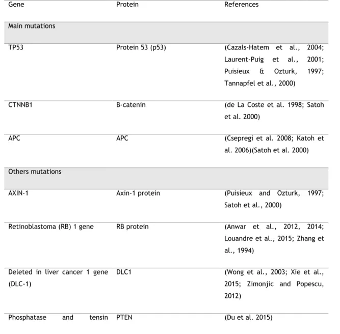
Table 2. Mutations responsible for hepatocellular carcinoma development.
Gene Protein References
Main mutations
TP53 Protein 53 (p53) (Cazals-Hatem et al., 2004;
Laurent-Puig et al., 2001; Puisieux & Ozturk, 1997; Tannapfel et al., 2000)
CTNNB1 Β-catenin (de La Coste et al. 1998; Satoh
et al. 2000)
APC APC (Csepregi et al. 2008; Katoh et
al. 2006)(Satoh et al. 2000) Others mutations
AXIN-1 Axin-1 protein (Puisieux and Ozturk, 1997;
Satoh et al., 2000)
Retinoblastoma (RB) 1 gene RB protein (Anwar et al., 2012, 2014;
Louandre et al., 2015; Zhang et al., 1994)
Deleted in liver cancer 1 gene (DLC-1)
DLC1 (Wong et al., 2003; Xie et al.,
2015; Zimonjic and Popescu, 2012)
homolog (PTEN)
Insulin-like growth (IGF) factor 2
IGF-2 (Enguita-Germán and Fortes,
2014)
Breast cancer type 2
susceptibility protein (BRCA2)
BRCA2 (Katagiri, Nakamura, and Miki
1996; Martins, Kedda, and Kew 1999)
Suppressor of cytokine signaling 1 (SOCS-1)
SOCS-1 (Nagai et al., 2001; Nagai et
al., 2002; Zhang et al., 2014)
SMAD family member 2 (SMAD2) SMAD2 (Dumont et al., 2003; Kawate
et al., 1999; Yakicier et al., 1999)
SMAD family member 4 (SMAD4) SMAD4 (Dumont et al., 2003; Kawate
et al., 1999; Yakicier et al., 1999)
c-myc c-myc (de La Coste et al., 1998;
Zimonjic and Popescu, 2012)
All these mutations contribute to cancer development through the regulation involved in cell proliferation, apoptosis and angiogenesis (Fujimori et al., 1991; Tsuda et al., 1992). These genetic mutations may lead to alterations in several signaling pathways, which may favor the development of hepatocellular carcinoma, mainly through the imbalance between proto-oncogene and tumor suppressor genes (Shang et al., 2015; Zhao et al., 2009). It can be highlighted the abnormal activation of Ras/ Raf/ MAPK (Galuppo et al., 2014), PI3K/ AKT/ mTOR (Zhou and Yeo, 2011), Wnt/β-catenin (Thompson and Monga, 2007) and Hedgehog signaling pathway (Berman et al., 2003). In addition, there are many other signalling pathways involved in the onset and progression of liver cancer, such as Notch (Strazzabosco and Fabris, 2012; Villanueva et al., 2012), IGF/IGF receptor (IGFR) (Enguita-Germán and Fortes, 2014) and EGF receptor signalling pathway (Berasain and Avila, 2014).
Among the several mutations associated with the development of hepatocellular carcinoma, numerous studies have pointed out the mutations in p53 and β-catenin as critical (Brito et al., 2012; reviewed by Gomes et al., 2013). The p53 is a tumor suppressor protein with important functions in cellular cytoplasm and nuclei, such as DNA damage repair, maintenance of the balance between cell proliferation and apoptosis, and angiogenesis inhibition (Fabregat, 2009, Ferreira et al., 1999). In normal cells, p53 is found in low concentrations but its levels increase in response to a variety of environmental stimuli, including ionizing radiation and
ultraviolet (UV) light, hypoxia, as well as growth factors deprivation and DNA damage (Ferreira et al., 1999; Bressa et al., 1991). Regarding the balance between cell proliferation and apoptosis, p53 is capable to activate signalling pathways that lead to apoptosis through several mechanisms, which include the downregulation of the anti-apoptotic protein B-cell lymphoma 2 (BCL-2) and the up-regulation of the pro-apoptotic Bcl-2 associated X protein (BAX) (Ferreira, Tolis, and Giaccone 1999) (Figure 4).
Figure 4. The function of p53. p53 expression increases in response to a variety of environmental stimuli in order to maintain the balance between cell proliferation and apoptosis. Adapted from (Hoffbrand et al., 2011).
However, this protein is often found mutated or with decreased expression in tumor cells of hepatocellular carcinoma (Ferreira, Tolis, and Giaccone 1999). The diminished expression of p53 in hepatocellular carcinoma leads to a decreased expression of pro-apoptotic proteins, contributing to the resistance to apoptosis (Ferreira et al., 1999; Fabregat, 2009).
β-catenin is a multitasking protein that exerts a crucial role in cell-cell adhesion. This protein is an integral structural component of cadherin-based adherence junctions, as well as the key nuclear effector of canonical Wnt signaling pathway. An imbalance in the structure and signaling properties of β-catenin can result in disease or deregulated growth, often associated with cancer and metastasis, as shown in Figure 5 (Ji et al., 2011; Valenta et al., 2012).
Figure 5. WNT/β-catenin pathway in hepatocellular carcinoma. In normal liver cells (“WNT OFF” state): β-catenin is sequestered by the multiprotein “destruction complex” in the cytoplasm, which consists of APC, AXIN, glycogen synthase kinase 3 (GSK3) and casein kinase (CK1). The “destruction complex” sequentially phosphorylates β-catenin, which triggers ubiquitination and degradation in the proteasome. The genes that respond to WNT remain inactive by binding of the protein Groucho regulatory protein gene T-cell factor/ lymphoid enhancer-binding factor (TCF/LEF). In hepatocellular carcinoma cells (activated or “WNT ON” state), the protein disheveled inhibits GSK3 phosphorylation activity and the “destruction complex” disintegrates. As a result, β-catenin accumulates in the cytoplasm and translocate to the nucleus, where it interacts with TCF/LEF to transcriptionally activate downstream target genes. Investigational agents targeting various components that have been tested in hepatocellular carcinoma are indicated in red boxes. epCAM, epithelial cell adhesion molecule; FZD, frizzled receptors; LRP, low-density lipoprotein receptor-related protein; SiRNA, small interfering RNA. Adapted from (Chua et al., 2014).
1.1.3. Diagnosis and therapy
The majority of hepatocellular carcinoma cases is detected in advanced stage due to the asymptomatic nature of this type of cancer (El-Serag and Rudolph, 2007; Morris et al., 2012; Yang and Roberts, 2010).
Regarding the diagnosis, in a first phase, it is evaluated a well-known tumor marker, the serum α-fetoprotein combined with abdominal ultrasound (El-Serag and Rudolph, 2007). Although the serum α-fetoprotein is the main marker of the disease, it presents low specificity and it is necessary to use complementary tests to confirm the diagnosis (El-Serag
and Rudolph, 2007). The complementary diagnostic methods used are the computed tomography, the magnetic resonance imaging and the liver biopsy (Bruix and Sherman, 2011). Surgical treatment as surgical resection (Roayaie et al. 2009, 2013) and liver transplantation (Takayama et al. 1998), are the most effective way to treat the patients with early stage hepatocellular carcinoma, are used also to local ablation as a treatment (Livraghi et al. 2008), although this is not as effective. When tumor mass is small, the resection or transplantation is not used, and usually, the treatment used is only ablative procedures (Galuppo et al., 2014).
Patients with intermediate stages of hepatocellular carcinoma, are considered for transarterial chemoembolization (TACE) (Bruix and Sherman, 2011), and recently, it was proposed a combination of TACE with sorafenib, a drug known as a tumor inhibitor, in the treatment of these patients in phase II (Galuppo et al., 2014).
Patients in the advanced stage the treatment options are extremely limited and the average life expectancy of the untreated patient is less than three months. At this stage, the available treatments are limited and ineffective, being the radiotherapy and chemotherapy used as treatment. Unfortunately, the treatment efficacy is limited by the radio- and chemoresistant profile of this type of cancer (Llovet and Bruix, 2003; Bruix and Sherman, 2005; Johnson, 2000). Chemotherapy is used only as a palliative treatment in order to relieve symptoms and improve the quality of life of patients. The patients are treated with doxorubicin, cisplatin and 5-fluorouracil (5-FU) in monotherapy or in combination (Giglia et al., 2010; Hua et al., 2014; Johnson, 2000). Radiotherapy just like the chemotherapy is used in treatment of hepatocellular carcinoma, but it is not particularly effective, due to the radioresistance displayed by this tumor, which may be a consequence of the heterogeneity, this tumor relative to the expression of certain genes namely TP53 gene, involved in proliferation pathways and cell death (Ma et al., 2010).
In last years, has been considerate the targeted therapy in the treatment of hepatocellular carcinoma, in an advanced stage of the disease. Several studies indicate that Sorafenib significantly increases the survival time of patients with hepatocellular carcinoma and has few adverse effects (Llovet and Bruix, 2008; Liu et al., 2006). This drug is an inhibitor of several tyrosine kinases, which activate signaling pathways associated to cell proliferation and angiogenesis (Liu et al., 2006).
Taking into account the absence of effective and selective therapeutic strategies for the treatment of hepatocellular carcinoma, which is mainly detected at an advanced stage, it is particularly important to find new therapies that allow reducing the mortality rate of this type of cancer.
1.1.4 Glycolytic metabolism
In addition to genetic mutations in hepatocellular carcinoma, the most fundamental metabolic alteration in tumor cells is the high rate of glycolysis, and the consequent increase in glucose uptake, a phenomenon firstly reported by Otto Warburg (Dayan et al., 2008; Kroemer and Pouyssegur, 2008; Warburg, 1956) and the dependency on the glycolysis for adenosine triphosphate (ATP) generation, especially in hypoxic environment (Gatenby and Gillies, 2004), as shown in Figure 6.
Figure 6. Schematic diagram of cancer cells glycolytic metabolism. After cellular glucose uptake via facilitated glucose transporters (GLUT), glucose is metabolized, originating pyruvate. The pyruvate is converted into lactate and transported to the extracellular medium by monocarboxylate transporter. Red cross evidence that pyruvate does not follow for oxidative phosphorylation, but for lactate production. Adapted from (Airley and Mobasheri, 2007).
The ‘‘Warburg effect’’ consists of an increase in glycolytic flux in cancer cells, as well as an increased activity of the enzymes involved, that is even maintained in conditions of high oxygen tension (‘‘aerobic glycolysis’’) and gives rise to an increased lactate production when compared to normal cells (Brahimi-Horn et., 2007; Oliveira et al. 2015; Warburg et al., 1924; Zhao et al., 2013). Therefore, it leads to an acidification of the environment, which promotes a favorable environment for the growth and survival of cancer cells due to the induction of angiogenesis, cell invasion and metastization (Gatenby and Gawlinski, 2003; Gatenby et al., 2006; Robey and Hay, 2009; Stern et al., 2002).
As mentioned previously, one of the changes associated with the Warburg effect is the alteration associated with the expression of several genes that may affect the cell growth of hepatocellular carcinoma. Several key enzymes in glycolysis, such as hexokinase (HK) II, phosphofructokinase (PFK) isoform 1, pyruvate kinase isoform M2 (M2-PK), lactate dehydrogenase (LDH
)
A (Hitosugi et al., 2010; Lv et al., 2011) and glucose transporters (GLUT) are up-regulated in hepatocellular carcinoma (Airley and Mobasheri, 2007; Amann et al., 2009; Young et al., 2011).Considering that ATP generation via glycolysis is far less efficient (two ATP per glucose) than through oxidative phosphorylation (36 ATP per glucose), cancer cells consume much more glucose than normal cells to sustain the ATP supply for their active metabolism and proliferation (Izyumov et al., 2004; Pinedo et al., 2003; Xu et al., 2005). For this reason, cells have to adopt a mechanism that allows them to increase glucose uptake and metabolism, and consequently, it has been consistently reported an increase in the expression of glucose transporters, mainly the isoform 1 (Amann and Hellerbrand, 2009; Amann et al., 2009). However, some studies have demonstrated that beyond the GLUT-1, other GLUT have an important role in the transport of the glucose, including GLUT-2, -3, -5 and -12 (Jadvar et al., 2009). GLUT-3 is overexpressed in cancer cells and it is associated with a poor prognosis (Baer et al., 2002), but the GLUT-1 is a better marker of poor prognosis than the GLUT-3 because the GLUT-3 was detected only in a limited number of cases (Karim et al., 2012). GLUT-2 has low affinity for glucose but is the most expressed GLUT isoform in the normal liver cells. Its expression is insulin-dependent (Mueckler 1994). It was also identified GLUT-5 as overexpressed in liver metastases (Rogers et al., 2002; Zamora-León et al., 1996). GLUT-12 has also been implicated in the enhanced glucose accumulation in some cancers, including breast (Rogers et al. 2002) and prostate cancer (Chandler et al., 2003).
It is well described that hypoxia is a strong modulator of cell energy metabolism and it is associated with human malignancies reviewed by (Gatenby and Gillies, 2004; Pelicano et al., 2006), especially in solid tumor tissues as the hepatocellular carcinoma. The availability of oxygen becomes increasingly smaller as tumor mass increases. Under such conditions, oxidative phosphorylation may not proceed normally because of insufficient oxygen, thus resulting in an increase of glycolysis. The consequences of increased glycolysis require further adaptation to environments with low glucose concentrations and high acid owing to the elevated production of lactate, which leads to acidification of tumor tissue and provides a microenvironment that promotes and selects cells with malignant behaviors. This acidification results in diffusion of H+ ions along concentration gradients into peritumoral normal tissue.
Normal cells are unable to survive under such conditions because these cells do not have a mechanism that allows them to adapt to extracellular acidosis (e.g. p53 mutations), whereas the tumour cells continue to proliferate (Gatenby and Gawlinski, 1996; Morita et al., 1992;
Hypoxia has been associated with drug resistance and reduced sensitivity to radiotherapy, due in part to the up-regulation of hypoxia-inducible factor 1 (HIF-1) (Semenza 1998), which activates the expression of important target genes involved in processes that promote tumor development, such as angiogenesis, glucose uptake, glycolysis, growth factor signalling, apoptosis, invasion and metastasis (Yasuda et al. 2004). However, cells with normal HIF-1 levels may display the glycolytic phenotype through of multiple cellular pathways, so that altered glucose metabolism (Osthus et al., 2000; Dong et al., 2015). The hypoxic environment and the acidification environment accelerates the cancer progression and the resistance to therapeutic strategies (Noguchi et al., 2000; Dong et al., 2015; Xia et al., 2012). The therapeutic resistance associated with the hypoxic environment is a common problem in the treatment of hepatocellular carcinoma, and the inhibition of glycolysis is a potential therapeutic strategy for hepatocellular carcinoma treatment (Liu et al. 2001).
Considering that cancer cells need high glycolytic activity in order to survive and grow, it was hypothesized that inhibition of glycolysis may severely abolish ATP generation in cancer cells and thus, may preferentially kill the malignant cells (Munoz-Pinedo et al., 2003; Izyumov et al., 2004). When glycolysis is inhibited, the intact mitochondria in normal and cancer cells allow them to use alternative energy sources, such as fatty acids and amino acids to produce metabolic intermediates to the Krebs cycle (Gatenby and Gillies, 2004; Xu et al., 2005). Taking into account that cancer cells produce less ATP through the electron transport chain, due to a hypoxic environment, the hepatocellular carcinoma cells have no other alternative than glycolysis to generate ATP. Therefore, using glycolytic inhibitors, that are particularly effective against cancer cells under hypoxic conditions, may reveal an effective therapy to treat hepatocellular carcinoma (Han et al., 2013; Lincet and Icard, 2014).
Glucose analogues (Liu et al., 2001; Liu et al., 2002), inhibition of the GLUT-1 (Amann and Hellerbrand, 2009; Pelicano et al., 2006), inhibition of glycolytic enzymes, such as HK II and LDH, reviewed by (Pelicano et al., 2006), and inhibition of pentose phosphate pathway (Fantin and Leder, 2006) are some of the strategies already tested to inhibit the tumorigenic process through the glycolysis inhibition.
Due to lack of effective therapies for the treatment of hepatocellular carcinoma, it is particularly important to find new therapies that allow reducing the mortality rate of this type of cancer. In this context, the human amniotic membrane (hAM) as a potential therapy for hepatocellular carcinoma has recently emerged (Mamede et al., 2014).
1.2. Human amniotic membrane
1.2.1. Structure and function
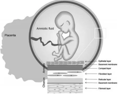
The placenta is a highly specialized organ that supports the normal development of the embryo/fetus through all the gestation period. This organ consists of two fetal membranes that do not fuse histologically and remain separated: an outer layer (chorion) and an inner layer (hAM or amnion). The hAM delimits the amniotic cavity, which is filled with the amniotic fluid.
The hAM is constituted by a simple epithelium, a thick basement membrane and a mesenchymal matrix that can be divided into compact layer, fibroblast layer, and intermediate (spongy) layer, as shown in Figure 7 (Niknejad et al., 2008; Mamede et al., 2012).
Figure 7. The placenta structure. The placenta is composed of two membranes: the chorion and the hAM. The hAM is constituted by three major layers: a single epithelial layer, a thick basement membrane, and an avascular mesenchyme. Adapted from (Kang et al., 2012a).
Histologically, the hAM varies from conception to maturity and several different patterns are often noted (Pollard et al., 1976). hAM is a thin and avascular membrane and their thickness can vary between 0.02 mm and 0.5 mm. The inner membranes of the placenta do not have blood vessels, nerves or muscles (Evangelista et al., 2008). The inner layer is constituted by a single homogeneous layer of cuboidal epithelial cells firmly fixed to the basement membrane, which is attached to a condensed acellular layer of mesenchymal membrane, composed of collagen type I, II and V reviewed by (Mamede et al., 2012). From a structural perspective, the amniotic membrane cells are connected to each other by numerous desmosomes (King
The epithelial layer is the innermost layer of the hAM, contacting with amniotic fluid. It is constituted by amniotic epithelial cells (hAECs) that express epidermal markers, such as EGF (Tabatabaei et al., 2014; Wang et al., 2011).The hAECs have a high number of organelles and several pinocytic vesicles and a big, irregular and homogeneous nucleus. In addition, the hAECs have many microvilli on their apical surface, which may be related to the role of hAECs in intracellular and transcellular transport, as well as in active secretory function (Pollard et al., 1976).
The basement layer is the intermediate layer of the hAM, being the thickest membrane found in human tissues, and serve as a permeability barrier to amniotic macromolecules and several molecules with a structural function, such as actin or spectrin, which will allow the maintenance of the membrane integrity. This layer is constituted mainly by collagen, elastin, laminin, fibronectin and proteoglycans. Collagens of type I, III, IV, V, and VI are the most abundant in basement layer and are responsible for a mechanical response (Malak et al. 1993). Elastin is present in hAM in high concentration and is capable of forming an elastic network (Bryant-Greenwood 1998). Laminin (Wolf et al., 1991; Akashi et al., 1999), fibronectin (Clamp, 1975; Bryant-Greenwood, 1998) and proteoglycans (Iozzo 2005) are responsible for the maintenance of the hAM integrity.
As shown in Figure 8, the mesenchymal layer can be divided into three different layers: compact layer, fibroblast layer, and intermediate (spongy) layer. The compact layer is the main fibrous skeleton of hAM and is rich in collagen, that provides to hAM a major tensile strength. The fibroblast layer is rich in human amniotic membrane mesenchymal cells (hAMCs), which derives from the mesodermal embryonic plate. The spongy layer is rich in proteoglycans and glycoproteins, which are responsible for the spongy appearance reviewed by (Parry and Strauss, 1998). This layer contains a non-fibrillar meshwork mainly constituted by type III collagen (Casey and MacDonald, 1996); reviewed by (Parry and Strauss, 1998).
Figure 8 . Histologic section of hAM, obtained through hematoxylin–eosin staining. hAM has three main layers: epithelial layer, basement membrane and mesenchymal layer, which can be divided into a compact layer, fibroblast layer, and spongy layer. Retrieved from (Riau et al.,2010).
The hAM has several metabolic functions during gestation, providing to the developing embryo/fetus protection against desiccation and an environment of suspension, where the embryo/fetus can grow free from pressures of the structures that surround (Sadler, 2000). The characteristic resistance of the hAM is mainly attributed to the layer of collagen type I, II and elastin. However, the elasticity of the hAM is mainly attributed to collagen type III (Sadler, 2000). Because of the presence of these collagens, one of the important properties of hAM is its resistance to proteolytic factors (Fukuda et al., 1999).
During the pregnancy, hAM has a crucial role in the transport of water, nutrients, and oxygen between the blood vessels of mother and the amniotic liquid (Evangelista, Soncini, and Parolini 2008), and it is also responsible for the regulation of pH in the amniotic fluid (Akashi et al. 1999). This regulation is dependent of the carbonic anhydrase enzymes, which are expressed in amniotic epithelial cells (Crescimanno et al., 1993; Mühlhauser et al., 1994). During the birth, the hAM plays an important role in the initiation and maintenance of uterine contraction, through the action of prostaglandins and enzymes integrated into the synthesis of the prostaglandins. The amniotic epithelium is the main source of prostaglandins, especially prostaglandin E2 (Okazaki et al., 1981) and also expresses prostaglandin-biosynthesis enzymes, such as phospholipase, prostaglandin synthase and cyclooxygenase (Bryant-Greenwood 1998). The expression of these enzymes is regulated by human chorionic gonadotropin (Toth et al., 1996) and IL-1β (Benedetto et al. 1990).
1.2.2. Properties of human amniotic membrane
The hAM has become a subject of study due to their attractive properties, which determine its application in several human diseases. Studies have shown that the hAM has the following properties: antimicrobial, antiviral, immunoregulatory, anti-adherent, anti-inflammatory, bacteriostatic, reepithelializing, anti-scarring, anti-angiogenic and it is nontumorigenic reviewed by (Toda et al., 2007). Also, the hAM has the ability to synthesize molecules with pro-apoptotic action (Mamede et al. 2012; Seo et al., 2008).
The antiangiogenic effect of the hAM is one of the main properties for its application in reconstructive medicine, such as ocular surface reconstruction (Kim and Tseng, 1995). The pluripotency of amnion-derived cells, associated to its low immunogenicity, gives hAM the potential of application for transplantation purposes reviewed by (Toda et al., 2007). In fact, some studies have shown a negligible immunological response after hAM transplantation (Akle et al., 1981; Kamiya et al., 2005).
Regarding the application of hAM as a potential anti-cancer therapy, the antiangiogenic, immunoregulatory, pro-apoptotic and nontumorigenic activity may largely contribute for the
The angiogenic switch process is important for tumor growth since the tumor cells have the ability to destroy the immunologic barrier for invasion and metastasis (Seo, Kim, and Kim 2008). Therefore, the hAM may be used as an antiangiogenic mediator and immunologic barrier, as well as the amniotic cell culture, which has anti-angiogenic properties and contains potent inhibitors of neovascularization (Hao et al., 2000; Shao et al., 2004). This antiangiogenic activity may be due to the antiangiogenic chemicals found principally in epithelial and mesenchymal cells of hAM, such as collagen XVIII, thrombospondin-1, IL-10, IL-1 receptor antagonist, and tissue inhibitor of metalloprotease 1, 2, 3 and 4 (Hao et al. 2000). In addition, it was found that the pigment epithelium-derived factor (PEDF), a potent antiangiogenic factor, is present at high levels in the basal membrane of hAM (Shao et al. 2004). In fact, the PEDF specifically inhibits endothelial cell growth, suppressing neovascularization in the cornea (Shao et al. 2004). The antiangiogenic mechanism of hAM can also be explained by the action of hAM as a physical barrier that prevents migration and vascular endothelial growth (Hao et al. 2000).
The immunoregulatory activity of the hAM inhibits the inflammatory response, namely through the epithelial cells that secrete soluble factors involved in inhibition of innate immune and adaptive immune system cells (Li et al., 2005). The hAM cells do not express human leukocyte antigen (HLA) -A, -B, -C, -DR or β2-microglobulin, and consequently, it presents low immunogenicity (Adinolfi et al. 1982). The hAM express differential no classical HLA, namely HLA-G, that is capable of inactivating lymphocytes and dendritic cells, as well as to suppress the host T cells by binding CD8 and HLA-G (Wang et al. 2006). Thus, it is thought that hAM can be used as a physical barrier against numerous factors that can initiate inflammatory processes, thereby maintaining the inflammatory cells in the infected area and thus reducing the number of inflammatory mediators involved in the tumoral response (Shimmura et al., 2001).
Concerning the pro-apoptotic activity, there are some studies showing that hAM secretes some pro-apoptotic factors, such as interferon gamma (IFN-γ). It was demonstrated that hAM induces apoptosis of interferon-activated macrophages (Li et al., 2006). Moreover, the hAM may accelerate apoptosis of polymorphonuclear neutrophils and reduce inflammation (Zhou et al., 2003).
1.3. Human amniotic membrane and cancer
Considering the attractive properties of hAM, such as immunoregulatory, anti-angiogenic and pro-apoptotic activities, in 2008 Seo et al. hypothesized that the hAM can be useful in the treatment of cancer.
In 2012, it was published a review article focusing on the attractive properties of hAM in cancer treatment (Mamede et al., 2012). Overall, the low immunogenicity of hAM suggests
that hAM will not be rejected by the host immune system after transplantation. The hAM may also act as a natural physical barrier around the tumor, preventing the supply of the nutrients and oxygen to the tumor cells through the synthesis of several antiangiogenic factors, which inhibits tumor growth (Mamede et al., 2012).
The majority of studies that evaluated the application of hAM in the treatment of cancer were performed using the hAMCs. Using direct or indirect co-cultures of hAMCs with hematopoietic and non-hematopoietic cancer cell lines, a decreased proliferation in cancer cells was observed in response to hAMCs treatment (Jiao et al., 2012; Khakoo et al., 2006; Magatti et al., 2012). Magatti and colleagues suggests the involvement of soluble inhibitory factors secreted by hAMCs and the cell-cell contact in the effect of hAMCs that is mediated by cell cycle arrest (Magatti et al. 2012), but another study also showed that the treatment with hAMCs inhibits the glioma grow through the activation of apoptosis by cell-cell contact (Jiao et al. 2012). However, several studies have demonstrated that hAMCs regulate tumor cell growth negatively and positively (Magatti et al. 2012). These inconsistencies may due to the heterogeneity of hAMCs (Klopp et al. 2011), yet that the tumor promotion or suppression appear to result in cell-to-cell interactions by direct contact or through secreted molecules such as growth factors, cytokines, and chemokines. For this, it is very important the study carefully the interaction between hAMCs and cancer cells.
Instead of using hAMCs there are studies using the hAECs, which are able to inhibit the tumor growth, namely breast cancer cells (Kang et al., 2012b; Niknejad and Yazdanpanah, 2014). Several studies indicated that the anti-cancer effects of hAECs were due to the cell-cell contact and soluble inhibitory factors secreted (Kang et al., 2012), or due to only soluble inhibitory factors secreted for hAECs by apoptosis induction (Niknejad and Yazdanpanah, 2014). Other studies indicate that the anti-cancer effects of hAECs were due to the cell-cell contact and secretion of soluble anti-angiogenic factors (Niknejad et al., 2013; Yazdanpanah et al., 2015). This effect of hAECs was due to the inhibition of heat shock protein 90 (Hsp90), which activates apoptosis, cell cycle arrest and inhibits angiogenesis (Niknejad et al., 2013b). HSP-90 is a strong inducer of cell cycle progression and also inhibits apoptosis in cancer cells. However, it seems that other signalling pathways could be involved in anti-cancer effects of the amniotic membrane, through amnion-derived cells that induce cytotoxicity by the secretion of cytokines such as tumor necrosis factor (TNF) -α and -β, macrophage colony-stimulating factor (M-CSF), IFN-γ and transforming growth factor beta (TGF-β) and IL-2, -4 and -3 (Kang et al., 2012a; Kucerova et al., 2007).
Considering the several studies on the anti-cancer properties of hAM, Niknejad hypothesized that, within hAM, different cell types may employ different mechanisms to inhibit cancer cell proliferation. The hAECs cells are responsible for the induction of apoptosis and angiogenesis inhibition in tumor cells, and the hAMCs cells are responsible for apoptosis induction and cell
cycle arrest of tumor cells, may leading to cell death by different mechanisms (Niknejad and Yazdanpanah, 2014).
In spite of these inconsistencies about the hAECs and hAMCs mentioned here, the amnion-derived cells have several advantages compared with the recurrent therapies for cancer and the regenerative medicine. However, further work is necessary to develop and prepare the amnion-derived cells for clinical applications.
Some studies also found that hAM-derived cells and their supernatant are able to induce apoptosis in glioma, cervical cancer and breast cancer cell lines (Jiao et al., 2012; Niknejad et al., 2014).
Taking into account, the attractive properties of the amnion-derived cells, in an innovative way, Mamede and colleagues have evaluated the protein extract of amniotic membrane (hAMPE) in cancer therapy, whose extract is obtained from epithelial and mesenchymal cells, as well as the extracellular matrix. In 2012, the effect of hAMPE in the metabolic activity was evaluated on 21 human cancer cell lines. It was demonstrated that the hAMPE can inhibit the metabolic activity of some human cancer cell lines. However, research about this cell line-dependent response to hAMPE treatment becomes indispensable, as shown in Table 3.
Table 3. Metabolic activity of several human cancer cell lines evaluated after incubation of hAMPE. Adapted from (Mamede et al., 2015).
Cancer Cell line Effect of hAMPE
Prostate PC3 Decreased 54% LNCaP No effect Breast HCC1806 Decreased 25-50% MCF7 No effect HCC1954 Stimulated Colon LS1034 Stimulated WiDr Decreased >50% C2BBe1 Stimulated
Lung A549 No effect
Pancreas PANC-1 Decreased >50%
MIA PaCa-2 Decreased 25-50%
Hepatocellular carcinoma HuH7 Decreased 25-50%
HepG2 Decreased >50%
Hep3B2.1-7 Decreased >50%
Others TFK1 (bile duct carcinoma) Stimulated
A375 (melanoma) Decreased 25-50%
MNNG/HOS (osteosarcoma) Decreased 25-50%
ECC-1 (endometrial) Decreased <25 %
HT-1376 (bladder) Decreased 25-50%
OE19 (esophageal) Decreased 25-50%
Since it was proven that hAMPE is able to inhibit the metabolic activity of several human cancer cell lines, namely in Hep3B2.1-7, HepG2 and HuH7 cell lines of hepatocellular carcinoma. Our research group has studied the pathways by which the hAMPE act on hepatocellular carcinoma. It was demonstrated that hAMPE is capable of inducing cytotoxicity and cell death in Hep3B2.1-7, HepG2 and HuH7 cell lines of hepatocellular carcinoma. The hAMPE decreased the metabolic activity, protein and DNA content in a dose and time-dependent, and induced cell morphology alterations in all cell lines of hepatocellular carcinoma, but the mechanism associated with cell death is dependent on the cell line, as suggested by in vitro and in vivo studies. The hAMPE is able to induce apoptosis in all hepatocellular carcinoma cell lines, but also induces necrosis in HepG2. The apoptosis in Hep3B2.1-7 and HepG2 cell lines is induced via membrane receptor-mediated mechanism, while in HuH7 cell line is induced via mitochondrial pathway (Mamede et al., 2015). In addition, the results showed that hAMPE have selective cytotoxicity because does not affect a non-tumorigenic human cell line (Mamede et al., 2015).
Recently, our research group investigated the cellular targets of hAM protein extracts (hAMPE) in hepatocellular carcinoma cell lines through in vitro studies. It was demonstrated that hAMPE is able to modify oxidative stress environment in hepatocellular carcinoma cell lines, as well as its cell cycle (Brito et al., 2012). The cellular and protein targets of the hAMPE were different between the hepatocellular carcinoma cell lines, such as DNA, p53, p21, β-catenin and multidrug resistance proteins. Thus, it is clear that the success of this treatment depends on of a personalized therapy based on the biological and genetic characteristics of the tumor (Mamede et al., 2016).
Chapter 2 - Aims
The attractive properties of hAM, such as its antiangiogenic, proapoptotic and anti-inflammatory activity, reviewed by (Mamede et al., 2012), encouraged to study the antitumor effects of hAM. Our research group demonstrated that hAMPE is capable of inducing cytotoxicity selectively, cell death, decreased the metabolic activity, protein and DNA content in a dose and time-dependent, and induced cell morphology and oxidative stress environment alterations in Hep3B2.1-7, hepG2 and HuH7 cell lines of hepatocellular carcinoma, as well as its cell cycle, but the mechanism associated to death type is dependent on cell line. Since there are no known fully signaling pathways and mechanisms of action of hAMPE, it is important to explore the effect of hAMPE.
The hAMPE induce death by apoptosis in all hepatocellular carcinoma cell lines for different pathways but induce only necrosis in HepG2. In addition, some targets of hAMPE were identified, such as DNA, p53, p21, β-catenin and multidrug resistance proteins.
Taking into account the results obtained so far by our research group, associated with increased glycolytic metabolism in cancer cells, this work aims to study the effect of hAMPE treatment in the glucose metabolism of hepatocellular carcinoma. In order to achieve this goal, three different human hepatocellular carcinoma cell lines were used to:
1. Evaluate the glucose consumption and lactate production in response to hAMPE treatment through proton nuclear magnetic resonance (1H NMR);
2. Determine the expression of glucose transporters, lactate dehydrogenase (LDH) and monocarboxylate transporter isoform 4 (MCT4), in response to hAMPE treatment by Western blot (WB) and/or fluorescence analysis.