ISSN 0103-8478
Degeneration rate of goat primordial follicles maintained in TCM 199 or PBS at
different temperatures and incubation times
José Roberto Viana Silva2* Alline Ferreira Brasil2 Regiane Rodrigues dos Santos2 Sônia Helena Furtado Costa2 Ana Paula Ribeiro Rodrigues2 Marcos Antônio Leal Ferreira2
Vanessa Porto Machado2 José Ricardo de Figueiredo1
Taxa de degeneração de folículos primordiais caprinos conservados em TCM 199 ou PBS em diferentes temperaturas e tempos de incubação
1Professor Adjunto, Universidade Federal do Ceará (UECE). 2Estudante de Pós-graduação, PPGCV - UECE.
*Autor para correspondência: Universidade Estadual do Ceará, Faculdade de Veterinária, Avenida Paranjana, 1700, Campus do Itaperi,
60740-000, Fortaleza-CE, Brasil. E-mail: roberto_viana@yahoo.com
ABSTRACT
The present work has investigated the degeneration rate of goat primordial follicles in situ after preservation in PBS or TCM 199 at different temperatures and incubation times. For each animal the ovarian pair was divided into 19 fragments. One ovarian fragment was taken randomly and immediately fixed (control). The other 18 ovarian fragments were randomly distributed in tubes containing PBS or TCM 199 and stored at 4º, 20º or 39ºC for 4, 12 or 24h. The storage of ovarian fragments in PBS or TCM 199 at 20ºC for 12h and 24h or at 39ºC, in all incubation times tested, increased significantly the percentage of degenerated primordial follicles (P<0.05). In contrast, for both media tested the degeneration rate of primordial follicles preserved at 4ºC for up to 24h and at 20ºC for 4h was similar to control values (P>0.05). In conclusion, this study shows that PBS was as efficient as TCM 199 in the preservation of goat primordial follicles in situ, being the best results observed at 4ºC.
Key words: primordial follicles, degeneration, goat, preservation.
RESUMO
O presente trabalho investigou a taxa de degeneração de folículos primordiais caprinos in situ após conservação em PBS ou TCM 199, em diferentes temperaturas e tempos de incubação. Para cada animal, o par ovariano foi dividido em 19 fragmentos. Um fragmento foi escolhido aleatoriamente e imediatamente fixado (controle). Os 18 fragmentos ovarianos restantes foram aleatoriamente distribuídos em tubos contendo PBS ou TCM 199 e conservados a 4, 20 or 39ºC por 4, 12 or 24h. A conservação de fragmentos ovarianos em PBS ou TCM 199 a 20ºC por 12 e
24h ou a 39ºC, em todos os tempos de incubação testados, aumentou significativamente a percentagem de folículos primordiais degenerados (P<0,05). Ao contrário, em ambos os meios testados, a taxa de degeneração de folículos primordiais conservados a 4ºC em todos os tempos testados e a 20ºC por 4h foi similar ao controle (P>0,05). Em conclusão, este estudo mostrou que o PBS foi tão eficiente quanto o TCM 199 na conservação de folículos primordiais caprinos in situ, sendo os melhores resultados observados na temperatura de 4ºC.
Palavras-chave:folículos primordiais, degeneração, cabra, conservação.
INTRODUCTION
Primordial follicles constitute 95% of the thousands of follicles present in the mammal ovaries. Although most of primordial follicles undergo atresia during growth and maturation in vivo, these follicles provide a valuable source for studies on in vitro
development of oocytes and eventual in vitro
conditions of ovaries, saline solution (CARVALHO et al., 2001), Braun-Collins (ANDRADE et al., 2001), coconut water solution (SILVA et al., 2000) and TCM 199 (FERREIRA et al., 2001) have been tested. However, these studies indicate only the overall incidence of follicular degeneration after preservation and considered all classes of preantral follicles (primordial, primary and secondary follicles together). The PBS is commonly used as a preservation medium for embryos (NIEMANN, 1991) and cumulus oocyte complex (JEWGENOW et al.,1998). TCM 199 is another medium that has been successfully used in the preservation of bovine ovaries (FIGUEIREDO et al.,1993) and cumulus oocyte complexes (TWAGIRAMUNGU et al.,1998), during transportation. However, there are currently no studies investigating the degeneration rate of goat primordial follicles in situ after preservation in PBS or TCM 199. The aim of this study was to evaluate the degeneration rate of goat primordial follicles after preservation in PBS or TCM 199 at different temperatures and incubation times.
MATERIAL AND METHODS
Ovaries (n=10) from 5 adult mixed breed goats were collected at a local slaughterhouse. The ovaries were stripped of surrounding fat tissue and ligaments and washed in 70% alcohol for approximately 10 seconds and then twice in 0.9% saline solution.
In the slaughterhouse, the ovarian pair from the same animal was divided into 19 fragments. One fragment was taken randomly and immediately fixed for histological examination (Control - Treatment 1 Time 0). The other 18 fragments were randomly distributed in tubes containing 2mL of Hepes TCM 199 + 1.50 mM Hepes (Cultilab-Brazil) or PBS (Cultilab-Brazil), at 4o, 20o or 39oC and stored for 4, 12 or 24h (Treatments 2 to 19). The temperatures were maintained using termosflasks filled with water at 4o, 20o or 39oC, and monitored through incubation time in each treatment, performed in five replication.
T h e c o n t r o l o v a r i a n f r a g m e n t w a s i n d i v i d u a l l y f i x e d i n C a r n o y f o r 2 4 h a n d dehydrated in a graded serie of ethanol, clarified with xylol and embbeded in parafin wax. The tissue was sectioned serially at a thickness of 7µm, and every 5th section was stained with periodic acid schiff (PAS) and hematoxylin. Each section was deparaffinated with xylol, rehydrated in graded alcohol, and examined by light microscopy (Leica) with 400x magnification.
The primordial follicles present one layer of flattened or flattened-cuboidal granulosa cells around the oocyte (HULSHOF et al., 1994). The basement membrane, presence or absence of pycnotic bodies and integrity of the oocyte and granulosa cells were evaluated. Based on these parameters, primordial follicles were classified as morphologically normal follicles (Figure 1), degenerated type 1 follicles (only the oocyte was degenerated; Figure 1) and degenerated type 2 follicles (degenerated oocyte and granulosa cells; Figure 1). These three classifications were assigned on a basis of atresia observed in the control and/or combined with changes that occurred as a result of storage.
The effect of media, temperatures and preservation periods on the percentage of degenerated primordial follicles was analyzed using a Chi-square test. Values were considered statistically significant when P < 0.05.
RESULTS
Approximately 100 primordial follicles were analyzed per treatment. In the treatments tested, including the control, morphologically normal primordial follicles present a spherical oocyte, with uniform cytoplasm and well organized granulosa cells, without pycnotic nucleus (Figure 1). The degenerated
Type 1 follicles sometimes had a retracted oocyte, with a pycnotic nucleus and strongly stained cytoplasm (Figure 1). The oocyte and granulosa cells degeneration was found in Type 2 degenerated follicles. The follicles with this kind of degeneration showed pycnosis at the oocyte level, and also disorganized granulosa cells detached, from basement membrane (Figure 1).
Figure 2 shows the percentage of degenerated primordial follicles stored in TCM 199 or in PBS. Compared to the control, it was observed that the percentage of degenerated primordial follicles maintained in both solutions at 4ºC for up to 24 h and at 20ºC for 4 h was similar to control (P>0.05). However, after preservation in both solutions at 20ºC for 12h and 24h or at 39ºC in all incubation times tested, the percentage of degenerated primordial follicles increased significantly when compared with control values (P<0.05). The increase of incubation time, from 4h to 12h and 24h did not affect significantly the percentage of degenerated primordial follicles after preservation in PBS or TCM 199 at 4ºC. Similar results were obtained in primordial follicles stored in both solutions at 20ºC and 39ºC, with the
increase of incubation time from 12 h to 24h. In contrast, primordial follicles stored in both solutions at 20ºC and 39ºC presented a significant increase in the percentage of degenerated primordial follicles with the increase of incubation time from 4h to 12h and from 4h to 24h.
With regard to the effect of temperature at the same incubation time, the results showed that in both solutions, in all incubation times tested, there was a progressive increase in the percentage of degenerated primordial follicles with the increase of temperature from 4º to 20º and 39ºC (P<0.05), except for the storage time of 4h, at which the temperature of 20ºC did not increase the percentage of degenerated follicles when compared with storage at 4ºC (P>0.05). When PBS was compared with TCM 199 at the same temperature and incubation time, no significant difference in the percentage of degenerated primordial follicles after incubation at 4oC, 20ºC and 39ºC for all incubation times tested was observed (P>0.05; Figure 2).
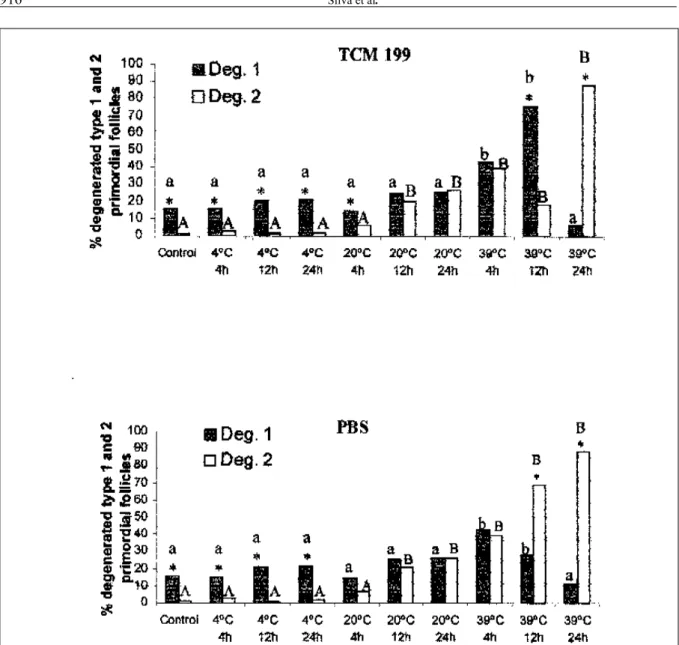
Figure 3 shows the distribution of degenerated Type 1 and Type 2 primordial follicles, in the control and after storage in TCM 199 (Figure
Figure 2 - Effect of temperature and incubation time on the percentage of goat degenerated primordial follicles preserved in TCM 199 and in PBS.
3A), and in PBS (Figure 3B). There was no significant difference in the percentage of degenerated Type 1 primordial follicles between the control and after storage in both solution at 4ºC and 20ºC at all incubation times tested, and at 39ºC for 24h. However, in ovarian fragments stored in both solutions at 39ºC for 4h and 12h a significant increase in the percentage of degenerated Type 1 follicles was observed when compared to control. On the other hand, a higher percentage of degenerated Type 2 follicles, compared
to control, was observed in the fragments stored in both solutions at 20ºC for 12h and 24h and at 39ºC at all incubation times tested.
There was a predominance of degenerated type 1 follicles in the control and in the fragments stored at 4oC, in both solutions, at all incubation times (P<0.05). In contrast, it was observed a predominance of degenerated type 2 follicles in the fragments stored at 39oC for 12h and 24h, in both solutions, except when the storage was performed
Figure 3 - Percentage distribution of the type 1 and 2 degenerated primordial follicles in goat ovaries, from the control and after storage in different treatments, in TCM 199 (A) and in PBS (B).
* indicates significant difference between the degeneration types in each treatment.
a,b different letters show significant differences in the percentage of degenerated type 1 follicles (Deg 1) found among different treatments and in the control (P<0.05).
in TCM 199 for 12h, where the percentage of degenerated type 1 was higher than degenerated type 2 (Figure 3).
DISCUSSION
This study evaluated specifically the sensitivity of primordial follicles to undergo degeneration after in vitro preservation and shows that the storage of ovarian fragments in PBS or TCM 199 at 20º or 39ºC increased the degeneration rate of goat primordial follicles. By contrast, the preservation of these follicles in both media at 4ºC maintained the level of degeneration similar to that observed in vivo
(control).
The storage of ovarian fragments in both solutions at 4ºC for up 24h and at 20ºC for 4h maintained the percentage of degenerated primordial follicles similar to that found in the ovary, immediately after animal death (control - time zero). Although the studies available did not evaluate the effect of media and temperature on the preservation of specifically primordial follicles, i.e. they considered all classes of preantral follicles together, the results from these studies demonstrated that the temperature of 4ºC has been successfully used in the follicular preservation for 24h in a solution poor in nutrients (Saline-CARVALHO et al., 2001) or hyperosmotic (Braun-Collins solution - SILVA et al., 2000) which shows that at 4ºC the composition of the medium is not a limiting factor. In addition, WOOD et al. (1997) preserved successfully domestic cat ovarian follicles at 4ºC for 48h. ROY & TREACY (1993) observed that a lower metabolic rate at low temperatures may be beneficial in maintaining viable human preantral follicles in vitro after isolation. The hypothermia provoked by low temperatures (4ºC), that might have reduced cellular metabolism, consequently minimizing the metabolic need and increasing the resistance of follicles to the reduction of nutrients and oxygen during preservation in vitro.
This work shows that when the primordial follicles were stored in PBS or in TCM 199 at 20ºC for 12 h and 24 h or 39oC at all incubation times, an increase in the percentage of degenerated primordial follicles was observed when compared to control. Similar results were observed with preservation of preantral follicles (without specific follicular type) at 20oC and 39oC for 12 and 24 h in saline (goat -CARVALHO et al., 2001; sheep - ANDRADE et al.,2001), TCM 199 (goat - FERREIRA et al., 2001) as well as in Braun-Collins or coconut water solution (goat - SILVA et al.,2000) showing that even in a poor
or rich nutrient media, a high degeneration rate were observed. The normal (39oC) or subnormal (20oC) metabolism associated with low oxygen tension in vitro
could result in a higher rate of follicular degeneration found in the treatments where the ovarian fragments were stored at 20oC and 39oC. JENNINGS et al.(1975) suggested that changes in the cellular membrane permeability induced by lack of oxygen, caused changes at a level of intracellular Na+,K+ and Cl-, that associated with changes in the distribution of Ca+ and increase of intracellular water, may lead to increased cellular volume and consequently cellular degeneration.
Overall, the increase in incubation temperature from 4ºC to 20ºC or 39ºC, as well as the increase in the incubation time from 4h to 12h after preservation at 20ºC and from 4 h to 24 h at 39ºC, in both solutions, increased significantly the percentage of degenerated follicles. At 20ºC or 39ºC the increase of the incubation period could cause firstly the depletion of intracellular energetic sources and then the nutrients and oxygen available in the preservation media, indispensable for the maintenance of follicular viability. Although preantral follicles are able to survive for a short periods under hypoxia conditions (SMITZ et al., 1996) and glycolisis can sustain its viability during a limited period, the presence of oxygen is necessary to assure the normal follicular development (BOLAND et al., 1994). Oxygen deficiency can lead to degeneration and death when the cellular metabolism is within the normal limit (39ºC) or close to it (20ºC). On the other hand, in contrast to our results, when goat preantral follicles were preserved at 20ºC for 4h in hyperosmotic solution (Braun-Collins solution - SILVA et al.,2000) a great percentage of degeneration was observed, showing that at this condition the osmolarity of the medium may be an important factor.
PBS for 48h. According DRIANCOURT & THUEL (1998) the oocyte is the first cell within the follicle to be affected by atresia, and whether oocyte atresia is related to oocyte defects or to an improper dialogue between the oocyte and its surrounding granulosa cells remains unclear. In addition, we show in previous study that the oocyte of goat preantral follicles were more sensitive to degeneration than granulosa cells (SILVA et al., 2001). In contrast, in both solutions after preservation at 39oC for 12h and 24h, there were significantly higher percentage of primordial follicles with degenerated oocyte and granulosa cells, i.e., Type 2 degeneration. Granulosa cells of the degenerated follicles were disorganized and enlarged in volume. Pycnotic nucleus and/or cytoplasm shrinkage were observed in the oocyte. According to CORTVRINDT et al. (1996), the oocyte retraction occurs due to the rupture of gap junctions that bind oocyte to the granulosa cells.
In conclusion, this study shows that PBS is as efficient as TCM 199 in the preservation of goat primordial follicles in situ. For both media, the storage of ovarian fragments at 20ºC or 39ºC increase significantly the degeneration rate of primordial follicles. The low degeneration rate of primordial follicles in situ after preservation at 4ºC may be usefull in assuring good quality of follicles after transportation to specialized laboratories, which could be important to provide healthy oocytes for use in culture and/or cryopreservation, thus enhancing the efficiency of animal reproduction in the future.
REFERENCES
ANDRADE, E.R. et al. Short term maintenance of sheep preantral follicles in situ in 0.9% saline and Braun-Collins solution. Small Ruminant Research, Amsterdam, v.41, p.141-149, 2001. BOLAND, M.I. et al. Characterization of follicular energy metabolism. Human Reproduction, Oxford, v.9, p.604-609, 1994.
CARROLL, J. et al. Extra-ovarian production of mature viable mouse oocytes from frozen primary follicles. Journal of Reproduction and Fertility, Cambridge, v.90, p.321-327, 1990. CARVALHO, F.C. et al. Effect of Braun Collins and saline solutions at different temperatures and incubation times on the quality of goat preantral follicles preserved in situ. Animal Reproduction Science, Amsterdam, v.66, p.195-208, 2001. C O RT V R I N D T, R . ; S M I T Z J . ; VA N S T E I RT E G H E M , A.C. A morphological and functional study of the effect o f s l o w f r e e z i n g f o l l o w e d b y c o m p l e t e i n v i t r o
maturation of primary mouse ovarian follicles. H u m a n R e p ro d u c t i o n, Oxf or d, v.11, p.2648- 2655, 1996.
DRIANCOURT, M.; THUEL, B. Control of oocyte growth and maturation by follicular cells and molecules present in follicular fluid. A review. Reproduction Nutrition and Development, Paris, v.38, p.345-362, 1998.
ERICKSON, G.F. An analysis of follicle development and ovum maturation. Seminars in reproductive endocrinology, v.4, p.233-254, 1986.
FERREIRA, M.A.L. et al. Effects of storage time and temperature on atresia of goat preantral follicles held in M 1 9 9 w i t h o r w i t h o u t i n d o l e - 3 - a c e t i c a c i d supplementation. Theriogenology, New York, v.55, n.8, p.1607-1617, 2001.
FIGUEIREDO, J. R. et al. Development of a combined new mechanical and enzymatic method for isolation of intact preantral follicles from fetal, calf and adult bovine ovaries. Theriogenology, New York, v.40, p.789-799, 1993.
HIRSHFIELD, A.N. Size frequency analysis of atresia in cycling rats. Biolology of Reproduction, Madison, v.38, p.1181-1188, 1988.
HULSHOF, S.C.J. et al. Isolation and characterization of preantral follicles from fetal bovine ovaries. Veterinary Quarterly, London, v.16, p.78-80, 1994.
JENNINGS, R.B.; GANOTE, C.E.; REIMER, K.R. Ischemic tissue injury. American Journal of Pathology, New York, v.81, p.179-197, 1975.
JEWGENOW, K. et al. Viability of small preantral ovarian follicles from domestic cats after cryoprotectant exposure and cryopreservation. Journal of Reproduction and Fertility, Cambridge, v.112, p.39-47,1998.
LUCCI, C.M. et al. Study of preantral follicle population in situ
and after mechanical isolation from caprine ovaries at different reproductive stages. Animal Reproduction Science, Amsterdam, v.56, p.223-236, 1999.
NIEMANN, H. Cryopreservation of ova and embryos from livestock: current status and research needs. Theriogenology, New York, v.35, p.109-124, 1991.
ROY, S.K.; TREACY, B.J. Isolation and long-term culture of human preantral follicles. Fertility and Sterility, New York, v.59, p.783-790, 1993.
SILVA, J.R.V.et al. Effect of coconut water and Braun-Collins solutions at different temperatures and incubation t i m e s o n t h e m o r p h o l o g y o f g o a t p r e a n t r a l f o l l i c l e s p r e s e r v e d i n s i t u. T h e r i o g e n o l o g y, N e w Yo r k , v. 5 4 , p.809-822, 2000.
SILVA, J.R.V. et al. Morphological and ultrastructural changes occurring during degeneration of goat preantral follicles preserved
in vitro. Animal Reproduction Science, Amsterdam, v.66, p.209223, 2001.
TWAGIRAMUNGU, H. et al. Media and time of oocyte transport influence their developmental competence for in vitro
production of bovine embryos. Theriogenology, New York, 1998, p.299.