Distinguishing mild cognitive impairment
from healthy aging and Alzheimer’s Disease:
The contribution of the INECO Frontal
Screening (IFS)
Helena S. MoreiraID1, Ana Sofia Costa2,3,4, A´ lvaro Machado2, São Luı´s Castro1, Ce´sar F. Lima1,5☯, Selene G. Vicente1☯
*
1 Centre for Psychology at University of Porto, Porto, Portugal, 2 Neurocognition Unit, Department of Neurology, Hospital de Braga, Braga, Portugal, 3 Department of Neurology, RWTH Aachen University, Aachen, Germany, 4 JARA Institute Molecular Neuroscience and Neuroimaging, Aachen, Germany, 5 Instituto Universita´ rio de Lisboa (ISCTE-IUL), Lisboa, Portugal
☯These authors contributed equally to this work.
*svicente@fpce.up.pt
Abstract
Executive functions are affected differently in healthy aging, Mild Cognitive Impairment (MCI) and Alzheimer’s Disease (AD), and evaluating them is important for differential diag-nosis. The INECO Frontal Screening (IFS) is a brief neuropsychological screening tool, developed to assess executive dysfunction in neurodegenerative disorders. Goals: We aimed to examine whether and how MCI patients can be differentiated from cognitively healthy controls (HC) and mild to moderate AD patients based on IFS performance. We also explored how IFS scores are associated with age, years of education, and depressive/ anxious symptoms (as assessed by the Hospital Anxiety and Depression Scale). Method: IFS total scores were compared between 26 HC, 32 MCI and 21 mild to moderate AD patients. The three groups were matched for age and education. The Area Under the Curve (AUC) was analyzed and optimal cut-offs were determined. Results: Healthy participants had higher IFS scores than both clinical groups, and MCI patients had higher scores than AD patients. IFS showed high diagnostic accuracy for the detection of MCI (AUC = .89, p<
.001) and AD (AUC = .99, p<.001), and for the differentiation between the clinical groups (AUC = .76, p<.001). We provide optimal cut-offs for the identification of MCI and AD and for their differentiation. We also found that, in general, higher education predicted higher IFS scores (no associations with age and depressive/anxious symptoms were observed). Alto-gether, these findings indicate that evaluating executive functions with the IFS can be valu-able for the identification of MCI, a high-risk group for dementia, and for differentiating this condition from healthy aging and AD.
a1111111111 a1111111111 a1111111111 a1111111111 a1111111111 OPEN ACCESS
Citation: Moreira HS, Costa AS, Machado A´, Castro SL, Lima CF, Vicente SG (2019) Distinguishing mild cognitive impairment from healthy aging and Alzheimer’s Disease: The contribution of the INECO Frontal Screening (IFS). PLoS ONE 14(9): e0221873.https://doi.org/10.1371/journal. pone.0221873
Editor: Stephen D. Ginsberg, Nathan S Kline Institute, UNITED STATES
Received: March 15, 2019 Accepted: August 17, 2019 Published: September 10, 2019
Copyright:© 2019 Moreira et al. This is an open access article distributed under the terms of the
Creative Commons Attribution License, which permits unrestricted use, distribution, and reproduction in any medium, provided the original author and source are credited.
Data Availability Statement: All relevant data are within the manuscript and its Supporting Information file.
Funding: This work was partially supported by grants from the Portuguese Foundation for Science and Technology (FCT) and the COMPETE 2020 program (CPUP UID/PSI/00050/2013, POCI- 01-0145-FEDER-0072, and NORTE-01-0145-FEDER-00026). HSM is supported by a grant from FCT (SFRH/BD/105201/2014). CFL is supported by an
Introduction
Executive function (EF) is an overarching term referring to the coordinated operation of spe-cific cognitive processes (e.g., planning, working memory, self-monitoring) that direct cogni-tion, emotion and motor activity during the accomplishment of goals, allowing individuals to respond adaptively to their environment [1]. Structural and functional neuroimaging studies [2,3] indicate that the prefrontal cortex is pivotal for EF, though other cortical (e.g., parietal areas [4]) and subcortical (e.g., cerebellum [5]) structures are also involved.
EF seems to follow an inverted U trajectory across the lifespan, declining in late adult years [6,7]. Cross-sectional [8] and longitudinal [9] studies show that EF are the first cognitive func-tions to decline with aging, and the magnitude of this decline is greater than the one observed for cognitive domains such as episodic memory, reasoning, and spatial abilities [7]. Working memory [10], inhibition, and planning [11,12] are the most affected EF processes, accounting for variance in cognitive domains such as learning and episodic memory [13,14]. This age-related decline in EF has been associated with structural and functional changes in frontal lobe areas [9,15,16]. Importantly, such decline is generally subtle and does not compromise auton-omy. When it does, there is a risk of association with neurodegenerative disorders.
Mild Cognitive Impairment (MCI) represents a stage of cognitive function between the expected decline seen in healthy aging and that seen in dementia, i.e., individuals with MCI have a more pronounced cognitive impairment than what would be expected for their age and education, but do not meet functional criteria for dementia [17,18]. Longitudinal studies indi-cate higher rates of progression to dementia in MCI patients vs. healthy controls [19,20], a finding highlighting the predictive value of this clinical entity. There is considerable heteroge-neity regarding the cause and prognosis of MCI. Most often, patients develop Alzheimer’s dementia (AD) [21]. However, MCI can also result from other degenerative conditions such as Parkinson’s disease [22] and frontotemporal lobar degeneration [23], or non-degenerative dis-orders such as cerebrovascular pathologies [24], brain tumors and medication [21]. Based on clinical presentation, MCI has been classified into four subtypes: amnestic MCI single domain, amnestic MCI multiple-domain, non-amnestic MCI single domain, and non-amnestic MCI multiple-domain [18,25]. Deficits in EF are common in amnestic multiple domain and non-amnestic single or multiple-domain MCI, with patients performing worse than HC in EF tasks [10,26]. These impairments affect patients’ functionality [27] and are associated with depres-sive and anxious symptoms [28]. Furthermore, MCI patients who present with executive and memory deficits combined are at a particularly high risk of progression to dementia (as com-pared to single domain amnestic or dysexecutive MCI) [29]. However, the study of EF in MCI remains a relatively poorly explored topic, particularly in what respects to comparisons with AD patients. Such studies are important for a better understanding of executive impairments in different conditions, and they can also contribute to better differential diagnosis.
AD is typically characterized by impairments in episodic memory [30,31], but discernable deficits in EF are also often present in components such as verbal inhibition [32,33], planning [12], abstraction [30] and working memory [34]. Executive dysfunction in AD patients is asso-ciated with increased frontoparietal brain activity in relation to task demands in functional MRI and positron emission tomography studies [35]. Deficits in EF impact on AD patients’ daily life, being associated with functional disability, with a greater need for care [36], and with the emergence of neuropsychiatric symptoms such as agitation, disinhibition [37], and psy-chotic symptoms [38].
The INECO Frontal Screening (IFS) [39] is a brief screening tool that assesses three execu-tive domains (response inhibition and set shifting, abstraction, and working memory) and presents good psychometric properties (for a review, see Moreira and colleagues [40]). The IFS FCT Investigator Grant (IF/00172/2015). The
funders had no role in study design, data collection and analysis, decision to publish, or preparation of the manuscript.
Competing interests: The authors have declared that no competing interests exist.
can discriminate controls from patients with frontotemporal dementia (FTD) [39,41,42] and AD [33,39,42], and it can also discriminate between some clinical groups, with FTD patients scoring lower than those with major depression [41] and AD [39,43]. To our knowledge, though, the utility of this tool in the context of MCI remains unknown. Previous studies indi-cate that the Montreal Cognitive Assessment (MoCA) [44], a screening tool for global cogni-tion, can distinguish MCI from HC and early stage AD patients [45,46]. Can EF, as assessed with the IFS, also distinguish MCI from the other groups? Another relevant issue concerns the potential effects of age and education on IFS performance, effects that might have implications for the use of this tool in MCI and in clinical practice more broadly. A number of studies report positive effects of education [33,47,48], but as for age results are mixed: some studies report negative effects of age on IFS performance [33], while others do not [47,48].
The goal of the current study was to investigate whether MCI patients can be differentiated from healthy controls and AD patients based on EF abilities, using IFS. The contribution of each IFS subtest was also analyzed, and optimal cut-off points to detect MCI and AD, as well as to differentiate these two clinical groups, were established. We hypothesized that MCI patients would present lower performance than HC on EF, as indexed by lower IFS scores, but higher performance than AD patients. Additionally, the influence of age, years of education and depressive/anxious symptomatology (as indexed by the Hospital Anxiety and Depression Scale; HADS) [49] on IFS performance was examined. We expected IFS performance to be negatively influenced by age and depressive/anxious symptoms and positively influenced by years of education.
Materials and methods
Participants
A total of 79 participants were included in this study: 26 HC, 32 MCI patients and 21 AD patients. Patients were retrospectively selected from a specialized memory outpatient clinic, and had their baseline neurological and neuropsychological assessment between 2013 and 2015 (all the results reported here are based on this initial baseline assessment). The Ethics Committee for Health of Hospital de Braga (Comissão de E´tica para a Sau´de Hospital de Braga) approved the retrospective analysis of the patients’ clinical information (CESHB 054/ 2016) and the study was conducted in agreement with the Helsinki Declaration. All data were anonymized before being accessed for research purposes. We were not required by the ethics committee to obtain informed consent from the patients, as the clinical data were entirely extracted from the available clinical records, i.e., no additional tests were specifically adminis-tered for the current study. This is in agreement with the Portuguese law N˚. 12/2005 of 26 January. Written informed consent was obtained from all HC participants.
Patients were consecutively included in the study if they had (1) criteria for MCI or AD, and (2) available information regarding the MMSE, MoCA, HADS and IFS total and subtest scores. They were classified as MCI using Petersen and colleagues [21] revised criteria: (1) objective impairment in formal neuropsychological measures (total score on MMSE and MoCA at least 1.5 SD below the demographically corrected mean) [50,45] and (2) preserved activities of daily living. According to the standardized neuropsychological assessment, 19 MCI patients were classified as “amnestic MCI” (multiple-domain) and 13 as “non-amnestic MCI” (single or multiple-domain). For each patient, the probable etiology of MCI was deter-mined by an experienced neurologist, based on detailed diagnostic work-up (including clinical history and neurological, neuropsychological, and functional and/or structural neuroimaging examinations). This information pointed to potential neurodegenerative pathology in most cases. Cases with clinical records of primary psychiatric disease, significant vascular pathology
and other focal lesions (e.g., traumatic brain injury) were excluded. Diagnosis of AD was based on standard diagnostic procedures, according to national and international diagnostic guide-lines for probable AD [51], and included the clinical history, neurological examination, routine blood tests, clinical imaging of the brain (computed tomography or magnetic resonance), and a comprehensive neuropsychological assessment (for protocol details see Costa et al. [52]). In the present study, the IFS was not considered for diagnostic purposes. For some patients, we also had information on cerebrospinal fluid biomarkers of AD (AΒ, total tau, and phospho-tau, AΒ/tau ratio) and/or functional imaging (fludeoxyglucose F18 positron emission tomog-raphy or single-photon emission computed tomogtomog-raphy). Most patients were over 65-years-old when diagnosed, and therefore potentially reflect sporadic rather than early-onset familial cases of AD [53]; only four of them were younger, but none of these had familial history of the disease and/or mutations in genes APOε4, PSEN1, PSEN 2 and APP. Only patients with mild-to-moderate dementia severity were included (stages 4 and 5 of Global Deterioration Scale) [54], all of them with MMSE scores above 10 (mild and moderate AD stage) [55].
The presence of depressive symptoms was not an exclusion criterion, given its prodromal value in MCI [56,57] and links with neuropathologic processes of AD [58]. However, in gen-eral, the magnitude of these symptoms was similar across groups (p = .79, seeTable 1).
HC participants were volunteers recruited in the community and they were matched with the MCI and AD patients for age and education. All of them were autonomous and had no known history of alcoholism/substance abuse, brain injury, neurological/psychiatric condi-tions, or other significant medical conditions that could affect cognition. For inclusion, normal performance on MMSE and MoCA, as indicated by scores above the age and education cor-rected cut-offs, was required [50,45].
Neuropsychological assessment
The sociodemographic and clinical data were collected through a clinical interview or obtained from patient records. The MMSE and MoCA were used as measures of global cognition. Symptoms of depression and anxiety were assessed with the HADS. All participants completed the Portuguese version of IFS [33]. The IFS takes around 10 minutes to administer and com-prises eight subtests that cover three domains (response inhibition and set shifting, abstraction, and working memory), and inspect the following specific processes: motor programming
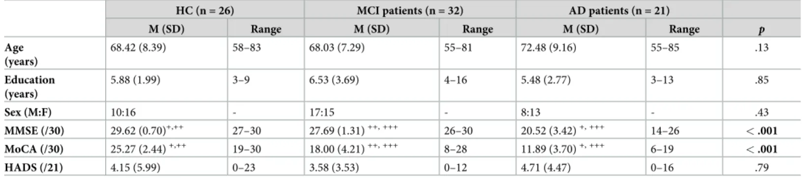
Table 1. Demographic and neuropsychological characteristics of Healthy Controls (HC), Mild Cognitive Impairment (MCI) and Alzheimer’s Disease (AD) participants.
HC (n = 26) MCI patients (n = 32) AD patients (n = 21)
M (SD) Range M (SD) Range M (SD) Range p
Age (years) 68.42 (8.39) 58–83 68.03 (7.29) 55–81 72.48 (9.16) 55–85 .13 Education (years) 5.88 (1.99) 3–9 6.53 (3.69) 4–16 5.48 (2.77) 3–13 .85 Sex (M:F) 10:16 - 17:15 - 8:13 - .43 MMSE (/30) 29.62 (0.70)+,++ 27–30 27.69 (1.31)++, +++ 26–30 20.52 (3.42)+, +++ 14–26 < .001 MoCA (/30) 25.27 (2.44)+,++ 19–30 18.00 (4.21)++, +++ 8–28 11.89 (3.70)+, +++ 6–19 < .001 HADS (/21) 4.15 (5.99) 0–23 3.58 (3.53) 0–12 4.71 (4.47) 0–16 .79
M, male; F, Female; MMSE, Mini Mental State Examination; MoCA, Montreal Cognitive Assessment; HADS, Hospital Anxiety and Depression Scale.
+
Differ from MCI (p < .001).
++
Differ from AD (p < .001).
+++
Differ from HC (p < .001).
(Luria’s motor series fist, edge, palm), sensitivity to interference (conflicting instructions), inhibitory control (Go/no Go), verbal inhibitory control (modified Hayling test), abstraction capacity (proverb interpretation), working memory for digits (backward digit span), verbal working memory (months of the year backward), and spatial working memory (modified Corsi block tapping test). The tasks motor series, conflicting instructions, and Go/no Go were taken from Frontal Assessment Battery (FAB) [59], a widely used screening test of EF. The other ones are specific to the IFS and were selected to optimize the sensitivity of this tool in comparison to the FAB. More comparative research is needed, but the FAB was found to dis-criminate between controls and AD patients less well than IFS (for a review see Moreira et al., [40]). IFS total score ranges between 0 and 30, with lower scores suggesting worse executive functioning.
Statistical analysis
Descriptive statistics were used for sample characterization. A multiple linear regression (enter method) was performed to examine the extent to which sociodemographic variables (educa-tion and age) and affective symptoms (anxious/depressive) influence IFS total score, even when the cognitive impairment associated to each diagnostic group is accounted for (i.e., Group was also included in the model). Comparisons between the three groups in IFS total and subtests’ scores were explored with one-way ANOVAs, and follow-up paired comparisons were Bonferroni corrected. Values of skewness and kurtosis were always below 2 and 7, respec-tively (skewness, range = -1.38–1.30; kurtosis, range = -1.19–1.14), suggesting that there is no large departure from normality in the data [60]. The diagnostic accuracy of the IFS total score for the detection of MCI and AD patients was assessed with receiver operation characteristics (ROC) curve analyses. The optimal cut-offs scores were selected based on the Youden index, with higher values indicating best sensitivity (the proportion of participants with cognitive impairment correctly identified as such) and specificity (the proportion of participants without cognitive impairment correctly identified as such). Finally, a discriminant analysis was carried out to determine the contribution of each IFS subtest for the group classification. All effects were considered significant atp < .05. Analysis were done using IBM SPSS1 software (ver-sion 24).
Results
Sample characterization and influence of age, education, depressive/
anxious symptoms on IFS scores
The characteristics of HC participants, MCI and AD patients are summarized inTable 1. The three groups did not differ in age [F (2,78) = 2.12, p = .13, ηp2= .05], years of education [F (2,78) = 0.85,p = .43, ηp2= .02), sex [χ2(2) = 1.7,p = .43] and in HADS total score [F (2,61) = 0.23,p = .79, ηp2= .01]. Note that the education level of our sample is lower than the observed in other similar studies [39,42,43], but it is representative of the majority of Portuguese elderly population. According to Censos 2011 [61], the majority of people over 65 years in Portugal completed only 4 years of formal education, corresponding to 1st Cycle of Basic Education (67.27%, 62.65%, and 56.80% of the people aged 65–69 years, 70–74 years, and 75–79 years, respectively).
MMSE scores significantly differed between the three groups, as expected [F (2,78) = 133.38,p < .001, ηp2= .78]; they were lower in the AD group than in both MCI (p < .001) and HC (p < .001) groups, and MCI patients also underperformed HC participants (p = .001). Please note that for MMSE there was a very low range in HC (between 27–30), reinforcing the
well documented celling effect of this measure [62]. Significant differences between groups were also found for MoCA total scores [F (2,75) = 76.84, p < .001, ηp2= .68]: the AD group presented the lowest scores, followed by MCI and HC groups, that also significantly differed from each other (allps < .001).
A multiple regression analysis was conducted with group, age, years of education and HADS total score as independent variables and IFS total score as dependent variable. The resulting model explained 73% of the IFS variance [adjustedR2= 0.73,F (4,61) = 41.13, p < .001]. Group was the strongest predictor (ß = 0.76, t = 11.19,p < .001), but education also sig-nificantly predicted unique variance in IFS performance (ß = 0.37, t = 5.22,p < .001), with more years of education being associated with better results in this tool. Neither age nor HADS total score were significant predictors (p = .67 and p = .83, respectively).
Sensitivity of the IFS to differentiate MCI patients from HC and AD
patients
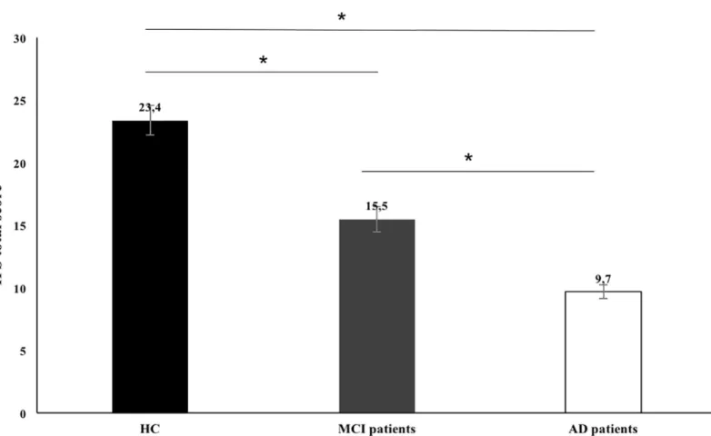
The performance of HC participants, MCI and AD patients on the IFS is depicted inFig 1. As hypothesized, significant differences were found between the three groups [F (2, 78) = 47.57, p < .001, ηp2= 0.56]. Specifically, MCI patients (M = 15.47; SD = 5.63; range = 4–27) had lower total scores than HC (M = 23.44; SD = 2.86; range = 16.5–29; p < .001) but higher than AD patients (M = 9.74; SD = 5.52; range = 3–20; p < .001). In order to determine if these dif-ferences remain significant after controlling for variables that we have shown to predict IFS
Fig 1. Total IFS total scores for control and clinical groups. HC, Healthy Controls; MCI, patients with Mild Cognitive Impairment (MCI); AD, patients with Alzheimer’s dementia. Error bars represent standard error.�p < .05.
performance (years of education), we conducted an analysis of covariance (ANCOVA). The group effect was confirmed even when this confounding variable was controlled [F (2,75) = 14. 58,p < .001, ηp2= .67], between all of the three groups: MCI patients vs. HC participants (p < .001), MCI patients vs. AD patients (p < .001) and AD vs. HC participants (p < .001). The same results were obtained when the ANCOVA also included sex as a covariate.
Since there were two identifiable subtypes within our sample of MCI participants (amnestic and non-amnestic), we also conducted an additional exploratory analysis to check if there were discernible differences between them in IFS total and subtests scores. No differences were found in IFS total score (p = .23) or subscores (ps > .74), apart from a marginally signifi-cant effect in the Conflicting Instructions subtest [with amnestic MCI performing slightly higher than non-amnestic,t(17.62) = 2.10, p = .05].
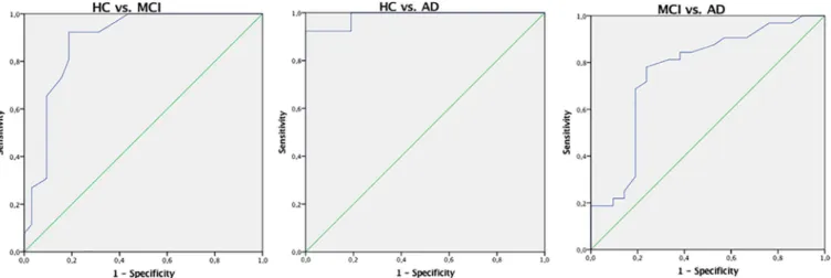
ROC curve analyses were carried out to evaluate the diagnostic accuracy of the IFS (seeFig 2). Regarding the discrimination between HC and MCI participants, this analysis generated an optimal cut-off of 20 points, with 92% sensitivity and 81% specificity (AUC of 0.89, CI = 0.80– 0. 98). In the discrimination between HC and AD participants, the optimal cut-off was 15 points, with 100% sensitivity of and 81% specificity (AUC of 0.99, CI = 0.96–1.00). A cut-off of 12.5 points reached the best sensitivity (78%) and specificity (76%) in the differentiation between clinical groups of MCI and AD patients (AUC of 0.76, CI = 0.62–0.90).
To examine the performance of the three groups in specific executive domains, the scores of each IFS subtest were compared (seeTable 2). The effect of group was largest for the Hayling test [F (2,76) = 44.58, p < .001, ηp2= .54], Proverbs interpretation [F (2,76) = 31.89, p < .001, ηp2= .46], Backward digit span [F (2,76) = 23.94, p < .001, ηp2= .39], and Motor series [F (2,76) = 19.74,ηp2= .34], suggesting that these were the subtests that were more sensitive to general variation across groups. For the Hayling test, Proverbs interpretation, and Backward digit span, AD and MCI patients scored lower than HC, but no significant differences were found between AD and MCI patients (Hayling test, HC vs. AD,p < .001; HC vs. MCI, p < .001 and MCI vs. AD,p = .66; Proverbs interpretation, HC vs. AD, p < .001; HC vs. MCI, p < .001 and MCI vs. AD,p = .26; Backward digit span, HC vs. AD, p < .001; HC vs. MCI, p < .001 and MCI vs. AD,p = .79). For the Motor series, significant differences were obtained between the three groups, with AD patients presenting the worst results, followed by MCI patients and by the HC (HC vs. AD,p < .001; HC vs. MCI, p < .001 and MCI vs. AD, p = .05). The remaining four subtests (Conflicting instructions, Go/no go, Verbal working memory and
Fig 2. Receiver operating characteristic (ROC) curves of the IFS in the differentiation between control and clinical groups, as well as between the two clinical groups. HC from MCI patients (left) and AD patients (center); MCI from AD patients (right).
Spatial working memory) were not sensitive to differences between HC and MCI patients, but they discriminated between HC and AD patients, and between MCI and AD patients (Con-flicting instructions:F (2,76) = 11.41, p < .001, ηp2= .21; HC vs. AD,p <. 001; HC vs. MCI,
p = .81 and MCI vs. AD, p = .001; Go /no go: F (2,76) = 9.03, p < .001; ηp2= .19; HC vs. AD,
p < .001; HC vs. MCI, p = 1.00 and MCI vs. AD, p = .003; Verbal working memory: F (2,76) = 16.32,p < .001, ηp2= .30; HC vs. AD,p < .001; HC vs. MCI, p = .21 and MCI vs. AD, p < .001; Spatial working memory:F (2,76) = 12.19, p < .001, ηp2= .24; HC vs. AD,p < .001; HC vs. MCI,p = .06 and MCI vs. AD, p = .02).
Finally, a discriminant analysis (enter method) was conducted to further understand how IFS scores could be informative about differences between groups. The discriminant function was able to correctly classify the group membership of 79% of the patients (Wilks’ Lambda = 0.25;χ2(16) = 98.77,p < .001). The three subtests that presented the highest contribution were: 81% in the Hayling test, 69% in the Proverb interpretation, and 63% in the Backward digit span. Note that these were the same subtests that showed larger effect sizes in the previ-ously presented comparisons across groups.
Discussion
EF are higher-order processes that are differentially compromised in healthy aging, MCI and AD. In previous studies, the IFS [39] has shown good discriminant accuracy between healthy participants and patients with dementia, and also between patients with different types of dementia. Nonetheless, its utility in MCI–a stage of cognitive decline that might progress to dementia—had not yet been explored. The present study provides four main new findings. First, MCI patients can be distinguished from healthy participants and early to moderate AD patients based on the IFS total score. Specifically, the MCI group had lower scores than con-trols and higher scores than patients with AD. Second, IFS performance was not influenced by age or depressive/anxious symptoms but was positively influenced by years of education. Nonetheless, its discriminative power in MCI vs. HC, MCI vs. AD and MCI vs. AD remains significant even when controlling for education. Third, cut-off scores of 20 and 12.5 provided optimal sensitivity and specificity in the identification of MCI vs. controls and of MCI vs. AD,
Table 2. Scores in IFS subtests of Healthy Controls (HC), Mild Cognitive Impairment (MCI) patients and Alzheimer’s Disease (AD) patients. HC (n = 26) MCI patients (n = 32) AD patients (n = 21)
p Post Hoc comparisons
IFS subtests M (SD) M (SD) M (SD)
Motor series 2.92 (0.39) 1.97 (1.03) 1.38 (0.97) < .001 HC > MCI > AD Conflicting instructions 2.69 (0.47) 2.41 (1.04) 1.38 (1.28) < .001 HC >AD; MCI > AD;
HC = MCI
Go/ no go 2.54 (0.71) 2.31 (1.06) 1.29 (1.38) < .001 HC >AD; MCI > AD; HC = MCI
Backward digit span 3.54 (1.00) 2.06 (0.98) 1.76 (0.83) < .001 HC >AD; HC > MCI; MCI = AD Verbal working memory 1.92 (0.27) 1.59 (0.71) 0.81 (0.93) < .001 HC >AD; MCI > AD;
HC = MCI
Spatial working memory 2.54 (0.81) 1.97 (1.09) 1.25 (0.63) < .001 HC >AD; MCI > AD; HC = MCI
Proverbs 2.21 (0.64) 0.92 (0.92) 0.55 (0.67) < .001 HC >AD; HC > MCI; MCI = AD
Hayling Test 5.15 (1.12) 1.88 (1.79) 1.33 (1.62) < .001 HC >AD; HC > MCI; MCI = AD IFS, INECO Frontal Screening
respectively. Fourth, performance on IFS subtests can also be informative to differentiate between healthy elderlies, MCI and AD patients. These conclusions are discussed in detail below.
The IFS total score accurately distinguished MCI patients from HC and AD patients. Find-ing that the IFS differentiates AD from HC adds to a number of previous studies, and high-lights again the utility of this screening tool in the context of this condition [33,39,42,43]. However, the current study is the first to provide evidence for the utility of IFS in the context of MCI: such utility is not only seen in analyses of score differences between groups, but also in subtests scores, discriminant analysis and analyses of diagnostic accuracy (AUC = .89 for MCI vs. HC, and AUC = .76 for MCI vs. AD). The existence of executive impairments in MCI has been documented before [26,63], but evidence for the utility of brief screening tools to assess them is limited. This finding is valuable both for research and clinical purposes, for instance for the neuropsychological characterization and newly diagnosed MCI patients (and monitoring of progression), but also for differential diagnosis (between MCI and AD). None-theless, we should note that AUC values were higher between MCI and HC than between MCI and AD, suggesting that the IFS might be particularly useful for the early detection of impair-ments in people at risk of developing dementia (and relatively less for the differentiation between MCI and AD patients). We found that a IFS cut-off of 20 reaches high values of sensi-tivity (.92) and specificity (.81) in the discrimination between MCI and HC. Such sensisensi-tivity and specificity values are comparable to those presented by MoCA [45], a well-established tool specifically developed for this purpose.
An analysis of subtests scores showed that these can also be informative. Subtests of verbal inhibitory control (Hayling subtest), abstraction (Proverbs), and verbal working memory (Backward digit span) were the most informative for differences between HC and MCI. How-ever, they were less sensitive to the differences between MCI and AD, i.e., between stages of subtle cognitive impairment and dementia. The Motor series subtest, in turn, was able to dis-criminate between the three groups. Tasks which explore sensitivity to interference (Conflict-ing instructions), inhibitory control (Go no-go) and work(Conflict-ing memory (verbal and spatial working memory subtests) were more valuable for the differentiation between MCI and AD, presenting relatively lower potential for the identification of subtle deficits of MCI in compari-son with HC. Thus, albeit the information provided by the IFS does not replace medical, com-prehensive neuropsychological and neuroimaging techniques, the examination of total and subtests scores produces relevant information that can significantly contribute to the differen-tial diagnosis between HC, MCI and AD patients. Which subtest(s) might be more informative will depend on the question being asked (e.g., discrimination between HC and MCI, or dis-crimination between HC and AD).
Contrary to our hypothesis, age did not predict the performance in IFS. This is not in line with the previous study of Moreira and colleagues [33], with a Portuguese normative sample, but is consistent with the recent study of Sanjurjo et al. [48] that aimed to evaluate the impact of demographic variables on IFS performance of healthy participants. Nonsignificant correla-tions between age and IFS total score were also found in the study of Ihnen and colleagues [47] with a dementia group. The absence of an age effect in IFS has been previously argued to reflect a celling effect of the measure [48], sometimes observed in the context of short screen-ing tools [64]. Nonetheless, studies with other classic executive measures such as task-switch-ing [8,65], verbal fluency, and the Wisconsin Card Sorting Test-Modified [66] also reported limited age effects. We should note, however, that the age range in the current study is some-what limited, for instance in comparison to the age range of a previous study by our group in which age effects on the IFS were found [33]. Moreover, our sample is relatively small and all participants were included in the multiple regression analysis regardless of their diagnostic
group (instead of including only the control ones)–this was done to increase statistical power, but it might have slightly biased the results. Thus, this question remains open and calls for more systematic research in the future. Intra-individual longitudinal analysis over time, for example, would be a more informative and stronger test to the influence of age on IFS perfor-mance. Education, in turn, significantly accounts for IFS performance, with more formal years of education being associated with higher scores. This effect has been consistently reported in previous studies with the IFS [33,47,48] but also with other executive screening measures such as the FAB [67]. Therefore, our study underlines the importance of considering education in the interpretation of the performance in executive tools [68]. Higher education levels seem to increase the contact with the evaluation contexts and to contribute to cognitive reserve, exert-ing a protective effect on the decline associated with healthy agexert-ing in EF [69]. Importantly, despite the influence of education, the discriminative power of this tool in the detection of EF deficits in MCI and in their differentiation from those of AD patients remains after accounting for this variable.
In an exploratory analysis, we compared the IFS total scores of patients with different MCI subtypes and found that they did not differ significantly. This is consistent with previous stud-ies that suggest that both amnestic domain and non-amnestic single and multiple-domain MCI subtypes presented executive deficits [26,29]. However, our groups were rela-tively small (n = 19 for amnestic MCI and n = 13 for non-amnestic MCI) and more studies with bigger samples will be essential to provide more reliable conclusions. Another issue that deserves more attention in future studies is the relationship between the IFS and measures of functional ability. It was established that performance in instrumental activities of daily living is highly related with EF [70]. Hence, it would be expected that performance in IFS could pre-dict the scores on functionality measures. This information is even more relevant since the dif-ferential diagnosis between MCI and dementia relies primarily in the decline in functionality [18,70].
Some limitations of our study should be acknowledged. In addition to the relatively small sample size, we did not have access to patients’ longitudinal data. Such information would be valuable particularly for MCI patients, not only to better clarify the aetiology of their cognitive impairment, but also to examine how the impairment changes over time and the rates of pro-gression to dementia. In future work it will be important to determine the extent to which brief tools such as the IFS can provide useful information to predict cognitive and functional outcomes of MCI patients over time.
Conclusion
This study produced evidence regarding the utility of the IFS for the identification of MCI, a well-established condition associated with a higher risk of progression to dementia. The early detection of this condition through a brief screening test enables prompting implementation of intervention strategies and managing of risk factors, thus holding potential to prevent a faster progression. Likewise, we found that performance in IFS can discriminate not only between normal aging and MCI and AD but also between these two clinical groups. The con-tribution of this tool for the differential diagnosis of these groups might be helpful in clinic set-tings where they are increasingly prevalent. Finally, our results underline the need of
normative data from different levels of education, in order to avoid biased interpretations.
Supporting information
S1 Dataset. Minimal anonymized dataset.
Acknowledgments
The authors would like to acknowledge the participants for giving their time and effort in par-ticipating in this study. They also thank Juliana Silva and Renata Ribeiro for help in the data collection.
Author Contributions
Conceptualization: Helena S. Moreira, Ana Sofia Costa, A´ lvaro Machado, São Luı´s Castro,
Ce´sar F. Lima, Selene G. Vicente.
Data curation: Helena S. Moreira, Ana Sofia Costa, A´ lvaro Machado.
Formal analysis: Helena S. Moreira, Ana Sofia Costa, Ce´sar F. Lima, Selene G. Vicente. Investigation: Helena S. Moreira, Ana Sofia Costa.
Supervision: Ana Sofia Costa, A´ lvaro Machado, São Luı´s Castro, Ce´sar F. Lima, Selene G.
Vicente.
Validation: Helena S. Moreira, Ana Sofia Costa, A´ lvaro Machado, São Luı´s Castro, Ce´sar F.
Lima, Selene G. Vicente.
Writing – original draft: Helena S. Moreira, Ana Sofia Costa, Ce´sar F. Lima, Selene G.
Vicente.
Writing – review & editing: Helena S. Moreira, Ana Sofia Costa, A´ lvaro Machado, São Luı´s
Castro, Ce´sar F. Lima, Selene G. Vicente.
References
1. Goldstein S, Naglieri JA, Princiotta D, Otero TM. Introduction: A History of Executive Functioning as a Theoretical and Clinical Construct. In: Goldstein S, Naglieri JA´ , editors. Handbook of Executive Func-tioning. New York: Springer; 2013. pp.24–44.
2. Yuan P, Raz N. Prefrontal cortex and executive functions in healthy adults: A meta-analysis of structural neuroimaging studies. Neurosci Biobehav Rev. 2014 May 42; 180–92.https://doi.org/10.1016/j. neubiorev.2014.02.005PMID:24568942
3. Robinson H, Calamia M, Gla¨scher J, Bruss J, Tranel D. Neuroanatomical Correlates of Executive Func-tions: A Neuropsychological Approach Using the EXAMINER Battery. J Int Neuropsychol Soc. 2013 Jun 13; 20(01):52–63.https://doi.org/10.1017/S135561771300060XPMID:23759126
4. Burzynska AZ, Nagel IE, Preuschhof C, Gluth S, Ba¨ckman L, Li S-C, et al. Cortical thickness is linked to executive functioning in adulthood and aging. Hum Brain Mapp. 2011 Jul 7; 33(7):1607–1620.https:// doi.org/10.1002/hbm.21311PMID:21739526
5. Marvel CL, Desmond JE. Functional Topography of the Cerebellum in Verbal Working Memory. Neuro-psychology Rev. 2010 Jun 22; 20(3):271–9.https://doi.org/10.1007/s11065-010-9137-7PMID:
20563894
6. Hirsiger S, Koppelmans V, Me´rillat S, Erdin C, Narkhede A, Brickman AM, et al. Executive Functions in Healthy Older Adults Are Differentially Related to Macro- and Microstructural White Matter Characteris-tics of the Cerebral Lobes. Front Aging Neurosci. 2017 Nov 30; 9.https://doi.org/10.3389/fnagi.2017. 00373PMID:29249957
7. Reuter-Lorenz PA, Festini SB, Jantz TK. Executive Functions and Neurocognitive Aging. In: Schaie KW, Willis SL, editors. Handbook of the Psychology of Aging. Oxford: Elsevier; 2016. pp. 245–262. 8. Aging Verhaeghen P. and Executive Control: Reports of a Demise Greatly Exaggerated. Curr Dir
Psy-chol Sci. 2011 May 24; 20(3):174–80.https://doi.org/10.1177/0963721411408772
9. Fjell AM, Sneve MH, Grydeland H, Storsve AB, Walhovd KB. The Disconnected Brain and Executive Function Decline in Aging. Cereb. Cortex. 2016 Apr 12.https://doi.org/10.1093/cercor/bhw082PMID:
27073220
10. Kirova A-M, Bays RB, Lagalwar S. Working Memory and Executive Function Decline across Normal Aging, Mild Cognitive Impairment, and Alzheimer’s Disease. Biomed Res Int. 2015; 2015:1–9.https:// doi.org/10.1155/2015/748212PMID:26550575
11. Amieva H, Phillips L, Della Sala S. Behavioral dysexecutive symptoms in normal aging. Brain Cogn. 2003 Nov; 53(2):129–32.https://doi.org/10.1016/s0278-2626(03)00094-0PMID:14607132
12. Espinosa A, Alegret M, Boada M, Vinyes SG, Valero S, Martine´z-Lage P, et al. Ecological assessment of executive functions in mild cognitive impairment and mild Alzheimer’s disease. J Int Neuropsychol Soc 2009 Jul 2; 15(05):751.https://doi.org/10.1017/s135561770999035xPMID:19570310
13. Angel L, Bastin C, Genon S, Salmon E, Fay S, Balteau E, et al. Neural correlates of successful memory retrieval in aging: Do executive functioning and task difficulty matter? Brain Res. 2016 Jan; 1631:53–71.
https://doi.org/10.1016/j.brainres.2015.10.009PMID:26541580
14. Princiotta D, DeVries M, Goldstein S. Executive Functioning as a Mediator of Age-Related Cognitive Decline in Adults. In: Goldstein S, Naglieri JA´ , editors. Handbook of Executive Functioning. New York: Springer; 2013.pp. 143–155
15. Fjell AM, Walhovd KB. Structural Brain Changes in Aging: Courses, Causes and Cognitive Conse-quences. Rev Neurosci. 2010 Jan; 21(3).https://doi.org/10.1515/revneuro.2010.21.3.187
16. Fernandez-Ruiz J, Peltsch A, Alahyane N, Brien DC, Coe BC, Garcia A, et al. Age related prefrontal compensatory mechanisms for inhibitory control in the antisaccade task. Neuroimage. 2018 Jan; 165:92–101.https://doi.org/10.1016/j.neuroimage.2017.10.001PMID:28988829
17. Petersen RC, Smith GE, Waring SC, Ivnik RJ, Tangalos EG, Kokmen E. Mild Cognitive Impairment. Arch Neurol. 1999 Mar 1; 56(3):303.https://doi.org/10.1001/archneur.56.3.303PMID:10190820
18. Petersen RC, Roberts RO, Knopman DS, Boeve BF, Geda YE, Ivnik RJ, et al. Mild Cognitive
Impairment: ten years later. Arch Neurol. 2009 Dec 1; 66(12).https://doi.org/10.1001/archneurol.2009. 266PMID:20008648
19. Bermejo-Pareja F, Contador I, Trincado R, Lora D, Sa´nchez-Ferro A´ , Mitchell AJ, et al. Prognostic Sig-nificance of Mild Cognitive Impairment Subtypes for Dementia and Mortality: Data from the NEDICES Cohort. J Alzheimers Dis. 2016 Feb 2; 50(3):719–31.https://doi.org/10.3233/JAD-150625PMID:
26757038
20. Roberts RO, Knopman DS, Mielke MM, Cha RH, Pankratz VS, Christianson TJH, et al. Higher risk of progression to dementia in mild cognitive impairment cases who revert to normal. Neurology. 2013 Dec 18; 82(4):317–325.https://doi.org/10.1212/WNL.0000000000000055PMID:24353333
21. Petersen RC, Caracciolo B, Brayne C, Gauthier S, Jelic V, Fratiglioni L. Mild cognitive impairment: a concept in evolution. J Intern Med. 2014 Mar; 275(3):214–28.https://doi.org/10.1111/joim.12190PMID:
24605806
22. Monastero R, Cicero CE, Baschi R, Davı` M, Luca A., Restivo V, et al.. . .Mild cognitive impairment in Parkinson’s disease: the Parkinson’s disease cognitive study (PACOS). J Neurol.2018 May; 265 (5):1050–1058.https://doi.org/10.1007/s00415-018-8800-4PMID:29478221
23. Domoto-Reilly K, Sapolsky D, Negreira A, Brickhouse M, Dickerson B. Mild cognitive impairment of frontotemporal lobar degeneration subtypes: Clinical and imaging characteristics. Alzheimers Dement. 2012 Jul; 8(4):P624.https://doi.org/10.1016/j.jalz.2012.05.1662
24. Consoli A, Pasi M, Pantoni L. Vascular mild cognitive impairment: concept, definition, and directions for future studies. Aging Clin Exp Res. 2012 Apr; 24(2):113–116.https://doi.org/10.1007/bf03325158
PMID:22842831
25. Winblad B, Palmer K, Kivipelto M, Jelic V, Fratiglioni L, Wahlund LO, et al. Mild cognitive impairment– beyond controversies, towards a consensus: report of the International Working Group on Mild Cogni-tive Impairment. J Intern Med. 2004 Aug 24; 256: 240–246.https://doi.org/10.1111/j.1365-2796.2004. 01380.xPMID:15324367
26. Brandt J, Aretouli E, Neijstrom E, Samek J, Manning K, Albert MS, et al. Selectivity of executive function deficits in mild cognitive impairment. Neuropsychology. 2009; 23(5):607–618.https://doi.org/10.1037/ a0015851PMID:19702414
27. Putcha D, Tremont G. Predictors of independence in instrumental activities of daily living: Amnestic ver-sus nonamnestic MCI. J Clin Exp Neuropsychol. 2016 May 31; 38(9):991–1004.https://doi.org/10. 1080/13803395.2016.1181716PMID:27240585
28. Rosenberg PB, Mielke MM, Appleby B, Oh E, Leoutsakos J-M, Lyketsos CG. Neuropsychiatric symp-toms in MCI subtypes: the importance of executive dysfunction. Int J Geriatr Psychiatry. 2010 Sep 15; 26(4):364–372.https://doi.org/10.1002/gps.2535PMID:20845402
29. Hessen E, Reinvang I, Eliassen CF, Nordlund A, Gjerstad L, Fladby T, et al. The Combination of Dysex-ecutive and Amnestic Deficits Strongly Predicts Conversion to Dementia in Young Mild Cognitive Impairment Patients: A Report from the Gothenburg-Oslo MCI Study. Dement Geriatr Cogn Dis Extra. 2014 Apr 9; 4(1):76–85.https://doi.org/10.1159/000360282PMID:24847346
30. Baudic S, Barba GD, Thibaudet MC, Smagghe A, Rem P, Traykov L. Executive function deficits in early Alzheimer’s disease and their relations with episodic memory. Arch Clin Neuropsychol. 2006 Jan; 21: 15–21.https://doi.org/10.1016/j.acn.2005.07.002PMID:16125364
31. Bejanin A, Schonhaut DR, La Joie R, Kramer JH, Baker SL, Sosa N. et al. Tau pathology and neurode-generation contribute to cognitive impairment in Alzheimer’s disease. Brain. 2017 Oct 7; 140: 3286– 3300.https://doi.org/10.1093/brain/awx243PMID:29053874
32. Amieva H, Phillips LH, Della Salla S, Henry JD. Inhibitory functioning in Alzheimer Disease. Brain. 2004 May 1; 127 (5): 949–964.https://doi.org/10.1093/brain/awh045PMID:14645147
33. Moreira H, Lima C, Vicente S. Examining Executive Dysfunction with the Institute of Cognitive Neurol-ogy (INECO) Frontal Screening (IFS). Normative Values from a Healthy Sample and Clinical Utility in Alzheimer’s Disease. J Alzheimers Dis. 2014 March 25; 42(1): 261–273. https://doi.org/10.3233/JAD-132348PMID:24840570
34. Belleville S, Chertkow H, Gauthier S. Working memory and control of attention in persons with Alzhei-mer’s disease and mild cognitive impairment. Neuropsychology. 2007 jul; 21(4): 458–469.https://doi. org/10.1037/0894-4105.21.4.458PMID:17605579
35. Firbank M, Kobeleva X, Cherry G, Killen A, Gallagher P, Burn DJ, et al. Neural correlates of attention-executive dysfunction in lewy body dementia and Alzheimer’s disease. Hum Brain Mapp. 2016 Mar; 37 (3):1254–70.https://doi.org/10.1002/hbm.23100PMID:26705763
36. Stokholm J, Vogel A, Gade A, Waldemar G. Heterogeneity in Executive Impairment in Patients with Very Mild Alzheimer’s Disease. Dement Geriatr Cogn Disord 2006; 22(1):54–9.https://doi.org/10.1159/ 000093262PMID:16682794
37. Chen ST, Sultzer DL, Hinkin CH. Executive dysfunction in Alzheimer’s: association with neuropsychiat-ric symptoms and functional impairment. J Neuropsychiatry Clin Neurosci. 1998 Nov; 10 (4): 426–432.
https://doi.org/10.1176/jnp.10.4.426PMID:9813788
38. Cummings JL. Cognitive and behavioral heterogeneity in Alzheimer’s disease: seeking the neurological basis. Neurobiol Aging. 2000 Nov; 21(6): 845–861.https://doi.org/10.1016/S0197-4580(00)00183-4
PMID:11124429
39. Torralva T, Roca M, Gleichgerrch E, Lo´pez P, Manes F. INECO Frontal Screening (IFS): A brief, sensi-tive, and specific tool to assess executive functions in dementia. J Int Neuropsychol Soc. 2009 Jul 28; 15(05):777.https://doi.org/10.1017/s1355617709990415PMID:19635178
40. Moreira HS, Costa AS, Castro SL, Lima CF, Vicente SG. Assessing Executive Dysfunction in Neurode-generative Disorders: A Critical Review of Brief Neuropsychological Tools. Front Aging Neurosci. 2017 Nov 9; 9.https://doi.org/10.3389/fnagi.2017.00369PMID:29170636
41. Fiorentino N, Gleichgerrcht E, Roca M, Cetkovich M, Manes F, Torralva T. The INECO Frontal Screen-ing tool differentiates behavioral variant—frontotemporal dementia (bv-FTD) from major depression. Dement Neuropsychol. 2013 Mar; 7(1):33–39.https://doi.org/10.1590/S1980-57642013DN70100006
PMID:29213817
42. Gleichgerrcht E, Roca M, Manes F, Torralva T. Comparing the clinical usefulness of the Institute of Cog-nitive Neurology (INECO) Frontal Screening (IFS) and the Frontal Assessment Battery (FAB) in fronto-temporal dementia. J Clin Exp Neuropsychol. 2011 Jul 14; 33(9):997–1004.https://doi.org/10.1080/ 13803395.2011.589375PMID:21923634
43. Custodio N, Herrera-Perez E, Lira D, Roca M, Manes F, Bae´z S, Torralva T. Evaluation of the INECO Frontal Screening and the Frontal Assessment Battery in Peruvian patients with Alzheimer’s disease and behavioral variant Frontotemporal dementia. eNeurologicalSci, 2016 Dec; 5: 25–29.https://doi. org/10.1016/j.ensci.2016.11.001PMID:29430554
44. Nasreddine ZS, Phillips NA, Be´dirian V, Charbonneau S, Whitehead V, Collin I, et al. The Montreal Cog-nitive Assessment, MoCA: A Brief Screening Tool For Mild CogCog-nitive Impairment. J Am Geriatr Soc. 2005 Apr; 53(4):695–699.https://doi.org/10.1111/j.1532-5415.2005.53221.xPMID:15817019
45. Freitas S, Simões MR, Alves L, Santana I. Montreal Cognitive Assessment: Validation Study for Mild Cognitive Impairment and Alzheimer Disease. Alzheimer Dis Assoc Disord. 2013; 27(1):37–43.https:// doi.org/10.1097/WAD.0b013e3182420bfePMID:22193353
46. Montiel JM, Cecato JF, Bartholomeu D, Martinelli JE. Evaluation of Montreal cognitive assessment for the differential diagnosis of mild cognitive impairment and Alzheimer’s disease in elderly patients with more than 5 years of schooling: Data from a Brazilian sample. Adv Aging Res. 2013; 2(4):121–129.
https://doi.org/10.4236/aar.2013.24018
47. Ihnen J, Antivilo A, Muñoz-Neira C, Slachevsky A. Chilean version of the INECO Frontal Screening (IFS-Ch): Psychometric properties and diagnostic accuracy. Dement Neuropsychol. 2013 Mar; 7 (1):40–47.https://doi.org/10.1590/S1980-57642013DN70100007PMID:29213818
48. Sierra Sanjurjo N, Saraniti AB, Gleichgerrcht E, Roca M, Manes F, Torralva T. The IFS (INECO Frontal Screening) and level of education: Normative data. Appl Neuropsychol Adult. 2018 Feb 12;1–9.https:// doi.org/10.1080/23279095.2018.1427096PMID:29432039
49. Zigmond AS, Snaith RP. The Hospital Anxiety and Depression Scale. American Psychological Associa-tion. 1983; 67(6): 361–370.https://doi.org/10.1037/t03589-000
50. Freitas S, Simões MR, Alves L, Santana I. The Relevance of Sociodemographic and Health Variables on MMSE Normative Data. Appl Neuropsychol Adult. 2014 Dec 22; 22(4):311–319.https://doi.org/10. 1080/23279095.2014.926455PMID:25531579
51. McKhann GM, Knopman DS, Chertkow H, Hyman BT, Jack CR, Kawas CH, et al. The diagnosis of dementia due to Alzheimer’s disease: Recommendations from the National Institute on Aging and the Alzheimer’s Association workgroup. Alzheimer Dem. 2011; 7: 1–7.https://doi.org/10.1016/j.jalz.2011. 03.005PMID:21514250
52. Costa AS, Rocha S, Machado A´ . Association of retrospective time estimation and severity of cognitive impairment. J Clin Exp Neuropsycho. 2016 May; 38(8):853–860.https://doi.org/10.1080/13803395. 2016.1167841PMID:27132467
53. Van Cauwenberghe C., Van Broeckhoven C., & Sleegers K. (2016). The genetic landscape of Alzhei-mer disease: clinical implications and perspectives. Genetics in Medicine, 18(5), 421.https://doi.org/ 10.1038/gim.2015.117PMID:26312828
54. Reisberg B, Ferris SH, de Leon MJ, Crook T. The Global Deterioration Scale for assessment of primary degenerative dementia. Am J Psychiatr. 1982 Sep; 139(9):1136–1139.https://doi.org/10.1176/ajp.139. 9.1136PMID:7114305
55. Doody RS, Massman P, Dunn JK. A Method for Estimating Progression Rates in Alzheimer Disease. Arch Neurol. 2001 Mar 1; 58(3).https://doi.org/10.1001/archneur.58.3.449PMID:11255449
56. Lyketsos CG, Lopez O, Jones B, Fitzpatrick AL, Breitner J, DeKosky S. Prevalence of Neuropsychiatric Symptoms in Dementia and Mild Cognitive Impairment. JAMA. 2002 Sep 25; 288(12):1475.https://doi. org/10.1001/jama.288.12.1475PMID:12243634
57. Modrego PJ, Ferra´ndez J. Depression in Patients With Mild Cognitive Impairment Increases the Risk of Developing Dementia of Alzheimer Type. Arch Neurol. 2004 Aug 1; 61(8).https://doi.org/10.1001/ archneur.61.8.1290PMID:15313849
58. Aznar S, Knudsen GM. Depression and Alzheimer’s disease: Is stress the initiating factor in a common neuropathological cascade. J Alzheimers Dis. 2011 Feb 26; 23: 177–193. https://doi.org/10.3233/JAD-2010-100390PMID:21098983
59. Dubois B., Slachevsky A., Litvan I., & Pillon B. F. A. B. (2000). The FAB: a frontal assessment battery at bedside. Neurology, 55(11), 1621–1626.https://doi.org/10.1212/wnl.55.11.1621PMID:11113214
60. Curran PJ, West SG, Finch JF. The robustness of test statistics to nonnormality and specification error in confirmatory factor analysis. Psychol Methods. 1996 Mar; 1(1):16–29. https://doi.org/10.1037/1082-989x.1.1.16
61. Statistics Portugal (2012). Censos 2011: Resultados Definitivos–Portugal. [Censos 2011: Definitive Results—Portugal]. Instituto Nacional de Estatı´stica, Lisboa.
62. Franco-Marina F, Garcı´a-Gonza´ lez JJ, Wagner-Echeagaray F, Gallo J, Ugalde O, Sa´nchez-Garcı´a S, et al. The Mini-mental State Examination revisited: ceiling and floor effects after score adjustment for educational level in an aging Mexican population. Int Psychogeriatr. 2009 Sep 7; 22(01):72.https://doi. org/10.1017/s1041610209990822PMID:19735592
63. Huey ED, Manly JJ, Tang M-X, Schupf N, Brickman AM, Manoochehri M, et al. Course and etiology of dysexecutive MCI in a community sample. Alzheimers Dem. 2013 Nov; 9(6):632–639.https://doi.org/ 10.1016/j.jalz.2012.10.014PMID:23452959
64. Hsieh S, Schubert S, Hoon C, Mioshi E, Hodges JR. Validation of the Addenbrooke’s Cognitive Exami-nation III in frontotemporal dementia and Alzheimer’s disease. Dement Geriatr Cogn Disord. 2013; 36 (3–4):242–250.https://doi.org/10.1159/000351671PMID:23949210
65. Kray J, Eber J, Lindenberger U. Age differences in executive functioning across the lifespan: The role of verbalization in task preparation. Acta Psycho. 2004 Feb; 115(2–3):143–65.https://doi.org/10.1016/j. actpsy.2003.12.001PMID:14962398
66. Isingrini M, Angel L, Fay S, Taconnat L, Lemaire P, Bouazzaoui B. Age-Related Differences in the Reli-ance on Executive Control in Working Memory: Role of Task Demand. Antal A, editor. PLOS ONE. 2015 Dec 23; 10(12):e0145361.https://doi.org/10.1371/journal.pone.0145361PMID:26700019
67. Appollonio I, Leone M, Isella V, Piamarta F, Consoli T, Villa ML, et al. The Frontal Assessment Battery (FAB): normative values in an Italian population sample. Neurol Sci. 2005 Jun; 26(2):108–116.https:// doi.org/10.1007/s10072-005-0443-4PMID:15995827
68. Branco LD, Cotrena C, Pereira N, Kochhann R, Fonseca RP. Verbal and visuospatial executive func-tions in healthy elderly: The impact of education and frequency of reading and writing. Dement Neurop-sychol. 2014 Jun; 8(2):155–161.https://doi.org/10.1590/S1980-57642014DN82000011PMID:
29213897
69. Rolda´n-Tapia MD, Ca´novas R, Leo´n I, Garcı´a-Garcia J. Cognitive Vulnerability in Aging May Be Modu-lated by Education and Reserve in Healthy People. Front Aging Neurosci. 2017 Oct 24; 9.https://doi. org/10.3389/fnagi.2017.00340PMID:29118710
70. Marshall GA, Rentz DM, Frey MT, Locascio JJ, Johnson KA, Sperling RA, Alzheimer’s Disease Neuro-imaging Initiative. Executive function and instrumental activities of daily living in mild cognitive impairment and Alzheimer’s disease. Alzheimers Dement. 2011 May; 7(3):300–8.https://doi.org/10. 1016/j.jalz.2010.04.005PMID:21575871