UNIVERSIDADE DE LISBOA
FACULDADE DE FARMÁCIA
CHARACTERIZATION AND FUNCTIONAL ANALYSIS OF
THE Gp1 ACCESSORY LYSIS PROTEIN IN
MYCOBACTERIUM SMEGMATIS INFECTION BY THE
MYCOBACTERIOPHAGE Ms6
Maria João Gracias Fernandes da Costa Catalão
DOUTORAMENTO EM FARMÁCIA
MICROBIOLOGIA
UNIVERSIDADE DE LISBOA
FACULDADE DE FARMÁCIA
CHARACTERIZATION AND FUNCTIONAL ANALYSIS OF
THE Gp1 ACCESSORY LYSIS PROTEIN IN
MYCOBACTERIUM SMEGMATIS INFECTION BY THE
MYCOBACTERIOPHAGE Ms6
Maria João Gracias Fernandes da Costa Catalão
Research Advisors:
Madalena M. V. Pimentel, Ph.D.
José A. F. Moniz Pereira, Ph.D.
DOUTORAMENTO EM FARMÁCIA
MICROBIOLOGIA
CHARACTERIZATION AND FUNCTIONAL ANALYSIS OF
THE Gp1 ACCESSORY LYSIS PROTEIN IN
MYCOBACTERIUM SMEGMATIS INFECTION BY THE
MYCOBACTERIOPHAGE Ms6
CARACTERIZAÇÃO E ANÁLISE FUNCIONAL DA
PROTEÍNA ACESSÓRIA DE LISE, Gp1, NA INFECÇÃO DE
MYCOBACTERIUM SMEGMATIS PELO
MICOBACTERIÓFAGO Ms6
Dissertação apresentada à Faculdade de Farmácia da Universidade de Lisboa para obtenção do grau de Doutor em Farmácia (Microbiologia)
Maria João Gracias Fernandes da Costa Catalão
2010
The studies presented in this thesis were performed at Centro de Patogénese Molecular, Faculdade de Farmácia da Universidade de Lisboa under the supervision of Professor Madalena Pimentel and Professor José Moniz Pereira. Maria João Gracias Fernandes da Costa Catalão was the recipient of a Ph.D. fellowship (SFRH/BD/24452/2005) from Fundação para a Ciência e Tecnologia (FCT), Lisbon, Portugal. This work was supported by grant PTDC/SAU-FCF/73017/2006 (to M. P.) from FCT, Portugal.
De acordo com o disposto no ponto 1 do artigo nº 40 do Regulamento de Estudos Pós-Graduados da Universidade de Lisboa, deliberação nº 961/2003, publicado em Diário da República – II Série nº 153 – 5 de Julho de 2003, a Autora desta dissertação declara que participou na concepção e execução do trabalho experimental, interpretação dos resultados obtidos e redacção dos manuscritos.
Contents
Preface ix Acknowledgments xiii Summary xv Sumário xvii Abbreviations xxiiiChapter 1: General Introduction
Objectives
1 65
Chapter 2: The Mycobacteriophage Ms6 Encodes a
Chaperone-Like Protein Involved in the Endolysin Delivery to the Peptidoglycan
67
Chapter 3: Functional Analysis of the Holin-Like Proteins of
Mycobacteriophage Ms6
107
Chapter 4: Sequence Analysis, Characterization and Lytic
Activity of Lysin A, the Cell Wall Hydrolase Encoded by the Mycobacteriophage Ms6
143
Preface
All the world´s a phage
c. William Shakespeare
March 24th, 1882. Robert Koch announced the discovery of the etiologic agent causative of tuberculosis, Mycobacterium tuberculosis. For this, was awarded the Nobel Prize in 1905. Although more than 100 years have passed, and despite the efforts of the scientific community, the introduction of many effective anti-tuberculosis agents and the use of a live attenuated vaccine (BCG), anti-tuberculosis remains the leading infectious cause of death worldwide killing over 2 million people every year. In addition, there has been an emergence of multidrug-resistant tuberculosis (MDR-TB) cases, the ultimate result of effective drugs ineffectively administered which in turns contributes to further spread of the disease. Containment of this growing problem is one of the great challenges of the humankind, and, unless poverty and unequal development are ended throughout the world, tuberculosis will continue to be an ineradicable public health problem.
The resurgence of tuberculosis, the difficulty of treating mycobacterial disease and the emergence of drug-resistant strains of M. tuberculosis has stimulated the renewed interest into mycobacteriophage research in order to investigate whether they could provide a complementary mean of therapy. Myco-bacteriophages are dsDNA viruses that infect mycobacteria. Following phage replication, lysis of the cell wall occurs, eventually destroying the host bacterium and releasing the progeny phage. The first mycobacteriophage was isolated in
1947, and since that time over 250 of these viruses have been identified. Mycobacteriophages have made a significant contribution to the knowledge of mycobacteria over the past sixty years, and following the development of typing techniques in the 1960s and 1970s they were widely used in epidemiological studies of tuberculosis. During the past two decades, mycobacteriophages have become important in molecular studies of mycobacteria, both in terms of studying phage biology and as tools in recombinant DNA technology, thus facilitating the investigation of mycobacterial pathogenesis. At present, their potential as diagnostics tools is also being realised with the development of exciting new techniques for rapid bacterial detection, drug susceptibility testing and mycobacterial recombineering, with the last one being an important contribution for the simple construction of potential vaccine strains of M. tuberculosis and the identification of its virulence determinants.
The ability of mycobacteriophages to lyse and kill mycobacteria also led to investigations of their potential as therapeutic agents against tuberculosis. Unfortunately however, early experiments on using lytic phages therapeutically during tuberculosis experimentally-infected animals not only failed to elicit cure, but in guinea pigs, had an adverse effect on their survival. Phage therapy is currently used in Eastern Europe and countries of the former Soviet Union and recent studies have confirmed that phages can be highly effective in treating many different types of bacterial infections, such as skin wounds, burns and ears infections or severe diarrhoea. However, treatment of systemic infections is the most challenging environment for bacteriophage action, with issues of compartmentation, host defense and kinetics, which are not present to the same degree in topical applications, creating real or imaginary barriers. In addition, killing intracellular pathogens such as M. tuberculosis presents a tough challenge as, in order to infect the target bacilli, the phages need to transverse the mammalian cell membrane, survive in the macrophage adverse intra-cellular environment
(acidification and presence of reactive oxygen species-ROS) or penetrate the granuloma.
When I started the Ph.D. program in the laboratory of Professor Madalena Pimentel and Professor Moniz Pereira, I continued the work initiated by former researchers of the lab and focused on investigating the role of a small accessory lysis protein restricted to mycobacteriophages. My first questions as a student who has just entered a new area of research, gave rise to additional new questions and to the obtainment of unexpected and exciting findings. The present work provides insight into the understanding of the molecular pathways involved in mycobacteriophage lysis, and more importantly, uncovers intriguing connections and links that warrant further investigations.
From the beginning, the purpose of my Ph.D. work, now presented and discussed in this thesis was to identify and characterize the role of the mycobacteriophage Ms6 Gp1 lysis protein during mycobacteria infection in view of the fact that, in addition to the endolysin, it is necessary for E. coli lysis without the requirement of holin activity to disrupt the cytoplasmic membrane. The functional analysis of the Ms6 holin-like proteins during mycobacterial infection came as a natural extension of this work. Chapter 1 provides a general, up-to-date review on the impact and the interest on mycobacteriophage research. In addition, preliminary data on the application of mycobacteriophages and the potential use of their lytic proteins in therapeutics is also discussed. We also focus on describing the mechanisms involved in bacteriophage-induced host cell lysis and the recent progress made in the field with the discovery of novel lysis models that include secretory endolysins endowed with signal peptides or signal-arrest-release (SAR) sequences and a new class of holins encoded by phages with SAR endolysins, pinholins. In Chapter 2, we provide evidence that Gp1 specifically interacts with the N-terminal region of the Ms6 endolysin and is involved in its translocation
across the cytoplasmic membrane in a holin-independent manner. In addition, construction of a mutant Ms6 phage defective for Gp1 synthesis showed that even though Gp1 is not vital for lysis is required for an efficient lysis of mycobacteria and a productive burst size. In Chapter 3, the role of the Ms6 holin-like proteins was further analysed, using a series of Ms6 mutant phages deleted for different lysis genes. Since Ms6 holins are not essential for mycobacterial lysis, their function in the Ms6 lysis model was discussed. Chapter 4 describes a remarkably finding observed during M. smegmatis infection by Ms6. We found that during infection, two endolysin forms are synthesized and both are required for the normal timing, progression and completion of host cell lysis. The implications of this new discovery are further examined. Finally, in Chapter 5 we integrate our overall findings, propose a general model for Ms6 lysis of mycobacteria and discuss specific future perspectives.
Even though the exact mechanism involved in Ms6 endolysin translocation in mycobacteria is still elusive, with this thesis we hope to elucidate and contribute to a better understanding of the mechanisms of mycobacteriophage lysis of mycobacteria. Ultimately, an increased knowledge of the exact pathways by which the mycobacteriophage lysis effectors are positioned next to their targets may result in the identification of mycobacteria new cell wall targets and in the promising therapeutic use of mycobacteriophage lysins.
Acknowledgments
As minhas primeiras palavras de agradecimento são dirigidas aos meus orientadores: ao Professor Moniz Pereira pelo enorme entusiasmo com que me recebeu, ainda aluna do 3º ano da licenciatura no seu grupo de investigação, por ter despertado em mim o interesse pela Microbiologia e pela Ciência e por tudo o que me ensinou. Obrigada pelas palavras de ânimo perante as minhas dificuldades e pela compreensão com que me permitiu chegar até aqui e me incentivou sempre a fazer mais e melhor. À Professora Madalena que me ensinou os primeiros passos na investigação, por ter confiado nas minhas capacidades e ter estado sempre disponível para me ajudar quando precisei. Agradeço ainda terem proporcionado as condições materiais necessárias para a execução do trabalho experimental desta tese.
À Dra. Paula Resende e ao Dr. João Vital, pela preocupação constante, por tudo o que me ensinaram e por todo o carinho e amizade que sempre me deram. Aos Professores do sub-grupo de Microbiologia, Prof. Helena Lourenço, Prof. Graciete, Prof. Mané, Prof. Isabel, Prof. Zé Miguel e Prof. João Gonçalves por terem contribuído, com tanto que me ensinaram, para o meu interesse pela Microbiologia. Às Professoras Elsa Anes e Paula Leandro por terem estado sempre disponíveis para me ajudar.
À Filipa, companheira de laboratório e grande amiga, por tudo o que vivemos juntas nos últimos cinco anos, por partilhares comigo as alegrias e as tristezas, por todo o teu apoio e disponibilidade e por te preocupares comigo como
ninguém! À minha querida amiga Cátia, companheira de aventuras e de desventuras, por teres sido o meu porto-de-abrigo nos dias de tempestade...Sem o teu apoio e amizade, tudo teria sido ainda mais difícil! Aos meus amigos que acompanharam todo o meu doutoramento: Zézinha, Inês, Miguel, Jo, Ritinha, Magda, Pedro, Sandra e Helena. Obrigada por me ouvirem, pela amizade e carinho. À Inês Vinga, Francisca Monteiro, Catarina Pinho e Catarina Milho por toda a ajuda e apoio, que em algum momento destes últimos quatro anos foram muito importantes. À Mariana e ao Fred pelos ensinamentos preciosos no início da minha aprendizagem na investigação. Ao Acilino, pela companhia e boa-disposição, e pelas conversas filosóficas. À Sylvie, pela companhia fim-de-semana fora no CPM, pela paciência com que me ouviste, por confiares em mim, pelo optimismo e energia contagiantes, por te teres tornado uma grande amiga! A todos os colegas do subgrupo de Microbiologia e do subgrupo de Bioquímica pela disponibilidade, ajuda e boa-disposição.
Aos meus queridos pais pelo apoio incondicional; ao estarem sempre presentes no meu pensamento, deram-me a força necessária para terminar esta tese. Saber que posso contar sempre com o vosso carinho e compreensão ajudou-me e ajudar-me-á sempre a superar todos os obstáculos e os momentos menos bons. Aos meus irmãos Patrícia e Vasco por todo o carinho e preocupação com as minhas digressões fora de horas ao CPM. Ao Francisco e ao recém-chegado Vasquinho, por me fazerem sorrir sempre!!!
Summary
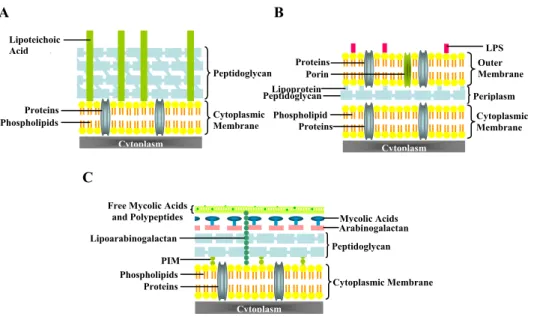
The majority of phages described to date (≈96%) is endowed with a tail and presents a double-stranded DNA (dsDNA) genome. Each infection cycle terminates with the strictly programmed and regulated lysis of the host brought about by two phage-encoded proteins, a murein-degrading enzyme, the endolysin, which is essential in achieving rapid hydrolysis of cell wall peptidoglycan, and a second membrane-embedded protein, the holin, which serves to release or activate the endolysin at a precisely defined time, thus bringing about an effective burst of the infected host. Targeting of endolysins to peptidoglycan can be achieved in two ways: through pore formation by canonical holins at a defined time or by continuous export of endolysins endowed with export signals during synthesis, assisted by the host Sec translocon. Mycobacteriophage Ms6 is a temperate bacteriophage that infects Mycobacterium smegmatis and possesses an unusual lytic cassette: in addition to the endolysin-holin lysis system encoded by genes lysA (gp2) and hol (gp4) respectively, the Ms6 lytic cassette comprises three accessory lysis proteins encoded by genes gp1, gp3 (lysB) and gp5 that are restricted to mycobacteriophages. Mycobacteria are Gram-positive bacteria that have evolved a complex cell wall, comprising a peptidoglycan-arabinogalactan polymer with covalently bound mycolic acids of considerable size, a variety of extractable lipids, and pore-forming proteins which provide an extraordinary efficient permeability barrier to noxious compounds and contribute to the high intrinsic resistance of mycobacteria to many drugs. To overcome the disadvantage that a complex cell
wall may represents for a successful infective cycle, mycobacteriophages have evolved new lysis strategies by acquiring, through their evolution, genes that likely confer a substantial selective advantage over those without them by providing faster and more complete lysis.
The present work underscores a new model for endolysin export in mycobacteriophage Ms6. Several lines of evidence indicate that gp1 encodes a secretion chaperone-like protein that binds the endolysin, assists the export to the extracytoplasmic environment independently of holin function and is required to accomplish an efficient lysis of M. smegmatis. Construction of different Ms6 derivatives deleted in different regions of the lysis operon demonstrated that the gene products of hol and gp5, although nonessential for phage viability, appear to play a role in controlling the timing of lysis. Remarkably, during M. smegmatis infection two endolysin forms (Lysin384 and Lysin241) are synthesized and both enzymes were shown to be essential for the normal timing, progression and completion of host cell lysis. In conclusion, this work highlights the role of the accessory lysis protein Gp1 and revealed alternative pathways for mycobacteria lysis, demonstrating that the presence of the mycobacterium-specific lysis factor Gp1, may confer a selective advantage not only for fitness under different environmental conditions but also as an alternative to lysis exclusively holin-dependent.
Keywords: Mycobacteriophages – Molecular chaperones – Holins – Endolysins -
Sumário
Mycobacterium tuberculosis, o agente etiológico da tuberculose,permanece como uma das principais causas de morbilidade e mortalidade no mundo. Esta realidade alarmante despertou um interesse renovado nas possibilidades terapêuticas dos bacteriófagos (vírus que infectam bactérias) e das sua proteínas líticas devido à capacidade de libertarem a progenia viral através da lise da célula hospedeira. A maioria dos fagos descritos até à data (≈96%) apresenta cauda e um genoma de DNA de cadeia dupla. Para induzirem uma lise efectiva do hospedeiro, estes bacteriófagos utilizam a estratégia “holina-endolisina” e sintetizam uma enzima, a endolisina, essencial para a hidrólise rápida do peptidoglicano e uma segunda proteína membranar, a holina, que permite a libertação ou activação da endolisina num período de tempo bem definido.
O bacteriófago Ms6 é um micobacteriófago temperado que infecta
Mycobacterium smegmatis. Como acontece com todos os fagos de DNA de cadeia
dupla, o seu operão lítico codifica para os dois genes essenciais para a lise do hospedeiro: a endolisina com actividade de amidase codificada pelo gene lysA (gp2) e a holina codificada pelo gene hol (gp4). Estes dois genes fazem parte de uma unidade de transcrição juntamente com os genes gp1, gp3 (lysB) e gp5. Estudos prévios demonstraram que a expressão dos dois primeiros genes do operão lítico (gp1 e lysA) na ausência da holina, é suficiente para induzir a lise de E. coli, um fenótipo não observado na ausência de gp1 ou com a expressão da endolisina apenas. gp1 é o primeiro gene do operão de lise do micobacteriófago Ms6 e
encontra-se localizado imediatamente a montante da endolisina. Codifica para uma pequena proteína de 77 aminoácidos que apresenta similaridade apenas com outras proteínas de micobacteriófagos, com função desconhecida. Apesar da inibição do crescimento de E. coli quando a proteína Gp1 é sobreexpressa, a estrutura secundária da sua sequência de aminoácidos não apresenta as características estruturais das holinas. A grande maioria das endolisinas descritas não possui uma sequência sinal para a secreção e dependem inteiramente das holinas para serem libertadas para o peptidoglicano. No entanto, estudos realizados revelaram a presença de um péptido sinal em N-terminal nas endolisinas de alguns fagos que infectam bactérias Gram-positivas como por exemplo, os bacteriófagos fOg44 de
Oenococcus oeni e φg1e de Lactobacillus plantarum. Para atingirem o peptidoglicano, as endolisinas destes fagos requerem a actividade do sistema de secreção Sec de E. coli para a sua exportação, e não a formação de lesões membranares pelas holinas. Particularmente interessante é o caso das endolisinas do fago P1 e 21 de E. coli que possuem uma sequência SAR (Signal-Arrest-Release Sequence) em N-terminal que lhes permite serem transportadas pelo sistema Sec até ao peptidoglicano. A análise bioinformática da sequência de aminoácidos de LysA não previu uma sequência sinal para a secreção não estando igualmente identificadas sequências SAR em endolisinas de outros micobateriófagos, o que parece indicar que a translocação desta proteína através da membrana citoplasmática está dependente dos outros genes de lise.
No presente estudo, investigou-se o envolvimento da proteína acessória de lise Gp1 no transporte da endolisina através da membrana citoplasmática e a função das holinas Hol e Gp5 na regulação do tempo de lise em M. smegmatis. Foi também estudada a função da endolisina durante a infecção das micobactérias e o seu espectro de actividade lítica. Na primeira parte do trabalho, os resultados obtidos demonstraram que em E. coli a co-expressão das proteínas Gp1 e LysA (endolisina) é suficiente para obter um efeito lítico na ausência da holina. No
entanto, Gp1 não pertence à classe das holinas, apresentando características estruturais e bioquímicas de chaperone molecular, como um baixo peso molecular (<15 kDa), um ponto isoeléctrico acídico e a capacidade de formação de dímeros. Experiências de interação de proteínas por crosslinking em E. coli, demonstraram que Gp1 interage com a endolisina LysA através de ligação com os primeiros 60 aminoácidos N-terminais da enzima, sendo esta região necessária e suficiente para a interacção. De facto, durante a infecção de M. smegmatis pelo Ms6 esta interacção também ocorre e a oligomerização de Gp1 parece ser necessária para este processo. A delecção de gp1 do operão de lise do bacteriófago Ms6 demonstrou que, embora Gp1 não seja essencial para a obtenção de um fenótipo de lise, é necessária para uma lise eficiente de M. smegmatis e para a libertação da progenia viral.
Tendo em consideração que tanto em E. coli como em M. smegmatis, a holina não é necessária para a lise, a sua função durante o ciclo de infecção foi estudada. A análise da sequência aminoacídica de Hol revelou a presença na região N-terminal da proteína de uma sequência SAR seguida de uma região transmembranar, características das pinholinas. Investigou-se ainda a função da proteína Gp5, que embora possuindo também características estruturais de holina, não é essencial para a lise de M. smegmatis. Utilizando a técnica de recombineering, construiram-se diferentes fagos derivados do Ms6, deleccionados em diferentes genes líticos e estudou-se o efeito dessas delecções no seu ciclo de replicação após a infecção dos hospedeiro. Observou-se que Hol e Gp5, embora não sejam essenciais para a viabilidade do bacteriófago, parecem ter um papel na regulação do tempo de lise: a delecção de hol causou uma lise precoce enquanto que a delecção de gp5 retardou o tempo de lise. Por outro lado observou-se também que a presença de ambas as proteínas durante a infecção é essencial para obter a lise do hospedeiro no tempo de lise programado.
Por fim, na terceira parte do trabalho, estudou-se a função durante o ciclo lítico das duas endolisinas (Lisina384 e Lisina241) codificadas pelo gene lysA. A análise da sequência nucleotídica de lysA revelou a presença de um gene de lise adicional codificado na mesma fase de leitura, e precedido por sinais de tradução (um codão de iniciação GTG e um local de ligação ao ribossoma). Embora a endolisina se tenha revelado essencial para a lise de M. smegmatis (um derivado do Ms6 deleccionado no gene lysA não é viável), bacteriófagos derivados do Ms6 defectivos na síntese da lisina384 ou da lisina241 embora sejam viáveis, apresentam um ciclo de infecção defectivo. A proteína Gp1 demonstrou ser essencial para a síntese ou estabilidade da lisina384 mas não da lisina241, embora não pareça participar na função final desta proteína, nem na sua activação ou conformação. Ambas as endolisinas possuem actividade lítica em diferentes bactérias Gram-positivas, Gram-negativas e micobactérias quando adicionadas exogenamente.
Em resumo, o presente trabalho demonstra a importância da proteína acessória de lise Gp1, não só na translocação da lisina384 através da membrana citoplasmática, mas também para uma lise eficiente de M. smegmatis. Por outro lado, estes estudos revelaram a existência, no micobacteriófago Ms6, de vias alternativas de lise através da presença, no seu genoma, de proteínas de lise adicionais (para além do sistema holina-endolisina) e exclusivas deste grupo de fagos. Devido à complexidade da parede celular micobacteriana e da barreira que poderá representar para um ciclo de infecção bem sucedido, estas proteínas de lise acessórias poderão conferir uma vantagem selectiva, não só em termos evolutivos durante a infecção em diferentes condições ambientais, mas também como uma alternativa viável para uma lise exclusivamente dependente da holina.
A caracterização dos mecanismos de lise das micobactérias pelos micobacteriófagos e um conhecimento das vias exactas pelas quais as enzimas efectoras da lise são posicionadas junto do seu substracto, poderá contribuir para a identificação de novos alvos terapêuticos na parede das micobactérias.
Palavras-chave: Micobacteriófagos – Tempo de lise – Chaperones moleculares –
Abbreviations
μg μl μm Å aa AG Amp ATCC ATP β-gal BCG BCIP BLAST bp BRED BS BS3 BSA cm CM Cryo-EM Cys CWBD DA DADA-PCR D-Ala D-Glu DNA ds Fig microgram microliter micrometer angstrom amino acid arabinogalactan ampicillinAmerican Type Culture Collection adenosine-5'-triphosphate
beta-galactosidase bacille Calmette-Guérin
5-bromo-4-chloro-3-indoxyl phosphate basic local alignment search tool base pair
bacteriophage recombineering of electroporated DNA
burst size
bis(sulfosuccinimidyl) suberate bovine serum albumin
centimeter
cytoplasmic membrane cryo-electron microscopy cysteine
cell wall binding domain dalton
deletion amplification detection assay-PCR
D-alanine D-glutamate
deoxyribonucleic acid double-stranded figure
FP GlcNac Gln Gly gp1 gp5 h His hol HRP ICTV IM IPTG Kan kb kbp kDa lacZ LB LIN Lpp LPS Lys lysA lysB MA m-DAP MDR-TB Met mg min ml mm mM m.o.i. MraY mRNA MurA MurNac Ni-NTA nm flanking primer N-acetil glucosamine glutamine glycine mycobacteriophage Ms6 Gp1 gene mycobacteriophage Ms6 Gp5 gene hour histidine
mycobacteriophage Ms6 holin gene horse radish peroxidase
International Committee on Taxonomy of Viruses inner membrane isopropyl β-D-1-thiogalactopyranoside kanamycin kilobase kilobase pair kilodalton β-galactosidase gene Luria-Bertani broth lysis inhibition lipoprotein lipopolysaccharide lysine
mycobacteriophage Ms6 endolysin gene
mycobacteriophage Ms6 mycolylarabinogalactan esterase gene mycolic acids meso-diaminopimelic acid multidrug-resistant tuberculosis methionine miligram minute mililiter milimeter milimolar multiplicity of infection phospho-N-acetylmuramoyl-pentapeptide transferase messenger ribonucleic acid
UDP-N-acetylglucosamine-enolpyruvyl transferase
N-acetylmuramic
nickel-nitrilotriacetic acid nanometer
OADC OD OM ONPG ORF PAGE PBS PCR p.f.u. PG PGRP phoA pI PIM p.m.f. PPIase PVDF RBS RNA ROS SAR SD SDS Ser SIV SP ss TB TDM TMD Tn Tris tRNA ts TTS Tyr wt Zn
oleic acid albumin dextrose complex optical density
outer membrane
ortho-nitrophenyl-β-D-galactopyranoside open reading frame
polyacrylamide gel electrophoresis phosphate-buffered saline
polymerase chain reaction plaque forming units peptidoglycan
peptidoglycan recognition protein alkaline phosphatase isoelectric point fosfatidilmio-inositol manoside proton-motive force peptidyl-prolyl cis-trans-isomerase polyvinylidene difluoride ribosome-binding site ribonucleic acid
reactive oxygen species signal-arrest-release Shine-Dalgarno
sodium dodecyl sulphate serine
simian immunodeficiency virus signal peptide
single-stranded tuberculosis
trehalose diester of mycolic acids transmembrane domain
transposon
tris(hydroxymethyl)aminomethane transfer ribonucleic acid
temperature-sensitive type III secretion system tyrosine
wild type zinc
1
General Introduction
General Introduction
1. The Bacteriophages: Biology and Interactions with Host Bacteria
Bacteriophages (phages) are bacterial viruses that play major roles in the ecological balance of microbial life and are the most abundant entities in the biosphere (Brüssow and Kutter, 2005). Over the course of evolution, bacteriophages have developed unique proteins that bind to and inactivate (or redirect), critical cellular proteins in bacteria, shutting off key metabolic processes to divert host metabolism to the production of progeny phages (Liu et al., 2004). The vast number on earth, estimated at 1031 (Brüssow and Kutter, 2005), has been appreciated only recently, but it is increasingly clear that phages exert enormous influence over the microbial world (Pedulla et al., 2003). The overall diversity of this population appears to be great: no genomically defined phage has been isolated more than once, and the relatively few sequenced phage genomes are highly varied and characterized by a high degree of mosaicism that likely arises from extensive horizontal genetic exchange (Hendrix et al., 1999; Hendrix, 2003; Hatfull, 2008a). The phage population is dynamic, turning over rapidly through constant attrition and subsequent amplification in permissive hosts (Pedulla et al., 2003). On a global scale, it is estimated that ~ 1025 phages initiate an infection every second (Pedulla
et al., 2003; Hendrix, 2005) and in each of those infections the phage encounters
DNA - of bacterial or prophage origin - with which it can potentially recombine to generate new genomic arrangements (Canchaya et al., 2003, Hatfull et al., 2006). The great majority of these phages (≈ 96%) are endowed with a tail and all of them present a double-stranded DNA (dsDNA) genome (Ackermann, 2003). There are a variety of different morphological types of bacteriophages and taxonomy is based on their shape and size as well as on their nucleic acid. The International Committee on Taxonomy of Viruses (ICTV) presently recognizes one order (Caudovirales), 13 families, and 31 genera of phages (Ackermann, 2006). Virions have binary, cubic, or helical symmetry, or are pleomorphic. A few types have a lipid-containing envelope or contain lipids as part of the particle wall (Ackermann,
Chapter 1
2005; Ackermann, 2006). The head (or capsid) is a protein shell often in the shape of an icosahedron that contains the viral genome that usually comprises dsDNA, but there are small phage groups with single-stranded (ss) DNA, ssRNA, or double-stranded (ds) RNA genomes. The tail may or may not be a contractile structure, usually connected to six tail fibres containing receptors at their tips that recognize attachment sites on the bacterial cell surface. Not all phages possess tails or tail fibres and here other attachment mechanisms are in place (Hanlon, 2007). Most of the bacteriophages of relevance belong to three families, the Siphoviridae,
Myoviridae and the Podoviridae, comprising 15 genera. The remaining phages
occupy 10 families, each with a small number of members (Ackermann, 2006). Upon infection of the bacterial host, phages can follow several life cycles: lytic, lysogenic, pseudolysogenic and chronic infections (Weinbauer, 2004). The life cycle of a temperate bacteriophage is shown in Figure 1. The virus encounters its bacterial host during random motion and attaches via specific receptors sites that may be any of a wide variety of cell surface components, including proteins, oligosaccharides, teichoic acids, peptidoglycan and lipopolysaccharides (Guttman
et al., 2005; Hanlon, 2007). In some cases, the attachment sites might be present on
the cell capsule, flagella or even conjugative pili. Initially the attachment is reversible but then becomes irreversible and is followed by transfer of phage genetic material into the host cell. Injection of the phage genome into the bacterial cell can occur by a variety of mechanisms, depending on the morphology of the virus, but can involve contraction of the tail and formation of a hole within the bacterial cell wall (Weinbauer, 2004; Hanlon, 2007). Many of the bases present on the phage DNA are chemically modified to confer protection against attack by cellular restriction and nuclease enzymes. The viral genome is generally transcribed by host cell RNA polymerase, producing early mRNA that has the effect of taking over the metabolic machinery of the bacterium, redirecting its metabolic processes to the manufacture of new virus components assembled into complete virions.
General Introduction
Following construction and assembly of new phage particles within the host cell, lysis of the bacterium generally occurs, with the release of the new phage progeny-lytic cycle (Guttman et al., 2005; Hanlon, 2007). In the lysogenic cycle, temperate phages do not automatically enter a lytic cycle but instead integrate their DNA into the host cell DNA after infection (Fig. 1). The bacterial cells are then termed lysogenic. When the bacterial DNA replicates, the phage DNA replicates at the same time and each daughter cell will contain the viral DNA (prophage). The prophage directs the synthesis of a repressor protein that blocks the transcription of its own genes and also those of closely related bacteriophages (Weinbauer, 2004).
Figure 1. Life cycle of a temperate bacteriophage. See text for more complete description.
Figure adapted from Hanlon, 2007.
Phage
Bacterial cell Adsorption and DNA injection Bacterial chromosome
DNA circularization
Lysogenic Pathway Lytic Pathway
Phage DNA integration into the host chromosome Celular division DNA replication Synthesis of viral components Late protein synthesis Assembly of phage components within the bacterial cell
Lysis of bacterial cell and release of phage progeny Phage DNA integrated replicates with the bacterial DNA
Prophage Early mRNA and protein synthesis
Lytic cycle induction
Chapter 1
The presence of a prophage can therefore confer upon a bacterial cell some sort of immunity to infection by other bacteriophages (Guttman et al., 2005; Hanlon, 2007). Cells may undergo several rounds of division but occasionally will spontaneously lyse and liberate progeny phage. Alternatively, a population of lysogenic cells may be induced to lyse by subjecting them to stress such as treatment with mutagenic agents or exposure to ultraviolet light. A chronic infection occurs, when a cell is infected and phage progeny is constantly released from the host cell by budding or extrusion without lysing it, while in a persistent infections (pseudolysogeny, phage carrier-state) phages multiply in a fraction of the population (Weinbauer, 2004).
When a prophage escapes regulation by the repressor, its DNA is cut free allowing it to embark upon a lytic cycle. However, excision of prophage DNA is often imprecise and bacterial genes adjacent to the prophage DNA may be incorporated into the infectious phage DNA and then transferred to subsequent host cells. This process is known as transduction and is responsible for the horizontal transfer of genes from one bacterial cell to another (Canchaya et al., 2003; Hanlon, 2007). The acquisition of prophages would be an irrelevant process for the evolution of pathogenic bacteria if phages did not transfer genes that confer selective benefit to the bacterial host as genes known to increase the survival fitness of lysogens (Brüssow et al., 2004). Lysogenic bacteria may possess other advantages in terms of the acquisition of genes conferring pathogenicity or increased virulence (lysogenic conversion) (Boyd and Brüssow, 2002; Brüssow et
al., 2004). Many bacteriophages carry virulence genes encoding proteins that play a
major role in bacterial pathogenesis. In addition, bacteriophage-bacteriophage interactions within the bacterial host cell also contribute significantly to the virulence of bacterial pathogens (Boyd et al., 2001; Roucourt and Lavigne, 2009) by influencing bacterial adhesion, colonization and infection, enhancing bacterial resistance to serum and phagocytosis, altering bacterial susceptibility to antibiotics
General Introduction
and encoding bacterial exotoxins (Wagner and Waldor, 2002). Important toxin genes known to have been acquired by transduction include those encoding the neurotoxin of Clostridium botulinum, the diphtheria toxin of Corynebacterium
diphtheriae, the Shiga toxins found in Escherichia coli O157 and the cholera toxin
in Vibrio cholerae (Waldor, 1998; Wagner and Waldor, 2002). Furthermore, the advent of whole genome sequencing of bacteria has revealed that prophage or phage-like elements are abundant and not only contribute to sequence diversity but have clearly played a significant role in evolution.
1.1. The impact of Mycobacterial Phages
Mycobacteriophages are viruses of the mycobacteria. The interest in these phages derives in large part from the medical significance and biological idiosyncrasies of their hosts (Hatfull, 2000; Hatfull, 2006). Mycobacteria are acid-fast staining bacteria with characteristic waxy cell walls that can be readily divided into two groups based on their growth rate: slow-growers such as Mycobacterium
tuberculosis and fast-growers such as Mycobacterium smegmatis. Several
mycobacterial species are important human and animal pathogens, the most notorious being M. tuberculosis and Mycobacterium leprae, the causative agents of tuberculosis and leprosy, respectively (Hatfull and Jacobs Jr., 1994; Hatfull, 2006). The extent of tuberculosis is alarming: M. tuberculosis is the leading cause of human mortality from a single infectious disease and the increased prevalence of multiple drug-resistant M. tuberculosis strains complicates its treatment (Zhang and Telenti, 2000; Hatfull, 2005a).
Mycobacteriophages have facilitated the development of mycobacterial genetic systems, methods for mycobacterial transformation (Hatfull, 2005b) and an assortment of applications including phage-based vectors (phasmids), integration-proficient plasmids, non-antibiotic-selectable markers, gene expression systems, transposon delivery vehicles (Hatfull, 2000), gene replacement strategies using
Chapter 1
nonreplicating vectors (Husson et al., 1990), long linear DNA fragments (Balasubramanian et al., 1996), incompatible plasmids (Pashley et al., 2003), counterselectable markers (Pelicic et al., 1996), specialized transducing shuttle plasmids (Bardarov et al., 2002) and, more recently, recombineering systems for mycobacteria (van Kessel and Hatfull, 2007; van Kessel and Hatfull, 2008) and mycobacteriophages (van Kessel et al., 2008; Marinelli et al., 2008). They have also provided insights into viral diversity and the evolutionary mechanisms that generate them and offer potential for the development of novel methodologies for the diagnosis, prevention, and treatment of mycobacterial diseases as well as revealing interesting biological features of their unusual hosts (Hatfull, 2006; Piuri
et al., 2009). The first mycobacteriophages were isolated and first investigated
more than 60 years ago prompted by their utility in phage-typing of clinical mycobacterial isolates (Hatfull et al., 2008). Over 200 different mycobacteriophages infecting a broad variety of mycobacterial hosts have been described: some mycobacteriophages (e.g., DS6A) are specific for the M.
tuberculosis complex, while others (e.g., Bxb1) are restricted to fast-growing
strains (such as M. smegmatis) and still others (e.g., D29) infect both fast- and slow growing mycobacteria (Hatfull, 2006).
Currently, more than 60 complete mycobacteriophage genomes have been sequenced and all are tailed phages belonging to either the Siphoviridae or
Myoviridae morphotypes; none are Podoviruses (Hatfull et al., 2010). The putative
gene products of these phages were grouped into “phamilies” of related sequences and the mycobacteriophages assorted into clusters and subclusters of related genomes, in an effort to provide an array of genetic tools for dissection and understanding of mycobacterial hosts and insights into viral diversity and evolution. Nine clusters of related genomes were revealed encompassing 55 of 60 genomes (clusters A-I) plus five singletons, mycobacteriophages Giles, Omega, TM4, Wildcat and Corndog (Fig. 2) (Hatfull et al., 2010). The length of
General Introduction
mycobacteriophage genomes vary considerably, from 41 kbp to 164 kbp. There is also a broad range of GC content, varying from 56,9% to 69,1% which is similar to the host’s, and genomes are typically replete with protein-coding genes with few non-coding spaces. The most common morphology is an isometric head with approximately 60 nm diameter and a long flexible tail (Hatfull et al., 2008; Hatfull
et al., 2010). In addition to the mycobacteriophage population being large and
varied, their genomes are characterized by extensive genetic mosaicism: each genome apparently contains a unique combination of individual modules, of which the majority corresponds to one or a small cluster of genes. Generation of these mosaic genomes reflects a high level of horizontal genetic exchange within the phage population, with illegitimate recombination events underlying the generation of new module boundaries (Pedulla et al., 2003; Hatfull et al., 2010). While the characteristic mosaic architecture of phage genomes can be explained by abundant horizontal genetic exchange events in their evolutionary history, little is known about which genomes participate in these events or the rates at which exchange occurs (Hatfull et al., 2008).
Chapter 1
Figure 2. Splitstree representation of mycobacteriophage relationships.
Mycobacteriophage predicted protein products were assorted into “phamilies” according to shared sequence similarities. Figure from Hatfull et al., 2010.
While a variety of genome architectures are seen among these mycobacteriophages, most have a cluster of genes involved in virion structure and assembly. There are typically 25 to 50 genes in these clusters, which are likely to be expressed as either a single or small number of operons. Many of these genes appear to be shared by different mycobacteriophages, although the extent of sequence similarity at the amino acid level is often sufficiently low to suggest that they departed from a common ancestor. The regions of these phage genomes that do not encode structural gene products presumably provide functions which are required for DNA metabolism, regulation, metabolic optimization of viral reproduction, and lysogenic conversion of their hosts. Of the protein-coding genes, approximately 50% have no detectable sequence similarity (at the amino acid level) to other known genes (including other mycobacteriophages genes). Approximately 90% have no detectable sequence similarity to genes outside of the mycobacteriophage group, indicating that these phages are richly populated with genes that have not been previously identified (Hatfull, 2006; Hatfull et al., 2008; Hatfull et al., 2010). The majority of newly sequenced phage genomes, including those from mycobacteriophages, contain a large proportion of genes of unknown function, as well as many genes whose general functions are known, but that have not been previously observed in phages (Hatfull et al., 2008; Hatfull et al., 2010). Recently, studies attempting to understand the origin and functions of viral ORFans (ORFs with no detectable sequence similarity to any other ORF in the databases) have been reported. A first genome-wide identification and analysis of ORFans in the bacteriophage world suggested that they are exclusively or highly phage-specific. Although phage ORFans play a lesser role in horizontal transfer to
General Introduction
bacteria, they may be among the major players contributing to the vast phage diversity. This fact also suggests that, despite the known propensity of phages for capturing and transducing fragments of host genomes, these processes have relatively small impact on the phage gene repertoire (Liu et al., 2006; Yin and Fisher, 2008). The presence of genes involved in host responses to bacterial infections as well as autoimmune diseases such as lupus suggests a more central role of bacteriophages in human diseases than previously recognized: mycobacteriophages Cjw1 gene 39 and Omega gene 61 encode close homologues of the leprosy and tuberculosis immunodominant antigen Lsr2 that is a potent stimulator of both cellular and humoral immune responses, suggesting a possible role for phages in mycobacterial virulence (Pedulla et al., 2003; Hatfull et al., 2005a). Phage Rosebush contains two genes (4 and 6) encoding homologues of enzymes involved in biosynthesis of tetrahydrobiopterin, a cofactor for a key enzyme in the host defence against mycobacterial infections, nitric oxide synthase (Hatfull et al., 2005a). Bxz1 gp220 encodes a homolog ~35% identical to the human Ro protein, a major target of the autoimmune response in Lupus and Sjogren´s diseases. The function of the Ro ribonucleoprotein is not known, but it is implicated in 5S RNA processing in Xenopus, dauer formation in Caenorhabditis
elegans and resistance to ultraviolet light in Deinococcus radiodurans; the presence
of a Ro homolog in Bxz1 raises the possibility that bacteriophages could act in concert with their hosts to stimulate autoimmunity (Pedulla et al., 2003; Hatfull et
al., 2005a). Conversely, toxin genes have not been identified in the sequenced
mycobacterial or mycobacteriophage genomes and consequently, have not been implicated in mycobacterial virulence. The role of phages in the virulence of mycobacterial pathogens remains unclear and the genomes of M. tuberculosis and
M. leprae contain no full-length prophages. However, both of the two sequenced
genomes of M. tuberculosis contain two small prophage-like elements, φRv1 and φRv2, at least one of which (φRv1) has an active integration system; both φRv1 and
Chapter 1
φRv2 contain capsid genes and could in principle form virus-like particles. Nevertheless, while all clinical isolates of M. tuberculosis appear to contain at least one of these elements, it is uncertain whether they play any role in the physiology of the host (Pedulla et al., 2003; Hatfull et al., 2005a; Hatfull, 2008b).
In summary, the mycobacteriophages are a remarkably diverse group of viruses whose characterization has provided helpful insights into the mosaic nature of bacteriophage genomes and the evolutionary mechanisms that give rise to them. There are huge numbers of uncharacterized mycobacteriophage genes that may have specific utilities such as the development of new tools for the genetic manipulation of mycobacteria or by providing new insights and approaches necessary to overcome the humankind’s most deadly microbial enemy, M.
tuberculosis. Indeed, the genes encoding for the lysis proteins of
mycobacteriophages represent an attractive direction for further work on their potential as natural anti-mycobacterial compounds.
2. Bacteriophage-Induced Host Cell Lysis
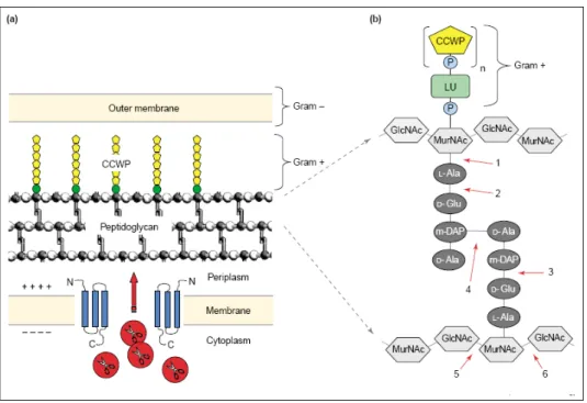
In general, bacteriophages must lyse their host cell to liberate the newly assembled progeny virions to the extracellular milieu (Young and Wang, 2006). Thus, lysis is a programmed event like all the other steps in the infectious cycle and of major importance regarding phage survival and ecological fitness (Wang, 2006). A sharply time-defined and efficient release of phage progeny is crucial to maximize both the burst-size and the opportunity to infect new hosts (Wang, 2006; São-José et al., 2007). The main barrier to host lysis is the continuous meshwork of peptidoglycan, a strong, stable structure that allows the bacterial envelope to withstand internal osmotic pressure (Young et al., 2000): compromising the cell wall is thus the fundamental goal for lytic processes. With the exception of filamentous phages that, as a result of their unique morphology and morphogenesis
General Introduction
can extrude through the envelope without fatal consequences for the host, all other phages must either degrade or otherwise compromise the peptidoglycan to cause lysis (Young et al., 2000; São-José et al., 2007). The filamentous phages constitute a large family of single-stranded DNA viruses that infect Gram-negative bacteria using pili as receptors. In contrast to other bacterial viruses, filamentous phage particles do not accumulate in the cytoplasm and cell lysis does not occur during phage progeny release. Instead, filamentous phages are produced by a concerted mechanism of assembly and secretion across both membranes of the Gram-negative cell (Russel, 1995; São-José et al., 2007).
From a mechanistic point of view, bacteriophages lyse the bacteria they infect by two essentially distinct modes:
1. Small single-stranded nucleic acid phages, with very limited genomes
under 6 kb, have a single phage protein to elicit lysis, presumably because of their restricted coding capacity (Bernhardt et al., 2002a). This protein with no apparent muralytic activity causes lysis by acting as a specific inhibitor of an enzyme in the multi-step pathway of murein biosynthesis. As this protein acts as a “protein antibiotic”, inhibiting cell wall synthesis and promoting a septal catastrophe as the cell attempts division, lysis is dependent on cell growth (Bernhardt et al., 2002a; Young and Wang, 2006). The only well characterized phages of this class are coliphages and among them there are three different prototypical lysis proteins: i) the E protein of the single-stranded (ss)-DNA bacteriophage φX174 (Microviridae), ii) the L protein of the ss-RNA bacteriophage MS2 (Leviviridae), and iii) the A2 protein of the ss-RNA bacteriophage Qβ (Alloleviviridae) (Young et al., 2000; Bernhardt et al., 2002a). For φX174 (Bernhardt et al., 2000) and Qβ (Bernhardt et
al., 2001a), it has been demonstrated unequivocally that the single lysis protein
produced, for which it was proposed the term amurin (Bernhardt et al., 2002a) acts as an inhibitor of a specific step in murein biosynthesis. However, the inhibited steps are different for the two phages: φX174 E and Qβ A2 inhibit separate
Chapter 1
enzymes, MraY and MurA, respectively, of the murein synthesis pathway (Young, 2005). Although the regulation of expression of the L protein of phage MS2 has been well studied and evidences indicate that interference with the stability of a large translation regulatory hairpin affects lysis timing, the L target remains unknown (São-José et al., 2007). The lytic capacity of E requires a host protein, SlyD (Roof et al., 1997) that is a peptidyl-prolyl cis-trans-isomerase (PPIase) or rotamase of the FKBP family (Roof et al., 1994; Hottenrott et al., 1997) that although is not the target of E lytic function, is required for protein stabilization (Bernhardt et al., 2000, Bernhardt et al., 2002b). The molecular target for the bacteriolytic E protein from bacteriophage φX174 is known to be the enzyme phospho-N-acetylmuramoyl-pentapeptide transferase (MraY), an integral membrane protein (Bernhardt et al., 2001b; Mendel et al., 2006) that catalyses the transfer of the MurNac pentapeptide from a UDP-MurNac pentapeptide to the polyisoprenoid carrier undecaprenylphosphate originating lipid I, the first membrane-bound murein precursor (Zheng et al., 2008). Phage Qβ A2 is a dual functional protein: it is simultaneously the Qβ capsid protein responsible for adsorption to the host pilus and the lysis effector (Karnik and Billeter, 1983; Bernhardt et al., 2001a). Genetic and biochemical characterization has identified its target as UDP-N-acetylglucosamine enolpyruvyl transferase (MurA), a soluble enzyme that catalyzes the first committed step of the murein precursor biosynthesis pathway (Bernhardt et al., 2001a).
2. Phages with double-stranded nucleic acid genomes use the
“holin-endolysin” strategy to achieve host cell lysis. The phage elaborates a soluble murein degrading enzyme, an endolysin, specifically dedicated to degrade the host cell wall, and a second membrane embedded protein, the holin, which serves to release or activate the endolysin at a precisely defined time (Young, 1992; São-José
et al., 2003). Moreover, the holin function is responsible for the crucial regulation
General Introduction
Genes controlling lysis are typically late-expressed and must be transcriptionally regulated (Young, 1992). Endolysins and holins encoded by different phages can be extremely diverse. Interestingly, pairwise combinations of different endolysins with related holins or similar endolysins with distinct holins are frequently found in the natural phage population. Still, it is apparent that phages have evolved additional means of fine-tuning the lytic schedule, through the synthesis of holin antagonists, referred to as antiholins. Thus, what characteristics would be appropriate for a system evolved to effect host lysis at a defined time? First, the lysis system should be as saltatory as possible and should not affect the productivity of the infected host, in terms of virion assembly, until the programmed time of lysis. Second, the lysis system should be very efficient and rapid once the infective cycle is terminated. Finally, the timer should be capable of being over-ridden in real time, should circumstances change during an infection. All these characteristics are exhibited by the holin-endolysin system of lysis and are mostly due to the properties of holins alone (Young and Wang, 2006).
2.1. Holins: Saltatory lethal membrane permeabilization
Holins are much more diverse and frequently unique with respect to their primary sequence (Young, 2002) and they may be defined as a single gene, encoding a putative small protein with at least one transmembrane domain (TMD) found in the vicinity of the endolysin gene (Wang et al., 2000; Young, 2002). Many holins genes have potential dual-start motifs which allow for the production of a shorter holin and a longer antiholin (Young, 2005). During the late phase of phage development, holins progressively accumulate in the cytoplasmic membrane of the host and while the proton-motive force (p.m.f.) is maintained, they assemble into oligomers and rafts of intrinsic stability (Young and Wang, 2006). Holins can be prematurely triggered by membrane depolarization with energy poisons such as cyanide and dinitrophenol (Gründling et al., 2001; Young, 2005). This observation
Chapter 1
led to a model for holin timing in which at a precise time programmed into its primary structure, the holin suddenly causes disruption of the membrane with non-specific hole formation and collapse of the membrane potential which sets the time of lysis by allowing the destruction of the cell wall by the released or activated phage encoded muralytic enzymes, the endolysins (São-José et al., 2003; Young and Wang, 2006).
2.1.1. The phage λ paradigm
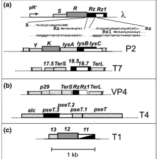
In lambdoid phages, all late genes are transcribed from a single promoter, designated PR´in bacteriophage λ (Young, 1992). The first genes of the late operon are the phage lysis genes: S, R, Rz and Rz1 (Fig. 3) (Young, 1992). The S gene encodes the holin (S105) and the antiholin (S107), as a result of translational initiation sites defined by codons 3 and 1 (Bläsi et al., 1989; Bläsi et al., 1990), respectively. The R gene encodes the endolysin which is a 18 kDa soluble murein transglycosylase that cleaves the glycosidic bond between N-acetylglucosamine and N-acetylmuramic acid residues, forming a cyclic product (Bieńkowska-Szewczyk et al., 1981). The DNA sequence in the lysis cassette contains two additional genes, designated Rz (Young et al., 1979) and Rz1, the later fully embedded in Rz in a +1 frame (Hanych et al., 1993; Kedzierska et al., 1996).
Rz/Rz1 gene products constitute a protein complex that somehow attacks the outer
membrane (OM), cleaving the oligopeptide linkages between the peptidoglycan and the outer membrane lipoprotein (Lpp) (Young and Wang, 2006; Berry et al., 2008).
General Introduction
Figure 3. Features of the lysis gene region in bacteriophage lambda (see text for further
details). Figure from São-José et al., 2003.
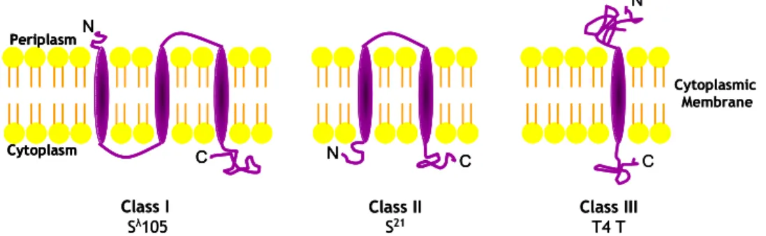
The phage λ S105 Holin
The S holin or S105, is a 105-residue integral cytoplasmic membrane protein with a three transmembrane domains (TMD) topology (N-out, C-in) (Gründling et
al., 2000a). Many S105 mutants have been isolated (Raab et al., 1986; Raab et al., 1988), including not only unconditional lysis defectives but also a plethora of alleles which accelerate or delay the onset of lysis. A particularly sensitive microdomain appears to be in the middle of TMD2. Both λSA52V and λSA52G are non-plaque-formers, but for opposite reasons. SA52V is unconditionally lysis-defective, whereas SA52G supports catastrophically early lysis (Raab et al., 1986; Raab et al., 1988; Johnson-Boaz et al., 1994). Similarly, at Cys51, substitution to a Ser accelerates lysis and replacement with a Tyr abolishes lysis. Thus, every potential TMD surface of S appears to be involved in setting the lysis clock (Gründling et al., 2000a; Gründling et al., 2000b). Although the sequences of the short periplasmic N-terminal domain and the cytoplasmic C-terminal domain are not essential for lysis, functional assembly of the λS holin requires periplasmic localization of its N-terminus (Graschopf and Bläsi, 1999a) and distribution of amino-terminal charged amino acids as well as the total amino-terminal net charge of S105 and S107 influence their lethal potential (Steiner and Bläsi, 1993). Furthermore, the C-terminal sequence of the λS holin constitutes a cytoplasmic regulatory domain which affects the timing of lysis although is not involved in the formation of the lethal membrane lesion nor in the “dual-start regulation” conserved in lambdoid holins (Bläsi et al., 1999). Strangely, however, although a G2H6G2 tag inserted near the membrane interface in the C-terminal cytoplasmic domain resulted in a functional lytic S protein, which has been the source of purified holin (Smith and Young, 1998), the simple addition of a hexahistidine tag
Chapter 1
in the C-terminal cytoplasmic domain disrupts holin function (Smith et al., 1998). Recently, it has been demonstrated that the N-terminus of S105 retains its formylated Met residue but that the N-terminus of S107 is fully deformylated. This supports the model that in S105, TMD1 inserts into the membrane very rapidly but that in S107, it is retained in the cytoplasm. Further, it reveals that, compared to S105, S107 has two extra positively charged moieties, Lys2 and the free N-terminal amino group, to hinder its penetration into an energized membrane. Moreover, an allele,
S105ΔTMD1, with TMD1 deleted, was found to be defective in lysis, insensitive to
membrane depolarization, and dominant to the wild-type allele, indicating that the lysis defective, antiholin character of S107 is due to the absence of TMD1 from the bilayer rather than to its ectopic localization at the inner face of the cytoplasmic membrane (White et al., 2010). λS holin has been shown by crosslinking studies, to form oligomers in the inner membrane of E. coli, but the ultimate degree of oligomerization in unknown (Zagotta and Wilson, 1990). Oligomerization does not depend on disulfide bridge formation (Gründling et al., 2000b) and the hydrophilic C-terminal part of the lambda S holin is non-essential (Rietsch et al., 1997). Oxidative dimer formation between S variants with single cysteines in the hydrophobic core of the TMD2 revealed that positions 48 and 51 are on a dimer interface (Gründling et al., 2000b). Furthermore, it has been suggested that the ability of S molecules to dimerize is not sufficient for the lytic step in holin function: oligomerization and a concerted conformational change which is equivalent to triggering of hole formation are also required: some lysis-defective alleles appear to be blocked at the monomer, dimer and oligomers stages (Gründling et al., 2000b).
Until recently, nothing was known about the nature of the λS membrane lesions. To calibrate the scale of the S-hole, a C-terminal fusion of the R endolysin with full-length β-galactosidase (β-gal) was constructed and the hybrid R-β-gal
General Introduction
product, an active tetrameric protein greater than 480 kDa in mass, was fully functional in lysis mediated by the S holin (Wang et al., 2003). This result suggested that at least some of the lesions created by the triggering of S are of size comparable to the large-scale lesions, in excess of 30 nm diameter (Wang et al., 2003; Young and Wang, 2006). In this study a holin lesion model was formulated in which holin protomers accumulate in the cytoplasmic membrane, oligomerize and ultimately form large two dimensional protein aggregates, designated as ‘death rafts’, from which lipid molecules are largely excluded by intimate interaction between individual holins via their transmembrane domains. Opening of an aqueous channel, with consequent local depolarization of the cytoplasmic membrane, triggers conformational change in the holins and subsequent dispersion of the subunits into the protein-bounded lesion in the cytoplasmic membrane (Wang et al., 2003). Latter, Savva and collaborators (2008) made use of electron microscopy and single-particle analysis to characterize the structures formed by λS holin in vitro. Purified S105 forms rings of at least two size classes, the most common having inner and outer diameters of 8.5 and 23 nm, respectively, and containing approximately 72 S105 monomers. The height of these rings, 4 nm, closely matches the thickness of the lipid bilayer (Krupovič and Bamford, 2008; Savva et al., 2008). More recently, cryo-electron microscopy (cryo-EM) analysis revealed that the scale of the holes is at least an order of magnitude greater than any previously described membrane channel, with an average diameter of 340 nm, some exceeding 1µm. The large holes can be viewed as supporting the notion that at the time of lethal triggering, the S105 holin exists in such large aggregates, leading to one or a small number of holes rather than many smaller holes distributed throughout the membrane (Dewey et al., 2010) as initially thought.
Chapter 1
The phage λ S107 Antiholin
The only essential genes for phage λ-mediated lysis are the S105 holin and the R endolysin but the presence of S107 delays lysis onset, allowing for a larger burst size and increasing effective hole formation after triggering (Gründling et al. 2000a). The λS107 antiholin and the λS105 holin are encoded in frame in the same S gene, and the proportion of S105 and S107 is for wild-type S, ~2:1 by the time of lysis (Chang et al., 1995). The differential expression of S107 and S105 is due to a structure directed initiation loop (sdi) overlapping the Shine-Dalgarno (SD) sequence in the S mRNA which hinders initiation of the S107 start codon (Bläsi and Young, 1996). The λS gene is characterized by a dual-start motif: the S105 holin and S107 antiholin share the same 105 amino acid sequence but S107 has an extra Met and Lys residues in the N-terminus (Fig. 3). These extra residues in S107 confer two extra positive charges comparing to S105, one charge by deformylation of Met1 and another by the Lys2 residue (Bläsi and Young, 1996): extra charges are known to hinder the translocation of the first TMD in an energized-membrane resulting in the altered topology of S107 compared to S105 (Graschopf and Bläsi, 1999b). The S105 holin has a three TMD topology whereas in the antiholin the first hydrophobic segment is unlikely to span the membrane. Furthermore, S107 exerts its inhibitory effect by dimerizing with S105, creating heterodimers which are either non-functional or of reduced non-functional capacity (Gründling et al., 2000c). The dissipation of membrane proton-motive force triggers the translocation of the first TMD of S107 which then becomes a topological homolog of S105 with similar hole/lesion-forming properties (Graschopf and Bläsi, 1999b).
Rz and Rz1 genes
The Rz and Rz1 genes and their equivalents are unique in biology: they are the only genes that share the same DNA in different reading frames but associated with the same phenotype (Hanych et al., 1993; Zhang and Young, 1999).
General Introduction
The Rz gene of bacteriophage λ belongs to the late operon which encompasses more than 25 genes for host cell lysis, phage DNA packaging and phage morphogenesis (Hanych et al., 1993). This third lysis gene was revealed by Tn903 mutagenesis of the λ phage which confers Mg2+-dependent lysis defect (Young et al., 1979). Cells infected with λRz:Tn903 in the presence of 10 mM MgCl2 do not undergo lysis; instead form spherical cells which are mechanically fragile and gradually lose refractility (Young et al., 1979). Until recently (Berry et
al., 2008) nothing was known about Rz function. It was suggested that Rz might
encode a murein-specific endopeptidase detected in lambda lysates (Taylor, 1971; Young et al., 1979; Bieńkowska-Szewczyk, 1980) which cleaves the oligopeptide crosslinks between outer membrane protein and the peptidoglycan (Zhang and Young, 1999). Rz encodes a 153 amino acid polypeptide with a hydrophobic N-terminus that is predicted to be either a secretory signal or N-terminal transmembrane domain by sequence analysis algorithms (Berry et al., 2008). Hanych et al. (1993), in an attempt to clone the Rz gene under a highly active promoter system, generated constructs in which an internal portion of the Rz gene was expressed. An unexpected 6.5 kDa polypeptide (Rz1) was observed to accumulate in the membrane fraction, which was found to result from the use of an internal, out-of-frame translational initiation site within the Rz gene. The sequence of the predicted Rz1 protein spans only 60 codons and contains a signal peptidase II cleavage site at Cys20 (Hanych et al., 1993; Zhang and Young, 1999). Rz1 processing was blocked by the signal peptidase II inhibitor globomycin, and by labelling with palmitate. The sequence of the mature Rz1 lipoprotein is very unusual containing almost 25% proline residues and the Rz1 protein is located almost exclusively in the outer membrane of E. coli (Kedzierska et al., 1996). It was demonstrated that, at physiological levels of expression, Rz and Rz1 are localized to inner membrane (IM) and outer membrane (OM), respectively, with their C-terminal domains predicted to lie in the periplasm (Berry et al., 2008).
Chapter 1
Furthermore, there is some genetic and phylogenetic evidence that the Rz and Rz1 proteins interact. First, the Rz/Rz1 equivalents from phage P2, lysB/lysC, complement defects in the lambda genes, but only as a cognate pair (Markov et al., 2004). In addition, yeast two-hybrid analysis of a library of phage T7 genes found multiple positives between clones with the last 10 codons of 18.7, the T7 Rz1 equivalent, and the last 50 codons of 18.5, the Rz equivalent (Bartel et al., 1996). These data suggest that Rz and Rz1 interact in a C-terminus to C-terminus fashion, which may account for the architectures of the embedded and overlapped Rz/Rz1 genes, in that these unusual arrangements minimize the likelihood of recombinational separation of the interacting domains (Summer et al., 2007). Finally, recently it was demonstrated that Rz and Rz1 proteins form a complex that improves the effectiveness of endolysin degradation of the murein layer and that function of this complex is lost if either protein is improperly localized. The OM is covalently attached to the cell wall by oligopeptide linkages between Lpp and the murein; a complex that spans the periplasm could conceivably push the IM away from the murein layer which might make endolysin-mediated murein degradation more efficient. In this study, a model for Rz/Rz1 function in host cell lysis was proposed: the first step is the temporally programmed permeabilization of the cytoplasmic membrane by the holin, resulting in the release of a cytoplasmic endolysin or the activation of a signal-anchor-release (SAR) endolysin. The second stage is the endolysin-dependent degradation of the murein layer followed by a third stage involving the fusion of the IM and OM mediated by the Rz-Rz1 complexes. The three steps of phage lysis mediated by holins, endolysins and Rz-Rz1 complexes form a sequential pathway in which holin function is required for endolysin function, which is in turn required for Rz-Rz1 function, although they are mechanistically independent (i.e. do not require heterotypic interactions with each other) (Berry et al., 2008).