UNIVERSIDADE DE LISBOA
FACULDADE DE MEDICINA VETERINÁRIA
MODULATING CHROMATIN STRUCTURE AND GENE EXPRESSION DURING AFRICAN SWINE FEVER VIRUS INFECTION – NEW STRATEGIES FOR AN EFFICIENT VACCINE RATIONAL
DESIGN
Gonçalo Daniel dos Santos Frouco
Orientador: Professor Doutor Fernando António da Costa Ferreira Co-orientador: Professor Doutor Carlos Lopes Vieira Martins
Tese especialmente elaborada para obtenção do grau de Doutor em Ciências Veterinárias na Especialidade de Sanidade Animal
Júri:
Presidente: Doutor Rui Manuel de Vasconcelos e Horta Caldeira Vogais:
Doutor Robert Michael Evans Parkhouse Doutor João Mário Brás da Piedade
Doutor Fernando António da Costa Ferreira Doutor Solange Judite Roque Coelho Alves Gil
Doutor José Alexandre da Costa Perdigão e Cameira Leitão
i ACKNOWLEDGMENTS
Along my PhD years, there are many people that contributed to the development of this work and whom I’m thankful.
I would like to thank my supervisor Professor Fernando Ferreira for the opportunity and for his guidance during these years. I’m grateful for his scientific ideas and the knowledge that he shared, and for the freedom that he gave me, allowing me to grow up as researcher. I also acknowledge my co-supervisor Professor Carlos Martins for giving me the opportunity to work in Infectious Disease Lab, for his precious advices and for sharing the expertise that he gain during his career dedicated to the study of African swine fever virus.
I would like to thank all the members of Infectious Disease Lab, Doctor Alexandre Leitão, Ju Silva, Rui Vieira, Paula Viana, João Coelho, Ferdinando Freitas and Margarida Simões. A special thanks to Doctor Alexandre Leitão for the critical analysis and his advices, and to my friends João Coelho, Ferdinando Freitas and Margarida Simões for their patience in teaching me lab techniques, for their help and scientific discussions, and for the good times spend in the lab.
To my friends and colleagues of CIISA with whom I had a lot of “hall discussions” sometimes important for the development of my work. I am grateful to Carla Carneiro, Clara Cartaxeiro, Joana Dias, Pedro Bule, Carla Mottola, Carolina Bento, Rui Seixas, Samuel Francisco, Inês Delgado, Ana Margarida Alho, Sandra Aguiar and Mariana Batista. Because moments of pleasure are always important a special thanks to Joana and Pedro for all the breaks that we did, and Ferdinando, Carolina, Carla and Clara for all the fun lunch times.
I would also like to thank my friends and family.
To João Fernandes for bringing me the dinner in my lab work nights. To Tiago Silva for his help during his summer internship and during the writing of this thesis. To Artur Silva for always having “Ben and Jerry’s” ice cream, for me, in his freezer.
To my mother, father and grandmother for their education and love, and to Sofia, Pedro and Clarinha for their absolute support. I’m extremely grateful for your strong support and for all the fun and tender moments that we’ve spend together during these years.
Finally, I left to the end the person that encourage me to do this PhD and always inspire me to go up one more step and to get out my comfort zone. Thank you Inês (and all our pets), for your unconditional support along these years, love and friendship, and for making me completely fulfilled. I appreciate every moment that we share and I’m sure that the future will bring us the realization to our plans.
Thank you all.
ii FUNDING
The present work was funded by the PhD fellowshipfrom FCT, Portugal (SFRH/BD/89426/2012) and from the European Union Seventh Framework Programme (FP7/2007-2013) under grant agreement n° 311931 - ASFORCE.
iii
Modulating chromatin structure and gene expression during African Swine Fever Virus infection – New strategies for an efficient vaccine rational design
ABSTRACT
African swine fever virus (ASFV) is a nucleo-cytoplasmic large DNA virus which infects all members of the family Suidae, causing a fatal disease of domestic swine and wild boar. Since no effective vaccine or treatment is available, ASF is considered a global threat for pig husbandry. The ASFV genome encodes among others, enzymes required for virion assembly, genome transcription and replication, including a putative histone-like protein, pA104R. In bacteria, these proteins perform topological modification of the chromosome (twisting, bending and folding), playing important structural and regulatory functions. Since ASFV has a large genome, a viral histone-like protein may be important for packaging its genome within the virion particle and/or for viral replication and transcriptional events. In this study, the ASFV-pA104R activity was characterized and its DNA-binding activities were evaluated. pA104R binds both to ssDNA and dsDNA, although having higher affinity to ds-DNA, over a wide range of temperatures, pH values, and salt concentrations and in an ATP-independent manner, with an estimated binding site size of about 14 to 16 nucleotides. The arginine residue located in pA104R’s DNA-binding domain, at position 69, also revealed to be important for an efficient DNA-binding. Additionally, since pA104R together with the viral type II topoisomerase, pP1192R, displayed DNA-supercoiling activity, a synergistic effect between these viral is proposed. The expression of pA104R was observed in the late phase of infection in infected cells with the Vero-adapted ASFV isolate Ba71V, co-localizing with cell nucleus and viral factories. siRNA experiments showed that the knockdown of A104R induce a reduction of viral progeny, copy numbers of viral genomes and transcription of a late viral gene, revealing that pA104R plays a critical role in viral DNA replication and gene expression. Results obtained on these studies prompted us to pursue the objective to generate a defective infectious single cycle (DISC) ASFV lacking the A104R gene. Recombinant virus was successfully obtained, however the complementary cell line previously developed did not support its replication. The antiviral activity of four HDACi against ASFV was also evaluated in this study. The results showing the abrogation of viral replication by NaPB open new insights on its use as an antiviral strategy to control ASFV spreading. Overall our data strongly support that pA104R plays an important role on ASFV replication opening a new window for the design of ASF control measures through the development of efficient and safe vaccines and antivirals.
Keywords: African swine fever virus, ASFV, histone-like protein pA104R, vaccine, defective infectious singe cycle viral particles, antivirals.
iv
Modulação da estrutura cromatínica e da expressão génica durante a infeção do Vírus da Peste Suína Africana – novas estratégias para o desenvolvimento de uma vacina eficaz
RESUMO
O vírus da peste suína africana (VPSA) é um vírus de DNA nucleo-citoplasmático que infeta todos os membros da família Suidae, causando uma doença com elevada mortalidade em suínos domésticos e nos javalis. Atualmente não existe uma vacina ou tratamento eficaz, tornando a peste suína africana (PSA) uma ameaça para a suinicultura mundial. O genoma do VPSA codifica aproximadamente 150 proteínas, algumas delas bem caracterizadas, estando envolvidas na transcrição, replicação ou na montagem do virião. No entanto, e apesar de todos os esforços realizados nas últimas décadas, a função biológica de numerosas proteínas virais não é ainda conhecida. Esta lacuna aliada à necessidade de um melhor entendimento sobre a biologia do VPSA e as suas interações com o hospedeiro têm contribuído em grande parto para a dificuldade no desenvolvimento de uma vacina eficaz contra PSA.
Por homologia de sequências proteicas, o genoma do VPSA codifica para uma proteína tipo histona (pA104R). Nas bactérias, estas proteínas são responsáveis por modular a topologia do DNA (torção, flexão e dobramento), desempenhando assim importantes funções estruturais e controlando a expressão de diferentes genes. O facto do genoma do VPSA codificar entre outras uma proteína viral semelhante a histonas bacterianas, reveste-se assim de enorme relevância pelo papel que que estas proteínas possam desempenhar na compactação do genoma na partícula viral e/ou para a sua replicação e transcrição. Neste contexto, este estudo pretendeu caracterizar o papel da VPSA-pA104R na replicação viral, tendo como objetivo contribuir para o conhecimento da biologia deste vírus e para averiguar se o gene A104R será um bom candidato para desenvolver uma vacina DISC (do inglês “defective infectious single cycle). Além disso, diferentes inibidores das histonas deacetilases (HDACs) foram testados como potenciais antivirais, eventualmente úteis no controlo da PSA Os principais objetivos deste trabalho foram assim os seguintes: (1) estudar VPSA-pA104R, através da clonagem, expressão, purificação e caracterização de sua atividade in vitro; (2) Compreender a relevância funcional de dois resíduos conservados de pA104R; (3) Avaliar os níveis de mRNA e proteína, bem como a localização intracelular de pA104R em células infetadas com VPSA, em diferentes tempos de infeção; (4) Desenvolver uma estratégia que permita a deleção da ORF A104R do genoma do VPSA e a obtenção de uma vacina DISC; (5) avaliar os níveis de acetilação das histonas das células infetadas para melhor compreensão do mecanismo de modulação dos mecanismos epigenéticos do hospedeiro pelo VPSA; (6) Avaliar o efeito dos inibidores das HDACs na infeção pelo VPSA.
v
Neste estudo, a VPSA-pA104R foi expressa num sistema procariota baseado em Escherichia coli. Após a sua purificação, a sua atividade foi caracterizada através de ensaios EMSA (do inglês “electrophoretic mobility shift assay”) e concluiu-se que esta proteína viral se liga tanto a DNA de cadeia simples como dupla, embora tenha maior afinidade para o de cadeia dupla, e estimou-se que o local de ligação seja cerca de 14 a 16 nucleótidos. Esta ligação ao DNA continua presente em variadas condições de temperaturas, pH e concentrações de sal e é independente de ATP. A perda de atividade da proteína mutada pontualmente no resíduo de arginina localizado na posição 69, revelou que este resíduo é importante para uma ligação eficaz ao DNA. Além disto, foi possível concluir que a carga positiva deste aminoácido é determinante para a capacidade de ligação do pA104R ao DNA. Adicionalmente, uma vez que se observou atividade de superenrolamento de DNA quando a pA104R e uma topoisomerase tipo II viral, pP1192R, foram adicionados a DNA plasmídeo relaxado, este trabalho suporta que o VPSA codifica de facto para proteínas necessárias a compactação do seu genoma e ainda é proposto a existência de um efeito sinérgico entre as duas proteínas virais acima descritas.
Como o objetivo de melhor compreender a importância da pA104R na infeção do VPSA, avaliou-se a dinâmica da sua expressão e a sua localização intracelular. A expressão de pA104R foi observada na fase tardia da infeção em células Vero infetadas com o isolado viral Ba71V, co-localizando com núcleo da célula e fábricas virais citoplasmáticas. Em relação à dinâmica de transcrição do gene A104R, apesar de ser típica de um gene tardio, foi possível detetar transcritos a partir das 2 horas pós-infeção (hpi). As experiências usando siRNA (do inglês “small interference RNA”) contra os transcritos do gene A104R, mostraram que a redução dos níveis de RNA deste gene induzem uma redução da progenia viral, do número de cópias de genomas virais e da transcrição de um gene viral tardio (B646L), revelando que o pA104R desempenha um papel crítico na replicação do DNA viral e na expressão de genes virais.
Atualmente, as únicas medidas de controlo do VPSA são baseadas na deteção precoce da doença e na rápida aplicação de medidas biossanitárias como o abate de animais infetados, controlo do movimento de animais e a vigilância. As tentativas falhadas até agora em obter uma vacina inativada ou atenuada permitem que novas estratégias, como as vacinas DISC ganhem espaço na investigação do VPSA como uma revigorante estratégia para controlar o VPSA. Os resultados obtidos neste estudo, anteriormente descritos, suportam a ideia que um mutante de deleção no gene A104R replicará o seu genoma nas células hospedeiras, mas não poderá compacta-lo dentro do virião, resultando num virião não-infecioso "vazio" que será incapaz de iniciar um segundo ciclo de infeção. Assim a infeção com este vírus mutante será capaz de estimular o sistema imunitário do hospedeiro, mas ao mesmo tempo será seguro, não produzindo progenia infeciosa. Assim outro objetivo deste trabalho foi então obter um vírus DISC deletado no gene A104R. Para isto, o vírus recombinante foi obtido por
vi
recombinação homóloga e uma linha Vero complementar, que expressa a pA104R, foi desenvolvida. Embora, o vírus recombinante tenha sido obtido com sucesso, a linha celular complementar desenvolvida não suporta a sua replicação e, como tal, a seleção e propagação do vírus recombinante não foi possível. Os baixos níveis de expressão de pA104R destas células quando comparados com os de células infetadas poderão explicar esta não complementação.
Com base em estudos anteriores que mostram que VPSA regula o estado epigenético da célula hospedeira, o grau de acetilação das histonas de células infetadas foi avaliado e a atividade antiviral de quatro inibidores das HDACs (NaPB, VPA, TSA e SAHA) contra a infeção pelo VPSA também foi testada neste estudo. O VPSA induz uma hipoacetilação dos resíduos de lisina 9 e 14 da histona H3 (H3K9K14). Esta modificação epigenética corrobora outras reportadas noutros estudos e todas elas estão classicamente correlacionada com o silenciamento de genes em células eucarióticas e pode indicar que o VPSA subverte diferentes mecanismos celulares, controlando o acesso da maquinaria de transcrição aos genes hospedeiros. Adicionalmente, um dos inibidores das HDACs testados, o NaPB, reverte este estado de hipoacetilação da histona e inibe a replicação do VPSA, interferindo com a expressão de gene virais tardios.
Os resultados obtidos neste estudo sugerem fortemente que a pA104R participa da modulação da topologia do DNA viral, estando envolvida na replicação, transcrição e/ ou compactação do DNA viral. Com o objetivo de desenvolver uma vacina DISC, o gene A104R poderá assim constituir um bom alvo a deletar. Esta nova estratégia poderá ser uma alternativa às tentativas até agora falhadas de obter uma vacina contra a PSA. Contudo, antes de uma vacina DISC ser realidade, um esforço científico no desenvolvimento de uma linha celular complementar será imperativo. Os resultados obtidos sugerem ainda que as HDACs celulares estão envolvidas no estabelecimento de infeção pelo VPSA e revelaram que o NaPB pode ser usado como uma estratégia antiviral adicional para controlar a propagação de vírus nas áreas de surto.
Palavras-chave: Vírus da peste suína africana, VPSA, proteína tipo-histona pA104R, vacinas, partículas virais “defective infectious singe cycle”, antivirais.
vii TABLE OF CONTENTS
Chapter I: Introduction………...1
1. African Swine Fever………..3
1.1. History and distribution………...………3
1.2. Epidemiology of African swine fever………...………4
1.2.1. African swine fever hosts………4
1.2.1.1. Domestic pigs………4
1.2.1.2. Wild suids………...………4
1.2.1.3. Soft ticks……….5
1.2.2. Transmission………..5
1.3. Strategies to control African swine fever……..……….6
1.3.1. Sanitary/ biosecurity control measures………...………..6
1.3.2. Development of ASF vaccine………..………..7
1.3.3. Development of antiviral agents………...………...8
1.4. Classification and morphology of African swine fever virus………8
1.4.1. Aetiology……….8
1.4.2. Structure and composition of ASFV particles………..8
1.5. The ASFV infectious cycle………10
1.5.1. Entry of ASFV into the host cell……….………11
1.5.2. ASFV gene expression and DNA replication…..………13
1.5.3. ASFV morphogenesis………...………16
1.5.4. Virion egress………...………16
2. ASFV histone-like protein……….16
2.1. Overview of the histone-like proteins………16
2.2. Biological function of bacterial histone-like proteins………...………17
2.3. Histone-like proteins classification………18
3. Viral infections and cellular epigenetic remodelling…….………20
3.1. Chromatin organization and structure………..………20
3.1.1. Histones and nucleosome structure……….………20
3.1.2. Euchromatin and heterochromatin………21
3.2. Chromatin remodelling……..………22
3.2.1. Histone modifications………22
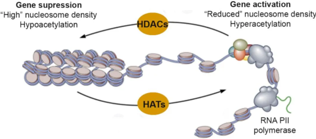
3.2.2. Histone deacetylases (HDACs) and histone acetyltransferases (HATs)…...23
3.2.2.1. Histone deacetylase inhibitors (HDACi) and histone acetyltransferase inhibitors (HATi)………..…24
3.2.2.2. HDACs and HATs enzymes and viral infections………26
4. Objectives……..………28
Chapter II: DNA-binding properties of the African swine fever virus pA104R, a histone-like protein involved in viral replication and transcription………29
2.1. Introduction………..………32
2.2. Material and Methods……….………33
2.2.1. Cloning, expression, and purification of recombinant A104Rwt, A104RR69A, A104RR69K and A104RP74A……….………33
2.2.2. Electrophoretic mobility shift assay (EMSA) ……….………34
2.2.3. Supercoiling assay……….34
2.2.4. Cells and viruses………35
2.2.5. Quantitative reverse transcriptionPCR (qRT-PCR)……..………35
2.2.6. Quantification of ASFV genomesby qPCR……….………36
2.2.7. Antibodies……….………36
2.2.8. Protein extraction and Western blotting………37
2.2.9. Immunofluorescencestudies………..………38
viii
2.3. Results…………...………39
2.3.1. pA104R forms distinct DNA-protein complexes in the presence of oligonucleotides with different length……….………39
2.3.2. pA104R binds both ssDNA and dsDNA in a wide range of temperatures, pH, and salt concentrations and in an ATP-independent manner………40
2.3.3. The arginine-69 residue is required for the DNA-binding activity of pA104R……….42
2.3.4. pA104R cooperates with ASFV-topoisomerase II (pP1192R) to modulate DNA supercoiling………...………43
2.3.5. The A104R gene encodes a late protein that localizes with viral DNA replicationsites………..………44
2.3.6. Knockdown of pA104R reduces viral infection………..………47
2.4. Discussion………48
Chapter III: Generation of a defective infectious single cycle African Swine Fever Virus particle lacking the A104R gene…….………51
3.1. Introduction…..……….53
3.2. Material and methods………...………..55
3.2.1. Cells and viruses……...………..55
3.2.2. Construction of Vero-pA104R cell line..………55
3.2.3. Immunofluorescence studies…………..………56
3.2.4. Protein extraction and western blotting analysis………56
3.2.5. Construction of plasmid transfer vector………57
3.2.6. Generation and purification of recombinant ASFV……….58
3.3. Results…………...………..………..59
3.3.1. A stable Vero cell line expressing ASFV-pA104R was established………..……….59
3.3.2. Vero-pA104R cell line supports ASFV infection although lower titres are obtained ……….…………..………....60
3.3.3. Deletion of the A104R gene from the Ba71V ASFV isolate………..……….……….61
3.4. Discussion…………..………..………62
Chapter IV: Sodium phenylbutyrate abrogates African swine fever virus replication by disrupting the virus-induced hypoacetylation status of histone H3K9/K14…………..65
4.1. Introduction.……….……….…67
4.2. Material and methods…….………….69
4.2.1. Cells and viruses…………..69
4.2.2. Drugs and cytotoxic assay……..…………..69
4.2.3. Drug treatment and viral infection…..……….…69
4.2.4. Antibodies………..……………70
4.2.5. Direct immunofluorescence studies……..………70
4.2.6. Western blotting analysis………..………...70
4.2.7. Data analysis………...……….71
4.3. Results……….…………...71
4.3.1. NaPB inhibits ASFV replication in a dose-dependent manner………71
4.3.2. NaPB does not inactivate extracellular ASFV particles…...………..72
4.3.3. NaPB inhibits the ASFV late protein synthesis and disrupts the virus-induced H3K9/K14 hypoacetylation status……….72
4.3.4. NaPB and enrofloxacin act synergistically to abolish ASFV replication………..…..74
ix
Chapter V: General Discussion, Conclusions and Future Directions………..…77
5.1. The role of pA104R in ASFV infection and modification of histone acetylation during ASFV infection………79
5.2. HDACi as potential drugs for the study of viral-host interaction and for the development of antiviral agents………..………82
5.3. The development of a DISC vaccine targeting pA104R………..…83
5.4. Concluding remarks………85
x LIST OF FIGURES
Figure 1. Dynamic of ASF infection……….7
Figure 2. Structure and protein composition of ASFV particle………..10
Figure 3. Early events duringASFV infection………..12
Figure 4. Accumulation kinetics for immediately early (blue, I215L), early (green, I73R), intermediate (yellow, I226R) and late (red, I329L) transcripts throughout ASFV infection……13
Figure 5. Effects of a gain-function HU mutant on colony morphology and nucleoid compaction………18
Figure 6. Architectural properties of histone-like proteins……….19
Figure 7. The organization of DNA within the chromatinstructure………...…………21
Figure 8. The dynamic state of histone acetylation/ deacetylation regulated by HDACs and HATs………..23
Figure 9. pA104R binds to DNA fragments with different lengths and shows a binding site size of about 14 to 16 nt………...39
Figure 10. pA104R binds to dsDNA with higher affinity than to ssDNA………..41
Figure 11. pA104R exhibits high DNA-binding affinity at a wide range of temperatures and pH values in an ATP-independent manner and is affected by ionic strength………42
Figure 12. Arg69 is needed for the efficient pA104R DNA-binding activity, in contrast to the Pro74 residue……….43
Figure 13. pA104R has DNA supercoiling activity in the presence of ASFV-topoisomerase II (pP1192R)……….43
Figure 14. The A104R gene, encoding for a late protein, is transcribed from 2 hpi………45
Figure 15. pA104R localizes within cell nuclei and cytoplasmic viral factories and is bound to Triton X-100-insoluble components/structures………46
Figure 16. siRNA-A104R_2 reduces the mRNA levels of A104R by 27% at 16 hpi…………...47
Figure 17. A104R mRNA knockdown inhibits ASFV infection………..48
Figure 18. Schematic overview of construction of Vero-pA104R cell line………56
Figure 19. Schematic diagram showing the methodology used to construct the plasmid transfer vector………..58
Figure 20. Schematic diagram representing the generation recombinant ASF viruses expressing GUS reporter gene………..58
Figure 21. ORF A104R is successfully integrated in transfected Vero cell genome…………..59
Figure 22. Complementary Vero cell line is expressing viral pA104R………..60
Figure 23. Isolation of recombinant virus using the plaques method………61
Figure 24. Isolation of recombinant virus using serial dilutions……….62
Figure 25. NaPB inhibits ASFV infection in a dose-dependent manner………..72
Figure 26. NaPB disrupts viral protein synthesis and alters the low acetylation levels of histone H3 at Lys9 and Lys14 induced by ASFV infection………...73
Figure 27. NaPB and enrofloxacin interact synergistically to inhibit ASFV infection…………..74
Figure 28. Proposed working model for the antiviral activity of NaPB on ASFV-infected cells…...83
xi LIST OF TABLES
Table 1. Genes encoding ASFV proteins involved in DNA replication, repair, nucleotide
metabolism, transcription and other enzymatic activities or host defence evasion………15
Table 2. Genes encoding structural proteins and other proteins involved in virus morphogenesis……….15
Table 3. Some histone modification and theirrole intranscription………22
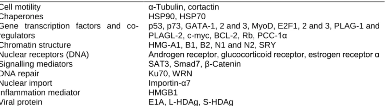
Table 4. Non-histone substrates of HDACs (short list)………...24
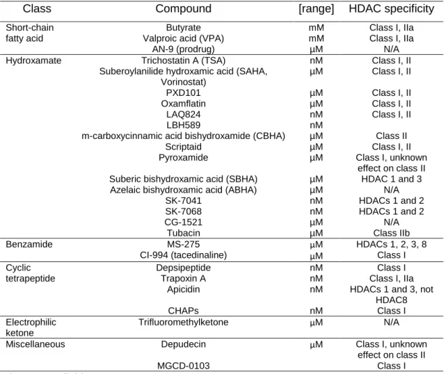
Table 5. Molecular characteristic and HDAC specificity of some HDACi………25
Table 6. Examples of viral proteins and their chromatin-associated targets………..27
Table 7. Primers used in the present study……….36
Table 8. Sequences of siRNAs used to knockdown expression of pA104R in ASFV-infected Vero cells………..38
xii LIST OF ABREVIATIONS
ADP adenosine 5′-diphosphate AraC cytosine arabinoside
ASF African swine fever
ASFV African swine fever virus
ATP adenosine 5′-triphosphate
bp base-pair
BSA bovine serum albumin
CbpA curved DNA binding protein A CbpB curved DNA binding protein B
cDNA complementary DNA
CPE cytopathic effect
DAPI 4′,6-diamidino-2-phenylindole DISC defective infectious single cycle DMEM Dulbecco’s modified Eagle medium DMSO dimethyl sulfoxide
DNA deoxyribonucleic acid
dsDNA double-stranded DNA
ssDNA single-stranded DNA
DPS DNA binding protein from starved cells
EBV Epstein-Barr virus
ECACC European Cell Culture Collection EDTA ethylenediaminetetraacetic acid EGCG (-)-epigallocatechin gallate
EGTA ethylene glycol-bis(2-aminoethylether)-N,N,N′,N′-tetraacetic acid EMSA electrophoretic mobility shift assay
ER endoplasmic reticulum
FBS fetal bovine serum
Fis factor for inversion stimulation FITC fluorescein isothiocyanate
GUS beta-glucuronidase
HBV Hepatitis B virus
hpi hour(s) post-infection
HAT histone acetyltransferases
HATi histone acetyltransferases inhibitors HDAC histone deacetylases
HDACi histone deacetylases inhibitors
HEPES 4-(2-hydroxyethyl)-1-piperazineethanesulfonic acid H-NS histone-like nucleoid structuring protein
HP1 Heterochromatin Protein 1
HRP horseradish peroxidase HSV Herpes simplex virus
HU histone-like protein from E. coli strain U93 H3K4me2 histone H3 dimethylated at lysine 4 H3K9K14 histone H3 lysine 9 and 14
H3K9K14Ac histone H3 lysine 9 and 14 acetylation H3K9me histone H3 methylated at lysine 9 H3K9me3 histone H3 trimethylated at lysine 9 IHF integration host factor
IPTG isopropyl-β-D-1-thiogalactopyranoside
kbp kilo base-pair
kDa kilo Dalton
Lrp leucine responsive protein MOI multiplicity of infection
xiii MTOC microtubule organization center
ncRNA noncoding RNA
NAP nucleo-associate protein NaPB sodium phenylbutyrate
NCLDV nucleo-cytoplasmic large DNA virus
nt nucleotides
OIE Office International des Épizooties – World Animal Organization
ORF open reading frame
PIPES piperazineN,N’-bis(2-ethanesulfonic acid) PBS phosphate-buffered saline
PBST PBS supplemented with 0.1% Tween-20 PCR polymerase chain reaction
PMSF phenylmethylsulphonyl fluoride PTM posttranslational modifications
RNA ribonucleic acid
rpm rotations per minute
RT room temperature
SAHA vorinostat
SDS sodium dodecylsulphate
SDS-PAGE sodium dodecylsulphate-polyacrylamide gel electrophoresis siRNA small interfering RNA
SFM scanning force microscopy
SMC structural maintenance of chromossomes StpA suppressor of the Td’ phenotype
SV40 Simian virus 40
TBE Tris-borate-EDTA
TCID50 tissue culture infectious dose 50%
TSA trichostatin A
Tx Triton X-100
VPA valproic acid
v/v volume per volume
wt wild-type
w/v weight per volume
1
CHAPTER I
3 1. African Swine Fever
1.1. History and distribution
African swine fever (ASF) is nowadays considered one of the most threatening diseases of pig husbandry. First described by Montgomery (1921) as an acute and fatal haemorrhagic fever affecting domestic pigs in Kenya, ASF remains endemic in most sub-Saharan African countries. The first spread of ASF outside Africa was reported in Portugal in 1957, as result of contaminated waste from airline flights which were provided to pigs near the Lisbon airport. Despite the prompt control and eradication of the disease, a further outbreak occurred in 1960 in Lisbon (Manso Ribeiro & Azevedo, 1961), with a subsequent spread to many areas of the Iberian Peninsula, where ASF remained endemic until 1995 (Sánchez-Vizcaíno, Mur, & Martínez-López, 2012). During the 1970s and 1980s ASF affected other countries in Europe, such as France, Italy, Malta, Belgium and Netherlands, as well as several Caribbean countries – Cuba, Dominican Republic and Haiti – and Brazil (Costard et al., 2009).
As consequence of control measures adopted, ASF was eradicated from those territories, with the exception of Sardinia, in Italy, where the disease persists up to date. However, in the African continent, the incidence of ASF increased during the 1990s and 2000s decade in endemic countries and entered in other regions not typically affected by the disease [Côte d’Ivoire (1996), Nigeria (1997), Togo (1997), Madagascar (1998), Ghana (1999), Burkina Faso (2003), Mauritius (2007) and Chad (2010)] (Sánchez-Vizcaíno et al., 2012). The increase of African swine fever virus (ASFV) circulating in Africa, together with the number of infected animals and contaminated products may contributed to the re-emergency of the disease in the European Continent via Georgia, in 2007 (Sánchez-Vizcaíno et al., 2012). The molecular analysis of the ASFV found in Georgia outbreak revealed a close relationship to virus circulating in Mozambique, Zambia and Madagascar (Rowlands et al., 2008), confirming the origin from South Eastern Africa or Madagascar of the disease in the Caucasus region. Since the first clinical cases were disclosed in the area surrounding the port of Poti (Black Sea), the virus was probably introduced through improperly disposed waste products from international ships carrying contaminated pork products, which were then used to feed pigs (Beltran-Alcrudo, Lubroth, Depner, & De La Rocque, 2008). From this initial outbreak the virus spread very quickly to other regions of Georgia, Armenia, Azerbaijan and Russia Federation, increasing the risk of introduction of ASF into the European Union (Sánchez-Vizcaíno, Mur, & Martínez-López, 2013; Gogin, Gerasimov, Malogolovkin, & Kolbasov, 2013). Due to the abundance of backyard pig units and areas of interaction between free-ranging pigs and wild boar, the illegal movement of infected animals and products, the lack of biosecurity measures and poor implementation of control measures, ASF is maintained in these regions and continued to spread into Ukraine (2012) and Belarus (2013) (Gallardo et al., 2015).
4
The first introduction of ASF in the EU was registered in Lithuania (2014), and rapidly further notifications were occurred in Poland, Latvia, Estonia, Czech Republic, Moldova and Romania (Gallardo et al., 2015; OIE, 2017). The recently re-introduction of ASF into EU area emphasizes the actual threat of this disease to the global pig husbandry.
1.2. Epidemiology of African swine fever 1.2.1. African swine fever hosts
1.2.1.1. Domestic pigs
The clinical and pathological signs of ASF in domestic pigs vary considerably depending on the virulence of the ASFV strain and host factors like its immunological status (Costard,
Mur,
Lubroth, Sanchez-Vizcaino, & Pfeiffer
, 2013). Acute infections with highly virulent virus strains are characterized by a massive apoptosis of lymphocyte and a haemorrhagic fever with an impairment of haemostasis and immune functions, and are associated with high morbidity and mortality rates (Blome, Gabriel, & Beer, 2013; Sánchez-Vizcaíno,Mur,
Gomez-Villamandos, & Carrasco
, 2015). Subclinically infected, chronically infected and recovered pigs are likely to play an important role in the ASF epidemiology, being responsible for the persistence of the disease in endemic areas, sporadic outbreaks and introduction into disease-free zones (Allaway, Chinombo, Edelsten, Hutchings, & Sumption, 1995; Gallardo et al., 2015).1.2.1.2. Wild suids
The warthog (Phacochoerus africanus) is considered the original vertebrate host and the most important reservoir of ASFV in Africa (Jori & Bastos, 2009; Costard et al., 2013). Infected adult warthogs are asymptomatic carriers of the disease, however viral replication and viremia occur in young animals which are infected by soft ticks carrying the virus, present in their burrows (Thomson, Gainaru, & Dellen, 1980).
The role of bushpigs (Potamochoerus larvatus), red river hogs (Potamochoerus porcus), and giant forest hogs (Hylochoerus meinertzhageni) in the ASF epidemiology remains not fully elucidated (Costard et al., 2009; Jori & Bastos, 2009). Although the bushpigs are susceptible to infection and develop sufficient levels of viremia to infect pigs and soft ticks, they do not show any clinical signs (Anderson, Hutchings, Mukarati, & Wilkinson, 1998; Oura, Powell, Anderson, & Parkhouse, 1998). Since these animals rarely interact to domestic pigs and soft ticks, due to the low densities of their population, their nocturnal habits and non-use of burrows, bushpigs are not considered important reservoirs of ASFV (Costard et al., 2013).
In Europe, wild boar (Sus scrofa) and feral pigs are highly susceptible to both natural and experimental ASFV infection (McVicar, Mebus, Becker, Belden, & Gibbs, 1981; Jori & Bastos, 2009) and, similarly to domestic pigs, the clinical signs of disease may vary considerably from peracute death to unapparent courses (Blome et al., 2013). Although, some studies revealed that the virus cannot persist in wild boar populations for long periods of time without the
re-5
infection by contact with infected domestic pigs and contaminated products (Laddomada et al., 1994; Mur, Martínez-López, & Sánchez-Vizcaíno, 2012), these animals facilitate the spread of the disease to free-range pigs (Costard et al., 2013; Gallardo et al., 2015).
1.2.1.3. Soft ticks
ASFV infected Ornithodoros spp. ticks, may harbor high levels of virus and a persistent infection (Boinas, Wilson, Hutchings, Martins, & Dixon, 2011; Burrage, 2013). These infected soft ticks may play an important role on ASF epidemiology, acting as vectors of the disease both for domestic and wild pigs and also being a long-term reservoir of the disease, as was suggested in Portugal when, in 1999, ASF re-emerged on a farm that had been affected previously and where infected ticks were found (
Beloin
et al., 2006; Costard et al., 2009; Boinas et al., 2011).1.2.2. Transmission
ASFV is transmitted by direct contact between infected and non-infected pigs (through blood, secretions and excretions), by consumption of meat and pork products from infected pigs, by bites of infected soft ticks, and through materials contaminated by blood, faeces, urine or saliva from infected animals (Guinat et al., 2016; Penrith & Vosloo, 2009). Infection of susceptible pigs normally occurs either orally or nasally, via tonsils or dorsal pharyngeal mucosa to the mandibular or retropharyngeal lymph nodes, from where the virus spread through viremia (Sánchez-Vizcaíno et al., 2009).
The virus is very resistant to inactivation and can persist and remain viable in the environment for long periods, thus enabling the transmission either by direct contact or via fomites (e.g. contaminated clothes and footwear, vehicles, equipment, bedding) for several weeks and turning the ASF control difficult (Costard et al., 2013; Davies et al., 2015).
In the sylvatic transmission cycle of ASF in Africa, which involves warthogs and ticks of the Ornithodoros moubata complex, there is not horizontal or vertical transmission between warthogs, thus the maintenance of infection is exclusively dependent on infected ticks (Jori & Bastos, 2009). This vector transmits the virus to young suckling warthogs, which have a short period of viremia, allowing the transmission to uninfected ticks during blood meals (Thomson et al., 1980). Since the transmission by direct contact seems improbable in this scenario, the transmission from sylvatic to domestic pigs, is attributed to the argasid ticks (Jori & Bastos, 2009).
Although, several studies indicate that Ornithodoros ticks can maintain ASFV infection for several months or even years and were responsible for ASF outbreaks in Iberian Peninsula (Basto et al., 2006; Boinas et al., 2011; Pérez-Sánchez, Astigarraga, Oleaga-Pérez, & Encinas-Grandes, 1994; Sanchez-Botija, 1963), their involvement in the current spread of the disease in the Caucasus, Russia and Eastern Europe is unlikely (Jori & Bastos, 2009; Guinat
6
et al., 2016). In this scenario, infected wild boars are an effective transmission route to domestic pigs by among others, direct contact and through ingestion of infected carcasses, being currently the greatest concern in the control of ASF nowadays in EU (Gabriel et al., 2011; Gallardo et al., 2015).
1.3. Strategies to control African swine fever 1.3.1. Sanitary/ biosecurity control measures
ASF is a viral swine disease, entailing high mortality rates and great sanitary and socioeconomic impact in affected countries, and it is listed as a notifiable disease by the World Organization for Animal Health (OIE) (Costard et al., 2013). Allied to its complex epidemiology and transmission and to a marked resistance of the ASFV in contaminated material and animal products (Sánchez-Vizcaíno et al., 2012), ASF control and eradication is complex and difficult due to the absence of a vaccine or an effective treatment against this disease. Thus, its control is based on the early detection and on an efficient laboratory diagnosis of the disease and the implementation of sanitary and biosecurity control measures, namely animal slaughter, movement restrictions, notification and surveillance (Wieland, Dhollander, Salman, & Koenen, 2011). The clinical diagnosis of the ASF is often complicated due to the similarity to other swine diseases and to the wide range of clinical forms of the disease, ranging from highly lethal to subclinical (Fig. 1). Highly virulent strains are usually responsible for the peracute and acute forms of the disease, characterised by high mortality rates after 1-4 and 4-9 days post-infection, respectively. These acute forms are characterised by a febrile syndrome with erythema and cyanosis of the skin, functional failures of internal organs, vomiting and haemorrhagic diarrhoea. In subacute forms of the disease, a persistent or fluctuating fever lasts for up to 20 days and the mortality rates are in the range of 30-70%, while in chronic forms clinical signs and lesions are not specific (delayed growth, emaciation, skin ulcers, arthritis, pneumonia, abortion) and could persist for several months (Blome et al., 2013; Gallardo et al., 2015; Sánchez-Vizcaíno et al., 2015).
7
Figure 1. Dynamic of ASF infection.
The picture summarizes the ASFV appearance in blood and antibodies after infection. The lethality of the different forms of the clinical disease, which ranges from acute to a subacute, as well as from recovered animals are also shown. Figure and legend were adapted from Gallardo et al. (2015).
1.3.2. Development of ASF vaccine
Despite all the efforts to develop an ASF vaccine, no vaccine is currently available to prevent and control the global spread of ASF. The complexity of the virus, with genes involved in the evasion of the host immune response, the variation extent of ASFV strains and the large gaps in knowledge concerning ASFV infection and immunity, contribute to the unsuccessful of all classical attempts to generate a vaccine using inactivated or live-attenuated virus (Rock, 2016). Experiments using traditional inactivated vaccines, such as inactivated infected cell extracts, supernatants of infected pig peripheral blood leukocytes, purified and inactivated virions, infected glutaraldehyde-fixed macrophages, and detergent-treated infected alveolar macrophage cell cultures, have failed to induce protection (Forman, Wardley, & Wilkinson, 1982; Mebus, 1988; Stone & Hess, 1967). Recently, Blome, Gabriel, & Beer (2014) also observed that adjuvants do not enhance the efficacy of these classical vaccines.
Nevertheless, studies showed that a solid immune response is induced in surviving pigs (Boinas, Hutchings, Dixon, & Wilkinson, 2004; Hamdy & Dardiri, 1984; Mebus & Dardiri, 1980) and animals infected with ASFV attenuated or moderately virulent develop protection against homologous virus (King et al., 2011; Lacasta et al., 2015; Leitão et al., 2001; Lewis et al., 2000; O’Donnell et al., 2015; Reis et al., 2016), suggesting that an effective vaccine against ASFV infection could indeed be feasible.
Unfortunately, significant safety issues (residual pathogenicity and potential for long-term viral persistence) were associated until now with live-attenuated vaccines (King et al., 2011; Lacasta et al., 2015; Lewis et al., 2000; Manso Ribeiro & Azevedo, 1961). Concerning ASF
8
subunit vaccines, although several ASFV proteins have been associated with protection (Lokhandwala et al., 2016; Lokhandwala et al., 2017), the available data shows that these vaccines are insufficient for the induction of a protective immunity in pigs (Argilaguet et al., 2013; Neilan et al., 2004; Rock, 2016).
1.3.3. Development of antiviral agents
In the absence of an effective vaccine against ASF, several antiviral agents have been identified. Although, some compounds have demonstrated strong anti-ASFV activity in vitro (e.g. rifamycin, fluoroquinolones, lauryl-gallate, methyl-b-Cyclodextrin, amiloride, sulfated polysaccharides, lysosomotropic agents, valproic acid, apigenin) (Cuesta-Geijo et al., 2012; Freitas, Frouco, Martins, Leitão, & Ferreira, 2016; García-Villalón & Gil-Fernández, 1991; Hakobyan et al., 2016; Hernaez & Alonso, 2010; Hurtado et al., 2008; Mottola et al., 2013; Salas, Kuznar, & Viñuela, 1983; Sánchez et al., 2012), in vivo studies are mandatory to reveal if they will be potential tools for the treatment of ASFV infection.
1.4. Classification and morphology of African swine fever virus 1.4.1. Aetiology
African swine fever is caused by African swine fever virus, a large, enveloped virus with an icosahedral morphology and a double-stranded DNA genome that ranges in length between isolates about 170 to 193 kbp (Tulman, Delhon, Ku, & Rock, 2009). ASFV encodes for between 151 and 167 open reading frames (ORFs) and is the only member of the Asfarviridae family (Dixon et al., 2012), sharing general features with other members of the nucleocytoplasmic large DNA virus (NCLDV) superfamily, or as proposed recently, members of the Megavirales order (Colson et al., 2013).
1.4.2. Structure and composition of ASFV particles
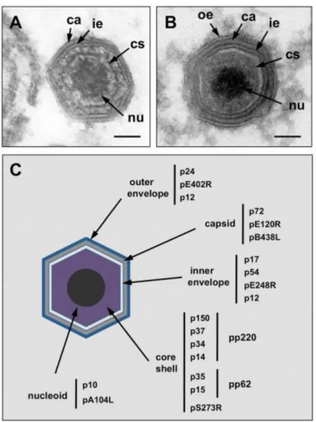
The ASFV virion has an icosahedral morphology with an average diameter of 200 nm and is comprised by more than 50 proteins with molecular weights ranging from 10 to 150 kDa (Esteves, Marques, & Costa, 1986; Salas & Andrés, 2013). The virions also contain enzymes and factors needed for early mRNA transcription and processing (Dixon, Chapman, Netherton, & Upton, 2013; Salas, Rey-Campos, Almendral, Talavera, & Viñuela, 1986).The viral particles have a complex multi-layered structure composed by a nucleoid surrounded by a thick protein layer designated core shell, an inner lipid envelope and the capsid. The external virions also contain an external membrane, the outer envelope (Fig. 2).
The external envelope is acquired by budding from the plasma membrane of the host cells (Breese & DeBoer, 1966). The virus attachment viral protein p12, the virus homologue of cellular CD2 (pE402R) and a cellular protein p24, which is present at the plasma membrane
9
of host cells, have been reported to be localized into this membrane (Sanz, Garcia-Barreno, Nogal, Vinuela, & Enjuanes, 1985; Carrascosa, Saastre, Gonzalez, & Viñuela, 1993).
The viral capsid, the outermost layer of the intracellular virions, is formed by about 2000 capsomers with the appearance of hexagonal prisms and is constituted mainly by the p72, encoded by the gene B646L. Another structural protein, pB438L, and the protein pE120R, which is involved in the transport of the viral particles from the factory to the plasma membrane, are other components of the capsid (Andrés, García-Escudero, Viñuela, Salas, & Rodríguez, 2001b; Epifano, Krijnse-Locker, Salas, Salas, & Rodríguez, 2006).
The inner envelope is derived from the endoplasmic reticulum (Rouiller, Brookes, Hyatt, Windsor, & Wileman, 1998) and is composed by the membrane proteins p54, p17 and pE248R (Rodríguez, García-Escudero, Salas, & Andrés, 2004; Rodríguez, Nogal, Redrejo-Rodríguez, Bustos, & Salas, 2009; Suárez, Gutiérrez-Berzal, Andrés, Salas, & Rodríguez, 2010). This membrane surrounds the core shell, a thick protein layer which domain is mainly constituted by the processing products of polyproteins pp220 and pp62, and also by pS273R (Andrés, Alejo, Simón-Mateo, & Salas, 2001a; Andrés, Alejo, Salas, & Salas, 2002).
The nucleoid is a structure of 80 nm and contains the viral genome and nucleoproteins such as the DNA-binding protein p10 and the histone-like protein pA104R (Munoz, Freije, Salas, Vinuela, & Lopez-Otin, 1993; Borca et al., 1996). This structure also comprises the transcriptional machinery for the synthesis and modification of early RNAs (Salas & Andrés, 2013).
10
Figure 2. Structure and protein composition of ASFV particle.
(A) Electron micrograph of an intracellular full ASFV particle and (B) of an extracellular mature ASF virion. (C) Illustration of the localization of ASFV structural proteins. Outer envelope (oe), capsid (ca), inner envelope (ie), core shell (cs) and nucleoid (nu). Figure and legend were adapted from Salas and Andrés (2013).
1.5. The ASFV infectious cycle
The natural target of ASFV are cells of the mononuclear phagocytic system, and its infectious cycle normally takes between 18 to 24 h post-infection (hpi), (Costa, 1990; Muñoz-Moreno, Galindo, Cuesta-Geijo, Barrado-Gil, & Alonso, 2015). A successful infection by ASFV, as by other viruses, is a process that consists of successive steps, including virus binding, internalization and uncoating, early transcription-translation, genome replication, late protein synthesis, virus particle morphogenesis and viral egress.
11 1.5.1. Entry of ASFV into the host cell
Different mechanisms have been proposed for viral adsorption and entry into the host cell: phagocytosis (Basta, Gerber, Schaub, Summerfield, & McCullough, 2010), macropinocytosis (Sánchez et al., 2012) and receptor-mediated endocytosis (Alcamí, Carrascosa, & Viñuela, 1989; Galindo et al., 2015; Hernaez & Alonso, 2010) (Fig. 3). Recently, two studies were developed and may have provided new insights about this topic. While Galindo et al. (2015) defend that the virus enters into macrophages by a classical clathrin- or lipid rafts/caveolae-mediated endocytosis, with special requirements for cholesterol, phosphoinositide-3-kinase (PI3K), actin dynamics and related signalling, Hernaez, Guerra, Salas, & Andrés (2016) showed that ASFV is internalized by both constitutive macropinocytosis and clathrin-mediated endocytosis (Fig. 3).
Concerning the receptor-mediated internalization, although the receptors for the virus remain unknown, the CD163 expression on the surface of swine macrophage has been correlated with susceptibility to ASFV infection (Sánchez-Torres et al., 2003). Additionally, several viral proteins such as p30, p12 and p54 bind to cell surface and are important for viral attachment and internalization (Angulo, Viñuela, & Alcamí, 1993; Gómez-Puertas et al., 1998).
Once inside the cell, ASFV particles move from early endosomes or macropinosomes to late, multivesicular endosomes where the virus becomes uncoated (Valdeira, Bernardes, Cruz, & Geraldes, 1998; Alonso et al., 2013). This gradual maturation of endosomes involves changes in their relative cytoplasmic position closer to the nucleus as well as their luminal environment. The intraluminal acidification of the late endosomes is required for a desencapsidation of the virions, a necessary step for uncoating and for a successful ASFV infection (Cuesta-Geijo et al., 2012). Upon virus uncoating, which involves the disruption of the outer membrane and the protein capsid, the inner membrane becomes exposed and fuses with the endosomal membrane, allowing the viral core egress into the cytosol to initiate replication (Hernaez et al., 2016) (Fig. 3).
ASFV virion reaches the replication site in the perinuclear area, close to the microtubule organization center (MTOC), through microtubules. In fact, the movement of ASFV particles relies on microtubules, and some studies showed a high affinity interaction between ASFV-p54 and the microtubule motor protein dynein, suggesting that this interplay facilitates the virus trafficking (Alonso et al., 2001; Hernaez, Escribano, & Alonso, 2006).
12
Figure 3. Early events during ASFV infection: ASFV enters cells using macropinocytosis (A) or clathrin
coated pits (B). Virus can also enter certain cells attached to phagocytosed red blood cells (C). Post-entry ASFV enters the endosomal–lysosomal system (D) from which it exits by fusing with the membrane and in doing so loses its external envelope (E). Virions are directed to perinuclear regions by microtubules through the interaction between dynein motors and the structural protein p54 (F). Viral matrix proteins and viral DNA enters the nucleus to initiate viral replication and co-incident with this is the phosphorylation and disassembly of nuclear lamins (G). Early viral gene expression and the initial stage of factory formation begins including the recruitment of vimentin and nuclear proteins, and presumably the movement of viral DNA to replication sites (H). Figure and legend were adapted from Netherton and Wileman (2013).
13 1.5.2. ASFV gene expression and DNA replication
After internalization, viral gene transcription is initiated, using enzymes packaged in the virion core (Salas, Rey-Campos, Almendral, Talavera, & Viñuela, 1986; Dixon et al., 2013). The transcription factors encoded by ASFV recognize a short sequence containing the viral promoter, localized upstream of each gene, that are specific for the different viral stages. ASFV genes expression are indeed strongly temporal regulated, being classified as immediately early, early, intermediate and late genes (Rodríguez & Salas, 2013) (Fig. 4).
Figure 4. Accumulation kinetics for immediately early (blue, I215L), early (green, I73R), intermediate
(yellow, I226R) and late (red, I329L) transcripts throughout ASFV infection. Primer extension assays were used to detect and measure steady state RNA levels for the different transcripts. The quantity of each transcript is plotted as the percentage of the maximum level. Figure and legend were adapted from Rodríguez and Salas (2013).
Immediately early and early genes are expressed prior to DNA replication, however it has been shown that while immediately early genes are repressed before the onset of DNA replication, the expression of early genes is detectable as early as 2 hpi, with a plateau of accumulation at 2-6 hpi, and can be detectable at late times of infection. ASFV early genes encode for enzymes involved in the nucleotide metabolism, DNA replication, regulation of host cell responses to infection, and for transcription factors that are necessary for intermediate and late gene expression (Almazán et al., 1992; Rodriguez, Salas, & Viñuela, 1992; Rodríguez & Salas, 2013).
On the other hand, the expression of intermediate and late ASFV genes are dependent on the viral DNA replication, and their expression is not detected when cells are infected in the presence of inhibitors of DNA replication, like cytosine arabinoside (AraC) (Rodríguez & Salas, 2013). Intermediate transcripts are first detected at 4-6 hpi, coincident with maximum expression of early genes, reaching a maximum level of accumulation at 6-8 hpi, and sharply
14
decreased at later times. The beginning of late gene expression is coincident with the maximum levels of intermediate mRNAs, reaching a maximum at 12-16 hpi and decreasing slowly thereafter. These two classes of genes codify for structural proteins of the virion, as well as polymerases and early transcription factors, required for the expression of the early genes, that will be packaged into the new virions (Rodríguez, Salas, & Viñuela, 1996; Rodríguez & Salas, 2013; Muñoz-Moreno et al., 2015).
ASFV DNA replication starts with a brief replication phase in the host cell nucleus, following a second replication phase in the cytoplasmic viral factories with a maximum peak at 8 hpi (García-Beato, Salas, Viñuela, & Salas, 1992a; Simões, Martins, & Ferreira, 2015b). Although the intranuclear phase of ASFV infection is still poorly understood, the early intranuclear replication disrupts nuclear structures and modifies the landscape of the host cell nucleus (Ballester et al., 2011; Simões et al., 2015b; Simões, Rino, Pinheiro, Martins, & Ferreira, 2015a). These events provide an environment that favours ASFV replication and may represent a mechanism by which ASFV controls cellular transcription and facilitate viral DNA replication.
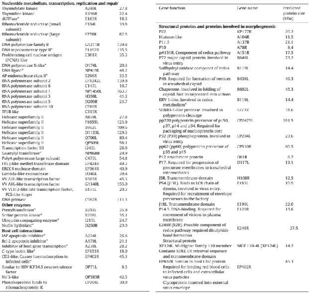
The ASFV genome encodes enzymes required for transcription and replication, and virion structural proteins. The known functions of ASFV viral proteins were reviewed in Dixon et al. (2013) and are shown in table 1 and 2.
15
Table 1. Genes encoding ASFV proteins
involved in DNA replication, repair, nucleotide metabolism, transcription and other enzymatic activities or host defence evasion. The gene nomenclature is shown in the central column and predicted molecular weight in the Benin 97/1 genome is shown on the right. Asterisks (*) indicate those for which functional data is available. Table and legend were adapted from Dixon et al. (2013).
Table 2. Genes encoding structural proteins
and other proteins involved in virus morphogenesis. Genes which encode known virus structural proteins and other proteins involved in virus morphogenesis are indicated. The gene name is indicated in the central column and predicted molecular weight in the right column. Table and legend were adapted from Dixon et al. (2013).
16 1.5.3. ASFV morphogenesis
The assembly of the ASFV particles takes place in viral factories, located in cellular cytoplasmic areas close to the nucleus and MTOC, which resemble aggresomes that are formed in response to misfolded proteins or even as antiviral defence (Wileman, 2007). These areas are enwrapped in a vimentin cage and surrounded by ER membranes (Andrés, García-Escudero, Simón-Mateo, & Viñuela, 1998) being mitochondria also recruited to their periphery during infection (Rojo et al., 1998).
The steps of ASFV morphogenesis include the formation of the inner envelope, the progressive formation of the capsid on the convex face of the virus inner envelope, the assembly of the core shell on the concave side of the envelope, and the formation of the nucleoid. Concerning the formation of the ASFV nucleoid, although the exact mechanism of this last step in morphogenesis is still unknown, some models suggest that the viral DNA is encapsidated, possibly together with ASFV nucleoproteins p10 and pA104R, and then condensed inside the assembling virus particles (Salas & Andrés, 2013).
1.5.4. Virion egress
Following mature virus particles assembly, intracellular virus are transported from viral factories to the cell surface through cellular microtubules, depending on the conventional kinesin and on the capsid protein ASFV-pE120R (Andrés et al., 2001b; Jouvenet, Monaghan, Way, & Wileman, 2004). Once at the cell surface, ASFV particles are released by budding to yield extracellular enveloped virions (Breese & DeBoer, 1966). On the other hand, cell lysis observed at late times of infection might represent an alternative mechanism of viral egress. ASFV virions can also induce unbranched actin projections that originate from the cellular plasma membrane, which may facilitate cell-cell viral spread (Jouvenet et al., 2006).
2. ASFV histone-like protein
2.1. Overview of the histone-like proteins
ASFV genome codes for a putative histone-like protein (pA104R), that shares a sequence identity of 25 to 30% with two families of bacterial histone-like proteins (HU and IHF), being the only histone-like protein encoded by a eukaryotic virus (Borca et al., 1996; Neilan et al., 1993). Histone-like proteins are small proteins present in bacteria, possessing superficial similarities with eukaryotic histone proteins (basicity, abundance, DNA binding properties and low molecular weight) (Luijsterburg, Noom, Wuite, & Dame, 2006). These proteins, also referred as nucleoid-associate proteins (NAPs), are associated with the bacterial DNA, compacting this structure to fit inside the bacterial cell and increasing the nucleoid stability (Pettijohn & Pfenninger, 1980). These DNA-binding proteins are thought to act as architectural components within the nucleoid and to modulate gene expression.
17
2.2. Biological function of bacterial histone-like proteins
Contrasting to eukaryotic organisms, where DNA in the nucleus is compacted by histones which organize DNA in a hierarchical process into the higher order structure that is chromatin, the bacterial chromosome is condensed to a form called the nucleoid, but does not possess any higher-order chromosomal organization and represents a comparatively open structure, accessible to DNA-binding proteins, RNA and DNA polymerases throughout the cell cycle (Hobot et al., 1985; Robinow & Kellenberger, 1994). In the case of Escherichia coli, 4.6 Mb of DNA with a contour length of approximately 1.6 mm must be contained and compacted within a cell that measures about 4 µm long and 1 µm wide (Dame, 2005). The packaging of DNA in bacteria is a complex process involving several different mechanisms including cellular confinement, macromolecular crowding, DNA supercoiling by DNA gyrase activity and histone-like protein interactions (Dame, 2005; De Vries, 2010).
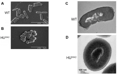
Histone-like proteins play an important role in the nucleoid compaction and in the control of its structure. Bacteria mutants lacking functional histone-like proteins have larger decondensed nucleoids (Graumann, 2001; Kano & Imamoto, 1990; Paull, Haykinson, & Johnson, 1993), whereas gain-function mutants result in severe nucleoid compaction and altered bacterial morphology (Fig. 5) (Kar, Edgar, & Adhya, 2005; Macvanin & Adhya, 2012; Spurio et al., 1992). Still the exact mechanism and the contribution of each nucleoid-associate protein to global nucleoid compaction is unclear (Dame & Tark-Dame, 2016).
Furthermore, histone-like proteins are known to perform topological modification of the chromosome (twisting, bending and folding), to regulate the function of promoters of individual operons and control the expression of many genes, affecting bacterial transcription on a global scale (Browning, Grainger, & Busby, 2010). Genes essential for cell viability, involved in bacterial virulence whose expression varies in response to different environmental stimuli, such as changes in temperature, pH, osmolarity, are genes reported to being affected by histone-like proteins (Berger et al., 2010; Blot, Mavathur, Geertz, Travers, & Muskhelishvili, 2006; Kar et al., 2005; Lang et al., 2007; McLeod & Johnson, 2001; Oberto, Nabti, Jooste, Mignot, & Rouviere-Yaniv, 2009).
Although the architectural properties of many histone-like proteins are well characterized in vitro, it has been difficult to investigate their roles in vivo due to functional, as well as the pleiotropic effect of deletion or overexpression of many of these proteins.
18
Figure 5. Effects of a gain-function HU mutant on colony morphology and nucleoid compaction.
Scanning electron microscopic pictures of wild-type E. coli cells (A) and mutant HU3842 (B). Thin section electron micrographs of wild-type E. coli cells (C) and mutant HU3842 (D). Figures and legend were adapted from Kar et al. (2005) and Macvanin & Adhya (2012).
2.3. Histone-like proteins classification
The major histone-like proteins have different DNA-binding properties and can be divided into two categories, those that have DNA sequence specificity and bind to specific locations of the bacterial chromosome, such as IHF (integration host factor), Lrp (leucine responsive protein), SMC (structural maintenance of chromosomes ), CbpA (curved DNA binding protein A), CbpB (curved DNA binding protein B); and those that do not recognise a particular DNA sequence and are located relatively uniformly throughout the nucleoid, as HU (histone-like protein from E. coli strain U93), H-NS (histone-like nucleoid structuring protein), StpA (suppressor of the Td' phenotype), Fis (factor for inversion stimulation) and Dps (DNA binding protein from starved cells) (Azam, Hiraga, & Ishihama, 2000; Prosseda et al., 2002). Interestingly, the intracellular concentration of these proteins varies in a growth phase dependent manner (Dorman, 2013).
The bacterial histone-like proteins can also be categorized according to their structural effect on DNA. Some NAPs (H-NS, SMC, Lrp) are DNA bridging proteins that are likely to play a role in DNA loops formation and stabilization by creating patches of bridged DNA segments along the DNA loop, while others (HU, IHF, Fis) have DNA bending properties and affect the arrangement of the nucleoid in loops (Dame, 2005; Luijsterburg et al., 2006) (Fig. 6). Dps, which is the predominant histone-like protein in cell stationary phase, also forms crystal-like structures with DNA, transforming the nucleoid into a transcriptionally inactive structure that is effectively protected against damage (Frenkiel-Krispin et al., 2004).
19
Figure 6. Architectural properties of histone-like proteins.
DNA bridging proteins (A-F): (A) Scanning force microscopy (SFM) image of a DNA loop formed by H-NS as consequence of DNA duplex bridging. (B) Low resolution model for DNA duplex bridging mediated by H-NS. (C) SFM image of Bacillus subtilis SMC that forms a rosette-like structure presumably by joining the head domains (D) Low resolution models for DNA duplex linking due to gathering or trapping by SMC. (E) EM image of the Bacillus subtilis LrpC–DNA complex (L) Low resolution model for DNA duplex bridging and wrapping mediated by Lrp. DNA bending proteins: (G) SFM image of an IHF–DNA complex (H) SFM image of HU–DNA complexes, when the presence of low concentrations of HU. (I) Low resolution model for DNA compaction by the binding of multiple HU/IHF molecules. (J) SFM image of Fis–DNA complexes with Fis bound at the nodes of supercoiled pUC18 plasmids. (k) Low resolution model for node formation due to Fis–Fis interactions. Alternative mechanisms of organization and compaction: (L) SFM image of Dps–DNA complexes. (M) Low resolution model of three-dimensional hexagonal Dps–DNA arrays. Figures and legend were adapted from Beloin et al. (2003); Ceci et al. (2004); Dame et al. (2005); Frenkiel-Krispin et al. (2004); Luijsterburg et al. (2006); Mascarenhas et al. (2005); Schneider et al. (2001); van Noort, Verbrugge, Goosen, Dekker, & Dame (2004).
20
3. Viral infections and cellular epigenetic remodelling
Viruses have co-evolved and share a unique relationship with their hosts, developing numerous mechanisms to evade host’s natural defences. Studies on the role of host cellular chromatin in the regulation of viral infection have uncovered the importance of viral-host chromatin interactions in the establishment of viral infection (Adhya & Basu, 2010; Knipe et al., 2013). Moreover, viral proteins by interacting with chromatin remodelling factors elicit various epigenetic changes in host cells in order to control gene expression and host innate immune antiviral defence processes, thereby promoting robust virus replication and pathogenesis. HIV, Epstein-Barr virus (EBV), hepatitis B virus (HBV), human papillomavirus, simian virus 40 (SV40), bovine leukemia virus (BLV), herpes simplex virus (HSV) and dengue virus are some currently known viruses that exhibit epigenetic immune evasion mechanism to survive and propagate in their host (Adhya & Basu, 2010).
3.1. Chromatin organization and structure 3.1.1. Histones and nucleosome structure
In eukaryotic cells, genomic DNA is packaged by histones into chromatin to fit inside the nucleus. The fundamental unit of chromatin is the nucleosome, consisting of 146 base pairs (bp) of DNA wrapped around a histone octamer containing two copies each of the histones H2A, H2B, H3 and H4 (Luger, Mäder, Richmond, Sargent, & Richmond, 1997). Linker histone (H1/H5) binding organizes an additional 20 bp of DNA to complete the nucleosome containing ~167 bp of DNA and it is responsible for forming the chromatosome structure (Thoma, Koller, & Klug, 1979; Woodcock, Skoultchi, & Fan, 2006). Each nucleosome is linked to the next by small segments of linker DNA, representing a primary packing structure. Further condensation is achieved through the formation of the more compact and repressive 30-nm chromatin fibre (secondary structure) and several more levels of higher-order chromatin organization (Jansen & Verstrepen, 2011; Luger, Dechassa, & Tremethick, 2012; Szerlong & Hansen, 2011) (Fig. 7).