Faculdade de Ciências
Departamento de Biologia Animal
Systematic comparison of the effects of α-synuclein
mutations on aggregation in Parkinson’s Diseases
Diana Fernandes Lázaro
Dissertação Mestrado em Biologia Humana e Ambiente
2012
Faculdade de Ciências
Departamento de Biologia Animal
Systematic comparison of the effects of α-synuclein
mutations on aggregation in Parkinson’s Diseases
Diana Fernandes Lázaro
Dissertação Mestrado em Biologia Humana e Ambiente
2012
Dissertation supervised by
Prof. Doutor Tiago Outeiro, PhD
Prof. Doutora Deodália Dias, PhD
Todas as afirmações efetuadas no presente documento são de exclusiva responsabilidade do seu autor, não cabendo qualquer responsabilidade à Faculdade de Ciências da Universidade de Lisboa pelos conteúdos nele apresentados.
Acknowledgements
I would like to start by thanking all my colleagues in the Outeiro lab, especially those who I worked with more closely.
To my supervisor Professor Tiago Outeiro who gave me this wonderful opportunity to begin making my dream of being a scientist come true. I am thankful for his support on the tough days, for his encouragement all along the way, for his patience with my PCR/mutations crisis and for those long discussions in his office. Thank you so much for your constant good mood in the lab, which really helps to relieve some stress.
To Ellen Gerhardt, I want to thank you for your help in the beginning of my work. I am appreciative for all of the molecular biology training and for helping with the analysis of my sequencing results.
To my dear Patricia Guerreiro, for the constant help. For always being there and for always being willing to help me. You were more than a colleague in the lab, you were the person who always provided the practical solution, the easy/simple way to get things accomplished, the advice… I have to thank you a lot for the minutes and hours that I took away from your work. Words cannot express how much I am in debt to you.
To Pauline Wales, for her support in those crazy days in the lab. I will never forget the long conversations that we had when things were not easy in the beginning. Thank you for supporting me and encouragement me on my work. I really have to say that I’m so lucky to working with you on your project.
I must also thank Sonja Reisenauer for helping me with adapting to my new life. I also want to thank Eva Rodrigues for letting me stay the first days in Göttingen in her house, and for sharing some of the challenges we faced during our work.
I would also like to address some words to my family and my closest friends, which I will write in Portuguese.
Queria agradecer a minha mana de coração, Inês Monteiro pelo companheirismo, amizade e partilha. Por teres estado sempre presente nos momentos em que mais precisei.
Aos meus dois grandes amigos e companheiro de guerra Vanessa Duarte e Gonçalo Ramos. Obrigado por todos os momentos de diversão e desatino e pela vossa amizade incondicional.
À minha querida e doce Ritinha Drago e à minha Cynthia Barroco pelo fantástico ano que passamos juntas, por entre tantos trabalhos e apresentações, partilhas e desabafos.
Ao meu amigo de longa data Júlio Barroco, pela força e inspiração que sempre me deste, por dizeres aquilo que precisava de ouvir e não aquilo que queria ouvir.
E por último, mas os mais importantes na minha lista de agradecimento, a minha família, Minha Mãe, Estela Lázaro, o meu Pai, Manuel Lázaro e o meu irmão Filipe Lázaro. A vocês quero vos agradecer do fundo do meu coração toda a força que me têm dado ao longe deste ano. Quero-vos agradecer esta oportunidade ímpar na vida que me concederam, acreditem que tenho dado o máximo todos os dias para poder alcançar os meus sonhos. As palavras serão sempre escassas e limitadas para expressar o laço que nos une.
A minha avó, pela inspiração, garra e coragem que sempre demostrou e demonstra diariamente. Obrigado por todo o carinho, amizade e ensinamento. E por último ao meu avô, que luta diariamente contra esta doença debilitante mas que por breves momentos reconheço o meu avô do antigamente… Quero que saibas que me inspiras a lutar ainda mais, a alcançar o melhor de mim para ajudar tantas pessoas na tua situação.
Obrigado a todos que ao longo da minha vida me tem inspirado, confortado e ajudado nas mais diversas situações.
Table of Contents
Table of Contents ...1 Sumário ...3 Abstract ...4 List of abbreviations ...5 Index of Figures ...7 Index of Tables...8 1. Introduction ...91.1. Protein Misfolding Diseases ...9
1.2. Quality control mechanisms ... 10
1.3. Synucleinopathies ... 12 1.4. Parkinson’s disease ... 12 1.5. Etiology of PD ... 14 1.5.1. Sporadic forms of PD ... 14 1.5.2. Familial forms of PD ... 15 1.5.2.1. Autosomal dominant PD... 16 1.5.2.2. Autosomal recessive PD ... 16 1.6. Alpha-synuclein ... 17
1.6.1. Familial aSyn mutations and their effects ... 18
1.6.1.1. A30P ... 18
1.6.1.2. E46K ... 19
1.6.1.3. A53T ... 19
1.6.2. aSyn post-translational modifications ... 19
1.6.2.1. Summary of known properties of the artificial aSyn mutants investigated in this study...19
1.6.2.1.1. E35K and E57K ... 19
1.6.2.1.2. Proline mutations ... 20
1.6.2.1.3. Mutants interfering with aSyn phosphorylation ... 20
1.6.2.1.4. Mutants interfering with aSyn sumoylation ... 21
1.7. aSyn aggregation model used in this study ... 22
1.7.1. SynT ... 22
1.7.2. Synphilin-1 ... 23
2. Objectives... 25
3.1. Primer design... 26
3.2. Generation of the constructs for the study ... 27
3.3. Transformation of XL10-Gold Ultracompetent Cells ... 28
3.4. Extraction of plasmid DNA and glycerol stocks ... 28
3.5. Agarose Gel Electrophoresis ... 28
3.6. Sequencing ... 29 3.7. Cell Culture ... 30 3.8. Cell transfection ... 30 3.9. Immunocytochemistry ... 31 3.10. Fluorescence Microscopy ... 31 3.11. Cell lysis ... 31 3.12. Protein quantification ... 31
3.13. Western Blot analysis ... 32
3.13.1. Gel preparation ... 32
3.13.2. Sample preparation ... 32
3.13.3. SDS-polyacrylamide gel electrophoresis (SDS-PAGE) ... 32
3.13.4. Transfer to nitrocellulose membrane ... 32
3.13.5. Western Blot ... 33
3.13.6. Statistical Analysis ... 33
4. Results ... 34
4.1 Generation of aSyn mutants ... 34
4.2 The effects of aSyn on inclusion formation ... 34
Familial PD mutants ... 35
Group of proline mutants ... 37
Lysine substitution mutants ... 39
Phosphorylation and SUMO mutants ... 40
4.3 Effect of mutations on the expression levels of aSyn ... 42
5. Discussion ... 44
6. Conclusion ... 48
7. Appendix ... 49
Sumário
O envelhecimento da população humana está associado ao aumento da incidência de doenças neurodegenerativas, como a doença de Parkinson (DP) e a doença de Alzheimer. O
misfolding e subsequente agregação de proteínas é uma característica patológica presente em
diferentes doenças neurodegenerativas. No entanto, permanece por esclarecer se tais eventos se tratam de uma causa ou uma consequência da progressão da doença.
O primeiro gene implicado na DP foi SNCA que codifica a proteína alfa-sinucleína (aSyn), principal proteína que compõe os corpos de Lewy (LB), agregados proteicos que se acumulam nos neurónios dos pacientes com DP. É igualmente conhecido que a sobre expressão da aSyn em vários modelos animais com DP resulta em citotoxicidade.
A maioria dos casos DP são esporádicos, no entanto cerca de ~5-10% de casos familiares estão ligados a mutações específicas em diferentes genes. Até ao momento, são conhecidas três mutações no gene que codifica aSyn e que se encontram igualmente associado à etiologia dos casos familiares de DP. No entanto, os mecanismos pelos quais cada mutação leva à doença, são ainda desconhecidos.
O objetivo deste projeto é investigar o efeito das diferentes mutações na formação de inclusões de aSyn ligada aos casos familiares (A30P, E4K e A53T), bem como mutações artificiais, conhecidas por interferirem com a biologia da agregação de aSyn (S129A, S129D, S87A, S87E, tripla prolina, Y125). Espera-se que esta análise comparativa forneça informações importantes sobre os mecanismos moleculares envolvidos no processo agregação/misfolding da aSyn, permitindo assim o desenvolvimento de novas e estratégias de intervenção na DP e outras sinucleinopatias
Palavras-chave: Doença de Parkinson, alfa-sinucleina, neurodegeneração, mutações, inclusões
Abstract
The aging of the human population is associated with an increased incidence of neurodegenerative diseases, such as Parkinson’s disease (PD) and Alzheimer’s disease (AD). Protein misfolding and aggregation is one of the pathological hallmarks present in different neurodegenerative disorders, but it is still unclear whether this is a cause or a consequence of the disease progression.
The first gene implicated in PD was SNCA. This gene encodes alpha-synuclein protein (aSyn), which is the main protein component of Lewy bodies (LB). LBs are protein inclusions that accumulate in living neurons of PD patients. It is known that, when overexpressed, aSyn results in an increase of cytotoxicity, in several cell-based and animal models of PD.
The majority of PD cases are sporadic and only ~5-10% are linked to familial cases and are associated with a specific gene mutation. To date, three mutations in the gene encoding aSyn are known, suggesting that it also plays a major role in the etiology of familial cases of PD. Nevertheless, the mechanisms through which each mutation leads to disease, are still not known.
The purpose of this study is to investigate the effect of inclusion formation of different familial aSyn mutations (A30P, E4K and A53T), as well as artificial mutations that are known to interfere with the normal biology and aggregation of the protein (S129A, S129D, S87A, S87E, Triple proline mutation, Y125). Ultimately, this comparative analysis will provide important insight into the molecular mechanisms involved in the misfolding/aggregation process and may enable the development of novel strategies, for intervention in PD and other synucleinopathies.
Keywords: Parkinson’s disease, alpha-synuclein, neurodegeneration, mutations, inclusion
List of abbreviations
Microgram µg Microliter µl Amino acid aa Alzheimer’s disease AD Alpha-synuclein aSyn Celsius C Dopamine DADesoxyribonucleic acid DNA
Double-stranded DNA dsDNA
Base pairs bp
Glucocerebrosidase GBA
Green fluorescent protein GFP
Neuroglioma cells H4
High-fidelity HF
Human Wild-Type hWT
kilo base kb
kilo Dalton kDa
Lewy bodies LB
Luria-Bertani medium LB medium
Leucine-rich repeat kinase 2 Lrrk2
Millimolar mM
1-methyl-4-phenyl-1,2,3,6-tetrahydropyridine MPTP
Non-amyloid component NAC
Sodium chloride NaCl
Phosphate buffered saline PBS
Parkinson’s disease PD
Paraformaldehyde PFA
Penicillin-streptomycin PS
Arginine to cysteine substitution at synphilin-1 residue 621 R621C synphilin-1
Radio Immuno Precipitation Assay buffer RIPA
Room temperature RT
Substantia nigra pars compacta SN
aSynuclein fused with a non-fluorescent GFP fragment SynT
Transgenic Tg
Tris acetate EDTA TAE
Tris buffered saline TBS
Tris buffered saline + Tween TBS-T
Tetramethylethylendiamin TEMED
Index of Figures
Figure 1. The ubiquitin proteasome system ... 11
Figure 2. Different levels of dopamine and acetylcholine in PD ... 13
Figure 3. Human aSyn ... 17
Figure 4. Aggregation pathway of aSyn ... 22
Figure 5. SynT model ... 22
Figure 6. Site of interaction between aSyn and synphilin-1 ... 24
Figure 7. Schematic representation of the synphilin-1 protein ... 24
Figure 8. Representation mutagenesis sites in the sequence of aSyn ... 27
Figure 9. SynT plasmid map ... 29
Figure 10. Representative images of the classification of cells ... 34
Figure 11. Effect of familial PD mutations on aSyn inclusions formation ... 36
Figure 12. Effect of R621C synphilin-1 and WT synphilin-1 in the formation of aSyn inclusion ... 37
Figure 13 Effect of proline mutations on aSyn inclusion formation ... 38
Figure 14 Effect of lysine substitutions on aSyn inclusions formation ... 39
Figure 15 Effect of post-translational modification mutants on aSyn inclusions formation ... 41
Figure 16 Immunoblot analysis of the levels of expression of aSyn PD mutants co-transfected with WT synphilin-1 or R621C synphilin-1 ... 42
Figure 17 Immunoblotting analysis of the levels of aSyn ... 43
Figure 18 Example of sequencing analysis... 52
Figure 19 Overview of the effects of all experiments regarding without inclusions group ... 53
Figure 20 Overview of the effects of all experiments regarding less than 5 inclusions group .... 53
Figure 21 Overview of the effects of all experiments regarding between 5-9 inclusions group . 54 Figure 22 Overview of the effects of all experiments regarding with more than 10 inclusions group ... 54
Index of Tables
Table 1 Summary of the PARK loci and risk genes for Parkinson’s disease ... 16
Table 2. Primers used for mutagenesis. ... 26
Table 3. PCR parameters used for the mutagenesis reactions. ... 27
Table 4. Groups of mutants used for the study.. ... 30
1. Introduction
1.1. Protein Misfolding Diseases
Life depends on the mechanisms by which a linear sequence of amino acids (aa) folds into a functional three dimensional (3D) protein. The process of protein folding is crucial to regulate biological activity (for example, to regulate cellular growth and differentiation) and also for targeting proteins to different cellular locations. Protein folding depends on the environment where it takes place and the precise mechanisms underlying how the polypeptide chain adopts the native architecture are still unknown (1, 2). During the life of the cell, proteins can often be misfolded as a result of stochastic fluctuations, the presence of certain mutations, stress conditions or metabolic challenges, like aging or cancer (3).
Disorders associated with alterations in the 3D conformation of proteins are known as proteopathies. These comprise diverse diseases related to misfolding, aggregation and inclusion body formation of one or more protein(s). Currently, proteopathies comprise over 40 diseases, including neurodegenerative diseases like Alzheimer’s Disease (AD), Parkinson’s Disease (PD), Huntington’s Disease (HD), prion encephalopathies, systemic diseases (type II diabetes, light chain amyloidosis) and a variety of other diseases (cystic fibrosis, amyotrophic lateral sclerosis, frontal temporal dementia) (4). In all these diseases, misfolding of a particular protein leads to its aggregation. Normally, these aggregates are formed by insoluble fibrillary aggregates, containing misfolded proteins with β-sheet formations (5).
The 3D conformation of a protein depends on its thermodynamic stability and kinetics. Amyloid fibrils are one possible type of aggregates, formed by most, if not all proteins. These species have a highly organized structure that confers them a unique kinetic stability. Once formed, they can persist for long periods, allowing deposition in tissues (1). Many aggregates contain a typical and very similar morphology with long, unbranched fibrils and with a core composed of β-sheets (6).
The different conformational states that proteins can have are highly regulated by the presence of molecular chaperones, proteolytic enzymes and other mechanisms. Failure of such regulatory mechanisms can compromise correct folding, leading to the onset and development of a misfolding disease.
The connection between protein misfolding, aggregation, and disease, is not clearly understood. At the cellular level, the onset of aggregation may be triggered by any factor that results in an increase in the concentration of the amyloidogenic precursor. In some cases, the deposition of protein aggregates may physically disrupt the functioning of cells and the respective tissues and organs. In other situations, the lack of functional protein, due to its recruitment to the aggregates, may have consequences in crucial cellular processes (7).
Mutations, environmental changes and chemical modification have all been implicated to favor amyloid formation in several pathologies (3). For example, protein mutations could promote the misfolding and aggregation of polypeptide sequences by interfering with the conformational stability of the protein (8). Thus, both a “gain of toxic function” as well as a loss of the protein’s normal function contribute to cell dysfunction and death that are observed in neurodegeneration (7).
1.2. Quality control mechanisms
As described above, the protein homeostasis (proteostasis) depends on a tight balance between folding and degradation (9). Proteins are in a continuous balance between conservation of the functional conformation, refolding of misfolded proteins and degradation of those that can not be refolded.
To maintain proteostasis, the cells rely on quality control mechanisms and proteolytic systems that aim at refolding, degrading or sequestering misfolded polypeptids and degradation. The two major cellular protein management strategies are the up-regulation of control components, and the sequestration of misfolded and/or aggregated proteins so they can be further processed/cleared (3).
Chaperones can promote the folding of newly synthesized polypeptides, their translocation across membranes and refolding. In cases where refolding is not possible, chaperones can direct proteins for degradation via the ubiquitin-proteasome pathway (10), preventing the formation of toxic aggregates (3).
The major proteolytic pathway in eukaryotes is the ubiquitin-proteasome system (UPS). The UPS is a very important extralysosomal protein degradation pathway, intervening in the maintenance of cellular homeostasis, protein quality control and regulation of essential processes such as cell cycle, DNA repairmen, transcription and signaling (Figure 1) (11, 12).
Figure 1. The ubiquitin proteasome system. Extralysosomal protein degradation pathway, where the target protein is ubiquitinated and destroyed by the proteasome machinery; image adapted from(13).
Degradation is controlled by an enzymatic ubiquitination cascade, i.e. the labeling of proteins with a polyubiquitin chain, which targets them for destruction in the 26S proteasome. Misfolded, aggregated proteins can also be degraded by another degradation pathway known as autophagy.
Autophagy is a conserved catabolic mechanism that occurs in eukaryotic cells, contributing to the turnover of cellular components (long-lived cytoplasmic proteins, plasma membrane and organelles) and to the maintenance of cellular integrity in post-mitotic tissues such as neurons, cardiac myocytes and skeletal muscle fibers, which are characterized by a very low replacement rate (14). Proteins and organelles targeted for degradation, via autophagy, are recognized, packageing/engulfed into autophagosome vesicles and fused with lysosomes -the site where contents are broken down (9).
Kroemer and colleagues described that autophagy is also a major degradation pathway for aggregation-prone proteins associated with neurodegenerative diseases. In certain mouse models, knockdown of autophagy genes Atg5 and Atg7 leads to aggregation and neurodegeneration (3).
When these and other systems fail, partially folded or misfolded proteins tend to aggregate (association of two or more non-native protein molecules). Aggregation can also lead to the formation of amorphous structures, largely driven by hydrophobic interactions, or to the formation of highly ordered, fibrillar aggregates called amyloid structures (15).
One of the major causes of failure of these systems is aging. Aged cells display compromised protein quality control components, leading to the inability to maintain metastable proteins in properly folded states, triggering neurodegenerative disease (16).
1.3. Synucleinopathies
Synucleinopathies are a group of neurodegenerative disorders that share a common pathologic lesion comprised of aggregates of alpha-synuclein (aSyn) protein in vulnerable populations of neurons and glial cells (17). The distribution of aSyn aggregates and type of affected cells are significantly different according to the disease (18)
The synuclein family is composed of soluble proteins with an acidic carboxyl terminus and five to six repeat motifs (KTKEGV), distributed throughout the amino-terminus (19). Synucleins are characterized by their small size, natively unfolded structure, highly charged state and low hydrophobicity (20).
In 1988, aSyn was the first member of the synuclein family to be described on the electric lobe of Torpedo californica for reacting with an antiserum against purified cholinergic vesicle (21). Because of its localization, within synapses and the nuclear envelope, the protein was termed synuclein (22). The protein became associated to several neurodegenerative disorders after the initial report of a non-Aβ component of Alzheimer’s disease (AD) amyloid precursor (NACP), consisting of a 35 aa polypeptide generated by cleavage of aSyn (7). Currently, there are three additional members of the synuclein family: β-synuclein (is highly homologous to aSyn), γ-synuclein (is absent from forebrain but is abundant in specialized neurons, such as dorsal root ganglia or nonneuronal tissues) and synoretin (23). The functions of the synuclein family members remain poorly understood, however, only aSyn has been directly implicated in neurodegenerative disease (19).
1.4. Parkinson’s disease
PD is the second most common neurodegenerative disease after AD, affecting around 1% of the population over the age of 65 (24) and 4-5% of the population by the age of 85 (25). For a long time PD was thought to be a “non-genetic” condition (26). Although most of the cases are sporadic (95%), in the last decade several mutations in particular genes have been linked to cause autosomal dominant or recessive inheritance of PD (27). This slow and progressive neurodegenerative disease results from a combination of several factors, including environmental, epigenetic and genetic.
The disease was originally described in the “Essay on the Shaking Palsy” by James Parkinson in 1817 (28) as a highly incapacitating and incurable disease (29). During the prodromal
phase, PD patients display non-motor dysautonomic clinical features such as hyposmia, constipation and daytime sleepiness (30), as well as hypotonia, paresthesia, cramps and seborrhea dermatitis (28).
The onset of PD usually occurs in mid to late adulthood and the most common motor symptoms are tremor, rigidity, akinesia (reduction in movement) and bradykinesia (slowed movement) (31, 32). These symptoms occur when the levels of dopamine, the chemical messenger responsible for inhibition of muscular contraction, become lower than levels of acetylcholine, which promotes muscular contraction. This leads to an imbalance of neurotransmitters, which are involved in activation of muscular contraction (33-35). Before the symptoms manifest, it is estimated that a 70% decrease in nigral dopaminergic neurons has already occurred (Figure 2) (32).
Figure 2. Different levels of dopamine and acetylcholine in PD. The imbalance between acetylcholine and dopamine promotes the muscular contraction symptoms, which are characteristic of PD; image adapted from(36).
Not all affected neurons in PD are dopaminergic. In later stages of the disease, other brain regions are also affected. These affected regions are responsible for the non-motor features of the disease, such as cognitive decline, olfactory deficits, psychosis, depression and, in some cases, hallucination episodes and also sleep disorders (17, 37). This presents serious problems for many patients treated with L-DOPA (3,4- dihyphroxy-L-phenlalanine) because, despite the fact that L-DOPA is the only effective treatment available, it does not relieve the non-motor symptoms (38).
Together with the degeneration of dopamine neurons in the ventral substantia nigra
pars compacta (SN) (39), there is accumulation of cytoplasmic ubiquitinated protein inclusions and lipids. These inclusions can be stained with hematoxylin/eosin to show an eosinophilic spherical body with a dense core, surrounded by a halo and are known as Lewy bodies (LB) (40). The LB was first described by Fritz Heinrich Lewy in 1912, who observed them in post mortem PD patients brains (41). This pathologic hallmark with fibrillar structure (38) can be present in the brainstem, basal forebrain, the autonomic ganglia and, appearing in higher concentrations, in the SN and locus coeruleus (42).
Dopamine
In addition to aSyn, LBs contain a variety of proteins such as torsin A, heat shock proteins and synphillin-1. aSyn in LBs is also known to be ubiquitinated and phosphorylated. This pathological hallmark of PD has been considered to be toxic, innocuous or protective, but the actual contribution to disease is still unknown (43). LBs are present in the surviving neurons in the brains of asymptomatic individuals (44) and in 10-15% of healthy, aged individuals (45, 46). As stated above, LBs are not exclusive in PD, as they are characteristic of Dementia with Lewy bodies (47), sporadic AD, Down’s syndrome and neurodegeneration with iron accumulation type 1 in the brain (48).
The development of PD involves several factors. Studies suggest that polymorphisms in the genes that are responsible for dopamine metabolism and transport, iron homeostasis, inflammation, mitochondrial dysfunction and exogenous or endogenous toxin metabolism might play a role in individual predisposition to developing PD (32).
With the progression of PD, components of the autonomic, limbic and somatomotor systems become damaged. In the first and second stages of the disease, inclusion body pathology is present in the medulla oblongata/pontine tegmentum and olfactory bulb/anterior olfactory nucleus. In stages 3-4, the SN and other areas of the midbrain and forebrain are affected and the first symptoms begin to manifest. In the end stages, 5-6 the process enters the neocortex and the disease manifests in all of its clinical dimensions (7).
1.5. Etiology of PD
1.5.1. Sporadic forms of PD
The majority of PD cases is sporadic/idiopathic and most likely caused by a combination of genetic and environmental factors. In sporadic cases, certain polymorphisms are known to contribute to an increased risk for the development of the disease. A specific polymorphic variant in the promoter region of the SNCA gene (REP1) increases the levels of gene expression and aSyn production (27). Variations at the 3’ untranslated region of the SNCA gene are also associated with a higher risk to develop PD and correlate with the levels of aSyn in blood and brain tissue (49).
The exposure to pesticides is considered to be one of the major environmental risk factors (50). Exposure to 1-methyl-4-phenyl-1, 2, 3, 6,-tetrahydropyridine (MPTP) or paraquat, for example, results in degeneration of dopaminergic neurons and chronic parkinsonism.
1.5.2. Familial forms of PD
The increasing number of genes and proteins linked to PD is uncovering a complex network of molecular pathways involved in its etiology, suggesting that common mechanisms underlie both familial and sporadic forms.
There are several genes directly linked to increased risk for typical PD (SNCA, LRRK2,
Parkin, PINK-1, DJ-1) and mutations in ATP13A2 cause autosomal recessive
Kufor-Rakeb-disease, a Parkinsonian syndrome with many other attributes (resume in Table 1) (50, 51) .
Locus Gene Map position Inheritance Disease onset Mutations
conclusive evidence for increase of risk for PD
PARK1/4 SNCA 4q21 dominant, rarely sporadic early onset A30P, E46K, A53T, gene duplications, triplications PARK8 LRRK2 12q12 dominant,
sporadic late onset
> 80 missense variants, including G2019S
PARK2 Parkin 6q25-q27 recessive,
sporadic early onset ~170 mutations PARK6 PINK 1 1p35-p36 recessive ~50 mutations, large deletions PARK7 DJ-1 1p36 ~15 mutations, large deletions
PARK9 ATP13A2 1p36 > 5 point
mutations
unknown relevance for increase of risk for PD
PARK3 unknown 2p13
dominant
late onset
not identified
PARK5 UCHL1 4p14 one mutation
PARK10 unknown 1p32 unclear not identified
PARK11 GIGYF2 2q36-q37
dominant 7 missense
variants
PARK12 Unknown Xq21-q25
unclear late onset
not identified
PARK13 OMI/HRTA2 2p13 2 missense
variants
PARK16 unknown 1q32 not identified
association with atypical Parkinsonism
PARK14 PLA2G6 22q12-q13 recessive juvenile dystonia Parkinsonism 2 missense mutations
PARK15 FBX07 22q12-q13 early onset
parkinsonian
3 point
1.5.2.1. Autosomal dominant PD
The first genetic mutation in the SNCA gene (A53T) was described in 1997 for being related with autosomal dominant PD. Subsequently, two other point mutations were described in families with autosomal PD, A30P and E46K (50). Multiplications of the SNCA gene are also causative of autosomal dominant PD, with a correlation between the total number of gene copies and the severity of the disease (27).
In 2004, LRRK2 was identified as another important gene linked to autosomal dominant PD.LRRK2 is a multidomain protein with kinase and GTPase activities. Its exact function is
currently unknownbut has been hypothesized to play a role in membrane trafficking and actin dynamics (52). So far, several mutations in this gene have been found in PD patients, but for many of them the pathogenic role remains to be investigated. The most frequent LRRK2 mutations implicated in PD is the G2019S missense mutation that promotes a gain of function of the kinase activity of the protein (50).
1.5.2.2. Autosomal recessive PD
Autosomal recessive mutations in genes such as Parkin, PINK1 and DJ-1 are the only ones to complete penetrance, meaning that the presence of homozygous or compound heterozygous mutations is sufficient per se to cause the parkinsonian phenotype (27).
Missense mutations and exonic deletions or insertions in the Parkin gene typically lead to early onset juvenile parkinsonism without LB formation (50).
Parkin is an ubiquitin E3 ligase, meaning that it prepares proteins for degradation by the
proteasome system. Parkin also acts upstream of PINK-1 in a pathway controlling mitophagy. Several autosomal recessive missense and nonsense mutations have been found in the PINK-1 gene, encoding a kinase with a mitochondrial targeting sequence, and were implicated to contribute to parkinsonism by disturbing mitochondrial fission and fusion events and function (50).
pyramidal syndrome
PD associated genes proposed by candidate gene approach
PARK16 SCA2 12q24.1 dominant unclear low range interrupted CAG expansions GBA 1q21 recessive
DJ-1 has been shown to display anti-oxidative and chaperone-like functions (53) and, in a complex with Parkin and PINK-1 it contributes to ubiquitination and degradation of unfolded proteins (54). Exonic deletions and point mutations in the DJ-1 gene have been associated with monogenic early-onset autosomal recessive PD (55).
1.6. Alpha-synuclein
aSyn is a ubiquitous protein found only in vertebrates (56), which is enriched in neural pre-synaptic terminals. It is absent in invertebrates, suggesting that aSyn may not be essential for synaptic transmission, however it could be involved in synaptic plasticity, like specialization of synapses (23).
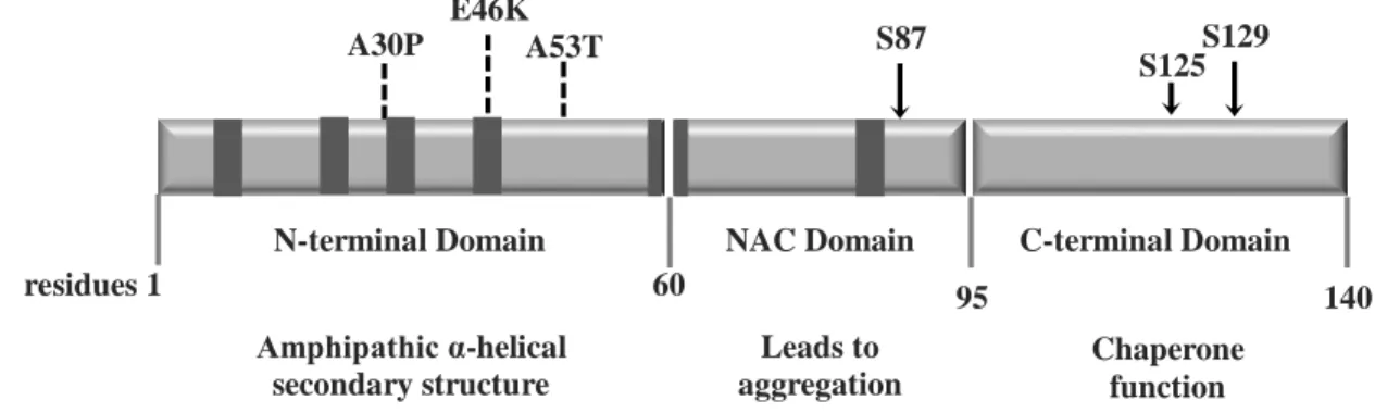
aSyn is a protein of 140 aa residues with three characteristic regions (Figure 3.) (56), and with a molecular weight of 14.5 kDa (57). The N-terminal domain is amphipathic and it is a highly conserved region (58), with several imperfect KTKEGV repeats, which is known to confer a propensity to form amphipathic α-helices, similar to the lipid-binding domain of apolipoprotein-like class (59). The hydrophobic central domain of aSyn is also referred to as the NAC region, which has been implicated in β-amyloid plaques in AD (60). The C-terminal part is highly variable in size and sequence, between species, is rich in proline and acidic aa (58), being negatively charged (61) (Figure 3).
Figure 3. Human aSyn. Schematic representing the structure of human aSyn showing the three distinct domains (N-terminal, NAC and C-terminal) and their corresponding functions. Amino acid positions are indicated in the bottom.
Dark bars inside protein domains represent the imperfect hexameric KTKEGV repeats. Arrows indicate the sites of
phosphorylation and the broken lines show the familial PD mutations.
The protein is known as natively unstructured, such that under physiological conditions (neutral solutions) it lacks a defined secondary structure (7, 43). Although it can assemble into β-sheet oligomers and fibrils in vitro reassembling the disease state, which is known to be toxic in cell culture and model organism (62). When binding to lipids, the secondary structure suffers a shift into an α-helical structure (43). aSyn can bind to vesicles and the plasma membrane via lipid raft (63). Amphipathic α-helical secondary structure Leads to aggregation Chaperone function N-terminal Domain NAC Domain C-terminal Domain
S87 A30P E46K A53T S129 residues 1 60 95 140 S125
aSyn can interact, for example, with synphilin-1 (enriched in neurons and present in synaptic vesicles), microtubules-associated protein tau (MAPT) and unphosphorylated tyrosine hydroxylase (TH), which decreases cellular dopamine content when aSyn bonds, inhibiting activation of TH (32). It is has also been described to be involved in the formation of synaptic vesicles in vitro and in vivo (64), and in stabilizing the effects of SNARES (65).
Nevertheless, the function of aSyn is not fully understood. When overexpressed, aSyn appears to disrupt vesicular transport since it inhibits trafficking between the endoplasmic reticulum and Golgi compartments (66, 67), causes mitochondrial deficits, impairs autophagy, mediates toxicity and increases sensitivity for oxidative stress (68, 69).
Currently, the exact link between the formation of aSyn inclusions and cytotoxicity remains unclear. Recent evidence suggests that aSyn dimers and oligomers play an important role in neurodegenerative disease and could be more toxic than the LB themselves (70). In the NAC region of aSyn, the lack of 11 aa or even the entire domain, reduces the propensity to form fibrils, meaning that the NAC domain is important for fibrillation (43) (71). The C-terminal region also showed interesting results when truncated. This aSyn truncation version resulted in assembled filaments more readily than the full length WT protein (71). These findings are important because 15% of aSyn present in LB is C-terminally truncated and are selectively recovered in insoluble fractions from synucleinopathy patients (71).
1.6.1. Familial aSyn mutations and their effects
Biophysical analyses have shown that the mutations associated with familial cases of PD (A30P, E46K and A53T) (Figure 3) do not seem to dramatically change the structural proprieties of the protein in solution. In addition, they also do not seem to interfere with the formation of α-helical conformation on the surface of SDS micelles. In spite of that, aSyn mutants strongly differ in their ability to form amyloid fibrils (38). Due to differences in aggregation kinetics the different mutations confer to aSyn protein, it is hypothesized that they might cause pathology through different mechanisms. There is currently great interest to firmly identify the most toxic aSyn species.
1.6.1.1. A30P
The A30P mutation reduces affinity for phospholipids displaying a debilitated membrane and vesicle binding capacity (72). In vitro, the A30P mutation seems to also reduce amyloid fibril formation (17) and to promote the accumulation of protofibrillar and oligomeric structures of lower order (73) while increasing cytotoxicity in different cell model systems (70).
Expressing human A30P in cultured rat brain cortical neurons disrupts aSyn transport, resulting in reduced axonal transport (74).
1.6.1.2. E46K
The E46K mutation elicits toxicity because it changes aSyn interactions by exposing the hydrophobic surface for potential intermolecular interactions, which may accelerate dimer formation and the subsequent generation of toxic oligomers (17). It is reported that the E46K mutation abolishes the ability of aSyn to bind to lipid vesicles (33, 75), triggers apoptosis of cultured dopaminergic neurons (75) and results in formation of pre-amyloid oligomers more rapidly than A53T and WT aSyn (34). Transgenic mice expressing human E46K aSyn develop detrimental age-dependent motor impairments, accumulate age-dependent neuronal inclusions and recapitulate the biochemical, histological, and morphological properties of LBs (76).
1.6.1.3. A53T
The A53T mutation expands the hydrophobic domain that confers gain-of-function toxicity by promoting the protein’s ability to adopt the β-sheet structure necessary for formation of oligomeric species (17). This mutant is believed to increase propensity for fibril and protofibril formation (28), accelerate aSyn oligomerization (29), increase toxicity (30) and accelerate the rate of in vitro aggregation, compared to that of human WT (hWT) aSyn and E46K (31). Moreover, A53T aSyn expression causes alterations in mitochondrial dynamics (77).
1.6.2. aSyn post-translational modifications
Several post-translational modifications of aSyn, such as nitration, phosphorylation, sumoylation, and phosphorylation have been described. While some studies show the mutations can increase the tendency of aSyn to misfold and aggregate, it is still unclear what the role of these modifications is in the context of PD (27).
1.6.2.1. Summary of known properties of the artificial aSyn mutants
investigated in this study
1.6.2.1.1. E35K and E57K
Single-point mutants, E35K and E57K, demonstrated a strongly decreased tendency to form fibrils compared with WT aSyn, as evidenced by time-resolved amyloid formation measured by Thioflavin T (ThT) binding (35) (used to predict the amyloid formation, as ThT
E57K form pronounced ring/pore-like structures, in contrast to WT aSyn (35). In animals, the most severe dopaminergic loss in the SN is observed with E35K and E57K oligomers of aSyn, which form fibrils very quickly (78).
1.6.2.1.2. Proline mutations
Two other mutations, A56P and triple proline (A30P/A56P/A76P -TP) seem to form a smaller amount of fibrils and demonstrate strongly reduced fibril elongation rates (38). Overexpression of A56P and TP aSyn in cells, flies, and rats, confers increased levels of neurotoxicity and impairs aSyn ability to form fibrils, providing strong evidence that soluble oligomers are the most toxic species in PD (38).
The substitution of alanine at position 76 for a proline residue was shown to inhibit the formation of β-pleated sheet structures and reduce the propensity of aSyn to polymerize. This A76P mutant did not result in a complete prevention of amyloid formation, suggesting that other steps are involved (79).
1.6.2.1.3. Mutants interfering with aSyn phosphorylation
Phosphorylation is an important reversible post-translational modification that regulates the structural and functional properties of proteins in health and disease. The role of phosphorylation in modulating the aggregation and fibrillogenesis of aSyn is currently the subject of intense investigation. The pathways involved are currently being investigated as putative targets for intervention in PD (39), because phosphorylation regions influence the affinity of aSyn for other proteins and thereby alter the biochemical and biological processes regulated by its interactions.
Phosphorylation of aSyn is known to occur on serine residues 129 (S129) and 87 (S87), as well as tyrosine residues 125 (Y125), 133 (Y133) and 136 (Y136) (38). However, whether phosphorylation promotes or inhibits aSyn aggregation and neurotoxicity in vivo remains unknown (39). Mimicking protein phosphorylation, by mutating specific serine residues to another aa could help elucidate some mechanisms associated with toxicity or aggregation.
S129 is located in the highly acidic C-terminal domain of aSyn, which remains highly flexible even within amyloid fibrils, making it difficult to understand the molecular mechanism of decreased neurotoxicity (38). S129-phosporylation (S129-P) is believed to promote the formation of cytoplasmic inclusions in vitro (39). However, there is a lack of correlation between S129-P and the levels of aggregation in vivo. It has been reported that S129-P increases the conformational flexibility of aSyn and inhibits its fibrillogenesis in vitro but does not perturb its membrane-bound conformation(40). The unphosphorylatable aSyn (S129A) showed opposite effects compared to WT aSyn and the phosphomimic S129D, decreasing the toxicity but
promoting the number of aSyn inclusions (80-82). The phosphorylation mimics (S129E/D) do not seem to fully reproduce the effect of phosphorylation on the structural and aggregation properties of aSyn in vitro (40). These data have important implications for the use of phosphomimics because several studies have reported that protein-protein interactions, mediated by phosphoserines, are not fully recapitulated when a serine is replaced by glutamate (40).
Studies in cell culture have shown three adjacent tyrosine phosphorylated residues (Y125, Y133, and Y136) that were also reported in human and Drosophila brain (83). Phosphorylation of these residues suppresses oligomerization. During aging, tyrosine phosphorylation is reduced, while LB from patients with PD show less phosphorylated Y125 (84). The lack of information on Y125 mutations is related to the difficult to detect Y125-P aSyn in human brains and the failing link between the progression the disease and the levels of aSyn phosphorylate in the tyrosine residue (85). The rapid dephosphorylation of aSyn in post-mortem tissues could explain the limited detecting of Y125-P aSyn in human brains (85).
S87, located in the NAC region of aSyn, is phosphorylated in in vivo models and increases the synucleinopathies (86). S87 is one of the few residues and phosphorylation sites that distinguish the human aSyn sequence from that of mouse and rat aSyn (87). S87-P might have a significant influence on the conformation of aSyn and affect the interaction of aSyn with proteins involved in its transport (39). It has also been shown to affect aSyn aggregation, alter the conformation of membrane bound aSyn and decrease its affinity to lipids (83).
1.6.2.1.4. Mutants interfering with aSyn sumoylation
In addition to phosphorylation, other post-translational modifications occur in aSyn and have gotten recent attention. One such modifications is the addition of the small ubiquitin-related modifier (SUMO), in a process known as SUMOylation. Several aggregation-prone proteins implicated in neurodegeneration were found to be SUMOylated, and sumoylation-deficient mutants demonstrate an enhanced tendency to aggregate in cell-based assays (88). The SUMOylated sites in aSyn are located in lysine 96 and arginine 102. Mutating these sites confers an increase propensity for aggregation and cytotoxicity in a cell-based assay and increased cytotoxicity in dopaminergic neurons of the SN, in vivo. Recent studies show that SUMOylation of aSyn inhibits its fibrillation and that mutation of the main aSyn SUMO acceptor sites leads to increased toxicity (88). In these studies, aggregates form as a result of proteasome impairment, such that inhibition of the proteasome, induces SUMOylation of aSyn and subsequent oligomerization (89). Indeed, SUMOylation is known to control protein-protein interactions, affects subcellular localization, stability and solubility of target proteins and also has been considerers to be a nuclear process (90, 91).
In the table in the appendix there is a list of all mutations studied in this work and the effects that have already been described in other models (Table 5).
1.7. aSyn aggregation model used in this study
Aggregation of aSyn is considered one of the crucial steps in PD and it is thought to precede via a seeding-nucleation mechanism. In vitro studies have revealed that aSyn aggregation is a nucleation-dependent process that initiates with the progression of monomer to oligomers to fibrils (Figure 4) (92). Recent studies support the hypothesis that pre-fibrillar intermediates (protofibrils) and not mature amyloid fibrils, may be the key toxic species in PD (15, 93, 94).
A way to study the impact of aSyn aggregation in vitro is via a well established aggregation assay initially described by McLean (95). In this model, overexpression of a tagged version of aSyn (SynT), together with symphilin-1 in a neuroglioma cell line (H4), promotes the formation of aSyn inclusions (95).
1.7.1. SynT
As shown in Figure 5, SynT is based on a fusion of aSyn with EGFP, at the C-terminal (aSynEGFP).
Figure 5. SynT model. This construction allows us to study aSyn aggregation in vitro, in mammalian cell lines. aSyn monomer ROS dimer Possible toxic species oligomer (protofibril) Lewy Body fibril
Figure 4. Aggregation pathway of aSyn. In normal cells, aSyn exits in an unfolded, monomeric state, and through disease-associated mechanisms, transitions to toxic species. Protofibrils may further oligomerize into fibrillar structures that ultimately are organized into LB.
83 amino acids Δ155
SynT
aSyn
EGFPThe model was discovered when H4 cells were transfected with aSynEGFP, and a widespread, diffuse distribution of the fusion protein was observed. However, immunostaining with an anti-GFP antibody failed to detect the inclusions, suggesting that EGFP had been cleaved (95). This data infers that the C-terminal region of EGFP was truncated (95). Interestingly, it was found that an 83 residue fragment which was left attached to aSyn could interfere with its C-terminus and promote its aggregation. These results were confirmed with other tags, expression of GFP-tagged tubulin or synaptophysin, which does not form aggregates, indicating that aSyn is the primary element driving aggregation in this model. (95). Additional proof that the tag is not simply causing aSyn aggregation derives from the fact that cells transfected with the A30P mutant display a decrease in aggregation formation, compared to WT aSyn.
In summary, this model is based on the expression of aSyn tagged with a truncated, non-fluorescent fragment of GFP (SynT).
Given the importance of local structural effects on the aggregation of aSyn, it will be crucial to elucidate how other alterations, such as those induced by aSyn mutations, might modulate its aggregation.
1.7.2. Synphilin-1
Synphilin-1 is a presynaptic protein that was first identified by yeast-two-hybrid screening as a protein that interacts with aSyn (96). It is composed of 919 amino acid residues and co-localizes with aSyn in LBs, in brains of PD patients (96). Association of synphilin-1 with aSyn is necessary for targeting to LB-like protein aggregates in cell culture (97).
Given the presynaptic location and its affinity for membranes and lipids (98) (more specifically, binding to lipid rafts), synphilin-1 also seems to be required for inclusion formation (99) and might act as an adaptor protein, anchoring aSyn to other proteins (like proteins involved in vesicular transport or cytoskeletal function) (31).
Recent studies show that synphilin-1 contains four ankyrin repeat domains and a coiled-coil domain in the central portion that specifically interacts/binds with aSyn, via the N-terminal residues of aSyn (Figure 6) (100). This specific interaction significantly promotes formation and accumulation of cellular inclusions that are probably comprised of aSyn (100).
Figure 6. Site of interaction between aSyn and synphilin-1. Schematic representation of the interaction between synphilin-1 and aSyn (100).
It was also found to be a substrate of the ubiquitin ligase Parkin (101).
In some cases of sporadic PD, a point mutation in synphiln-1 (R621C) was reported (96, 102). C621 mutation is located towards the C-terminal of synphilin-1 and might interfere with the cytoprotective function of synphilin-1 (Figure 7)(101) .
In neuroblastoma cells, the R621C mutant synphilin-1 displays a reduced capacity to form intracytoplasmic protein inclusions compared with WT synphilin-1. The conformational alteration that R621C synphilin-1 promotes interferes with protein aggregation and exerts misfolding protein stress (101). Synphilin-1 is degraded by the proteasome and its inhibition
increases the tendency of synphilin-1 to form inclusions in HEK293 cells. The fact that WT synphilin-1 is more sensitive to proteasome inhibition than R621C synphilin-1 might indicate differences in the recognition by the ubiquitin– proteasome system (102).
In transgenic mice, synphilin-1 can attenuate A53T aSyn-induced motor abnormalities and decrease astroglial reaction and neuronal degeneration, suggesting that synphilin-1 may play a neuroprotective role against A53T aSyn toxicity when there is no loss of dopamine neurons in the SN (99). However, it is not clear whether synphilin-1 triggers cytotoxicity by itself or if it acts as an accelerator that boosts cellular stress responses, induced by an interaction partner (99).
Figure 7. Schematic representation of the synphilin-1 protein. Different structural features are highlighted. The R621C mutation is localized in a highly conserved region.
N C 1 Ankyrin-like 919 ATP/GTP binding Coiled-coil PKAKDEDSKKILRQLLGKEISENVCTQEKL
^
621 C2. Objectives
Synucleinopathies are a group of disorders that share, as a pathological hallmark, the misfolding and aggregation of aSyn. Thus, the main goal of this project was to provide insight into the molecular determinants of aSyn inclusion formation in cultured cells. To this end, the main aims were:
Aim 1. To generate the constructs to express mutant forms of aSyn
We generated, in a systematic way, constructs encoding for the different mutant forms of aSyn which were tested throughout the project.
Aim 2. To investigate the effects of the different mutations on aSyn inclusions in in vitro models
We expressed the different forms of aSyn in cell culture models and assessed inclusion formation by immunocytochemistry and fluorescence microscopy.
Aim 3. Explore if the R623 synphilin-1 mutation influences aSyn inclusions formation
We co-expressed synphilin-1 (WT or R621C synphilin-1) together with aSyn and compared their effects on aSyn inclusion formation.
3. Materials and Methods
3.1. Primer design
In order to generate the different constructs we generated primers for each mutation in the study. Using two different web-based applications (Primer-X and Agilent) conditions were optimized (temperature melting and GC percentage), to design the primers (Table 2).
Primer name Sequence 5' to 3' Tm bp
A30P forward GGG TGT GGC AGA AGC ACC AGG AAA GAC AAA AGA 66.1 33 A30P reverse TCT TTT GTC TTT CCT GGT GCT TCT GCC ACA CCC 66.1 33 E35K forward CAG AAG CAG CAG GAA AGA CAA AAA AGG GTG TTC TCT 64.1 36 E35K reverse AGA GAA CAC CCT TTT TTG TCT TTC CTG CTG CTT CTG 64.1 36 E46K forward TAG GCT CCA AAA CCA AGA AGG GAG TGG TGC ATG G 83 34 E46K reverse CCA TGC ACC ACT CCC TTC TTG GTT TTG GAG CCT A 83 34 A53T forward G AGT GGT GCT AGG TGT GAG GAC AGT GGC TGA GAA GAC 69.8 37 A53T reverse GTC TTC TCA GCC ACT GTC GTC ACA CCA TGC ACC ACT C 69.8 37 A56P forward GGT GTG GCA ACA GTG CCT GAG AAG ACC AAA G 66.3 31 A56P reverse CTT TGG TCT TCT CAG GCA CTG TTG CCA CAC C 66.3 31 E57K forward GTG GCA ACA GTG GCT AAG AAG ACC AAA GAG C 65.1 31 E57K reverse GCT CTT TGG TCT TCT TAG CCA CTG TTG CCA C 65.1 31 A76P forward TGA CGG GTG TGA CAC CAG TAG CCC AGA AG 66.9 29 A76P reverse CTT CTG GGC TAC TGG TGT CAC ACC CGT CA 66.9 29 S87A forward AAG ACA GTG GAG GGA GCA GGG GCC ATT GCA GCA G 87 34 S87A reverse CTG CTG CAA TGG CCC CTGCTC CCT CCA CTG TCT T 87 34 S87E forward ACA GTG GAG GGA GCA GGG GAA ATT GCA GCA GC 85 32 S87E reverse GCT GCT GCA ATTTCC CCT GCT CCC TCC ACT GT 85 32 K96R forward GCC ACT GGC TTT GCT AGA AAG GAC CAG TTG GGC 68.5 33 K96R reverse GCC CAA CTG GTC CTT TCT GAC AAA GCC AGT GGC 68.5 33 K102R forward AAG GAC CAG TTG GGC AGG AAT GAA GAA GGA GCC 67.3 33 K102R reverse GGC TCC TTC TTC ATT CCT GCC CAA CTG GTC CTT 67.3 33 Y125D forward GAT CCT GAC AAT GAG GCT GAT GAA ATG CCT TCT GAG G 66.7 37 Y125D reverse CCT CAG AAG GCA TTT CAT CAG CCT CAT TGT CAG GAT C 66.7 37 Y125F forward GGA TCC TGA CAA TGA GGC TTT TGA AAT GCC TTC TGA 64.1 36 Y125F reverse TCA GAA GGC ATT TCA AAA GCC TCA TTG TCA GGA TCC 64.1 36 S129G forward GAC AAT GAG GCT TAT GAA ATG CCT GGT GAG GAA GGG TAT C 67.1 40 S129G reverse GAT ACC CTT CCT CACCAG GCA TTT CAT AAG CCT CAT TGT C 67.1 40 S129D forward GGC TTA TGA AAT GCC TGA TGA GGA AGG GTA TCA AG 81 35 S129D reverse CTT GAT ACC CTT CCT CATCAG GCA TTT CAT AAG CC 81 35 S129A forward CTT ATG AAA TGC CTG CTG AGG AAG GGT ATC 78 30 S129A reverse GAT ACC CTT CCT CAG CAG GCA TTT CAT AAG 78 30 Table 2. Primers used for mutagenesis. Sequence, melting temperature (Tm) based on a [Na+]] of 50 mM and the
3.2. Generation of the constructs for the study
Using the SynT construct as a template, the mutations were introduced by site-directed mutagenesis (QuickChange II Site-Directed Mutagenesis Kit, Agilent Technologies). In Figure 8 there is a schematic representation of the DNA (upper panel) and aa sequences of aSyn where the mutagenesis was performed.
A.
B.
Figure 8. Representation mutagenesis sites in the sequence of aSyn. A. DNA sequence. B. Protein sequence. The same colors used to mark the point mutations in the primers in Table 2 were used to highlight the mutations in the DNA and protein aa sequence of aSyn.
The mutagenesis kit allows us to introduce point mutations by PCR using the DNA polymerase PfuUltra high-fidelity (HF). The PCR conditions used are shown in
Table 3.
Segment Cycles Temperature Time
1 1 95ºC 1 min
95ºC 50 sec
2 16* 58ºC 50 sec
68ºC 6.30 min**
3 1 68ºC 10 min
Table 3. PCR parameters used for the mutagenesis reactions. *depending on the type of mutation that was performed; **1min/kb plasmid.
Following PCR, the product was treated with Dpn I and the reaction was immediately incubated at 37ºC for 1 hour. Dpn I is an endonuclease that specifically recognizes sequence 5´-Gm6ATC-3´ and targets methylated and hemimethylated DNA. Addition of this enzyme will digest the parental DNA template (DNA is dam+ methylated and therefore susceptible to Dpn I digestion.) and leave only the newly synthesized DNA.
3.3. Transformation of XL10-Gold Ultracompetent Cells
One microliter of the previously described Dpn I-treated DNA was added to XL10-Gold ultracompetent cells that were thawed on ice. This transformation reaction was incubated on ice for 30 minutes. One heat pulse was applied for 45 seconds at 42ºC, and was followed by a 2 minute incubation on ice. 500 µl of S.O.C medium (2% Tryptone, 0.5% Yeast Extract, 10 mM NaCl, 2.5 mM KCl, 10 mM MgCl2, 10 mM MgSO4, 20 mM glucose) was added and the tubes were subsequently incubated at 37°C for 1 hour. Finally, half of the transformation mixture was plated on LB-agar plates containing ampicillin and then incubated at 37°C overnight (ON).
3.4. Extraction of plasmid DNA and glycerol stocks
Random colonies were picked from the LB-agar plates and used to inoculate liquid LB medium supplemented with ampicillin (Sigma-Aldrich) (269 µM). After 12-16 hours shaking at 225rpm at 37°C, the bacterial cultures were harvested by centrifugation and the plasmid preparation was performed using Invisorb® Spin Plasmid Mini Two kit (Invitek).
The purity and DNA yield were confirmed by spectrometric measurement using the Nanodrop (ND-1000 Spectrometer V3.5.2).
Additionally, glycerol stocks of the bacterial cultures were prepared, for further plasmid multiplication. In a 1:1 dilution, bacterial culture was mixed with 80% glycerol and immediately stored at -80°C.
3.5. Agarose Gel Electrophoresis
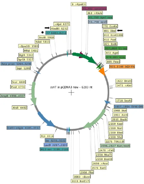
Once DNA was purified, enzymatic digest was performed with XbaI and Hind III (Fermentas) enzymes to verify the positive colonies. These enzymes were chosen according to sites flanking the SynT sequence in our plasmid (Figure 9).
Figure 9. SynT plasmid map. Black arrows indicate the two chosen enzyme for the enzyme digestion; The black dot indicates the primer used for DNA sequencing.
Reactions were performed at 37°C for one hour and the digestion products were subsequently separated by agarose gel 1.5% (w/v) (peqLab), in 1x TAE (40mM Tris, 20mM acetic acid, 1mM EDTA) with ethidium bromide (Sigma-Aldrich), in a horizontal electrophoresis tank. The samples and marker (GeneRuler™ 1kb DNA Ladder) were loaded and the gel was run at 130 V for 25 minutes.
3.6. Sequencing
The samples sequenced to confirm the mutations introduced. We used a T7 primer (MWG Biotech AG) that anneals to the T7 promoter region (marked with a dot, Figure 9) in the plasmid. The results were analysed with Sequencher 4.9.
3.7. Cell Culture
Human neuroglioma cells (H4) were maintained in OPTI-MEM® (Life Technologies) supplemented with 10% Fetal Bovine Serum and 1% Penincillin/Streptomycin. The cells were grown at 37°C in an atmosphere of 5% CO2. 24 hours prior to transfection, 250,000 cells/well were plated in 35mm Ibidi dishes, so that the cells are 60-80% confluent upon transfection.
3.8. Cell transfection
On the day of transfection, FuGENE® 6 Transfection Reagent (Promega) was added to Opti-MEM® serum-free and incubated for 5 minutes.
Plasmid DNA and FuGENE® 6 reagent were mixed in a 1:3 ratio. Plasmids encoding aSyn and synphilin-1 were added in equal amounts (2 µg), and incubated for 30 minutes. Finally, the mixture was added to each dish and incubated for 48 hours. After the transfection, the cells were subjected to immunocytochemistry, for studying aSyn inclusions. H4 cells were also plated in 12 or 24-well plates (Costar) for western blot analysis.
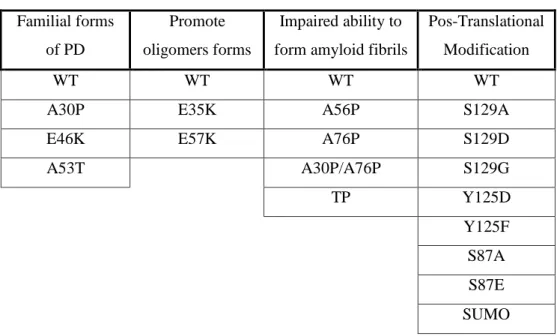
To facilitate the study, the mutations were divided in four categories as is shown in Table 4.
Familial forms of PD
Promote oligomers forms
Impaired ability to form amyloid fibrils
Pos-Translational Modification
WT WT WT WT
A30P E35K A56P S129A
E46K E57K A76P S129D
A53T A30P/A76P S129G TP Y125D Y125F S87A S87E SUMO
Table 4. Groups of mutants used for the study. The group with familial form of PD was transfected with either WT or R621C synphilin-1.
3.9. Immunocytochemistry
After 48 hours, the cells were washed two times with DPBS (PAN) and fixed with 4% paraformaldehyde (PFA) for 10 minutes at room temperature (RT). After washing again three times with DPBS, the cells were permeabilized with 0.5% Triton X-100 (Sigma-Aldrich) for 20 minutes at RT. After blocking in 1.5% normal goat serum (PAA)/DPBS for 1 hour, cells were incubated with primary antibody (mouse anti-aSyn, 1:1000, BD Transduction Laboratory and/or anti-v5 1:1000, Abacam) overnight or for 3 hours and secondary antibody (donkey anti-mouse IgG-Alexa488 and/or goat anti-rabbit IgG-Alex 568, 1:1000, Invitrogen) for 2 hours. Finally, cells were stained with Hoescht (1:5000 in DPBS, Molecular Probes) for 5 minutes, washed and maintained in DPBS for fluorescent microscopy.
3.10. Fluorescence Microscopy
3.10.1. Quantification of aSyn inclusions
A total of three independent experiments were performed for each mutation. Transfected cells were detected and scored based on the aSyn inclusions pattern and classified into four groups: cells without inclusions, less than five inclusions (<5 inclusions), between five to nine inclusions (5-9 inclusions) and more than ten inclusions ( 10 inclusions). Results were expressed as the percentage of the total number of transfected cells.
3.11. Cell lysis
Cells are lysed to release the proteins of interest, such that they can migrate individually through a separating gel.
Forty-eight hours after transfection, cell culture plates were placed on ice and 100 µl of cold RIPA lysis buffer (50mM Tris, 150mM NaCl, 0,1% SDS, 1% NP-40, 0,5% Deoxycholate, protease inhitors supplement and pH adjusted 8) was added to each well. Cells were subsequently scraped off the dish with a cell scraper and gently transferred to a pre-cooled eppendorf tube. The samples were stored at -80ºC.
3.12. Protein quantification
The protein concentration was determinate by Bradford protein assay.
For the standard curve, 200µl Bradford solution was mixed with 50µl of the respective standard protein solution (0; 0.1; 0.25; 0.5; 1; 2; 4; 6 mg/ml (BSA in H2O bidest.)). 1µl of each sample was diluted in 49 uL H2O for 150 uL Brafdord reagent (Bio-RAD) and measured at 595
standard samples for the standard curve and the protein concentration of the samples was calculated by .
3.13. Western Blot analysis
3.13.1. Gel preparation
A 12% polyacrylamide gel was prepared by mixing 24.8% (v/v) separating buffer (150 mM Tris base, 0,4 % (w/v) SDS pH adjusted to 8.8 with HCl) 39.6% (v/v) acrylamide/bisacrylamide solution, 0.5% (v/v) TEMED (Applichen), 34,6% (v/v) distilled H2O (ddH2O). Ammonium persulphate (APS) (Schleicher/Schüll bioscience) was added to initiate the crosslinking reaction (0.5 % v/v). This solution was immediately transferred into a cast, overlaid with isopropanol and incubated for 15 minutes. Upon polymerization, the isopropanol was removed. The stacking gel (7,5 %) was composed of: 24,8 % (v/v) stacking buffer (50 mM Tris base, 4 % (w/v) SDS pH adjusted to 6.8 with HCl), 24.8 % (v/v) acrylamide/bisacrylamide, 49.4 % (v/v) ddH2O, 0.5 % (v/v) TEMED and 0,5 % APS. After addition of APS, the mixture was immediately overlaid on the separating gel and a gel comb was inserted.
3.13.2. Sample preparation
Each sample was prepared by combining 30µg of total protein with protein sample buffer (PBS 4x, 1 M Tris-HCl pH 6.8, β-Mercaptoethanol, 20% SDS, Glicerol 100%, Bromophenol blue) to a total volume of 20µl. All samples were boiled at 100ºC for 5 minutes.
3.13.3. SDS-polyacrylamide gel electrophoresis (SDS-PAGE)
Samples and loading protein ladder (10-250 kDa) (Bio-Rad) were loaded and ran in 1x running buffer (250 mM Tris base, 200 mM Glycine, 1% SDS) at 100 V for 60 minutes.
3.13.4. Transfer to nitrocellulose membrane
The proteins were transferred to a nitrocellulose membrane (Whatman).
Sponges and blotting paper were soaked in 1x transfer buffer (0.025 M Tris base, 0.192 M Glycine, 20 % methanol (v/v)). The transfer was carried out for 90 minutes at 0.2A.
3.13.5. Western Blot
Membranes were blocked with 5% skim milk powder (Fluka) in 1xPBS-Tween (50mM Tris,150mM NaCl, 0,05% Tween, pH=7,5) for 60 minutes at RT.
Membranes were further incubated for 3 hours (or ON at 4°C) with the primary antibody, either mouse anti-asyn (BD Transduction Laboratory) or mouse anti-β-actin (Sigma-Aldrich) at 1:1000 in 3% BSA TBS-Tween.
After washing three times in TBS-Tween for ten minutes, the membranes were incubated in anti-mouse horseradish peroxidase-conjugated (GE Healthcare) at 1:10000 in 3% milk/TBS-Tween. Membranes were again washed three times.
Detection was made using enzyme linked chemiluminescence (ECL) detection reagents, based on the horseradish peroxidase secondary antibody conjugate catalysis of luminol in alkaline conditions. Luminol Reagent and Peroxide Solution (Millipore), mixed 1:1, were applied to the membrane 1 minute before scanning with α-imager FluoroChem software (producer, place) was utilized for reconstructing a picture of the membrane and the bands with positive antibody staining.
The digital images obtained were analyzed using ImageJ software (http://rsbweb.nih.gov/ij/download.html) to quantify the amount of protein in samples.
3.13.6. Statistical Analysis
For statistical evaluation of the data, Graphpad PRISM 5 (San Diego California USA) software was used. T-test was performed to analyze differences between the various groups. Significance was assessed for p ≤ 0.05, whereas * corresponds to p ≤ 0.05, ** corresponds to p ≤ 0.01 and *** corresponds to p ≤ 0.001.


![Table 2. Primers used for mutagenesis. Sequence, melting temperature (Tm) based on a [Na +] ] of 50 mM and the number of base pairs (bp) are listed](https://thumb-eu.123doks.com/thumbv2/123dok_br/15580470.1049159/31.893.149.744.345.1083/table-primers-mutagenesis-sequence-melting-temperature-number-listed.webp)