Laboratório de Genética
Psoriasis is more than a skin disorder. An
ACE study
Matilde José Sousa Bandeira
Laboratório de Genética
Psoriasis is more than a skin disorder. An
ACE study
Matilde José Sousa Bandeira
Orientado por:
Prof. Dr. Manuel Bicho
Psoriasis is more than a skin disorder. An ACE study.
Abstract
Introduction: Psoriasis is now known to be a systemic autoimmune disease with an associated increased cardiovascular risk. This seems to rely on systemic inflammation. Angiotensin-converting enzyme (ACE) has been associated with oxidative stress, a state characteristic of cardiovascular disorders and this enzyme’s activity is higher in psoriasis. Our aim is to assess cardiovascular risk in psoriatic patients and its association with ACE polymorphism and activity.
Methods: Our population consisted of 1155 individuals (64 psoriatic patients, 1091 controls) whose ACE genotype and activity were determined. The cardiovascular risk was estimated through lipid profiles. PASI score was used as a severity measurement.
Results: Psoriatic severity was associated with a higher cardiovascular risk estimated by lower HDL concentrations (r=-0.496, p=0.007) and higher triglyceride levels (r=0.421, p=0.020), as well as TC/HDL and LDL/HDL ratios (r=0.612, p<0.001 and r=0.437, p=0.023, respectively) which have been shown to have stronger predictive value for cardiovascular disorders
Conclusions: Psoriatic patients should be treated as a risk group for cardiovascular diseases and new markers for severity and this associated risk may be of worthy pursuit. Further studies are crucial to enhance our knowledge on inflammation and psoriasis.
Keywords: ACE, Psoriasis, Cardiovascular Risk, Lipid profile
Resumo
Introdução: A psoríase é atualmente vista como uma doença sistémica autoimune com um risco cardiovascular associado, cuja correlação parece advir de um estado de inflamação sistémica. A enzima conversora de angiotensina (ECA) associa-se a stress oxidativo, característico da patologia cardiovascular e apresenta níveis de atividade superiores na psoríase. O nosso objetivo é avaliar este risco e a sua associação com o polimorfismo e atividade do ECA.
Métodos: A amostra consiste em 1155 indivíduos (65 psoriáticos, 1091 saudáveis), dos quais se determinou o genótipo do ECA e a sua atividade. O risco cardiovascular foi estimado a partir do perfil lipídio e a severidade da doença pelo índice PASI.
Resultados: A severidade da psoríase associou-se a diminuição do HDL (r=-0.496, p=0.007) e aumento dos triglicerídeos (r=0.421, p=0.020), bem como dos índices CT/HDL e LDL/HDL (r=0.612, p<0.001 e r=0.437, p=0.023, respetivamente), os quais traduzem melhor valor preditivo para doenças cardiovasculares.
Conclusões: Os doentes psoriáticos devem ser tratados como um grupo de risco para patologia cardiovascular e deverão ser procurados novos marcadores para a severidade da doença e risco associado. Novos estudos são essenciais para melhorar o conhecimento nesta área.
Palavras chave: ECA, Psoríase, Risco Cardiovascular, Lípidos
O Trabalho Final exprime a opinião do autor e não da FML.
Psoriasis is more than a skin disorder. An ACE study.
Introduction
Psoriasis affects approximately 2% of the population worldwide [1, 2], accounting for 12 million individuals within the USA and Europe [3], with a higher prevalence at higher latitudes and also more prevalent among caucasians [4].
Psoriasis can present at any age and in different ways. It can be classified according to morphologic appearance including plaque, inverse, erythrodermic, pustular, and guttate forms, sometimes with nail manifestations as well, including nail dystrophy and psoriatic lesions of the nail bed [2, 5]. The most common clinical variant is psoriasis vulgaris, representing 90% of all cases. This type of psoriasis is usually manifested as well circumscribed red or salmon pink papulosquamous plaques covered by silvery scales surrounded with normal skin. Manifestations are generally symmetrically distributed in the body, specially on the extensor area of the elbows and knees, on the scalp, and lumbosacral region [1, 6], although any or all portions of the body surface may be affected.
Patients with psoriasis have a higher prevalence of numerous comorbidities, such as pulmonary, renal, hepatic, rheumatologic and cardiovascular diseases [7] with a clear majority of the literature revealing a correlation between psoriasis and cardiovascular risk, linking it with disorders such as obesity, hypertension, dyslipidemia, diabetes mellitus (DM) and metabolic syndrome. This epidemiologic association seems to be stronger as psoriasis is more severe [8]. Even though there are multiple definitions of disease severity, it is accepted by most that mild psoriasis has less than 3% body surface area involved, moderate psoriasis between 3 and 10% and severe psoriasis more than 10% body surface area involvement, as proposed by the National Psoriasis Foundation [9].
Obesity, a well known cardiovascular risk factor, has been studied as a comorbidity in patients with psoriasis and the association has been shown to grow with the severity of psoriasis, with an adjusted odds ratio (OR) of 1.79 (95% CI [1.55,2.05]) for patients with severe psoriasis and 1.27 (95% CI [1.24,1.31]) among the ones with mild psoriasis [10]. Despite there being a definite connection between the two it is not yet established wether obesity predates or comes after the development of psoriasis. Just as the definition of OR itself explains, it does not establish a cause-effect relation in either direction and it could even be that the association is due to a third property,
which is a contributing factor for both psoriasis and obesity, or even a common underlying pathophysiology. In fact there is both evidence of a BMI increase in patients after they were diagnosed with psoriasis [11], pointing to obesity as secondary to the disease, and strong evidence that indicate that obesity is a risk factor for the development of psoriasis, with both hip circumference and waist-hip ratio associated with higher risk of psoriasis [12, 13].
This “psoriasis’ obesity” is believed to be an important connection to all other cardiovascular disorders and risk factors among these patients. There is even a tendency for central adiposity as shown in a study which illustrated that children and adolescents with psoriasis are more prone to obesity and that this obesity is more likely abdominal [14], and waist circumference, waist-hip ratio or any other central adiposity measures are presumed to be more relevant to cardiovascular risk than an increased BMI [15, 16]. In a 24 week study there was much more significant results in reducing disease severity when cyclosporine therapy was combined with a low-calorie diet as opposed to cyclosporine alone [17], suggesting that obesity may also affect treatment efficacy.
Pathophysiologically, psoriasis seems to share many cytokines with obesity, such as TNF-α and IL-6. There is a shown rise of both serum TNF-α levels and TNF-α receptors as BMI and waist circumference increase [18] and there is also higher levels of c-reactive protein (CRP) in psoriatic populations as well as a positive association between its levels and BMI [19], being not only increased in active stages of disease but also during remission compared to controls [20].
In regards to hypertension, there have been studies that suggest a correlation between psoriasis and hypertension as well as the difficulty to control it [21]. This correlation seems to be severity dependent [22].
Dyslipidemia has also been associated with psoriasis. Triglyceride, total-cholesterol, and LDL-cholesterol levels were found to be higher in psoriatic patients [23, 24], and these changes were seen at the onset of the disease [25], leading to the possibility that dyslipidemia may precede psoriasis. It is however unclear whether this lipid profile changes are due to the increased incidence of obesity in psoriatic populations and its influence in lipid metabolism or actually independently associated with psoriasis. Adipocytes release a variety of pro inflammatory molecules including some cytokines such as TNF-α and IL-6. These molecules are shown to not only increase cardiovascular risk [26] but also to affect free fatty acids, cholesterol and lipid levels [27]. There have been a few studies that found an atherogenic profile with decreased total HDL levels and higher levels of small LDL and HDL particles in psoriatic patients, measured by nuclear magnetic resonance spectroscopy, and these were shown to be associated with increased aortic inflammation,
which suggests that HDL particle characteristics may be an important participant in the cardiovascular risk seen in psoriasis [28, 29]. The same study found a lower HDL efflux capacity in psoriatic patients, which is associated with non calcified coronary atherosclerosis, non related to the HDL levels [30]. This decrease in cholesterol efflux may be explained by another study that describes psoriatic HDL as having diminished phospholipid and cholesterol components, which results in the reduction of its capacity to promote cholesterol efflux from macrophages. This efflux capability was negatively correlated with psoriasis area and disease severity [31].
Another important comorbidity is the metabolic syndrome, that in a way correlates most of the comorbidities discussed above. It is directly linked to intra-abdominal obesity, and is perceived as a setting of chronic systemic inflammation that meets at least three of the following: abdominal obesity, impaired glucose regulation, hypertriglyceridemia, reduced HDL and hypertension [32]. The association has been proved in many studies, with abdominal obesity as the most common criteria met by the psoriatic group, followed by hypertriglyceridemia and decreased levels of HDL [33].
This abdominal obesity must be recognized almost as an endocrine organ that is in fact able to release many bioactive peptides (adipokines) such as TNF-α, IL-6, leptin and adiponectin that coordinate inflammation, glucose and lipid metabolism and endothelium function [34].
Adipocytes may also interact directly with macrophages that are increased in the dermis and which, by releasing TNF-α, will influence gene transcription in adipose tissue, augmenting the levels of leptin in decrement of adiponectin levels which is an important anti-inflammatory molecule that also functions in regulating insulin sensitivity, and block insulin signaling, becoming a major contribute for insulin resistance [35]. This insulin resistance was shown to be present in the dermal endothelium of both psoriatic plaques and “normal” skin in patients with psoriasis and it promotes endothelial expression of adhesion molecules and inflammation by shifting anti-/pro-atherogenic insulin-dependent pathways towards the latter [36]. Furthermore, it has also a role in regulating keratinocytes, since some of the cytokines that cause this shift and the consequent insulin resistance, namely IL-1β, are also responsible for disrupting insulin-dependent keratinocyte differentiation and thus boosting keratinocyte proliferation [37], one of the most discussed features of psoriatic skin.
TNF-α, besides inducing insulin resistance, increasing production of adhesion molecules by endothelial cells and having a positive correlation with BMI as discussed above, also augments free fatty acids which further induce insulin resistance, all of which are crucial steps for endothelial damage and atherogenesis [38]. Additionally, IL-6 too induces insulin resistance and increases
adhesion molecules production. It also stimulates the liver to release fibrinogen and CRP, all of which further encourage atherosclerosis [39].
Adiponectin seems to work against these actions, augmenting insulin sensitivity, decreasing TNF-α production and monocyte cell adhesion and reducing transformation of macrophages into foam cells [40], which is a feature of atherosclerosis [41]. It is positively correlated to weight loss and high levels of adiponectin have been shown to confer lower risk for myocardial infarction (MI) in men [42]. This adipokine’s plasma levels seem to be negatively correlated with BMI, as opposed to leptin, which increases with both BMI and waist circumference. Adiponectin decreases more in relation to visceral adiposity than to subcutaneous adipose tissue [43]. Adiponectin was also found to be decreased in hypertension and ischemic heart disease [44, 45].
Leptin, on the other hand, seems to have opposite effects. In a study that compared different ethnicities, individuals at high risk for cardiovascular diseases and metabolic syndrome had higher levels of plasma leptin [46], which goes hand to hand with a study that found increased leptin to affect carotid intima-media thickness (CIMT) [47]. Leptin was also shown to be higher in patients with psoriasis, working as a severity marker [48].
Finally, another comorbidity that has been associated with psoriasis is type II diabetes. A recent study confirmed that patients with psoriasis had higher rates of DM and this higher risk was once again severity dependent [49], as it was shown before [10, 50]. This association could be due to the many insulin resistance mechanisms explained before, mainly driven by TNF-α, however psoriasis seems to be associated with a diabetes susceptibility locus which might suggest a stronger correlation from the base [51], and the association between psoriasis and DM is shown to still be present when adjusted for BMI [52].
Psoriasis has been, as discussed above, linked to various comorbidities and it’s also associated with an increased mortality, specially in cases of severe psoriasis [53, 54]. This increased risk of death has been said to have numerous etiologies including infection, kidney disease and dementia however to be mainly due to cardiovascular problems [55] and markedly seen in young patients with severe psoriasis in which the risk of MI is significantly higher. [56]
It is also important to note the association between psoriasis and both CIMT and coronary artery calcification (CAC), which seem to be good parameters when in search for a cardiovascular risk measurement [57-60]. A study in 2016 showed a significant adjusted association between CIMT and PASI (psoriasis area and severity index), which is a score of affected body area and
severity in psoriasis [61]. The mean CAC was also shown to be increased in other studies when comparing psoriatic patients to controls [62].
One of the mechanisms proposed to link psoriasis and cardiovascular disorders is inflammation and despite the fact that, for a long time, psoriasis was seen as a skin disease, there has been growing and now significant evidence of it being a systemic disorder with an underlying systemic inflammatory process. We should therefore perceive psoriases as a common, chronic systemic disease most frequently manifested by skin lesions [5].
Inflammation has been associated with cardiovascular risk, on one hand because it plays a central role in atherosclerosis, which is another mechanism that has been conjectured as a link between psoriasis and cardiovascular diseases [50], and on the other hand because it has been suggested that chronic inflammation itself may lead to an exacerbation of cardiovascular complications, with a possibility that this effect might actually work through vascular damage as well. A study using murine models with psoriasiform dermatitis without any comorbidities showed aortic root inflammation in 33% of the affected group compared with 0% in controls (p=0.04) and concluded that skin-specific inflammation promotes both vascular inflammation and an increase in thrombotic rates and that treatment against this skin inflammation attenuates these pathways [63]. In fact, systemic treatments targeting psoriatic underlying inflammation have been studied and shown to improve both psoriasis and the cardiovascular disorders. Methotrexate was proven to significantly reduce vascular disease risk, specially when given in lower doses [64], which could be explained by the fact that in lower doses methotrexate’s anti-inflammatory properties might outweigh its influence in homocysteine levels, that are shown to augment cardiovascular risk [65, 66], supported by the same study in which the association of folic acid to the high dose treatment improved the outcomes as well [64]. There was also a study in which etanercept had a lowering influence on CRP levels [19] and this inflammatory protein has been shown to add prognostic information in the prediction of future cardiovascular events beyond that given by all lipid measures [67]. In 2011, another study demonstrated that an anti-psoriatic treatment improved endothelial cell disfunction [68].
Atherosclerosis has an undeniable bond with psoriasis as it is significantly more prevalent in psoriatic patients with an adjusted OR (for gender, race, BMI, smoking status, hypertension, HDL and CRP) of 2.67 (95% CI [1.20,5.92]), having an even stronger association when adjusted for age, since there is an excess of atherosclerosis in younger psoriatic groups [69], and because both plaques have similar inflammatory cell infiltrates in their content [70]. Psoriasis is a Th1, Th17 and Th22 inflammatory disease, with expansion and activation of these T cells subsets, antigen
presenting cells and the subsequent cytokines [71, 72] and these Th1 and Th17 cascades are common to atherosclerosis [70]. The activation of these pathways and its cytokines such as IL 17 and IL 6, increased in psoriasis, also help further progression of atherosclerotic plaques and are accordingly found increased in cases of unstable coronary artery disease [73, 74]. Monocytes, that are known to promote arterial smooth muscle proliferation and transformation of macrophages into foam cells within the vessel wall [75, 76] have an increased aggregation that is characteristic of psoriatic inflammation [77]. Furthermore, raised levels of monocyte chemoattractant protein-1 and macrophage-derived chemokine are associated with cardiovascular risk, specifically atherosclerotic plaque instability and rupture [78] and are found increased in skin lesions and serum of psoriatic patients [79].
Insulin resistance is yet another mechanism suggested to contribute to cardiovascular diseases in psoriasis, however it is thought to be induced by chronic inflammation [80] and it seems to play a massive role in atherosclerosis [81, 82], so it might just be a part of the same extensive mechanism where changes like insulin resistance, atherosclerosis and dysregulation of adipose tissue, that lead to the many comorbidities discussed above, are driven by cytokines and other pro-inflammatory mechanisms, which appear to be over regulated in psoriasis. These changes seem to further amplify inflammation, creating a sort of vicious cycle between psoriasis and cardiovascular diseases [83].
A relevant and currently discussed part of psoriasis and its development is the genetic background. Besides the commonly discussed genes involved in psoriasis (PSORS1-13, D2S134) [84] there seem to be different genetic factors that contribute to disease susceptibility [85]. One example of these might be the angiotensin-converting enzyme (ACE) insertion/deletion (I/D) polymorphism.
ACE is a carboxypeptidase that cleaves angiotensin I into the vasoactive angiotensin II in the endothelium surface and that also degrades bradykinin, a strong vasodilator, providing a double vasoconstrictive effect. Angiotensin II increases formation of reactive oxygen species, stimulates the synthesis of cytokines including IL6 and IL8 [86, 87], which are present in the pathogenesis of both psoriasis and cardiovascular diseases as discussed previously, and promotes insulin resistance [88], which suggests that higher levels of ACE would be in concordance with the pathophysiology of psoriasis. On the other hand, bradykinin has a pro-inflammatory effect as well, enhancing synthesis of IL6 and IL8 [89], indicating that lower levels of ACE or even ACE inhibition could favor the inflammatory state seen in psoriasis. ACE-inhibitors have in fact been associated with
exacerbations and even appearance of psoriasis [90, 91]. Favoring the first, higher ACE levels suggest a state of oxidative stress, characteristic of cardiovascular disorders [92].
The ACE gene is located on chromosome 17q23 and has 26 exons and 25 introns. On intron 16 a polymorphism was detected, defined by an insertion or a deletion of a 287 bp non coding Alu repeat sequence [93] that has been shown to affect ACE levels and function [94]. Serum ACE concentrations were found higher in DD homozygotes, with the I/D polymorphism accounting for 47% of the variability in these concentrations [95].
Multiple studies showed an increment in serum ACE activity in patients with psoriasis when compared to healthy controls [96], which decreased to identical levels as those seen in controls after treatment for psoriasis. Various psoriatic treatments were studied and there was no significant difference on how ACE levels dropped in each case [97], suggesting that ACE could be an useful measurement for therapeutic efficacy.
A study using murine models showed that inflammation reduced the ACE2/ACE ratio, with a clear decrease in ACE-2 levels and a trend to increase ACE gene expression [98], pointing to the possibility that higher levels of ACE in patients with psoriasis could be due to the inflammatory state that this disease involves [99] and the consequent shift in the ACE2/ACE balance, which is also favored by the fact that any type of treatment decreased ACE levels to levels identical to those seen in healthy controls. ACE-2 was discovered to balance the pro-inflammatory and vasoconstrictive effects of the ACE/angiotensin II/AT1 axis, through the ACE2/angiotensin-(1-7)/ Mas counter-regulatory axis [100].
Because of the seen alterations in ACE levels in psoriatic patients and also the common pathways that seem to exist between the two, its polymorphism has been studied in this population. Studies are conflicting in the matter, showing either no association between the ACE I/D polymorphism and the risk of psoriasis [101], or contradictory results regarding which genotype is associated with a risk for psoriasis:
• within the Asian ethnicity a few studies show that the ACE II genotype and I allele might confer susceptibility to psoriasis [102, 103];
• studying a Turkish population, the distribution of the ACE I/D polymorphism didn’t have significant differences in psoriatic patients when compared to controls, nonetheless the I allele was found to be significantly associated with familial psoriasis. Furthermore, onset was earlier in patients with familial psoriasis carrying the I allele [104];
• in an Austrian study, the analysis of all psoriatic patients showed a significantly higher prevalence of the II genotype than in controls, with a stronger association when restricting the study to patients with early-onset psoriasis [105];
• a greek study found, however, the D allele and ID genotype to be more common in patients with early onset psoriasis [106].
In terms of cardiovascular risk, besides the well-known factors such as smoking, hypertension and DM, there are individual inherited factors that play a role in the development of cardiovascular diseases, which is supported by the fact that only a fraction of patients with said risk factors have in fact a cardiovascular event. The ACE I/D polymorphism has been studied and proposed to be one of these inherited factors. One study with a 518 patient population showed a clear association between the D allele and a higher rate of major cardiovascular events with a hazard ratio of 1.64 (95% CI [1.27,1.98] P < 0.001) [107]. The DD genotype was also found to be associated with an increased risk of left ventricular hypertrophy [108] and both the DD and the ID genotypes have shown significant associations with the development of coronary artery disease [109].
This study’s purpose is to correlate the cardiovascular risk in psoriasis with the ACE I/D polymorphism and more importantly this enzyme’s activity levels, whose association to psoriasis despite having conflicting results may be one of the genetic and molecular factors that significantly influences cardiovascular risk within this group of patients. In order to assess the cardiovascular risk, as many have done before, the lipid profile was used with its many parameters that have been shown to have a good predictive value regarding cardiovascular disorders [110]. In fact, abnormalities in lipoprotein metabolism are one of the key factors in atherogenesis (the basis for a great number of cardiovascular disorders), shown to represent around 50% of the modifiable risk of developing cardiovascular disease [111].
Methods
This study utilizes data from 64 patients with psoriasis, whose blood samples were analyzed by the genetics lab team from Faculdade de Medicina da Universidade de Lisboa and were granted for the purpose of this study by Ângela Gil, Masters in Human Biology and Genetics. These blood samples belong to patients with psoriasis from the Dermatology department in Hospital de Santa Maria - Centro Hospitalar Lisboa Norte, and were provided by Paulo Filipe, PhD MD and Joana Antunes, MD.
The control group includes data from 1091 individuals obtained in Clínica de Endocrinologia, Diabetes e Metabolismo de Lisboa and granted by Mário Rui Mascarenhas, PhD MD and Joana Ferreira, PhD student MD.
With the blood samples, DNA extraction, quantification and amplification were performed using a modified salting out method, the NanoDrop spectrophotometer, PCR with specific primers for the ACE polymorphism and the GeneAmp PCR System 2700 thermal cycler. Afterwards, a gel electrophoresis allowed a determination of the patients’ genotype based on how the DNA fragments moved through the matrix of agarose reflecting their size (Restriction Fragment Length Polymorphism technique) and therefore wether that sample had two larger phenotype fragments as in the II genotype, two smaller ones (DD genotype) or one of each in the ID genotype (see attachment 1).
The enzyme activity was also determined by recording the decrease in absorbance of furylacryloyl-phenylalanyl-glycyl-glycine (FAPGG) at 346 nm for 15 minutes at 37ºC, since ACE also mediates this substrate’s cleavage.
In addition to the ACE genotype, the data collected from both the psoriatic and the control group included age, gender, BMI (only available for the control group), total cholesterol (TC), HDL, LDL and triglycerides with missing information in a few individuals. Further calculations were performed to obtain the nonHDL cholesterol levels (TC - HDL) and the LDL/HDL ratio. 50 of the 64 psoriatic patients also had their PASI score calculated.
The statistic analysis was performed with the IBM SPSS Statistics 24.
Results and discussion
This study’s populations display a clear difference between the gender frequencies, with 76.7% males to 23.3% females in the control group and 29.7% males to 70.3% females in the psoriatic group. In order to surpass this discrepancy all tests comparing the two populations were either performed separately for female and male subjects or adjusted for gender. In terms of age, it is shown in table 1 that both groups are similar with a calculated non significant correlation using a t-test and therefore no major adjustment is required.
Table 2 shows the ACE genotype frequencies in the psoriatic and control groups. The control group is in the Hardy-Weinberg equilibrium as seen by the chi-square test (χ2=0.718, p=0.698). It
was also checked for the psoriatic group which was likewise found to be distributed according to the equilibrium (χ2=0.204, p=0.903).
Psoriatic patients Controls
Age female 53.18±13.03 (n=17) 54.71±13.95 (n=837) male 51.73±13.06 (n=40) 55.61±12.75 (n=254) total 52.16±12.95 (n=57) 54.92±13.68 (n=1091)
Table 1: analysis of the characteristic age with mean±standard deviation and number of cases
Psoriatic patients Controls
ACE I/D polymorphism II 11.5% (n=7) 13.3% (n=59) ID 41.0% (n=25) 43.8% (n=194) DD 47.5% (n=29) 42.9% (n=190) p=0.777 ACE allele I 32.0% (n=39) 35.2% (n=312) D 68.0% (n=83) 64.8% (n=574) p=0.480
Table 2: frequencies of the ACE polymorphism and alleles and respective number of cases; p-values from chi-square test; significance level defined as p≤0.05
In terms of genotype frequencies, the DD genotype appears to be more frequent in psoriatic patients. However, once a chi-square test is executed, no correlation is shown to be statistically significant. The same for the analysis of both alleles (also in table 2), with the D allele in higher frequency in the psoriatic group but, once again, the chi-square test not showing a significant correlation between them and the disease. This result, despite being negative, supports some of the data discussed above where a lack of consistency in results is predominant [101-106]. There is also no significant difference between both genders.
Once analyzing ACE activity instead of the genotype, this enzyme’s activity is found to be significantly higher in female psoriatic patients, with a strong tendency to also be increased in male psoriatic patients when compared to controls (table 3). When adjusting for gender, the entire psoriatic population was still found to have significantly higher ACE activity levels than controls.
When analyzed if the ACE activity was influenced by its genotype (table 4) using both psoriatic patients and controls, a higher activity level corresponded to the DD genotype and an intermediate level was found in the heterozygote population. As mentioned before, this result is in agreement with previous studies [94, 95], despite being also known that the genotype only accounts for about half of the variability of the enzyme’s levels.
Psoriatic patients Controls
ACE activity female 17.38±7.67
(n=6) 11.62±6.59 (n=220) p=0.037 male 19.83±14.66 (n=14) 12.76±6.96 (n=56) p=0.063 total 19.09±12.80 (n=20) 11.85±6.67 (n=276) p=0.015 p=0.049*
Table 3: means for ACE activity displaying the standard deviation and number of cases for each subgroup; p-values calculated with t-test or Mann Whitney test according to the distribution of the samples; significance level defined at
p≤0.05; *p-value adjusted for gender with linear regression
ACE activity ACE I/D polymorphism II 7.75±3.31 (n=24) ID 11.27±5.62 (n=101) DD 14.57±8.70 (n=93) p<0.0001
Table 4: ACE activity for each ACE genotype with mean±standard deviation and number of cases; p-value calculated with a kruskal-wallis
Once analyzing this correlation in both groups separately, the association appears to be stronger in the control group, which might support the idea that the increase in ACE activity in psoriatic patients compared to healthy subjects could be due to other mechanisms and not dependent on the ACE genotype which in fact seems to have similar frequencies in both groups. When using a two-way ANOVA test to check the influence of psoriasis adjusted for the genotype it is still significant (p=0.004). In conclusion, genetics seem to play a smaller role in the variability of ACE activity in individuals with psoriasis versus healthy subjects and this might be due to some kind of stimuli that maintains ACE levels higher in psoriatic patients regardless of their genotype.
A study in 2008 demonstrated using murine models that the skin, besides playing a known important role in the adaptation to environmental changes (with its vascular plasticity), is in fact a critical mediator of systemic responses to environmental oxygen [112]. This study showed a response to hypoxia through the epidermal transcription factor HIF-1α which induces nitric oxide release to increase erythropoietin expression. Transposing this to psoriatic patients, it is possible that their skin suffers from hypoxia due to the cellular hyperproliferation (characteristic in this disease) which would then lead to the mentioned release of NO and could explain the differences seen in ACE activity when comparing to the control group. Higher ACE levels suggest an oxidative stress state which is present in many cardiovascular disorders and is in fact one of the precipitating factors for atherogenesis [92, 113].
Considering the lipid profile as the cardiovascular risk measurement for this study and pursuing the analysis of the possible association between ACE and cardiovascular disorders, only a weak correlation could be found between ACE activity and LDL/HLD ratio (r=0.133, p=0.05). No other correlation between this enzyme’s activity and different cholesterol measures was found to be significant when analyzing all subjects.
The means of ACE activity are different when dividing the subjects into two groups considering the lipid concentration found to be the limit for pathologic levels. For example, looking at the subjects with a considered normal total cholesterol, a lower mean of ACE activity is found when comparing them to the subjects with a TC ≥ 190 mg/dl, and the same goes for the other lipid parameters. However, these differences in means are nonsignificant. It is felt that a larger psoriatic sample with their ACE activities measured would be of importance to assess the true influence of this enzyme in the cardiovascular risk seen in these patients.
In this study, more parameters within the lipid profile were measured, such as the mentioned LDL/HDL ratio, TC/HDL ratio and non-HDL cholesterol levels. It has been established that LDL cholesterol levels are not informative enough regarding the lipoprotein metabolism and these “atherogenic indices” have been defined in order to optimize this predictive capacity. LDL becomes even less reliable, in terms of its calculation, when triglyceride concentration exceeds 300mg/dl [110], which is true for a part of the individuals suffering from dyslipidemia. LDL may also underestimate the magnitude of the lipoprotein abnormality in these patients since it does not account for VLDL or IDL, which is where the non-HDL outweighs LDL predictive value [110].
Total cholesterol on its own is also a flawed indicator since it displays no difference between an increase in HDL cholesterol or an increase in LDL. Total cholesterol, HDL, and TC/HDL ratio were compared between healthy subjects and myocardial infarction survivors and the ratio was found to have less overlap of populations [114].
When it comes to HDL, reduced concentration levels mean reduced anti-atherosclerotic trait and are associated with various risk factors. It probably implicates independent risk [115] and is critical to assess, giving the ratios a good predictive value [110].
In conclusion, the weak correlation between ACE activity and LDL/HDL ratio may be more meaningful than is apparent, however the lack of other correlation with HDL, non-HDL and TC/ HDL ratio makes it less likely that a clear association exists between ACE activity and a worse lipid profile.
The fact that the control group consists of a population of patients followed in an endocrinology clinic raises the question as to wether it is a good sample of the non-psoriatic population, since in terms of obesity, DM and hypertension this population may have a higher prevalence than the general population. The statistical analysis should therefore be adjusted for such pathologies.
Despite the lack of information regarding BMI in the psoriatic group, it is clear that the control group has a very large proportion of individuals with increased BMI. Once excluded the population with a BMI≥30kg/m2, there is a tendency for an increase in nonHDL cholesterol with a
higher ACE activity (r=0.146, p=0.082). In the same circumstances, the correlation between ACE activity and LDL/HDL ratio becomes slightly stronger (r=0.209, p=0.016). One would expect such modification considering the fact that obesity is an inflammatory disease as well as psoriasis and could therefore have a stronger influence on the cardiovascular risk masking the effect of psoriasis or in this case ACE levels. Even these levels may be influenced by the coexistence of obesity. In fact, in 2002, a study showed a decrease in serum ACE related to weight loss in obese subjects
[116]. Although these results are not significant since this variable was only accounted for in the control group, they are mentioned as an indication of the possible positive changes in this study’s results if this exclusion criterion could be met for all subjects involved.
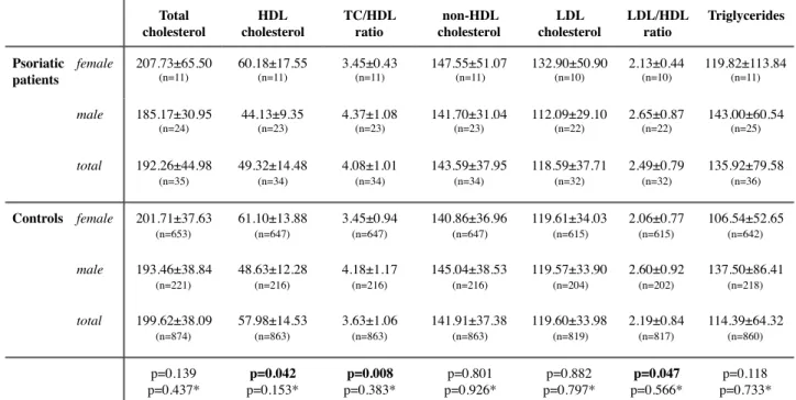
When trying to assess the differences in cardiovascular risk between psoriatic patients and controls, even though lower levels of HDL, higher TC/HDL and LDL/HDL ratios appear to be associated with psoriasis, once adjusted for gender and age these associations become statistically nonsignificant (table 5).
If the obese population (BMI≥30kg/m2) is excluded the results are more promising. It is
important to remember that this exclusion criterion could not be met in the psoriatic group because of lacking information. However, since obesity has so many interactions with what is being analyzed, it is felt to be of interest to show the massive change in outcomes.
A significant association was found between lower levels of HDL and psoriasis as well as with higher levels of ACE activity (table 6), after adjustment for age and gender. There is also a strong tendency for an increase in TC/HDL ratio and triglyceride levels in psoriasis, which comes in agreement with many of the basis of this study as well as previous studies [117].
Total cholesterol HDL cholesterol TC/HDL ratio non-HDL cholesterol LDL cholesterol LDL/HDL ratio Triglycerides Psoriatic patients female 207.73±65.50 (n=11) 60.18±17.55 (n=11) 3.45±0.43 (n=11) 147.55±51.07 (n=11) 132.90±50.90 (n=10) 2.13±0.44 (n=10) 119.82±113.84 (n=11) male 185.17±30.95 (n=24) 44.13±9.35 (n=23) 4.37±1.08 (n=23) 141.70±31.04 (n=23) 112.09±29.10 (n=22) 2.65±0.87 (n=22) 143.00±60.54 (n=25) total 192.26±44.98 (n=35) 49.32±14.48 (n=34) 4.08±1.01 (n=34) 143.59±37.95 (n=34) 118.59±37.71 (n=32) 2.49±0.79 (n=32) 135.92±79.58 (n=36) Controls female 201.71±37.63 (n=653) 61.10±13.88 (n=647) 3.45±0.94 (n=647) 140.86±36.96 (n=647) 119.61±34.03 (n=615) 2.06±0.77 (n=615) 106.54±52.65 (n=642) male 193.46±38.84 (n=221) 48.63±12.28 (n=216) 4.18±1.17 (n=216) 145.04±38.53 (n=216) 119.57±33.90 (n=204) 2.60±0.92 (n=202) 137.50±86.41 (n=218) total 199.62±38.09 (n=874) 57.98±14.53 (n=863) 3.63±1.06 (n=863) 141.91±37.38 (n=863) 119.60±33.98 (n=819) 2.19±0.84 (n=817) 114.39±64.32 (n=860) p=0.139 p=0.437* p=0.042 p=0.153* p=0.008 p=0.383* p=0.801 p=0.926* p=0.882 p=0.797* p=0.047 p=0.566* p=0.118 p=0.733*
Table 5: means for the different parameters of the lipid profile with standard deviation and number of cases for each subgroup; p-values calculated with t-test or Mann Whitney test according to the distribution of the samples; significance level defined at p≤0.05; *p-values adjusted for gender and age using linear regression
This result reinforces the idea that obesity does have an influence when it comes to assessing the ACE and psoriatic effect on lipid profile and therefore BMI should be calculated in both populations (controls and psoriatic) for a future study. It would also be relevant to inquire the subjects of both groups about their usual medication and exclude or adjust these findings to the intake of statins, other cholesterol lowering agents or any cardiovascular risk altering medication. Diabetes and hypertension are also comorbidities that influence cardiovascular risk and might be associated with psoriasis as discussed above and therefore should be considered in both the psoriatic and control groups and used to adjust statistical results. The clinical information provided regarding the psoriatic group did not include such diagnosis.
Total cholesterol HDL cholesterol TC/HDL ratio non-HDL cholesterol LDL cholesterol LDL/HDL ratio Triglycerides ACE activity Psoriatic patients f 207.73±65.50 (n=11) 60.18±17.55 (n=11) 3.45±0.43 (n=11) 147.55±51.07 (n=11) 132.90±50.90 (n=10) 2.13±0.44 (n=10) 119.82±113.84 (n=11) 17.38±7.67 (n=6) m 185.17±30.95 (n=24) 44.13±9.35 (n=23) 4.37±1.08 (n=23) 141.70±31.04 (n=23) 112.09±29.10 (n=22) 2.65±0.87 (n=22) 143.00±60.54 (n=25) 19.83±14.66 (n=14) t 192.26±44.98 (n=35) 49.32±14.48 (n=34) 4.08±1.01 (n=34) 143.59±37.95 (n=34) 118.59±37.71 (n=32) 2.49±0.79 (n=32) 135.92±79.58 (n=36) 19.09±12.80 (n=20) Controls f 200.54±36.89 (n=403) 63.87±14.42 (n=397) 3.27±0.83 (n=397) 137.07±35.39 (n=397) 118.13±33.26 (n=379) 1.95±0.72 (n=379) 96.20±43.07 (n=396) 11.61±6.75 (n=144) m 195.77±42.56 (n=130) 51.63±12.79 (n=128) 3.96±1.09 (n=128) 144.17±40.96 (n=128) 122.04±37.08 (n=122) 2.48±0.89 (n=121) 129.20±86.06 (n=128) 13.41±7.97 (n=34) t 199.37±38.37 (n=533) 60.89±14.98 (n=525) 3.44±0.95 (n=525) 138.80±36.91 (n=525) 119.09±34.23 (n=501) 2.08±0.80 (n=500) 104.26±58.32 (n=524) 11.95±7.01 (n=178) p=0.143 p=0.473* p<0.001 p=0.011* p<0.001 p=0.064* p=0.479 p=0.758* p=0.943 p=0.873* p=0.007 p=0.233* p=0.024 p=0.060* p=0.023 p=0.003*
Table 6: means for the different parameters of the lipid profile with standard deviation and number of cases for each subgroup when excluded subjects with BMI≥30kg/m2; p-values calculated with t-test or Mann Whitney test according to the distribution of the samples; significance level defined at p≤0,050;
*p-values adjusted for gender and age using linear regression; f-female, m-male, t-total
Image 1: on the left, HDL concentrations in psoriatic patients with mild (54.26±14.72) versus moderate to severe disease (41.67±11.04); on the right, triglyceride levels in patients with mild psoriasis (113.38±66.66) compared to patients with moderate to severe disease (163.56±44.95)
In order to estimate an association between psoriasis severity and cardiovascular risk, two subgroups of psoriatic patients were defined, knowing that PASI≤10 accounts for a mild psoriasis and PASI>10 includes both moderate to severe disease [118]. The results in table 7 show a significant association with some of the features in the patient’s lipid profile. The HDL level is significantly lower in psoriatic patients with moderate to severe disease when comparing to mild psoriasis (image 1). TC/HDL ratio was found to be higher in patients with a more severe disease, as well as the triglycerides level that is shown to be significantly higher when PASI>10, all of which when adjusting for age and gender. A tendency was also found for an increase in LDL/HDL ratio with psoriatic severity, after the mentioned adjustment.
PASI was not found to be significantly correlated with the ACE genotype (one-way ANOVA test p=0.116) with a nonsignificant higher frequency of the II genotype in psoriatic patients with moderate to severe disease (OR=2.25, p=0.338).
Total cholesterol HDL cholesterol TC/HDL ratio non-HDL cholesterol LDL cholesterol LDL/HDL ratio
Triglycerides ACE activity
PASI ≤10 196.60±46.20 (n=20) 54.26±14.72 (n=19) 3.76±0.89 (n=19) 143.74±39.51 (n=19) 121.42±37.41 (n=19) 2.31±0.69 (n=19) 113.38±66.66 (n=21) 10.24±8.22 (n=5) PASI >10 196.44±38.42 (n=9) 41.67±11.04 (n=9) 4.88±1.02 (n=9) 154.78±32.57 (n=9) 121.13±36.60 (n=8) 3.01±0.89 (n=8) 163.56±44.95 (n=9) 17.20±11.03 (n=4) p=0.814 p=0.604* p=0.028 p=0.022* p=0.012 p=0.011* p=0.279 p=0.228* p=0.811 p=0.559* p=0.035 p=0.089* p=0.021 p=0.020* p=0.327 p=0.297*
Table 7: means for the different parameters of the lipid profile with standard deviation and number of cases for each subgroup; p-values calculated with t-test or Mann Whitney test according to the distribution of the samples; significance level defined at p≤0.05; *p-values adjusted for gender and age
Image 2: graph showing association between TC/HDL ratio and psoriasis severity (estimated by PASI); pearson’s correlation; significance level defined at p≤0.05
r=0.612, p<0.001
Considering the fact that once PASI is divided into two groups there is some information that is lost in the analysis, the continuous variable was used to check for correlations with the pearson or spearman test (depending on the distribution). This showed that PASI is inversely associated with HDL cholesterol (r=-0.496, p=0.007) and positively correlated with LDL/HDL ratio (r=0.437, p=0.023) and triglycerides levels (r=0.421, p=0.020), indicating a decrease in HDL cholesterol with a concomitant increase in LDL/HDL ratio as well as in triglycerides with psoriasis severity.
As can be seen in image 2, PASI is also strongly correlated to TC/HDL ratio (r=0.612, p<0.001), which leads to believe that psoriasis severity is in fact associated with an increase in cardiovascular disease as the TC/HDL ratio is one of the lipid indices with higher predictive value [114, 119, 120].
Conclusions
This study found an association between psoriasis and a higher ACE activity level, independent of its genotype. This increase in ACE activity suggest an oxidative stress state that is characteristic of cardiovascular disorders since it is one of the precipitating factors in atherogenesis. The genotype was strongly associated with ACE activity as also found in previous studies, although no association between the ACE I/D polymorphism and this disease or its severity could be demonstrated in our population. It is felt that a larger psoriatic sample with their ACE activities measured would be of importance to assess the true influence of this enzyme in the cardiovascular risk seen in these patients, specially since there appears to be a nonsignificant increase in ACE activity in the population with increased cholesterol levels.
Psoriatic severity was associated with a higher cardiovascular risk estimated by a lower HDL concentrations (r=-0.496, p=0.007) and higher triglyceride levels (r=0.421, p=0.020), as well as TC/ HDL and LDL/HDL ratios (r=0.612, p<0.001 and r=0.437, p=0.023, respectively) which have been shown to have a stronger predictive value for cardiovascular diseases.
Further studies are crucial in order to keep enhancing our knowledge on the overlap of inflammation and psoriasis as a systemic autoimmune chronic disease. In a similar study, it would be important to have more information, with special relevance in using a larger psoriatic population sample and having better information regarding BMI and other comorbidities that alter cardiovascular risk, as mentioned before, as well as medication that could affect our lipid profile levels and underestimate the difference between populations. It would also be of value to have a control group that better represents the normal population, which might not be found within the hospital/clinic environment.
It would also be interesting to assess lipid function and form in psoriatic patients as they may have deeper disturbances in this metabolism than just merely a quantitative issue, as can be perceived in the literature.
Psoriatic patients should be seen as a risk group for cardiovascular disorders and controlled accordingly. New and better markers for severity and for assessing this risk, as ACE might be, may be of worthy pursuit.
Acknowledgments
Ao Professor Doutor Manuel Bicho, pelo conhecimento, orientação e apoio. Aos demais membros do laboratório de genética, bem como dos serviços de Dermatologia e Endocrinologia, pelos dados fornecidos.
Aos meus pais, por tudo e ainda mais. Ao Afonso e à Cara, por todas as aventuras e passeios e pela inspiração para investigar e saber.
Aos meus amigos, por estarem do meu lado, e ao Filipe, por tanta paciência durante todo este processo.
References
1. Langley, R.G., G.G. Krueger, and C.E. Griffiths, Psoriasis: epidemiology, clinical features, and quality of life. Ann Rheum Dis, 2005. 64 Suppl 2: p. ii18-23; discussion ii24-5.
2. Nestle, F.O., D.H. Kaplan, and J. Barker, Psoriasis. New England Journal of Medicine, 2009. 361(5): p. 496-509.
3. Menter, A., et al., Exploring the association between cardiovascular and other disease-related risk factors in the psoriasis population: the need for increased understanding across the medical community. J Eur Acad Dermatol Venereol, 2010. 24(12): p. 1371-7.
4. Parisi, R., et al., Global epidemiology of psoriasis: a systematic review of incidence and prevalence. J Invest Dermatol, 2013. 133(2): p. 377-85.
5. Gottlieb, A.B. and F. Dann, Comorbidities in Patients with Psoriasis. The American Journal of Medicine, 2009. 122(12): p. 1150.e1-1150.e9.
6. Griffiths, C.E. and J.N. Barker, Pathogenesis and clinical features of psoriasis. Lancet, 2007. 370(9583): p. 263-71.
7. Yeung, H., et al., Psoriasis severity and the prevalence of major medical comorbidity: a population-based study. JAMA Dermatol, 2013. 149(10): p. 1173-9.
8. Yim, K.M. and A.W. Armstrong, Updates on cardiovascular comorbidities associated with psoriatic diseases: epidemiology and mechanisms. Rheumatology International, 2016: p. 1-9.
9. Pariser, D.M., et al., National Psoriasis Foundation clinical consensus on disease severity. Arch Dermatol, 2007. 143(2): p. 239-42.
10. Neimann, A.L., et al., Prevalence of cardiovascular risk factors in patients with psoriasis. J Am Acad Dermatol, 2006. 55(5): p. 829-35.
11. Herron, M.D., et al., Impact of obesity and smoking on psoriasis presentation and management. Arch Dermatol, 2005. 141(12): p. 1527-34.
12. Naldi, L., et al., Cigarette Smoking, Body Mass Index, and Stressful Life Events as Risk Factors for Psoriasis: Results from an Italian Case–Control Study. Journal of Investigative Dermatology, 2005. 125(1): p. 61-67.
13. Setty, A.R., G. Curhan, and H.K. Choi, Obesity, waist circumference, weight change, and the risk of psoriasis in women: Nurses' Health Study II. Arch Intern Med, 2007. 167(15): p. 1670-5.
14. Paller, A.S., et al., Association of pediatric psoriasis severity with excess and central adiposity: an international cross-sectional study. JAMA Dermatol, 2013. 149(2): p. 166-76.
15. Asia Pacific Cohort Studies, C., Central obesity and risk of cardiovascular disease in the Asia Pacific Region. Asia Pac J Clin Nutr, 2006. 15(3): p. 287-92.
16. Després, J.-P., Body Fat Distribution and Risk of Cardiovascular Disease. An Update, 2012. 126(10): p. 1301-1313.
17. Gisondi, P., et al., Weight loss improves the response of obese patients with moderate-to-severe chronic plaque psoriasis to low-dose cyclosporine therapy: a randomized, controlled, investigator-blinded clinical trial. Am J Clin Nutr, 2008. 88(5): p. 1242-7.
18. Moon, Y.S., D.H. Kim, and D.K. Song, Serum tumor necrosis factor-alpha levels and components of the metabolic syndrome in obese adolescents. Metabolism, 2004. 53(7): p. 863-7.
19. Strober, B., et al., Effects of etanercept on C-reactive protein levels in psoriasis and psoriatic arthritis. Br J Dermatol, 2008. 159(2): p. 322-30.
20. Chodorowska, G., D. Wojnowska, and M. Juszkiewicz-Borowiec, C-reactive protein and alpha2-macroglobulin plasma activity in medium-severe and severe psoriasis. J Eur Acad Dermatol Venereol, 2004. 18(2): p. 180-3.
21. Armstrong, A.W., et al., Psoriasis and hypertension severity: results from a case-control study. PLoS One, 2011. 6(3): p. e18227.
22. Takeshita, J., et al., Effect of psoriasis severity on hypertension control: a population-based study in the United Kingdom. JAMA Dermatol, 2015. 151(2): p. 161-9.
23. Akhyani, M., et al., The lipid profile in psoriasis: a controlled study. J Eur Acad Dermatol Venereol, 2007. 21(10): p. 1330-2.
24. Tekin, N.S., et al., Accumulation of oxidized low-density lipoprotein in psoriatic skin and changes of plasma lipid levels in psoriatic patients. Mediators Inflamm, 2007. 2007: p. 78454.
25. Mallbris, L., et al., Psoriasis is associated with lipid abnormalities at the onset of skin disease. J Am Acad Dermatol, 2006. 54(4): p. 614-21.
26. Fantuzzi, G., Adipose tissue, adipokines, and inflammation. J Allergy Clin Immunol, 2005. 115(5): p. 911-9; quiz 920.
27. Jung, U.J. and M.S. Choi, Obesity and Its Metabolic Complications: The Role of Adipokines and the Relationship between Obesity, Inflammation, Insulin Resistance, Dyslipidemia and Nonalcoholic Fatty Liver Disease. Int J Mol Sci, 2014. 15(4): p. 6184-223.
28. Mehta, N.N., et al., Abnormal lipoprotein particles and cholesterol efflux capacity in patients with psoriasis. Atherosclerosis, 2012. 224(1): p. 218-21.
29. Yu, Y., et al., Aortic vascular inflammation in psoriasis is associated with HDL particle size and concentration: a pilot study. Am J Cardiovasc Dis, 2012. 2(4): p. 285-92.
30. Salahuddin, T., et al., Cholesterol efflux capacity in humans with psoriasis is inversely related to non-calcified burden of coronary atherosclerosis. Eur Heart J, 2015. 36(39): p. 2662-5.
31. Holzer, M., et al., Psoriasis alters HDL composition and cholesterol efflux capacity. J Lipid Res, 2012. 53(8): p. 1618-24.
32. National Cholesterol Education Program Expert Panel on Detection, E. and A. Treatment of High Blood Cholesterol in, Third Report of the National Cholesterol Education Program (NCEP) Expert Panel on Detection, Evaluation, and Treatment of High Blood Cholesterol in Adults (Adult Treatment Panel III) final report. Circulation, 2002. 106(25): p. 3143-421.
33. Love, T.J., et al., Prevalence of the metabolic syndrome in psoriasis: results from the National Health and Nutrition Examination Survey, 2003-2006. Arch Dermatol, 2011. 147(4): p. 419-24.
34. Ronti, T., G. Lupattelli, and E. Mannarino, The endocrine function of adipose tissue: an update. Clin Endocrinol (Oxf), 2006. 64(4): p. 355-65.
35. Davidovici, B.B., et al., Psoriasis and systemic inflammatory diseases: potential mechanistic links between skin disease and co-morbid conditions. J Invest Dermatol, 2010. 130(7): p. 1785-96.
36. Schluter, K., et al., Insulin Resistance May Contribute to Upregulation of Adhesion Molecules on Endothelial Cells in Psoriatic Plaques. Acta Derm Venereol, 2016. 96(2): p. 162-8.
37. Buerger, C., et al., Interleukin-1beta interferes with epidermal homeostasis through induction of insulin resistance: implications for psoriasis pathogenesis. J Invest Dermatol, 2012.
132(9): p. 2206-14.
38. Nguyen, M.T., et al., JNK and tumor necrosis factor-alpha mediate free fatty acid-induced insulin resistance in 3T3-L1 adipocytes. J Biol Chem, 2005. 280(42): p. 35361-71.
39. Gani, D.K., et al., Evaluation of C-reactive protein and interleukin-6 in the peripheral blood of patients with chronic periodontitis. J Indian Soc Periodontol, 2009. 13(2): p. 69-74.
40. Whitehead, J.P., et al., Adiponectin--a key adipokine in the metabolic syndrome. Diabetes Obes Metab, 2006. 8(3): p. 264-80.
41. Yu, X.H., et al., Foam cells in atherosclerosis. Clin Chim Acta, 2013. 424: p. 245-52. 42. Pischon, T., et al., Plasma adiponectin levels and risk of myocardial infarction in men. Jama, 2004. 291(14): p. 1730-7.
43. Takahashi, M., et al., Plasma leptin levels and body fat distribution. Horm Metab Res, 1996. 28(12): p. 751-2.
44. Mallamaci, F., et al., Adiponectin in essential hypertension. J Nephrol, 2002. 15(5): p. 507-11.
45. Kumada, M., et al., Association of hypoadiponectinemia with coronary artery disease in men. Arterioscler Thromb Vasc Biol, 2003. 23(1): p. 85-9.
46. Lilja, M., et al., Higher leptin levels in Asian Indians than Creoles and Europids: a potential explanation for increased metabolic risk. Int J Obes (Lond), 2010. 34(5): p. 878-85.
47. Ciccone, M., et al., Plasma leptin is independently associated with the intima-media thickness of the common carotid artery. Int J Obes Relat Metab Disord, 2001. 25(6): p. 805-10.
48. Cerman, A.A., et al., Serum leptin levels, skin leptin and leptin receptor expression in psoriasis. Br J Dermatol, 2008. 159(4): p. 820-6.
49. Edson-Heredia, E., et al., Prevalence and incidence rates of cardiovascular, autoimmune, and other diseases in patients with psoriatic or psoriatic arthritis: a retrospective study using Clinical Practice Research Datalink. J Eur Acad Dermatol Venereol, 2015. 29(5): p. 955-63.
50. Sommer, D.M., et al., Increased prevalence of the metabolic syndrome in patients with moderate to severe psoriasis. Arch Dermatol Res, 2006. 298(7): p. 321-8.
51. Wolf, N., et al., Psoriasis is associated with pleiotropic susceptibility loci identified in type II diabetes and Crohn disease. J Med Genet, 2008. 45(2): p. 114-6.
52. Brauchli, Y.B., S.S. Jick, and C.R. Meier, Psoriasis and the risk of incident diabetes mellitus: a population-based study. Br J Dermatol, 2008. 159(6): p. 1331-7.
53. Gelfand, J.M., et al., The risk of mortality in patients with psoriasis: results from a population-based study. Arch Dermatol, 2007. 143(12): p. 1493-9.
54. Ogdie, A., et al., Risk of mortality in patients with psoriatic arthritis, rheumatoid arthritis and psoriasis: a longitudinal cohort study. Ann Rheum Dis, 2014. 73(1): p. 149-53.
55. Abuabara, K., et al., Cause-specific mortality in patients with severe psoriasis: a population-based cohort study in the U.K. Br J Dermatol, 2010. 163(3): p. 586-92.
56. Gelfand, J.M., et al., Risk of myocardial infarction in patients with psoriasis. Jama, 2006. 296(14): p. 1735-41.
57. Vliegenthart, R., et al., Coronary Calcification Improves Cardiovascular Risk Prediction in the Elderly. Circulation, 2005. 112(4): p. 572-577.
58. Polonsky, T.S., et al., Coronary Artery Calcium Score and Risk Classification for Coronary Heart Disease Prediction: The Multi-Ethnic Study of Atherosclerosis. Jama, 2010.
303(16): p. 1610-6.
59. McClelland, R.L., et al., 10-Year Coronary Heart Disease Risk Prediction Using Coronary Artery Calcium and Traditional Risk Factors: Derivation in the MESA (Multi-Ethnic Study of Atherosclerosis) With Validation in the HNR (Heinz Nixdorf Recall) Study and the DHS (Dallas Heart Study). J Am Coll Cardiol, 2015. 66(15): p. 1643-53.
60. Bots, M.L., et al., Carotid Intima-media Thickness Measurements: Relations with Atherosclerosis, Risk of Cardiovascular Disease and Application in Randomized Controlled Trials. Chin Med J (Engl), 2016. 129(2): p. 215-26.
61. Bańska-Kisiel, K., et al., Carotid intima-media thickness in patients with mild or moderate psoriasis. Advances in Dermatology and Allergology, 2016. 4(4): p. 286-289.
62. Ludwig, R.J., et al., Psoriasis: a possible risk factor for development of coronary artery calcification. British Journal of Dermatology, 2007. 156(2): p. 271-276.
63. Wang, Y., et al., Chronic skin-specific inflammation promotes vascular inflammation and thrombosis. J Invest Dermatol, 2012. 132(8): p. 2067-75.
64. Prodanovich, S., et al., Methotrexate reduces incidence of vascular diseases in veterans with psoriasis or rheumatoid arthritis. J Am Acad Dermatol, 2005. 52(2): p. 262-7.
65. van Ede, A.E., et al., Homocysteine and folate status in methotrexate-treated patients with rheumatoid arthritis. Rheumatology (Oxford), 2002. 41(6): p. 658-65.
66. Ganguly, P. and S.F. Alam, Role of homocysteine in the development of cardiovascular disease. Nutr J, 2015. 14.
67. Ridker, P.M., et al., Non-HDL cholesterol, apolipoproteins A-I and B100, standard lipid measures, lipid ratios, and CRP as risk factors for cardiovascular disease in women. Jama, 2005. 294(3): p. 326-33.
68. Boehncke, S., et al., Systemic therapy of plaque-type psoriasis ameliorates endothelial cell function: results of a prospective longitudinal pilot trial. Archives of Dermatological Research, 2011. 303(6): p. 381-388.
69. Santilli, S., et al., Visualization of atherosclerosis as detected by coronary artery calcium and carotid intima-media thickness reveals significant atherosclerosis in a cross-sectional study of psoriasis patients in a tertiary care center. J Transl Med, 2016. 14(1): p. 217.
70. Armstrong, A.W., et al., A tale of two plaques: convergent mechanisms of T-cell-mediated inflammation in psoriasis and atherosclerosis. Exp Dermatol, 2011. 20(7): p. 544-9.
71. Lowes, M.A., M. Suárez-Fariñas, and J.G. Krueger, Immunology of Psoriasis. Annu Rev Immunol, 2014. 32: p. 227-55.
72. Mahil, S.K., F. Capon, and J.N. Barker, Update on psoriasis immunopathogenesis and targeted immunotherapy. Semin Immunopathol, 2016. 38: p. 11-27.
73. Hashmi, S. and Q.T. Zeng, Role of interleukin-17 and interleukin-17-induced cytokines interleukin-6 and interleukin-8 in unstable coronary artery disease. Coron Artery Dis, 2006. 17(8): p. 699-706.
74. Wang, Z., et al., Increased Th17 cells in coronary artery disease are associated with neutrophilic inflammation. Scand Cardiovasc J, 2011. 45(1): p. 54-61.
75. Ziats, N.P. and A.L. Robertson, Jr., Effects of peripheral blood monocytes on human vascular cell proliferation. Atherosclerosis, 1981. 38(3-4): p. 401-10.
76. Chen, Y.L., Y.J. Chang, and M.J. Jiang, Monocyte chemotactic protein-1 gene and protein expression in atherogenesis of hypercholesterolemic rabbits. Atherosclerosis, 1999. 143(1): p. 115-23.
77. Golden, J.B., et al., Chronic Psoriatic Skin Inflammation Leads to Increased Monocyte Adhesion and Aggregation. J Immunol, 2015. 195(5): p. 2006-18.
78. Liu, X.L., et al., Local Gene Silencing of Monocyte Chemoattractant Protein-1 Prevents Vulnerable Plaque Disruption in Apolipoprotein E-Knockout Mice. PLoS One, 2012. 7(3).
79. Mehta, N.N., et al., Modulation of cardiometabolic pathways in skin and serum from patients with psoriasis. J Transl Med, 2013. 11: p. 194.
80. Park, M.H., et al., Age-related inflammation and insulin resistance: a review of their intricate interdependency. Arch Pharm Res, 2014. 37(12): p. 1507-14.
81. Boehncke, W.H. and S. Boehncke, Cardiovascular mortality in psoriasis and psoriatic arthritis: epidemiology, pathomechanisms, therapeutic implications, and perspectives. Curr Rheumatol Rep, 2012. 14(4): p. 343-8.
82. Rajwani, A., R.M. Cubbon, and S.B. Wheatcroft, Cell-specific insulin resistance: implications for atherosclerosis. Diabetes Metab Res Rev, 2012. 28(8): p. 627-34.
83. Azfar, R.S. and J.M. Gelfand, PSORIASIS AND METABOLIC DISEASE: EPIDEMIOLOGY AND PATHOPHYSIOLOGY. Curr Opin Rheumatol, 2008. 20(4): p. 416-22.
84. Chandra, A., et al., Genetic and epigenetic basis of psoriasis pathogenesis. Molecular Immunology, 2015. 64(2): p. 313-323.
85. Al Robaee, A.A., Molecular genetics of Psoriasis (Principles, technology, gene location, genetic polymorphism and gene expression). Int J Health Sci (Qassim), 2010. 4(2): p. 103-27.
86. Kranzhofer, R., et al., Angiotensin induces inflammatory activation of human vascular smooth muscle cells. Arterioscler Thromb Vasc Biol, 1999. 19(7): p. 1623-9.
87. Takahashi, T., et al., Participation of reactive oxygen intermediates in the angiotensin II-activated signaling pathways in vascular smooth muscle cells. Ann N Y Acad Sci, 2000. 902: p. 283-7.
88. Taniyama, Y., et al., Mechanisms of reactive oxygen species-dependent downregulation of insulin receptor substrate-1 by angiotensin II. Arterioscler Thromb Vasc Biol, 2005. 25(6): p. 1142-7.
89. Hayashi, R., et al., Bradykinin stimulates IL-6 and IL-8 production by human lung fibroblasts through ERK- and p38 MAPK-dependent mechanisms. Eur Respir J, 2000. 16(3): p. 452-8.
90. Wolf, R., A. Tamir, and S. Brenner, Psoriasis related to angiotensin-converting enzyme inhibitors. Dermatologica, 1990. 181(1): p. 51-3.
91. Cohen, A.D., et al., Drug exposure and psoriasis vulgaris: control and case-crossover studies. Acta Derm Venereol, 2005. 85(4): p. 299-303.
92. Husain, K., et al., Inflammation, oxidative stress and renin angiotensin system in atherosclerosis. World J Biol Chem, 2015. 6(3): p. 209-17.
93. Rigat, B., et al., PCR detection of the insertion/deletion polymorphism of the human angiotensin converting enzyme gene (DCP1) (dipeptidyl carboxypeptidase 1). Nucleic Acids Research, 1992. 20(6): p. 1433-1433.
94. Danser, A.H., et al., Angiotensin-converting enzyme in the human heart. Effect of the deletion/insertion polymorphism. Circulation, 1995. 92(6): p. 1387-8.
95. Rigat, B., et al., An insertion/deletion polymorphism in the angiotensin I-converting enzyme gene accounting for half the variance of serum enzyme levels. J Clin Invest, 1990. 86(4): p. 1343-6.
96. Ryder, K.W., et al., Serum angiotensin converting enzyme activity in patients with psoriasis. Clin Chim Acta, 1985. 153(2): p. 143-6.
97. Huskic, J., et al., Serum and tissue angiotensin converting enzyme in patients with psoriasis. Coll Antropol, 2008. 32(4): p. 1215-9.
98. Hanafy, S., M. Tavasoli, and F. Jamali, Inflammation alters angiotensin converting enzymes (ACE and ACE-2) balance in rat heart. Inflammation, 2011. 34(6): p. 609-13.
99. Raff, M., H. Bardach, and M.M. Muller, [Angiotensin-converting enzyme (ACE). Increased serum activities in patients with dermatological diseases (author's transl)]. Arch Dermatol Res, 1981. 270(3): p. 341-5.
100. Guang, C., et al., Three key proteases--angiotensin-I-converting enzyme (ACE), ACE2 and renin--within and beyond the renin-angiotensin system. Arch Cardiovasc Dis, 2012.
105(6-7): p. 373-85.
101. Coto-Segura, P., et al., Lack of association between angiotensin I-converting enzyme insertion/deletion polymorphism and psoriasis or psoriatic arthritis in Spain. Int J Dermatol, 2009.
48(12): p. 1320-3.
102. Chang, Y.C., et al., Association between the insertion/deletion polymorphism of the angiotensin I-converting enzyme gene and risk for psoriasis in a Chinese population in Taiwan. Br J Dermatol, 2007. 156(4): p. 642-5.
103. Yang, K., et al., Angiotensin-converting enzyme insertion/deletion polymorphism and susceptibility to psoriasis in a Chinese population. J Renin Angiotensin Aldosterone Syst, 2014.
15(1): p. 39-43.
104. Ozkur, M., et al., Association of insertion/deletion polymorphism of the angiotensin-converting enzyme gene with psoriasis. Br J Dermatol, 2004. 151(4): p. 792-5.
105. Weger, W., et al., The angiotensin-converting enzyme insertion/deletion and the endothelin -134 3A/4A gene polymorphisms in patients with chronic plaque psoriasis. Exp Dermatol, 2007. 16(12): p. 993-8.
106. Veletza, S., et al., Assessment of insertion/deletion polymorphism of the angiotensin converting enzyme gene in psoriasis. J Dermatol Sci, 2008. 49(1): p. 85-7.
107. Wu, C.K., et al., Renin-Angiotensin System Genes Polymorphisms and Long-Term Prognosis in Taiwanese Patients with Hypertension and Coronary Artery Disease. Acta Cardiol Sin, 2013. 29(1): p. 28-36.
108. Cosenso-Martin, L.N., et al., Angiotensin-converting enzyme insertion/deletion polymorphism, 24-h blood pressure profile and left ventricular hypertrophy in hypertensive individuals: a cross-sectional study. Eur J Med Res, 2015. 20(1).
109. Dhar, S., et al., Polymorphism of ACE gene as the genetic predisposition of coronary artery disease in Eastern India. Indian Heart J, 2012. 64(6): p. 576-81.
110. Millán, J., et al., Lipoprotein ratios: Physiological significance and clinical usefulness in cardiovascular prevention. Vasc Health Risk Manag, 2009. 5: p. 757-65.
111. Yusuf, S., et al., Effect of potentially modifiable risk factors associated with myocardial infarction in 52 countries (the INTERHEART study): case-control study. Lancet, 2004.
364(9438): p. 937-52.
112. Boutin, A.T., et al., Epidermal sensing of oxygen is essential for systemic hypoxic response. Cell, 2008. 133(2): p. 223-34.
113. Stocker, R. and J.F. Keaney, Jr., Role of oxidative modifications in atherosclerosis. Physiol Rev, 2004. 84(4): p. 1381-478.
114. Kinosian, B., H. Glick, and G. Garland, Cholesterol and coronary heart disease: predicting risks by levels and ratios. Ann Intern Med, 1994. 121(9): p. 641-7.
115. Ascaso, J., et al., Management of dyslipidemia in the metabolic syndrome: recommendations of the Spanish HDL-Forum. Am J Cardiovasc Drugs, 2007. 7(1): p. 39-58.
116. Harp, J.B., S.A. Henry, and M. DiGirolamo, Dietary weight loss decreases serum angiotensin-converting enzyme activity in obese adults. Obes Res, 2002. 10(10): p. 985-90.
117. Pietrzak, A., et al., Lipid Disturbances in Psoriasis: An Update. Mediators of Inflammation, 2010. 2010: p. 13.
118. Mrowietz, U., et al., Definition of treatment goals for moderate to severe psoriasis: a European consensus. Arch Dermatol Res, 2011. 303(1): p. 1-10.
119. Shai, I., et al., Multivariate assessment of lipid parameters as predictors of coronary heart disease among postmenopausal women: potential implications for clinical guidelines. Circulation, 2004. 110(18): p. 2824-30.
120. Hsia, S.H., et al., A population-based, cross-sectional comparison of lipid-related indexes for symptoms of atherosclerotic disease. Am J Cardiol, 2006. 98(8): p. 1047-52.
Attachments
Attachment 1:
Image 3: electrophoresis gel for the ACE i/d polymorphism; the first column shows a DD fenotype with a fragment of 190bp and the second shows the heterozygous phenotype with the D fragment and the I
fragment with 477bp; II genotype not displayed in this set; performed in the lab in May 2015