Universidade de Lisboa Faculdade de Ciências Departamento de Biologia Vegetal
Screening and isolation of compounds with antimicrobial activity produced by
multi-resistant bacteria.
Dissertação
Bruno Miguel Prazeres do Espírito Santo
Mestrado em Microbiologia Aplicada
2014
Universidade de Lisboa Faculdade de Ciências Departamento de Biologia Vegetal
Screening and isolation of compounds with antimicrobial activity produced by
multi-resistant bacteria.
Dissertação orientada por Doutor Ricardo P. M. Dias (BioFIG) e Prof. Dr. Francisco Dionísio (FCUL)
Bruno Miguel Prazeres do Espírito Santo
Mestrado em Microbiologia Aplicada
Screening and isolation of compounds with antimicrobial activity produced by
multi-resistant bacteria.
Bruno Miguel Prazeres do Espírito Santo
Master Thesis
2014
This thesis was fully performed at the Centre for Biodiversity, Functional and
Integrative Genomics (BioFIG), Faculty of Sciences of the University of Lisbon
(TecLabs-FCUL) under the direct supervision of Dr. Ricardo P. M. Dias.
Prof. Dr. Francisco Dionísio was the internal designated supervisor in the scope
of the Master in Applied Microbiology of the Faculty of Sciences of the University
of Lisbon.
i
Acknowledgements
My sincere thanks go to everyone who, directly or indirectly, made the realization of this master’s thesis possible. In all honestly, a long term project such as a thesis is always the reflection of the support from all the people who took the time and effort to help in any way they could. For that I’m truly thankful.
I would like to begin by thanking Professor Rogério Tenreiro, for welcoming me and giving the opportunity to preform my studies at the BioFIG.
Secondly, I would like to show all my gratitude to my external supervisor Dr. Ricardo Dias, for all the patience, guidance, support and helpful conversation which always stimulated my scientific criticism that helped me evolve as a scientist, as well as solving some problems found “on the road”.
I’m very thankful to Professor Francisco Dionísio, for the availability and for accepting being my internal supervisor.
I would like to thank Dr. Alice Martins for taking the time to assist me with the chemical components of the thesis, without whose help I would be completely lost in the world of analytical chemistry. The exceptional guidance provided and the unwearying availability was crucial for the development of this thesis. I can’t thank you enough. Additionally I’d like to thank Sofia Alves for taking upon her time to stop her thesis work to inject my GC-MS samples, and thank you for the help “deciphering” the results.
Thanks to Professor Ana Tenreiro and Dr. Sandra Chaves for suggesting the work developed by Dr. Ricardo Dias as a possible thesis theme. Additionally I would like to thank for all the help in the laboratory and for taking the time to answer all my questions.
I also like to appreciate the support and help rendered by my friends and fellows at the BioFIG research lab including Claudia Luís, Marta Pacheco, Luís Sobral and David Ferreira for their shared experiences, both professional and personal, which always allowed me to preform my work either better or with a happier face! Thank you all.
Last but definitely not the least, I’d like to thank my family and friends for all the support they have always given me and encouragement to always keep moving forward. A special thanks to my parents, for the brilliant education they gave me, which helped me (so far ☺) throughout the 24 years of my life.
ii
Abstract
In the late 20s, after Sir Alexander Fleming discovered the penicillin, a large interest in the search for compounds with antimicrobial activity produced by microorganisms emerged. This event marked the beginning of a new era of antibiotic discovery, where numerous scientist would screen for antibiotics in microorganisms, mostly from the soil.
Previous work undertaken at LMB-BioFIG, suggest that ΔphoQ mutation in
Stenotrophomonas maltophilia D457 strain is associated to an increased antimicrobial activity.
The main focus of this work was to: determine the existence of compounds with antimicrobial activity produced by the aforementioned mutant; determine its spectrum of activity and optimize the culture conditions that are optimal to its production. To answer these questions, a series of biologic susceptibility assays were performed, with both the parental D457 strain and D457ΔphoQ strain. Namely the cross-streak assay, agar-well diffusion assay and drop-diffusion assay. For the later, different culture conditions for the producing strains were tested in order to better understand which conditions favour the compound production, and also, sequential extractions with different organic solvents with different polar characteristics were made to fractionate the obtained batches regarding the polarity of its content, and tested for biologic activity.
Finally, in an attempt to unveil the compound of interest, protein quantification, thin layer chromatography and gas chromatography-mass spectrometry analysis were performed on all extracts to better elucidate the nature and structure of the compound.
This work revealed that the compound of interest is of non-polar nature produced in low concentrations, around 0.1 g/L. Was only present in the ethyl acetate fraction, with inhibitory activity against the test strains. In neither biologic assay did the parental strain show any inhibitory activity. Additionally, it was revealed by protein quantification assays that the mutant strain secreted almost 3-fold more protein than the parental strain. The results obtained by GC-MS suggest that the compound of interest resembles with the 2,3-dihydroxybenzoic acid, with possible presence of cyclic structures, such as aromatic rings in its molecular structure. However, further analytical procedures must be undertaken to clarify and confirm the structure of the secreted compounds by D457ΔphoQ strain.
iii
Resumo
No final dos anos 20, após Sir Alexander Fleming ter descoberto a penicilina, emergiu um grande interesse na pesquisa de compostos com atividade antimicrobiana a partir de microrganismos. Este evento marcou o início de uma nova era na descoberta de antibióticos, onde vários cientistas focaram os esforços na pesquisa de antibióticos em microrganismos, especialmente com origem no solo em particular espécies do género Penicillium e
Streptomyces. No entanto, nem todos os microrganismos são suscetíveis a estes compostos.
Determinadas bactérias e fungos, possuem uma enorme diversidade de mecanismos que conferem resistência a esses compostos. Estes mecanismos de defesa podem basear-se em reações enzimáticas que levam à alteração da estrutura do composto que levam à sua inativação, em aumento de número de bombas de efluxo membranares que rapidamente expulsam os compostos do meio intracelular ou finalmente em alterações no local de ligação do composto que levam à interrupção da ligação entre o antibiótico e o local de ação. A presença dos referidos mecanismos é de elevada relevância medica quando presentes em bactérias patogénicas, uma vez que dificulta o tratamento de doenças pelos métodos convencionais. Atualmente, o número de mortes associadas a infeções causadas por bactérias multirresistentes ronda os 25000 apenas na União Europeia, e este estigma custa cerca de 1,5 mil milhões de euros todos os anos.
Stenotrophomonas maltophilia D457 é uma bactéria gram-negativa, popular por ser um
organismo com uma exímia capacidade de resistência a antibióticos e metais pesados, ainda, na qualidade de agente patogénico oportunista, está vulgarmente associado a infeções em pacientes cujo sistema imunitário se encontra debilitado e a infeções originadas em hospitais. As infeções mais frequentes causadas por este agente patogénico são associadas a septicemias causadas por cateteres e infeções respiratórias em doentes com fibrose cística.
Trabalho desenvolvido previamente no LMB-BioFIG, sugere que uma mutação de
frame-knockout mutação do gene phoQ na estirpe Stenotrophomonas maltophilia D457 está
associada a um aumento na produção de um composto com atividade antimicrobiana. O gene
phoQ faz parte de um sistema de dois componentes PhoP-PhoQ regulador de virulência em
diversas bactérias patogénicas. O foco principal deste trabalho foi: determinar a existência de compostos com atividade antimicrobiana produzidos pelo mutante mencionado anteriormente; determinar qual o seu espectro de atividade e otimizar as condições de cultura que são mais favoráveis para a sua produção. Para responder a estas questões, diversos ensaios de atividade biológica foram realizados com ambas as estirpes, quer a parental D457 quer a mutante D457ΔphoQ. Nomeadamente ensaios de cross-streak, difusão de poços de agar e difusão de gotas. Para este último, diferentes condições de crescimento das estirpes produtoras foram testados para determinar quais as condições que favoreciam a produção
iv
dos compostos com atividade antimicrobiana, ainda, extrações sequenciais usando diferentes solventes orgânicos, (diclorometano, acetato de etilo e metanol) foram realizadas com o intuito de fracionar os produtos obtidos do crescimento das estirpes produtoras de acordo com a polaridade dos seus conteúdos, culminando no teste da sua atividade biológica contra estirpes da biblioteca do laboratório cuja característica principal é a sua elevada resistência a antibióticos.
Finalmente, na tentativa de desvendar qual o composto de interesse, ensaios de quantificação de proteína, cromatografia em camada fina e cromatografia gasosa-espectrometria de massa foram realizados a todos os extratos para melhor elucidar a natureza e a estrutura do composto.
Este trabalho revelou que o composto de interesse é de natureza apolar e é produzido em baixas concentrações, menos de 0,1 g/L. Encontra-se apenas presente na fração de acetato de etilo. Esta fração revelou atividade biológica contra diversas estirpes teste, nomeadamente, Acinetobacter haemolyticus VR, Stenotrophomonas maltophilia D457R,
Pseudomonas aeruginosa PAO1, Escherichia coli ESLB, Bacillus cereus B4379 e Enterococcus faecalis ATCC 29212. Em nenhum ensaio de atividade biológica, a estirpe
parental revelou inibição de crescimento das estirpes teste. Ensaios de quantificação de proteína revelaram que o mutante apresenta uma maior produção de proteínas comparativamente com a estirpe parental, num grau de grandeza 3 vezes superior à parental. Os resultados do GC-MS sugeriram que o composto com atividade biológica se assemelha com o ácido 2,3-dihidroxi benzoico e poderá possuir estruturas cíclicas como anéis aromáticos na sua estrutura. No entanto, adicionais procedimentos analíticos serão necessários para clarificar e confirmar a estrutura dos compostos produzidos pelo mutante ΔphoQ.
v
Table of Contents
Table of Contents
Acknowledgements
... iAbstract
... iiResumo
... iiiTable of Contents
... v1. Introduction
... 11.1 Microbes and Antibiotics ... 1
1.2 Antibiotic resistance ... 2
1.2.1 Mechanisms of action vs. resistance ... 3
1.3 Research and development of antibiotic agents ... 5
1.4 Stenotrophomonas maltophilia ... 9
1.4.1 Stenotrophomonas maltophilia D457 vs. ΔphoQ ...10
2. Thesis goals
...123. Materials and Methods
...133.1 Bacterial strains and growth conditions ...13
3.2 Swimming and Swarming ...14
3.3 Expression of compounds with antimicrobial activity ...14
3.3.1 Liquid medium ...14
3.3.2 Solid medium ...15
3.4 Active Compounds Isolation ...15
3.4.1 Liquid-Liquid Extraction ...15
3.4.2 Solid-Liquid Extraction...16
3.4.3 Extract Concentration ...16
3.5 Biological Activity Assays...16
3.5.1 Cross-Streak Assay ...16
3.5.2 Agar-Well Diffusion Method ...17
vi
3.6 Structure Elucidation ...18
3.6.1 Thin layer chromatography (TLC) ...18
3.6.2 Gas chromatography-Mass spectrometry (GC-MS) ...18
3.7 Protein Quantification ...18
3.7.1 Spectrum analysis and Protein Quantification by UV ...19
3.7.2 Biuret assay ...19
4.
Results and discussion
...204.1 Growth characteristics ...20
4.2 Biologic Assays ...21
4.3 Protein quantification ...25
4.4 Thin layer chromatography assay ...27
4.5 Gas chromatography-Mass spectrometry analysis ...28
5. Conclusion
...316. Bibliography
...321
1.
Introduction
1.1 Microbes and Antibiotics
Even though chemical warfare is considered, by modern society, the most dangerous and politically incorrect way of resolving conflicts, bacteria and fungi don’t share the same opinion. In fact, microorganisms are veterans in this subject. Since very long, we’re aware that
Penicillium rubrum, Penicillium notatum Lactococcus lactis and Streptomyces griseus produce
very important molecules that are used to compete against other species in the environment (Fleming 1929; Rogers 1928; Schatz et al. 1944). In 1942, Dr. Selman Waksman used for the first time the term “antibiotic” to refer a substance with the ability to kill bacteria and fungi or slow eventually to a stop their growth (Waksman 1947).
Generally, the production of these molecules grant a fair advantage when microbes are put across other species that are competing for the same food/energy resources. Yet, the production of these antibiotics doesn’t come without a cost. The microorganism that produces a certain antibiotic, has to be, at some point, resistant to that same compound otherwise he would die. Both the production and resistance to a certain compound, requires extra energetic and material burdens to the microorganism making him less fit in some occasions. For example, when two strains, one producing and one sensitive to the antibiotic, are put together, obviously the producing strain is going to be the fittest and consequently will have higher growth rates than the sensitive, because the toxin/antibiotic produced will eliminate or at least slow the competitors’ growth. On the other hand, if a producing strain and a resistant-only strain are put together, the tables will turn and the producing strain won’t be as fit anymore, because the resistant-only bacteria doesn’t have the extra energetic and material cost from producing the antibiotic and as it is resistant to the antibiotic it won’t have any effect on its growth. The remaining scenario is when a sensitive strain and a resistant-only strain are put across, the one who will reproduce more will be the one with less energetic/material costs, which in this case, is the sensitive. This is the basic concept of the “rock-paper-scissors” evolutionary game (Lenski & Riley 2002). So it appears that there is some kind of “love-hate” relation between microbes and antibiotics.
Currently, the ability to produce antibiotics present in some microorganisms is being exploited by the pharmaceutical industry. Probably the most popular case of society exploring these incredible features dates back the late 1920’s when Dr. Alexander Fleming discovered that when he cultured Penicillium rubrum in an appropriate substrate the fungi would exude a substance with anti-microbial properties, later named Penicillin (Fleming 1929). This event triggered a large interest in researching of novel antibiotics produced by microbes marking the
2
beginning of modern era of antibiotic discovery (Beutler 2010). This discovery was a major step forward in drug discovery and healthcare, as it made possible to cure many former life-threatening bacterial infections. Additionally, the discovery of penicillin stimulated the interest in the screening for antibiotics from other living organisms, as is the case of streptomycin from
Streptomyces. However we are now at a point that the amount of effort that is being put into
the discovery of novel antibiotics is not following the increase of antibiotic resistant strains, and this growing threat is of high importance to public health (Hirschler 2013).
1.2 Antibiotic resistance
The World Health Organization (WHO), has repeatedly stated that we are in a situation that requires immediate attention. Multidrug resistant bacteria, commonly called as super-bugs, currently kill over 25,000 patients yearly in the European Union alone and around 1.5 billion euros per year are spent trying to prevent more causalities (Norrby et al. 2009). Coupled with the lack of interest of drug companies to research for new antibiotics, the bad use of current antibiotics in health care is drastically increasing the rate that these super-bugs are being selected. Difficult access to quality-assured medicines and uninformed society often leads to patients to take incomplete courses of prescribed treatment or in other cases to resort to lower-quality medicines, these two instances combined create an optimum set of conditions to select and preserve multidrug-resistant strains (Sun, Jackson, Gordon A Carmichael, et al. 2009; Togoobaatar et al. 2010).
While in most developed countries the health services can be trained and debriefed in order to regulate the prescribing of antimicrobials, the same cannot be as easily applied to more remote and sub-developed regions, where the lack of training and supervision of health personnel, low to none methods of rapid diagnostic facilities to support treatment decisions and, most disturbing of all, where economic incentives are given to those who prescribe and deliver medicines as well as inappropriate marketing of pharmaceuticals that stimulates improper medicine prescribing, often contributes to the bad use of antibiotics (Sun, Jackson, Gordon A. Carmichael, et al. 2009). This negligence towards antimicrobial use lead us to the state that we are today were the old generation of antibiotics are becoming less effective and the new ones are beginning to fail too early. In the year 2000 Linezolid - a promising antibiotic from the oxazolidinone class used to treat serious infections caused by Gram-positive bacteria resistant to several other antibiotics - was approved by the Food and Drug Administration (FDA) and a year later the first resistant strain emerged in a oncologic ward of an hospital in the United States (Schmidt 2004; Bozdogan & Appelbaum 2004).
3
Hospitals are the perfect stage for the drug-resistant pathogens to arise, mostly because there are two main mechanisms for a sensitive strain to become resistant to a given antibiotic. For a sensitive strain to become resistant, one of two things have to occur: either a spontaneous mutation in its genome or horizontal gene transfer (HGT) from contact with a second naturally or previously acquired resistant strain (Palmer & Kishony 2013). Basal mutation rates are drastically increased when sub-lethal concentrations of drugs are given to treat an infection, this happens when a patient decides to not complete the full cycle of treatment. It was shown that when given sub-lethal concentrations of bactericidal antibiotics, such as β-lactams, quinolones and aminoglycosides to E. coli and S. aureus, these bacteria would produce reactive oxygen species (ROS) (Kohanski et al. 2007). Certain ROS, including hydroxyl radicals, can easily damage de DNA and lead to the accumulation of mutations (Kohanski et al. 2010). Error-prone DNA polymerases are activated when breaks are found in the DNA as a part of the SOS stress response system. These breaks are induced by sub-lethal doses of certain families of antibiotics like quinolones. The repair of these breaks increases the chance of mutation which might lead to antibiotic resistance (Cirz et al. 2005). Sub-lethal doses of antibiotic can also increase the rate of HGT, even low environment concentrations resulting from anthropogenic activities like agriculture, livestock or other industrial activities (Gillings 2013). The transference of mobile genetic elements, such as transposons, insertion sequences, integrons, integrating conjugative elements or pathogenicity islands through transformations, conjugation or transduction is strongly induced when sub-lethal concentrations of antibiotics in general (Aminov 2011). This facilitates the diffusion of previously existing resistance genes to microorganisms that were previously sensitive.
1.2.1 Mechanisms of action vs. resistance
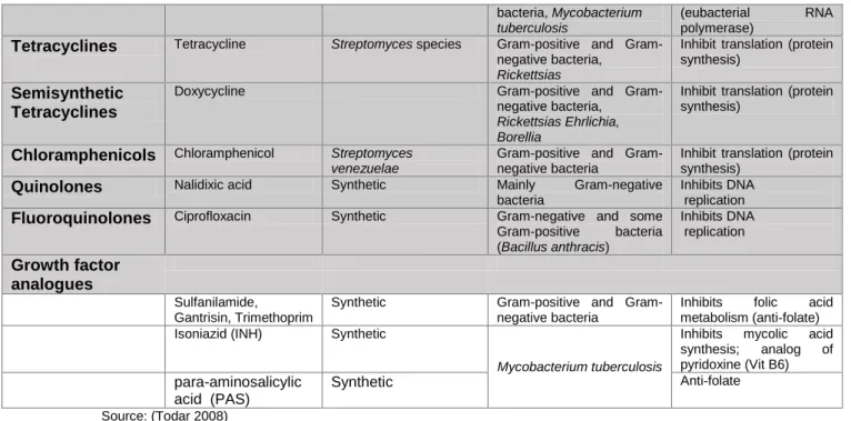
Different antibiotics can possess different mechanisms of action. The table 1 below show the different families of antibiotics with its chemical class, biological source, spectrum of activity and mode of action.
Facing such diversity in antibiotics and mechanisms of action, bacteria managed to come up with an even greater diversity of mechanisms which confer resistance to these molecules. There are three major strategies that bacteria possess to become resistant to antibiotics. Firstly, some microorganisms are capable of changing the antibiotic’s chemical structure. This mechanism is present in bacteria who produce β-lactamases, these enzymes cleave the β-lactam ring of the penicillin, or other β-lactams, which leaves the antibiotic obsolete (Petrosino et al. 2009). Secondly, some bacteria have an increased the number of efflux pumps in their cellular membrane. These efflux pumps can rapidly expel the antibiotic that enter the cell, keeping the internal antibiotic concentration to a minimum (Webber 2002). Lastly, bacteria also have the ability to modify chemically or genetically the target site of the
4
antibiotic, once the target is modified the antibiotic cannot bind and resistance is acquired (Nikaido 2009).
Some antibiotics are associated with more than one mechanism of resistance. It has been described all three mechanisms of resistance in Tetracycline, its noticeable why bacteria are highly adaptive microorganisms. Bacteria easily uptake tetracycline, however active efflux pumps can just as easily discard any molecule of tetracycline who enters the bacteria (Levy 1992). Another way tetracycline can become obsolete is by a bacteria producing a protein which protects the ribosome blocking the association of the antibiotic with the ribosome, allowing it to resume protein synthesis (Taylor & Chau 1996). Finally, it has been also described inactivation of the molecule by enzymatic activity. Even though it’s the rarest of all three, it has been described in human gut Bacteroides a gene named tet (X) which codifies a 44-kDa cytoplasmic protein which chemically alters tetracycline in the presence of oxygen and NADPH (Speer et al. 1991; Chopra & Roberts 2001).
Table 1 - Chemical class of antibiotics and their characteristics
Chemical class Example Source Spectrum Mode of action
Β-Lactams
Penicillins Penicillin G P. notatum Gram-positive Bacteria Inhibits steps in cell wall synthesis and murein assembly.
Cephalosporins Cephalothin Cephalosporium species
Semisynthetic β-Lactams
Aminopenicillins Ampicillin, Amoxicillin positive and Gram-negative bacteria
Inhibits steps in cell wall synthesis and murein assembly.
Clavulanic Acid Clavamox (Clav. Acid + Amoxicillin)
Streptomyces clavuligerus
Suicide inhibitor of β-Lactamases.
Monobactams Aztreonam Chromobacter violaceum
Inhibits steps in cell wall synthesis and murein
assembly.
Carboxypenems Imipenem Streptomyces cattleya
Peptides
Polypeptides Polymyxin Bacillus polymyxa Gram-negative bacteria Damages cytoplasmic membranes
Bacitracin Bacillus subtilis Gram-positive bacteria Inhibits steps in murein (peptidoglycan)
biosynthesis and assembly
Glycopeptides Vancomycin Streptomyces orientales
Gram-positive bacteria,
Staphylococcus aureus
Lincomycins Clindamycin Streptomyces
lincolnensis
positive and Gram-negative bacteria anaerobic Bacteroides Inhibits translation (protein synthesis) Aminoglycosides
Streptomycin Streptomyces griseus positive and Gram-negative bacteria
Inhibit translation (protein synthesis) Gentamicin Micromonospora
species
positive and Gram-negative
Peudomonas
Macrolides Erythromycin Streptomyces erythreus Gram-positive and
Gram-negative bacteria not enteric, Neisseria,
Legionella, Mycoplasma
Inhibit translation (protein synthesis)
Polyenes
Amphotericin B Streptomyces nodosus Fungi (Histoplasma) Inactivates membranes containing sterols Nystatin Streptomyces noursei
Rifamycins Rifampicin Streptomyces
mediterranei
positive and Gram-negative
5
Source: (Todar 2008)
1.3 Research and development of antibiotic agents
There are several phasesduring antibiotic research and development (R&D) that a new drug applicant (NDA) has to be submitted to before being accepted into the market. A novel approach in drug discovery is trending where small scientific of academic groups focus on the
early stages of antibiotic development, namely the preclinical stage (Livermore 2011) and once promising results regarding a NDA are found, a bigger, industrial company, previously associated with the research group, takes control of the project, mainly because the next phases require a bigger monetary investment (Tralau-Stewart et al. 2009; Rosenblatt 2013). During the first phase of drug discovery, the most important issue to be addressed is making sure that the NDA is active against a broad range of organisms and also that it isn’t toxic when administered. Initial susceptibility testing begins with a regular “dip-disk” test or a “drop test”, which is applied to a freshly inoculated lawn of a bacterial strain to an agar plate. After the plates are incubated at optimal growth conditions halos of inhibition are measured to assess the presence of antimicrobial activity (Dougherty & Pucci 2012). Toxicity assays are usually
bacteria, Mycobacterium
tuberculosis
(eubacterial RNA polymerase)
Tetracyclines Tetracycline Streptomyces species positive and
Gram-negative bacteria,
Rickettsias
Inhibit translation (protein synthesis)
Semisynthetic Tetracyclines
Doxycycline positive and Gram-negative bacteria,
Rickettsias Ehrlichia, Borellia
Inhibit translation (protein synthesis)
Chloramphenicols Chloramphenicol Streptomyces
venezuelae
positive and Gram-negative bacteria
Inhibit translation (protein synthesis)
Quinolones Nalidixic acid Synthetic Mainly Gram-negative
bacteria
Inhibits DNA replication
Fluoroquinolones Ciprofloxacin Synthetic Gram-negative and some
Gram-positive bacteria (Bacillus anthracis) Inhibits DNA replication Growth factor analogues Sulfanilamide, Gantrisin, Trimethoprim
Synthetic positive and Gram-negative bacteria
Inhibits folic acid metabolism (anti-folate) Isoniazid (INH) Synthetic
Mycobacterium tuberculosis
Inhibits mycolic acid synthesis; analog of pyridoxine (Vit B6)
para-aminosalicylic acid (PAS)
Synthetic Anti-folate
Fig. 1 - The research and development process. (Rosenblatt 2013)
6
performed with human cell lines or in more advanced phases of development on animals in certified facilities.
When working with natural products, one important aspect is the complexity of the fermented broths. Once antimicrobial activity is observed in a sample, the active molecule must be identified and isolated from the complex broth which contains several different molecules. Occasionally, the molecule(s) of interest is(are) in much lower concentration than most of the remaining constituents, which makes the isolation process particularly difficult (Gray et al. 2006). To elucidate the chemical structure of the biologically active molecule present in a fermentation broth, series of assays have to be conducted. Firstly the compound must be isolated via solvent extractions, ion-exchange methods, gel filtration, and chromatography, and only after, resorting to nuclear magnetic resonance (NMR) and X-ray crystallography we are able to clearly identify the molecular structure (Exarchou et al. 2005; Exarchou et al. 2006; Tatsis et al. 2007). All these procedures combined can take several months to complete (Gootz 1990).
After all the time and work invested, the isolated molecule isn’t ready to move up to the clinical phases. It is still possible to properly change the molecule’s structure in order to improve its activity, pharmacokinetics, toxicity and to avoid becoming obsolete by mechanisms of resistance (Dougherty & Pucci 2012). During this phase efforts are focused on understanding the Structure-Activity Relationship (SAR) to elucidate which modifications result in a better and more stable antibiotic. Only after demonstrating the efficiency and stability of the lead compound, it will undergo further clinical trials (FDA 2006). The clinical trials are divided in 3 phases: during the first trial phase a drug is given to a small group of healthy volunteers (20 to 80 volunteers) to determine safety and dosing; in the second the same drug is then given to slightly bigger group of sick patients (100 to 500) in order to test for efficacy and further safety; during the third phase the universe of the patients who are administered the drug is greatly increased (1000 to 5000) for further efficacy and safety testing and to compare the results of the tested drug with other standard or experimental drugs (National Institutes of Health 2001). After passing these 3 phases the drug is green-lit for commercialization, and sometimes a determined period of time is established by the FDA called post-market surveillance studies period to keep under watch the effects of the drug in the general population (National Institutes of Health 2001).
However, drug development does not cease once a drug enters clinical trials. The FDA pointed out 5 major aspects in their guidelines made available in 2009 that have to be addressed in parallel during early clinical trials so that a NDA can be vetted to move through all the clinical phases. The first aspects that have to be meticulously questioned are the
7
demonstration of in vitro and in vivo activity against targeted pathogens; secondly, a study of which culture conditions interfere with the assessment of antimicrobial activity is required to establish quality control parameters; comprehending how the body fluids and secretions interact with the in vitro activity of the NDA is of major importance as well a full description of the NDA’s mechanism of action or inhibition (MOA/MOI), killing efficiency, potential for resistance development and cross resistance to other antimicrobials. This is especially relevant when dealing new antibiotics with new MOA (CDER 2009), as the behaviour of the drug can be more unpredictable, finally, the possible interactions with other antimicrobial agents or any other drugs has to be carefully studied (Dougherty & Pucci 2012). Following these guidelines provided by the FDA will, in the long run, increase the success rate of an NDA.
During the final phase of clinical trials, the main focus point is quality control, together with in vitro antimicrobial assessment (CDER 2009). Data from the quality control assessments of both the clinical trials and the in vitro assays as well as the results of antimicrobial efficiency are presented in a final report to the authorities so an antibiotic can be approved or delayed for more testing. The time scale, from lead isolation and optimization to commercialization, takes in average a total of 14 years in the US (DiMasi et al. 2003) during this time a massive amount of money is invested and if, for some reason, the drug fail to meet the requirements to enter the market all the invested money is lost. In fact, the attrition rates in the road of antibiotic discovery and development are incredibly high. In average, out of 10,000 substances compounds screened only around a 250 will show antimicrobial activity and will go through pre-clinical trials, but unfortunately only about 5 qualify as candidates during the testing in the pre-clinical trials. This means that from discovery to approval each compound can has a 99% chance of not being approved (European Comission 2008). The most common issues associated with such high attrition rate are poor pharmacokinetics and toxicity (Kassel 2004). It’s extremely important being able to identify as early as possible which candidates are more likely to be dropped out later in the process. The ability to identify such candidates allows to avoid wasting money and resources on a candidate that will not be approved.
Rules and techniques have been developed to help identify molecules with propensity to have drug-like characteristics. In 1997 Christopher A. Lipinski elaborated a set of five rules, later called “Lipinski’s Rule of Five”. When the characteristics of a drug candidate obeyed the 5 rules, Lipinski would consider the candidate to have “drug-likeness” and would be likely that it would be orally active in humans. The rules are as follows: the molecular structure of the candidate should not have more than 5 hydrogen bond donors and 10 hydrogen bond acceptors; its molecular mass should be lower than 500 Dalton and should not have an octanol-water partition coefficient (logP) greater than 5 (Lipinski 2004; Lipinski et al. 2001).
8
After proving the relevance of this rule of thumb, other rules were created in the same genera, like the “Rule of Three” which is an adaptation for lead compounds. According to this rule, a compound which do not comply with more than one of the rules bellow isn’t considered to have lead-likeness, and the rules are: octanol-water partition coefficient log P not greater than 3; molecular mass less than 300 Daltons; not more than 3 hydrogen bond donors; not more than 3 hydrogen bond acceptors; not more than 3 rotatable bonds. Statistics shows that candidates who obey to this rule of thumb have lower attrition rates during clinical trials (Leeson & Springthorpe 2007; Congreve et al. 2003).
Another approach to reduce attrition rates is by preforming ADME (absorption, distribution, metabolism and excretion) assessment in the early stages of drug development. Information gathered with these assays, allow to understand the critical reasons why most drug candidates are abandoned. That information can be used to either help during the development phase, by tailoring the molecule to better suit the requirement needs or to abandon the research as early as possible (Kassel 2004; Wang & Urban 2004). There is a wide range of information that can be collected from ADME assays. The rate at which a drug is dissolved under the various conditions of the human body, which helps understand how easily a NDA is entering the organism; this can be assessed with simple assays like the “saturation shake-flask” (Saal & Petereit 2012). Membrane permeability to the NDA helps understand if the drug is able to diffuse through barriers, such as membranes present in our gastro-intestinal tract, in order to enter the transportation systems, these assays are regularly performed in vitro using human cell lines or other mammal cell lines (Gleeson 2008). In vitro or in silico determination of the ionization constant (pKa, the concentration at which both neutral species and ionized from are equally distributed) helps further understanding of its solubility and permeability, also, pKa data aids better understanding the binding mechanisms of therapeutic events which might lead to optimization of chemical reactions (Wang & Urban 2004). Lypophilicity (LogP and LogD) data is important to predict solubility, permeability and also transport mechanism, once again is the “saturated shake-flask” is used but with a more lipophilic solution, however more advanced techniques are available to determine de LogP and LogD of a NCE like liposome chromatography, immobilized artificial membrane and chromatography approaches (Wohnsland & Faller 2001; Wang & Urban 2004). Chemical stability is tested by incubating the NCE in various extreme conditions like pH and temperature, followed by a quantification of the molecule as a whole using detection systems like LC-MS or HPLC, this is important to assure that the molecule stays unchanged and active as it goes through all the different conditions inside the human organism (Kibbey et al. 2001; Wang & Urban 2004). The main determinant of drug concentration in the blood is the hepatic metabolism stability, because even though a drug is orally absorbed there must not be any
9
degradation by the liver enzymes (Obach 2001; Wang & Urban 2004). The most reliable data on this subject is usually gathered from human experiment in clinical trials due to the complex and extreme conditions present in our organism. Still, in vitro models are being studied using a vast array of liver enzymes, microsomes, isolated hepatocytes, so that lead optimization initiate before the clinical trials.
In the past, many scientists and companies searched for new active molecules in natural products, not only antibiotics but also other molecules with possible medicinal use. During a long time, 39 % of the drug substances available in the pharmaceutical industry were natural products or inspired by a natural compound, and 60-80% of the antibiotics and anti-cancer drugs were derived from natural products (Cragg et al. 1997). Though it seems that this trend is changing. Between 2001 and 2008 was registered a drop of 30% in natural-product-based development projects (Harvey 2008). This is due to advances in high-throughput screening technologies. Such technologies, allowed us to unveil a broader range of synthetic molecules in a more cost-effective way with the vastest therapeutic indications, such as, anti-cancer, anti-infective/antibiotic, anti-diabetic, anaesthetic, analgesics etc. (Ganesan 2008) . Still, the pharmaceutical industry, in general, is withdrawing from the use of natural products in drug research because of the difficulties that they come across. Even though natural products are easily assimilated by the our organism, there are still some downsides, for example the complexity of the molecular structure of the products, the slowness of working with biological material which sometimes can also interfere with intellectual property rights and, in some cases, the availability of the raw materials (Lam 2007; Rishton 2008). These mentioned issues combined with the fact that in the past couple of years, several drug companies abandoned the research for antibiotics and focused on more profitable lines of drug development, such as drugs for chronic diseases like cancer, reduces the chances of discovering new and more efficient antibiotics (Hirschler 2013).
1.4 Stenotrophomonas maltophilia
Since 1947, when first isolated from pleural fluid, this bacteria have seen its name corrected three times. The first name attributed in 1947 was Bacterium bookeri and soon was re-named to Pseudomonas maltophilia (Hugh & Leifson 1963). Later, through RNA analysis it was concluded that it had more resemblance with the Xanthomonas genus, and so it was named X. maltophilia (Swings et al. 1983). It was only in 1995 through DNA and rRNA studies plus sequencing and mapping of the 16s rRNA genes that the distinction of S. maltophilia and other members of the genus became clear (Nesme et al. 1995).
This gram-negative bacteria is an opportunistic pathogen, usually associated to infections in patients who already have their immune system debilitated. However, It’s
10
importance as nosocomial pathogen is increasing noticeably In the last years (Denton & Kerr 1998a; Brooke 2012). The most regular infections caused by this pathogen are usually associated with catheter-related bacteraemia/septicaemia and respiratory infections in cystic fibrosis patients. Other forms disease manifestation are pneumonia, bloodstream bacteraemia, soft skin and tissue infections, osteomyelitis, meningitis, endocarditis, urinary tract infection, biliary sepsis among others (Brooke 2012). This bacteria can be found not only in hospitals but practically anywhere in the world, from shower caps, tap water, natural bodies of water, soil, fruits, vegetables, raw meat, fish (Denton & Kerr 1998). Currently, the most alarming fact about this bacteria is that genomic analysis revealed that this microorganism have a remarkable capacity for drug and heavy metal resistance. Several genetic determinants which confer resistance to different classes of antibiotics via alternative mechanisms have been identified in its genome (Crossman et al. 2008).
1.4.1 Stenotrophomonas maltophilia D457 vs. ΔphoQ
Bacteria do not possess specialized organs to evaluate its surrounding environment; however, it present mechanisms that allow to process the information from its surrounding environment, such as, pH, O2 concentration, luminosity, and other external conditions. These
mechanisms are usually associated with two-component systems (TCS) (Stock et al. 2000). It has been discussed that these TCS are possible targets for antimicrobial agents (Barrett & Hoch 1998; Stephenson & Hoch 2002; Furuta et al. 2005; Watanabe et al. 2003).
The phoPQ operon is one of the two-component systems which are related to the regulation of virulence in many gram-negative bacteria. This regulation is mediated by the stimulation of the histidine sensor kinase PhoQ by extracellular conditions, namely the concentration of bivalent ions and changes in pH, and the response regulator PhoP (Véscovi et al. 1997; Garcia Véscovi et al. 1994). Described responses to these types of stimuli include alteration of lipopolysaccharide biosynthesis and increased resistance to several antibiotics (Zwerschke 2012). Also it has been reported that pathogenic bacteria become more virulent in the presence of low concentrations of Mg2+ (Soncini et al. 1996). During the phenotypic
assessment of a novel S. maltophilia D457 mutant by frame knockout of the phoQ gene, evident changes on the morphology, growth and motility patterns were revealed. When cultured in the same media, the D457ΔphoQ mutant showed morphologic changes and increased swimming and swarming mobility compared with the D457 parental of a magnitude 32 fold and 15 fold for swimming and swarming respectively (Zwerschke 2012).
However, what was more interesting was the apparent evidence of an antibacterial substance that was being produced by the D457ΔphoQ mutant but not produced by the parental strain. The main purposes of antibiotic resistance mechanisms is conferring antibiotic
11
tolerance to bacteria which produce antibiotics. Hence, the remarkable natural antibiotic resistance that S. maltophilia possess might be related with antibiotic production itself. It has been described production of nonribosomal peptides by species from the same genus as S.
maltophilia (Royer et al. 2004; Birch & Patil 1985). Nonribosomal peptides are molecules with
a vast array of biologic functions, being antimicrobial activity one of the most relevant in modern medicine (Schwarzer et al. 2003). Evidence gathered by previous work undertaken regarding the production of a putative compound with microbial activity by S. maltophilia D457ΔphoQ mutant, combined with published work which reveals production of potent antimicrobial across species in the same genus, and with recent in depth genome studies that suggest that the genus Xanthomonas might be a promising reservoir for bioactive nonribosomal peptides (Royer et al. 2013).
12
2. Thesis goals
Previous studies have showed that several species from the same genus of
Stenotrophomonas maltophilia are capable of producing compounds with high antimicrobial
activity (Hashidoko et al. 1999; Birch & Patil 1985). Additionally, previous work suggested that a frame knock-out mutation in the phoQ gene of S. maltophilia, would generate severe phenotypic changes in comparison to the parental strain. In addition to this phenotypic changes, there was also the possibility that the mutant would start producing a compound with antimicrobial characteristics. (Royer et al. 2013). Taking this into account, we hypothesised that a knockout frame mutation in the phoQ gene of S. maltophilia D457, might induce the activation of a metabolic pathway which otherwise would be down-regulated or inhibited, and the result would be a production of compounds with antimicrobial activity.
In this work, we aimed to understand the differences between the mutant strain and the parental strain, regarding the production of the compound with antimicrobial activity to determine if the presence of such compounds were exclusive to the mutant. The best culture conditions at which this, or these, compound(s) is(are) produced and its yield, as well as its spectrum of activity against drug resistant strains were assessed by culturing both the parental strain and the mutant in different conditions and submitting the broths in which they were grown to susceptibility tests. Moreover, knowing the chemical characteristics of this/these unknown compounds was of our interest since it would help understand what chemical structures would confer biological activity to these compounds. This information would be relevant to the R&D community in order to develop molecules with the same characteristics. Therefore, GC-MS analysis was performed in the attempt to elucidate the chemical characteristics, including molecular structure of the unknown compounds.
13
3. Materials and Methods
3.1 Bacterial strains and growth conditions
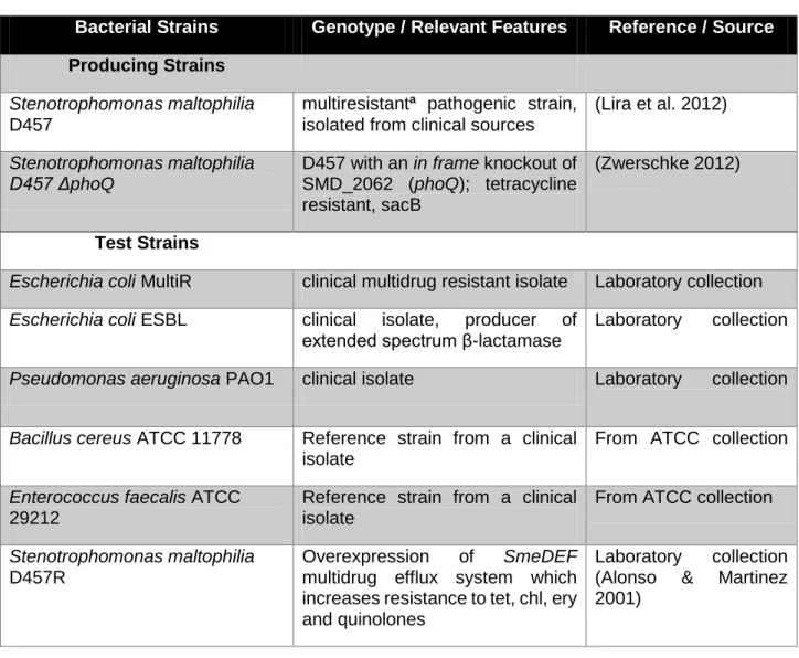
The bacterial strains used in this study are listed in table 2. The strains were divided in two categories: producing strains and test strains, the first being the strains which are being screened for production of compounds with antimicrobial activity and the later the strains which the samples/extracts are tested on. Each bacterial strain was stored in cryovials at -80º Celsius in a glycerol solution at a concentration of 20%. Fresh cultures were started by inoculating 5 millilitre (ml) of lysogeny broth (LB),(10 grams per litre (g/L) of tryptone, 5 g/L of yeast extract and 5 g/L NaCl) (Bertani 1951), with 15 microliter (μl) of stock culture stored at -80º C. All strains were incubated over night at 37 ºC in an incubator with orbital shaking at 200 rpm. For further use, the strains were transferred to appropriate medium and conditions as described below. Every manipulation of stocks and cultures was performed in aseptic environments using either a Bunsen burner or a Telstar Bio II A laminar flow chamber.
Table 2- Strains used in this work.
Bacterial Strains Genotype / Relevant Features Reference / Source
Producing Strains
Stenotrophomonas maltophilia
D457
multiresistanta pathogenic strain, isolated from clinical sources
(Lira et al. 2012)
Stenotrophomonas maltophilia D457 ΔphoQ
D457 with an in frame knockout of SMD_2062 (phoQ); tetracycline resistant, sacB
(Zwerschke 2012)
Test Strains
Escherichia coli MultiR clinical multidrug resistant isolate Laboratory collection
Escherichia coli ESBL clinical isolate, producer of
extended spectrum β-lactamase
Laboratory collection
Pseudomonas aeruginosa PAO1 clinical isolate Laboratory collection
Bacillus cereus ATCC 11778 Reference strain from a clinical
isolate
From ATCC collection
Enterococcus faecalis ATCC
29212
Reference strain from a clinical isolate
From ATCC collection
Stenotrophomonas maltophilia
D457R
Overexpression of SmeDEF
multidrug efflux system which increases resistance to tet, chl, ery and quinolones
Laboratory collection (Alonso & Martinez 2001)
14
Staphylococcus aureus MRSA Clinical β-lactam resistant isolate Laboratory collection
(Monteiro et al. 2012)(Monteiro et al. 2012)(Monteiro et al. 2012)(Monteiro et al. 2012)(Monteiro et al. 2012)(Monteiro et al. 2012)
Acinetobacter haemolyticus VR Clinical isolate, vancomycin
resistant
Laboratory collection
3.2 Swimming and Swarming
Motility assays were conducted in order to assure that the ΔphoQ mutant strain was still phenotypically stable. By comparison of swimming and swarming patterns between D457 and ΔphoQ was possible to monitor the stability of the phenotype.
The swimming and swarming assays were mainly preformed like previously described (Rashid & Kornberg 2000). The swimming motility was carried out by inoculating a single colony of an over-night culture grown in lysogeny agar (LA, 1.5% wt/vol) at 37 ºC to the centre of a LA plate that contained 0.3% (wt/vol) of commercial agar. The plates were sealed with Parafilm® M to prevent dehydration and incubated at 37 ºC for 16 hours. For the swarming motility assay, 1 µl of over-night culture grown in LB at 37 ºC 200 rpm was transferred to the centre of a LA plate with 0.8% [wt/vol] commercial agar. The plates were sealed and incubated at 37 ºC for 16 hours and further 48 at room temperature. After the incubation period, the diameter of the bacterial growth in both swimming and swarming plates was measured. Both assays were made in triplicate.
3.3 Expression of compounds with antimicrobial activity
Different conditions were tested in order to obtain the best production yield of compounds with antimicrobial activity. In this study, Stenotrophomonas maltophilia ΔphoQ and D457 strains were grown in, solid and liquid, LB and M9 minimal medium supplemented with casamino acids and glucose.
3.3.1 Liquid medium
Two 500 ml Erlenmeyer flasks with 200 ml of either LB medium or M9 minimal medium supplemented with glucose and casamino acids were inoculated with 25 µl of pure over-night (16 hours) cultures of the Stenotrophomonas maltophilia D457ΔphoQ mutant strain and
15
Stenotrophomonas maltophilia D457 parental strain, and incubated for 24, 48 and 72 hours in
a MaxQ 4000 orbital shaker at 30 ºC 200 rpm. For each medium, the same volume of non-inoculated medium were incubated in the same conditions and used as controls.
3.3.2 Solid medium
Batches composed of six plates of lysogeny agar medium (LA, 1.5 % commercial agar) each with 15 ml of medium were inoculated with 100µl of a pure over-night (16 hours) culture of each producing strains separately, with the optical density (OD) at 600 nm adjusted to 0.02 units. Cells were seeded across the medium using sterile glass spheres by shaking vigorously for about 30 seconds, in order to grow in a uniform layer of cells. The plates were incubated for 24, 48 and 72 hours at 30 ºC.
Equal amount of M9 minimal medium plates supplemented with casamino acids and glucose plates were inoculated in the exact same conditions. For each medium, the same amount of non-inoculated plates were incubated in the same conditions and used as controls.
3.4 Active Compounds Isolation
To test higher concentrations of the antimicrobial compound, to have a less chemically complex sample for further analysis and to determine the polarity of the active compound(s) sequential extractions involving three organic solvents, Dichloromethane (DCM), Ethyl Acetate (EA) and methanol (MeOH) were performed. The following extractions were only targeted for extracellular compounds, hence the cellular pellets were discarded.
3.4.1 Liquid-Liquid Extraction
Cultures growing in Erlenmeyer’s as mentioned in 3.3.1, were centrifuged in a
Beckman J2-21 centrifuge, at 8500 rpm, and 4 ºC, for 15 minutes, and further filtered using
sterile Minisart® 0.45 µm filters in order to completely remove any suspended cells. Subsequently, the recovered supernatant was poured into a chemically sterile pear-shaped separatory funnel, and the partitioning was carried out according to the Kupchan method, with slight modifications (VanWagenen et al. 1993). The first organic solvent added was DCM in a 1:1 proportion. The mixture was shaken vigorously for 5 minutes and left for settling until both phases were completely distinct and able to collect the organic solvent. This procedure was repeated 3 times. After the last DCM extraction, the same volume of EA was added and the extraction was carried out as described above. Finally, a spoonful of anhydrous sodium sulphate was added to each of the fractions to remove any water that might have been dragged
16
in the separation process. Then, both fractions were filtered (Machery-Nagel® MN 615 filter paper) to remove the sodium sulphate.
3.4.2 Solid-Liquid Extraction
Producing strains growing in solid medium as described in 3.3.2, were carefully removed from the agar and discarded. Then, the cleaned agar was cut in squares with 2 cm height and transferred to a 500 ml Erlenmeyer flask. DCM (200 ml) was added to the Erlenmeyer and the extraction was carried out by shaking the flask for one hour at 200 rpm, at room temperature. The liquid phase was then recovered and filtered (Machery-Nagel® MN 615 filter paper) to remove any traces of agar from the DCM fraction and anhydrous sodium sulphate added. The extraction process was repeated twice. After the last extraction with DCM, the same volume of the second organic solvent (EA) was added to the Erlenmeyer flask containing the agar and the sequential extraction was concluded in the same conditions as referred above. Finally, the extraction was completed with MeOH in the same conditions, except for the addition of anhydrous sodium sulphate.
3.4.3 Extract Concentration
The resulting solutions from the sequential extractions were concentrated at 40 ºC, under low pressure (640 mbar for DCM fraction, 200 mbar for EA fraction and gradually lowering the pressure until reaching 40 mbar for MeOH and H2O fractions), in a BUCHI rotary
evaporator. After complete dryness, the residue was suspended in 200 µl of DMSO for further biological assays, and/or suspended in 200 µl of the respective organic solvent, from witch 50 µl were reserved for further analysis by thin layer chromatography (TLC) and gas chromatography-mass spectrometry (GC-MS).
3.5 Biological Activity Assays
3.5.1 Cross-Streak Assay
A simple cross-streak assay was performed as described by Williston (Williston et al. 1947), with slight modifications. In this assay, a fresh culture of the producing strain was streaked horizontally in the centre of the LA medium plates for 3 days and incubated at 37 ºC. After 72 hours, the test strains were streaked at right angles and further incubated for another 16 hours at 37 ºC. The inhibition
Producing strain
Test strains
Test strains
Fig. 2 - Schematic
representation of the cross-streak assay layout.
17
was evaluated by measuring the length of the inhibition zones (mm).
3.5.2 Agar-Well Diffusion Method
Crude samples of the filtered supernatant mentioned in 3.3.1 were tested for the presence of inhibitory compounds using a modified agar well-diffusion method described by Paik and Glatz (Paik & Glatz 1995). For this assay, the medium was poured into the petri dishes about 5 mm deep, in a horizontal laminar flow chamber. The plates were dried for 2 hours in said laminar flow chamber, to facilitate sample diffusion. Then, each plate was divided in four equally sized quadrants and a 5mm well was cut in their centre. Afterwards, 50 µl of each sample was transferred into the well and the test strains streaked vertically and horizontally around the well. The plates were incubated at 37 ºC and after 16 hours observed for growth inhibition. Wells which were filled with non-inoculated medium served as controls.
3.5.3 Drop Diffusion Method
The samples resolved in DMSO mentioned in 3.4.3 were tested for antimicrobial activity using the drop diffusion method mentioned by Hili and Evans (Hili et al. 1997), with slight modifications. After diluting overnight cultures of the test strains to and OD600 of 0.02, 100µl of culture was spread
in LA plates using glass pearls so only a single layer of cells would grow in the medium. After allowing the cells settle and the plates dry for 10 minutes, a single 5 µl drop of each sample was carefully placed in the respective test area and the plates were incubated at 37ºC for 24 hours, after which
the diameter of the inhibition the inhibition zones was measured. For each sample a minimum of 3 replicates was performed.
Fig. 3 – Schematic
representation of the Drop Diffusion Method layout.
18
3.6 Structure Elucidation
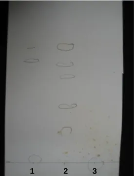
3.6.1 Thin layer chromatography (TLC)
Analytical TLC was carried out on silica gel UV254 plates with 0.2 mm thickness, 20 x
20 cm, (Alugram® Xtra SIL G Aluminium, Macherey-Nigel). Before a single drop was placed on the TLC plates, they were cut into 12 x 7.5 cm parts, allowing 6.5 cm of effective migration distance. After allowing the drop to dry, plates were developed using a mixture of DCM and methanol (10:1) as mobile phase, and placed in a vertical glass chamber at room temperature. After migration, the plates were dried under pressurised air and the spots were observed under UV light at 254 and 366 nm. Then, the plates were pulverized with a solution of concentrated sulphuric acid (10%) in ethanol and, finally, burned with a Power Plus heat gun, in order to permanently reveal the spots.
3.6.2 Gas chromatography-Mass spectrometry (GC-MS)
Samples obtained in 2.3.3 were analysed by GC-MS, in order to tentatively identify the bioactive compound(s). The analysis was performed using a Thermo Scientific Trace GC Ultra gas chromatography equipment, coupled to a Thermo Scientific ITQ 900 ion trap mass spectrometer. The mass range for this run was 50 to 400 (m/z) in positive ion mode. GC analysis were performed on a Bruker fused silica column (30.0 m x 0.25 mm I.D., 0.25 µm film thickness; 5% phenyl / 95% dimethylsiloxane) and helium was used as gas career. Mass analysis were performed by the standard impact electron ionization mode. The GC run was performed with and oven temperature of 200 ºC starting from 50 ºC (15 ºC/min increase), with a 1/10 split rate at 12 ml/min with an inlet temperature of 250 ºC. Chromatograms and mass spectra data were collected and analysed with the Thermo Scientific Xcalibur program.
3.7 Protein Quantification
For protein quantification purposes the strains were grown in a minimal medium to reduce the interferences caused by medium constituents, therefore ΔphoQ and D457 strains were grown in M9 agar plates as described in 3.3.2. After 72 hours of incubation, two solid-liquid extractions were performed with methanol as described in 3.4.2. After the methanolic fractions were filtered, phenylmethanesulfonyl fluoride (PMSF) dissolved in isopropanol was added to each Erlenmeyer to an end concentration of 1mM to prevent protein degradation during the evaporation process of the MeOH (Thermo Scientific 2014). The fractions were evaporated as described in 3.4.3 with a starting pressure of 350 mbar and gradually reducing the pressure to 40 mbar, since the methanolic extraction dragged a considerable amount of water from the M9 agar plates.
19
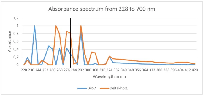
3.7.1 Spectrum analysis and Protein Quantification by UV
In order to obtain absorbance spectrums of the methanolic extracts obtained in 3.7, the samples were diluted (1:50) to produce on-scale readings, and 1 mL of each sample was transferred into an individual quartz cuvette. After zeroing the UNICAM UV2 spectrophotometer with a water filled quartz cuvette, the wavelength range tested was 228 to 700 nm with 2.0 nm bandwidth and at a variable speed controlled by the spectrophotometer’s
Inteliscan feature. The absorbance spectrums were recorded and analysed with MS Excel
2013.
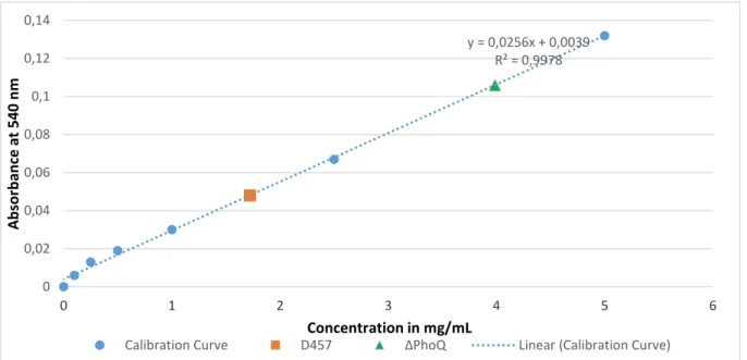
3.7.2 Biuret assay
Protein quantification via the biuret method was performed in a 96 well polystyrene microplate. In this assay, 40 µl of water (used as control), 40 µl of standard solution of bovine serum albumin (BSA) in 7 different concentrations (0 to 5 mg/mL) and 40 µl of the two samples prepared in 3.7 were transferred in triple to the microplate wells (table in annex). After transferring all the solutions into its respective well, 200 µL of biuret reagent (300 mL of 10% (w/v) NaOH to 500 mL of a solution containing 0.3% copper sulphate pentahydrate and 1.2% sodium potassium tartrate, then diluted to 1 L of H2O MilliQ) was added to each well and the
mixture homogenized with the micropipette. The microplate was then wrapped with tin foil to protect from light and incubated for 20 minutes at room temperature for colour development. The values of absorbance were measured with the microplate reader Zenyth 3100 at 540 nm with steering at room temperature and the obtained absorbance readings were plotted against the calibration curve in order to determine the protein concentrations.
20
4. Results and discussion
4.1 Growth characteristics
The motility assays described in 3.2, allowed the assessment of the phenotypic stability of the mutant. This was possible because previous work showed that the mutant ΔphoQ had increased motility over the parental D457, which was verified regularly during the assessments.
Fig. 4 - Motility differences between the D457 and the ΔphoQ strain. Each experiment was
performed in triple and the error bars represent the standard deviation for each data set.
The mobility assessment allowed to verify the integrity of the mutant by comparing the growth pattern of the two strains. In this case the mutant showed, as expected, a significant increase of motility capabilities. The growth diameter of the mutant in the swimming assay was in average 13 times higher than the parental strain, 74 mm and 5.6 mm respectively. The mutant strain also showed a significant increase in swarming motility compared to the parental strain, the ΔphoQ mutant strain managed to travel 31.3 mm, while the D457 parental strain 9 mm. Additionally, this assay allowed us to observe the production of a putative enzyme with agarase characteristics. The production of this putative agarase was only present in the mutant strain ΔphoQ and this event was able to be reproduced several times, however what triggered such event is still not of our knowledge. In average, only 1 out of 3 plates would show
Fig. 5 – Production of a putative agarase by the
mutant S. maltophilia ΔphoQ grown in LA 0.3 % agar.
0 10 20 30 40 50 60 70 80 90 Swimming Swarming Diame ter in m ill im etres (m m ) D457 ΔphoQ
21
agar degradation, always starting from the periphery of the plate and slowly dissolving the agar leading to accumulation of gas in this area. Even though we never registered agar degradation by the parental strain D457, according to KEGG database this strain contains in its genome two genes which putatively encode for two agarases. One being the alpha-agarase and the other the beta-agarase, EC 3.2.1.158 and EC 3.2.1.81 respectively (Kanehisa & Goto 2000; Kanehisa et al. 2014). The first one was first isolated from a Thalassomonas sp., and is responsible for endohydrolysis of (1->3)-alpha-L-galactosidic linkages in agarose, yielding agarotetraose as the major product (Ohta et al. 2005). The second one, firstly isolated from a marine Vibrio, hydrolyse the (1->4)-beta-D-galactosidic linkages in agarose randomly. Both use agarose as main substrate but they can use other substrates like agarohexaose, neoagarohexaose and porphyran (Jam et al. 2005). The described mutation in the virulence related PhoQ operon, results, as shown, in a noticeable alteration in the bacteria’s phenotype. Additionally, to this cellular changes wouldn’t be surprising if changes at a metabolic level would arise as well from this mutation. The production of a putative agarase is evidence that the mutation triggered the activation of certain, previously inactivated, metabolic pathway(s). Because the phoP-phoQ operon is directly related with the regulation of virulence (Kasahara et al. 1992), would be plausible if the metabolism of a toxin would be affected by a mutation in one of the genes, either up-regulated of down-regulated. Taken this hypothesis into account, screenings for antimicrobial agents were conducted.
4.2 Biologic Assays
To properly assess the antimicrobial activity of the unknown compound produced by the ΔphoQ mutant, four different biologic assays were performed. Results obtained from the first three assays, namely the “dot assay”, cross-streak assay and agar-well diffusion assay, did not suggest the production of a compound with antimicrobial activity by any of the two strains (data not shown). In the cross-streak assay, antimicrobial activity is detected when one of the streaks of the test strain fail to grow near the previously streaked producing strain, which in this case did not occur, all the test strains managed to grow from the periphery of the LA plate to the area near the producing strain. The same was observed in the “dot-assay” and agar-well diffusion methods, in these two cases a halo of inhibition was expected to be observed if a high enough concentration of antibiotic was produced by the producing strain. Since no inhibition was observed with these approaches, no actual conclusion could be made about whether or not the strains produced a biological active molecule.
There are several possible justifications for the absence of inhibition zones in this particular case, the first being that the organism do not produce a antimicrobial compound to the extracellular space, the second one is that the organism does not produce the compound at all or maybe he does produce a biologically active compound but only in small quantities,
22
leading to a very low extracellular concentration of compound which can lead to a misinterpretation of its actual presence/absence.
Consequently, to increase the compound(s) concentration, the organic extracts resolved in DMSO were submitted to the biological susceptibility assay Drop Diffusion assay. Since molecules tend to dissolve according to the solvent’s polarity (polar molecules dissolve in polar solvents and non-polar molecules dissolve in non-polar solvents), this method also allowed to understand the polarity of the unknown compound(s), as each of the four solvents used in the extraction process, had its own level of polarity. Starting from the least polar solvent the sequence goes as follows: dichloromethane, ethyl acetate, methanol and water. The extracts obtained in 3.4.3 of the two different stains could be described as a light yellow powder, except for the extract relative to the mutant grown for 72 hours in LA, which had a dark yellow or orange colour. The extracts were dissolved in DMSO to a final concentration of 10 mg/ml and were used in the biologic drop-diffusion assay. Results obtained from these assays showed that the ethyl acetate extract from the LA medium inoculated with ΔphoQ mutant had clear inhibition zones around the spot where the 5 µL of sample was applied. Most of the inhibition halos were the same size, around 10 to 11 mm. The biggest inhibitory halo was observed on the EA extract of ΔphoQ, grown for 72 hours in LA medium, against E.
faecalis ATCC 29212 with 32 mm. Small inhibition zones were also occasionally visible in the
ethyl acetate fractions of solid M9 minimal medium inoculated with ΔphoQ mutant. No inhibition zones against the test strains were observed for the remaining fractions (dichloromethane, methanol or water) of ΔphoQ mutant, and organic extracts of non-inoculated medium
Fig. 6 –Photos of the Drop-Diffusion assay, taken to the two most inhibited strains E. faecalis
ATCC29212 (A) and S. maltophilia D457 (B). The big halos of inhibition are relative to the AE fraction of 72 hours grown ΔphoQ in LBA.