2011
Margarida Fernandes
Resende
Co-culture of SSEA4
+-hASCs derived Osteoblasts
and Endothelial Cells
Co-cultura de Osteoblastos e Células Endoteliais
derivadas de SSEA4
+-hASCs
2011
Margarida Fernandes
Resende
Co-culture of SSEA4
+-hASCs derived Osteoblasts
and Endothelial Cells
Co-cultura de Osteoblastos e Células Endoteliais
derivadas de SSEA4
+-hASCs
Dissertação apresentada à Universidade de Aveiro para cumprimento dos requisitos necessários à obtenção do grau de Mestre em Biomedicina Molecular, realizada sob a orientação científica da Doutora Manuela Estima Gomes, Professora auxiliar convidada da Escola de Engenharia da Universidade do Minho e co-orientação da Doutora Odete Abreu Beirão da Cruz e Silva, Professora auxiliar da Secção Autónoma de Ciências da Saúde da Universidade de Aveiro.
o júri
presidente Prof. Doutora Margarida Sâncio da Cruz Fardilha
Professora auxiliar convidada da Secção Autónoma de Ciências da Saúde da Universidade de Aveiro
Prof. Doutora Maria Manuela Estima Gomes
Professora auxiliar convidada da Escola de Engenharia da Universidade do Minho
Prof. Doutora Odete Abreu Beirão da Cruz e Silva
Professora auxiliar com agregação da Secção Autónoma de Ciências da Saúde da Universidade de Aveiro
Prof. Doutor Alexandra Margarida Pinto Marques Investigador Auxiliar da Universidade do Minho
Prof. Doutora Ana Gabriela Henriques
Professora auxiliar convidada da Secção Autónoma de Ciências da Saúde da Universidade de Aveiro
agradecimentos À Dra Manuela Gomes e Dra Alexandra Marques pela introdução no mundo da Engenharia de Tecidos e Células Estaminais.
A Prof.Odete da Cruz e Silva pela força e compreensão.
Aos 3B’s Research Group por me acolherem durante esta fase final do meu percurso académico.
A todos os meus colegas dos 3B’s pelos bons momentos, pelo apoio, incentivo e boa disposição, dentro e fora do “CCL”. Em especial à Sílvia, Ana Regina, Pedro, Alessandra e Praveen.
Aos amigos que apesar de longe estiveram sempre por perto. Em especial às Joanas Vieira, Rocha e Oliveira, Rita, Igor e Miguel.
Aos meus pais pelo apoio incondicional, pelo optimismo e confiança, pelos “brainstormings” de fim-de-semana que um dia terão os seus efeitos na minha vida e na do meu irmão. Ao meu irmão por tudo.
palavras-chave Co-cultura; SSEA4; hASC; Tecido Adiposo; Osso; Osteoblastos; Células Endoteliais; Differenciação Osteogénica; Angiogénese.
resumo A comunicação celular entre Osteoblastos (OBs) e Células Endoteliais (ECs) é bidireccional e ocorre a dois níveis: comunicação indirecta e comunicação directa, sendo esta última mediada por proteínas através de “gap junctional communication”. O estudo in vitro dessas interacções é desenvolvido através de sistemas de co-cultura, onde pelo menos dois tipos de células são cultivados simultaneamente e submetidos ao mesmo microambiente, sendo espectável que este mimetize mais especificamente o ambiente encontrado in vivo. O tecido adiposo humano é actualmente reconhecido como uma fonte de células estaminais. Uma subpopulação específica de células estaminais isoladas a partir da Fracção Vascular do Estroma do Tecido Adiposo, positiva para o marcador associado de pluripotência SSEA4 – SSEA4+-hASCs – demonstrou ter capacidade de diferenciação osteogénica e endotelial, possibilitando a obtenção de OBs e ECS a partir da mesma fonte inicial. Esta tese reporta o estabelecimento de um modelo de co-cultura de SSEA4+ -hASCs precondicionadas para a linhagem osteogénica (SSEA4+-hASCs OBs) e para células endoteliais derivadas da mesma subpopulação. Foi avaliada a forma como o meio de cultura e as proporções de cada tipo celular influenciam os fenótipos osteogénico e endotelial das células em co-cultura.
Os resultados demonstraram que o meio de cultura EGM suplementado com factores de diferenciação osteogénica permitiu a manutenção do potencial de diferenciação da subpopulação SSEA4+-hASCs nas duas linhagens. Adicionalmente, os resultados evidenciam um potencial de mineralização e angiogénico mais consistente nas condições de co-cultura compostas por uma maior proporção de células endoteliais. Em conclusão, a condição de co-cultura com a proporção SSEA4+-hASCs OBs: SSEA4+-hASCs derived ECs de 1:3 em EGM OST revelou melhores resultados no que diz respeito ao fenótipo osteogénico e permitiu uma manutenção consistente da estabilidade fenotípica das células endoteliais. O modelo de co-cultura estabelecido tem grande potencial para permitir ultrapassar a limitação de vascularização apresentadas
keywords Co-cultures; SSEA4; hASC; Adipose Tissue; Bone; Osteoblasts; Endothelial Cells; Osteogenic Differentiation; Angiogenesis.
abstract The crosstalk between Osteoblasts and Endothelial cells is bidirectional and occurs at two levels: indirect cell communication and direct cell communication mediated by proteins at gap junctions structures. The in vitro study of these interactions rely on co-culture systems where at least two cell types are cultured at the same time and submitted to the same microenvironment that is expected to mimic the in vivo settings. Human adipose tissue has been recognized as a potential source of stem cells for regenerative medicine, with multilineage differentiation potential. Recently, it was found that a specific subpopulation of adipose stem cells isolated from the Stromal Vascular Fraction of the Adipose Tissue, positive for the pluripotency associated marker SSEA4 – SSEA4+-hASCs – can give rise to both osteoblastic and microvascular endothelial-like cells, enabling the obtention of these two cell types from the same cell source.
This thesis reports the establishment of a co-culture model for the simultaneous culture of two cell types – SSEA4+-hASCs preconditioned osteoblasts (SSEA4+-hASCs OBs) and SSEA4+-hASCs derived endothelial cells. The influence of the cell culture media and the proportion of each cell type influenced the osteogenic and angiogenic outcomes from the system.
Our results show that EGM OST culture media allowed to the maintenance of the osteogenic and endothelial differentiation potential of this subpopulation. Concerning the osteogenic and angiogenic potential of the co-cultures, it was shown that both were more consistent on the conditions with higher proportion of ECs in the system.
In conclusion, the co-culture condition with the cell ratio OBs:ECs of 1:3 in EGM OST revealed the most efficient osteogenic outcome parameters as well as great stability of endothelial phenotype along the culture period.
The established co-culture system has a great potential to overcome the vascularization limitations of the current Bone Tissue Engineering strategies.
LIST OF FIGURES ... 5 LIST OF TABLES ... 6 ABBREVIATIONS ... 7 I. INTRODUCTION ... 9 INTRODUCTION ... 11 1. BONE TISSUE ... 12 2. 2.1. Overview of bone biology, structure and function ... 12
2.2. Bone formation pathways ... 13
2.3. Bone repair ... 15
TISSUE ENGINEERING APPROACHES FOR BONE REGENERATION ... 17
3. 3.1. Stem cells overview ... 18
ADIPOSE TISSUE... 19
4. 4.1. Principles of Adipose Tissue Physiology ... 19
4.2. Adipose-derived Stem cells (ASCs) ... 20
4.3. Differentiation Potential of ASCs... 21
4.3.1. Osteogenic Lineage Differentiation... 22
4.3.2. Endothelial Lineage Differentiation ... 23
RELEVANCE OF CELL-TO-CELL INTERACTIONS IN BONE TISSUE 5. ENGINEERING ... 24
5.1. Cell-to-cell communication ... 24
5.2. Cell-to-cell communication between osteogenic and endothelial cell lineages ... 26
REFERENCES ... 28
6. II. MATERIALSANDMETHODS ... 41
HARVESTING, ISOLATION, SELECTION AND CULTURE OF STEM CELLS FROM 1.
HUMAN ADIPOSE TISSUE ... 43
1.2. Immunomagnetic beads-based cell separation ... 45
PREPARATION OF THE IMMUNOMAGNETIC BEADS ... 45
2.
2.1.1. Selection of the hASCs sub-population expressing SSEA-4 (SSEA-4+hASCs) ... 46
2.2. Cell culture ... 46
2.2.1. hASCs and SSEA4+hASCs expansion ... 46
2.2.2. Endothelial Commitment ... 47
2.2.3. Osteogenic Commitment ... 47
CO-CULTURESYSTEM ... 48
3.
3.1. Optimization of the culture conditions ... 48
3.2. Establishment of the Co-culture ... 49
CHARACTERIZATIONTECHNIQUESANDMETHODOLOGIES ... 51
4.
4.1. Cell Morphology ... 51
4.2. Cell Proliferation ... 51
4.3. Alkaline Phosphatase activity quantification ... 52
4.4. Alkaline Phosphatase staining ... 52
4.5. Alizarin Red staining ... 53
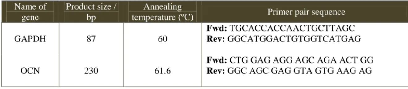
4.6. Gene Expression Analysis of Osteocalcin ... 53
4.6.1. mRNA extraction and cDNA synthesis ... 53
4.6.2. Quantitative Real Time PCR ... 54
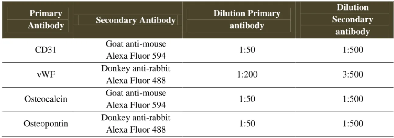
4.7. Immunocytochemistry ... 55
REFERENCES ... 57
5.
III. OPTIMIZED CONDITIONS FOR CO-CULTURING PRE-OSTEOBLASTS AND ENDOTHELIAL CELLS DERIVED FROM SSE4+-HASCS ... 61
INTRODUCTION: ... 63
1. MATERIALS AND METHODS ... 65
2. 2.1. Cell culture ... 65
2.3. Alkaline Phosphatase activity and staining ... 66
2.4. Alizarin Red staining ... 67
2.5. Real Time-RT PCR ... 67
2.5.1. Quantitative Real Time PCR ... 68
2.6. Immunocytochemistry ... 68
RESULTS ... 69
3. 3.1. Definition of Co-culture Conditions ... 69
3.2. Co-culture Performance ... 70 3.2.1. Co-culture Morphology ... 70 3.2.2. Cell Proliferation ... 70 DISCUSSION ... 78 4. REFERENCES ... 81 5. IV. GENERAL CONCLUSIONS ... 85
GENERAL CONCLUSION ... 87 1.
L
IST OFF
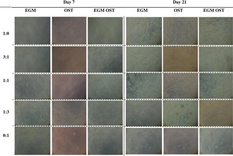
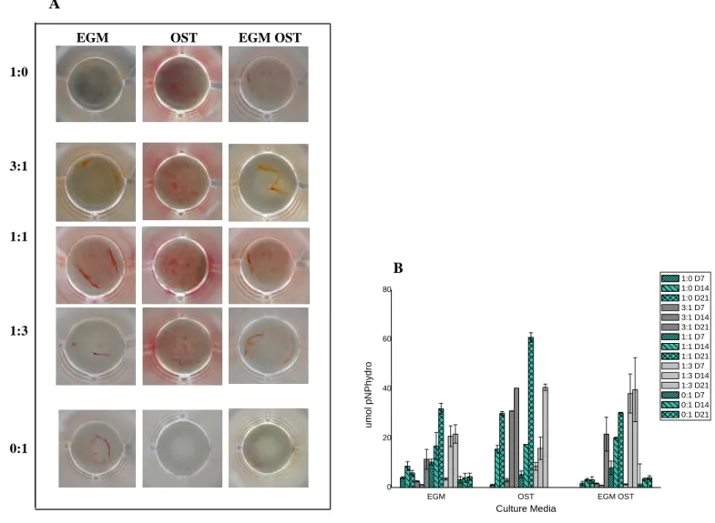
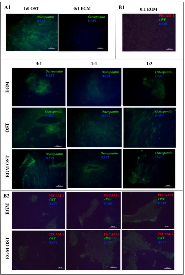
IGURESFigure 1. Optical micrographs showing the morphology of hASCs OBs and SSEA4+-hASCs derived ECs in monoculture and in co-culture, cultured in EGM, OST and EGM OST after 7 and 21 days after cell seeding. At day 7 is still possible to detect the cell morphology differences between both cell types in co-culture (3:1 , 1:1 and 1:3) in EGM and EGM OST; however, not visible in OST media. ... 71 Figure 2. Amount of dsDNA that correlates with cell number quantified along culture – 7, 14 and 21 days – in monocultures of hASCs committed into the osteogenic lineage and SSEA4+-hASCs derived ECs in three different culture media – EGM, OST and EGM OST. and in co-culture of SSEA4-hASCs committed cells onto endothelial and osteogenic lineage. ... 72 Figure 3. (A) Alkaline Phosphatase Staining at days 14 and 21 of culture in co-culture at different cell ratios – 3:1| 1:1| 1:3 – in three culture media – EGM, OST and EGM OST. Positive Control – monoculture of SSEA4+-hASCs committed into the osteogenic lineage; Negative control: SSEA4+-hASCs derived ECs. (B) Amount of hydrolised p-nitrophenol phosphate that correlates with the ALP activity quantified along culture – 7, 14 and 21 days – in monocultures of SSEA4+-hASCs committed into the osteogenic lineage and SSEA4+-SSEA4+-hASCs derived ECs and in co-culture at different cell ratios – 3:1| 1:1| 1:3 – in three different culture media – EGM, OST, and EGM OST. ... 74 Figure 4. (A) Alizarin Red staining results after 21 days of culture, to perform the evaluation of calcium-rich deposits and mineralization of the extracellular matrix in the systems: in monocultures of SSEA4+-hASCs committed into the osteogenic lineage and SSEA4+-hASCs derived ECs (as controls, positive and negative, respectively) and in co-culture at different cell ratios – 3:1| 1:1| 1:3 – in three different culture media – EGM, OST, and EGM OST. ... 75 Figure 5. Osteocalcin relative expression calculated after quantitative Real Time RT-PCR. Results were first normalized against GAPDH and then against the monoculture of SSEA4+-hASCs OBs after 7 days of culture and are presented as fold-change... 75 Figure 6. (A1 and A2) Immunostaining of SSEA4+-hASCs OBs and ECs in monoculture and in co-culture for Osteopontin (green) and Osteocalcin (red) markers. (B1 and B2) SSEA4+-hASCs derived ECs in monoculture and SSEA4+-hASCs OBs and ECs in co-culture in EGM and EGM OST for the analysis of vWF (green) and PECAM-1 (red); No signs of staining were found for the conditions in OST media. ... 77
L
IST OFT
ABLESTable 1. Summary of the constituents of each cell culture media used in this work: α-MEM for cell expansion of hASCs and hASCs; EGM 2-MV for endothelial committement of SSEA4+-hASC fraction. ... 48 Table 2. Summary of the constituents of each cell culture media used for the co-culture system: EGM 2-MV; OST - α-MEM supplemented with osteogenic differentiation factors; EGM OST - resulting from the supplementation of EGM 2MV media with osteogenic differentiation factors at the same concentrations used for preparing Osteogenic media... 50 Table 3. Co-culture set-up for cell ratio: all the proportions corresponds to the ratio SSE4+hASCs OBs : SSEA4+hASCs derived ECs. ... 50
A
BBREVIATIONSα-MEM Alpha-Minimal Essential Medium
ALP Alkaline Phosphatase
AR Alizarin Red
ASC Adipose Stem Cell
BAT Brown Adipose Tissue
BBE Bovine Brain Extract
BMSC Bone Marrow-derived Mesenchymal Stem Cell
CAM Cell-Adhesion Molecules
CD Cluster Differentiation
CD31 See PECAM-1
cDNA Complementary DNA
Cx43 Connexin 43
DAPI 4, 6-Diamidino-2-phenyindole dilactate
diH2O Distilled water
DMF N,N-Dimethylformamide
EC Endothelial Cell
ECM Extracellular Matrix
EGM Microvascular Endothelial Growth Medium
EGM OST Microvascular Endothelial Growth Medium supplemented with osteogenic differentiation factors
EPC Human Umbilical Cord blood Endothelial cells
ESC Embryonic Stem Cells
FACS Fluorescent-activated Cell Sorting
FBS Fetal Bovine Serum
GDF-5 Growth and Differentiation Factor 5
GAPDH Glycetaldehyde-3-phosphate Dehydrogenase
hEGF Human Epidermal Growth Factor
HIF Hypoxia-Inducible Factor
HUVEC Human Umbilical Vein endothelial cells hOEC Human Outgrowth Endothelial cells
Ig Immunoglobulin
MSC Mesenchymal Stem Cell
OCN Osteocalcin
OPN Osteopontin
OST Osteogenic Media
pNP p-nitrophenol
pnPP p-nitrophenyl phosphate
PECAM-1 Platelet Endothelial Cell Adhesion Molecule
PCR Polymerase Chain Reaction
PTH Parathyroid Hormone
R3-IGF-1 Human recombinant analog of insulin-like growth factor-I with the substitution of Arg for Glu at position 3
RT Room Temperature
SSEA4+-hASCs OB SSEA4+-hASCs derived Osteoblasts SSEA4+-hASCs ECs SSEA4+-hASCs derived Endothelial Cells
SVF Stromal Vascular Fraction
TE Tissue Engineering
TGF-β Transforming Growth Factor-beta VEGF Vascular Endothelial Growth Factor
vWF von Willebrand factor
INTRODUCTION
1.
Adult stem cells are undifferentiated cells present mostly in a variety of tissues and organs and that predominantly show the property of self-renewal and capacity to differentiate into major specialized cell types. For this reason adult stem cells have been widely studied for application in the field of tissue engineering (TE) and regenerative medicine. Up to now several sources of adult stem cells have been identified and one of the most promising is adipose tissue. In fact it represents an abundant and accessible source of adult stem cells with the ability to differentiate along multiple lineage pathways including osteogenic and endothelial.
Tissue engineering strategies are generally based on a supportive material (scaffold) where stem cells are seeded and eventually cultured to obtain a functional tissue substitute. Nevertheless, the success of any TE strategy is largely dependent on controlling the biology of the cells at the site of repair or regeneration, since are the cells that constitute and co-ordinate the basic structure and function of tissues. Regarding bone tissue engineering, the establishment of a concise vascular supply of bone constructs is one of the major pitfalls and therefore a hurdle for the clinical application of such engineered constructs. The intraosseous vasculature is important for several bone physiological processes. Its role is evident on the skeletal development and growth by affecting both bone modelling and remodelling processes and isfound essential to guarantee the viability and functionality of the construct upon implantation and hence, its success in the regeneration of the tissue defect.
Endothelial cells (EC) are the main cellular mediator of the vascular network formation and in the sequence of a well-developed vascular network, primary osteoblasts (OB) undergo calcification and differentiation into osteocytes, enabling a healthy bone formation. Therefore, culturing ECs as a homogenous population or combined with OBs is a promising approach to solve the lack of vascularization in bone tissue engineered constructs. This thesis is centred on a specific subpopulation of cells existing within stromalvascular fraction (SVF) of adipose tissue which can be differentiated into the osteogenic and endothelial lineages, and which was used to develop a co-culture model that has a great potential for application in bone tissue engineering approaches.
BONE
TISSUE
2.
O
VERVIEW OF BONE BIOLOGY,
STRUCTURE AND FUNCTION2.1.
Bone tissue is a dynamic highly vascularized tissue with the capacity of constant remodeling by undergoing local resorption and rebuilding. The matrix producing OB, the tissue resorbing osteoclast and the osteocytes, which accounts for 90% of all cells in the adult skeleton, are three of the main cell types that can be found in bone tissue. All perform crucial functions in bone homeostasis. [2]
Osteocytes are the most abundant cell type and rely enclosed within the lacuna-canalicular network of bone. They can be viewed as highly specialized and fully differentiated OBs. From each osteocyte a network of cytoplasmic processes extends through cylindrical canaliculi to blood vessels and other osteocytes. These cells are involved in the control of extracellular concentration of calcium and phosphorus, as well as in the adaptive remodeling behavior via cell-to-cell interactions in response to local environment. Considering that these cells are the principal cell in the adult bone, it appears that osteocyte veritable three-dimensional syncytium may drive the spatial and temporal recruitment of the cells that form and resorb bone.[3]
Osteoblasts are bone forming cells derived from mesenchymal stem cells (MSCs) present among the bone marrow stroma that have one of three fates: can incorporate in its own osteoid and differentiate into an osteocyte, remains quiescence into a lining cell, or undergo apoptosis. Functionally, OBs are the cells that produce and lay down the bone ECM and that regulate its mineralization/calcification. They are located at the bone surface together with their precursors, forming a tight layer of cells, and are highly anchorage dependent, relying on the extensive cell-matrix and cell-cell contacts to maintain their cellular function and responsiveness to metabolic and mechanical stimuli. [4-6] During its life-span, the OBs produce between 0.5-1.5µm of osteoid per day, the unmineralized organic matrix that subsequently undergoes mineralization giving the bone its strength and rigidity.[2]
Osteoclasts are multinucleated giant cells of the hematopoietic descent; their precursors are located in the monocytic fraction of the bone marrow and functionally, they are the main responsible for the bone resorption process, which is required for bone morphogenesis during development, for the continual repair of microdamage in the skeleton and for the adaptation of bone to mechanical load. Osteoclasts bind to bone matrix peptides via integrin receptors in their membrane linking to bone matrix peptides. This causes them to become polarized, with the bone resorbing surface developing a ruffled border that forms when acidified vesicles that contain matrix metalloproteinases are
transported via microtubules to fuse with the membrane. Theprotons and proteases components are secreted to demineralise and degrade the bone matrix, respectively.[2, 6, 7]
Osteodifferentiation occurs along successive stages leading mature OBs which initially involves three different phases: proliferation, matrix maturation and mineralization, each of them characterized by a specific pattern of gene expression. [8, 9] The first phase – proliferation phase – is essentially associated with cell growth and cell cycle regulatory genes, along with extracellular matrix (ECM) biosynthesis. At the post-proliferative period the ECM starts to be mineralized and the activity of alkaline phosphatase (ALP), which is responsible for promoting crystal formation in matrix vesicles by removing nucleation inhibitors, increases. Mineralization of ECM occurs after a period of matrix production and occurs in three steps: nucleation of calcium phosphate crystals, crystal growth by precipitation and conversion into octacalcium phosphate and finally hydroxyapatite.[10] Ultimately, OBs will acquire the typical osteocyte morphology, become entrapped in their own bone matrix, and progressively stop producing matrix. The life span of osteocytes is probably largely determined by bone turnover, when osteoclasts resorb bone and either “liberate” or destroy osteocytes.[10]
In adults, bone tissue exists in two forms, trabecular bone and compact bone, distinguishable by the spatial orientation of its mineral and organic constituents, and by their distinct characteristic locations in the skeleton. [8, 10]
The main functions of bone include structural body support, protection of vital organs, mineral reservoir acting in mineral homeostasis. Also have a front line role in hematopoiesis and endows the body with the capability of movement. [8]
Considering its functional relevance and the ever-growing incidence of skeleton/bone disorders, most of them resulting from the imbalance between breakdown and formation of new bone, it is evident how patients’ quality of life can be affected. In fact, bone tissue lost occurs in a wide variety of clinical situations, for which currently used therapies are clearly insufficient.
B
ONE FORMATION PATHWAYS2.2.
Bone formation occurs as a result of coordinated cell proliferation, differentiation, migration, and remodelling of the ECM that occurs by two different processes: endochondral ossification and intramembranous ossification.[11] In both cases MSCs first have to aggregate to form mesenchymal condensations that are situated at the location of future skeletal elements and
prefigure its shape.[12] In most cases, the cells of the mesenchymal condensations will differentiate into chondrocytes, expressing characteristic cartilaginous matrix genes such as type II collagen,
type IX collagen, type XI collagen, aggrecan, chondromodulin-1 and matrilin-3, and that form the
growth template of the future bone. [11] In the center of the condensations that will give rise to bone through endochondral ossification, mesenchymal cells differentiate into chondrocytes, forming a cartilaginous template, that once formed leads to further differentiation of the innermost chondrocytes into hypertrophic chondrocytes. The mesenchymal cells that remain in the periphery of the growth plate form a structure called perichondrium, whose cells start to differentiate into OBs to form, around the cartilaginous core, a mineralized structure called the bone collar. Once the hypertrophic chondrocytes are fully differentiated, they are already surrounded by a calcified ECM and dye from apoptosis. [13]
One of the most important functions of the mineralized ECM is to favor the vascular invasion from the bone collar through a vascular endothelial growth factor (VEGF)-dependent mechanism. This mechanism serve two different purposes: to attract chondroclasts that will degrade the ECM surrounding the hypertrophic chondrocytes, and to bring in osteoblast progenitors derived from the bone collar. The hypertrophic chondrocytes express and synthesize a number of VEGF isoforms, VEGF-A, VEGF-B, VEGFC, VEGF-D and also express neuropilin-1, VEGF receptor-2 and VEGF receptor-3, comprising an autocrine/paracrine system that will further encourage inwards penetration of the vascular endothelium.[14, 15]This sequence of events lead to the development of the primary ossification centres in which the cartilaginous ECM will then be replaced by a bone ECM, rich in type I collagen. The ossification process will move on centrifugally, consuming most of the cartilage template, recruited osteoblasts (OBs) replace the degraded cartilage with trabecular bone, and a bone marrow is thus formed.[16-18] These highly coordinated events lead to longitudinal bone growth and bone formation between epiphyses and metaphyses regions at each end of bones of bearing joints and long bones.[8, 13]
At the same time, there is a radial growth of the diaphysis, the region between the epiphyses and metaphyses, and of parts of the metaphyses by intramembranous bone formation with direct apposition of cortical bone by OBs from the inner layer of the periosteum. This type of ossification involves the condensation and differentiation of mesenchymal cells into OBs, which began to synthesise and secret osteoid.[19] Gradually, more osteoid is produced, followed by the complete mineralization. As a result, more osteoblasts become trapped within their lacunae and start the differentiation into osteocytes. This is closely coordinated with resorption of bone by osteoclasts on the inner cortical endosteal surfaces and lateral metaphyseal surfaces to maintain the relative proportions of the marrow cavity to the cortices and the overall shape of the bone as it grows.[19]
This third type of bone formation is known as appositional formation and occurs during bone enlargement and bone remodelling. [10]
Some common regulators of both types of ossification are angiogenic growth factors. In intramembraneous ossification there is an invasion of capillaries transporting MSCs which directly differentiate into OBs.[11] In endochondral ossification there is an intrinsic relationship between chondrogenesis and osteogenesis which is dependent on the vascularisation status of the cartilage template. MSCs/precursor cells undergo chondrogenic differentiation, become hypertrophic and initiate angiogenic growth factors secretion, namely several isoforms of VEGF [14] thus determining vascular vessel invasion responsible for the consequent recruitment of bone forming cells.[20, 21] Moreover,vasculature and more specifically ECs secrete several factors that control the recruitment, proliferation and differentiation of either osteoblasts or osteoclasts, highlighting the point that angiogenesis and osteogenesis are not dissociated processes.[22, 23]
Bone vasculature is also decisive for the remodelling process. This process comprises two phases: resorption of pre-existing bone by osteoclasts and de novo bone formation by OBs through transitory specialized anatomical structures, called basic multicellular units (BMUs). BMUs are located in the vicinity of blood vessels, which growing rate is coordinated with the advance of BMUs, this indicate the crucial role of vasculature in bone remodelling and consequently the importance of vascular cells, such as ECs and pericytes in this process.[24, 25]
B
ONE REPAIR2.3.
The intraosseous vascular system of adult bone tissue is mainly composed by diaphyseal or feeding arteries, superior and inferior metaphyseal arteries, superior and inferior epiphysial arteries and periosteal vessels.
When bone fracture occurs there is a disruption of the marrow architecture and the blood vessels structure within and around the fracture site is also compromised.
Fracture healing process results of the combination of both endochondral and intramembranous ossification, the first occurring usually external to periosteum in regions that are mechanically less stable and immediately adjacent to the fracture site, while the last one occurs internal to periosteum at the proximal and distal edges of the callus.[26, 27] The repair process itself is comprised of four overlapping phases: the initial inflammatory response, soft callus formation, hard callus formation, initial bone union and bone remodeling. The immediate inflammatory response leads to the
recruitment of MSCs that subsequent differentiate into chondrocytes that produce cartilage and OBs.[26, 27]
At the molecular level, fracture repair is driven by the three main classes of factors: (1) pro-inflammatory cytokines, (2) the transforming growth factor-beta (TGF-β) superfamily and other growth factors, and (3) the angiogenic factors.[28, 29] Understanding how those factors affect fracture or postsurgicalhealing is essential to the development ofmolecular approachesintended to enhance bone healing after surgeryor traumatic injury.
Pro-inflammatory cytokines like interleukins (IL) -1 and -6 and tumor necrosis factor (TNF) -α have been shown to be involved in the initiation of the repair cascade.[30] Their expression profiles vary along the repair process, being secreted by different cell types according to the primary or later stages, playing a central role over the timing of the immune response, and the beginning of local tissue repair and regeneration. TNF-α, IL-1α and IL-6 expression is primarily localized inflammatory cells during the early periods of inflammation (within 24 hours of injury) and in mesenchymal and osteoblastic cells later during healing process, at the transition from chondrogenesis to osteogenesis during endochondral maturation.[31, 32] Moreover, carry out central functions in the induction of downstream responses to injury by having a chemotactic effect on other inflammatory cells, enhancing ECM production and stimulating angiogenesis in the injury site. [33]
During this early stage of the healing process, angiogenesis also takes place, being a prerequisite for further progression of the regeneration cascade. In fact, the formation of the callus is a physiological reaction that requires the existence of an adequate blood flow.[34, 35] Therefore, the reconstruction of an intact intraosseous vascular structure is an early event during fracture repair. [36, 37] The biochemical environment and particularly hypoxia also has an important role in this process because of the effect that the latter has in the secretion, by OBs, of signaling molecules that regulate ECs proliferation, differentiation and secretion of osteogenic growth factors, namely hypoxia-inducible factor (HIF).[38] As result, committed osteoprogenitor cells of the periosteum and undifferentiated multipotent MSCs are activated and differentiate into chondrocytes and fibroblasts that produce a semi-rigid soft callus, stabilizing the fracture and serving as a template for primary bone formation. [27] The new bone is known as the hard callus and is composed of woven bone.
In regions that are mechanically less stable, endochondral ossification occurs, enhanced by the soft tissues surrounding the fracture site. During this process, MSCs are recruited and begin to proliferate by day 3 after fracture. From day 7 to 21 the chondrogenic differentiation occurs
followed by its proliferation, resulting in the formation of the soft callus. These cells synthesize and release cartilage-specific matrix and once it achieves mechanical stability, the cartilage undergoes hypertrophy and mineralization in an organized special manner at the primary ossification centres. [39] VEGF-A is secreted by the hypertrophic chondrocytes inducing sprouting angiogenesis from the perichondrium, which then leads to the recruitment of OBs, osteoclasts and heamatopoietic cells. It also leads to the removal of the calcifying hypertrophic chondrocytes by chondroclasts and woven bone formation occurs after the MSCs recruitment and differentiation into OBs, which replaces the degraded cartilage.[34] Eventually, the soft callus is replaced by laminar bone and undergoes remodeling into the original cortical and/or trabecular bone configuration following the pathway observed in the growth plate.[16, 17, 40]
TISSUE
ENGINEERING
APPROACHES
FOR
BONE
REGENERATION
3.
Tissue engineering approaches are based on seeding and in vitro culturing of cells harvested preferentially from an autologous source, within a structural support material or scaffold prior to implantation. The effectiveness of those strategies are ultimately highlighted on the stability and functional restore of the damaged tissue, which will be in certain way influenced by the exogenous source of cells and by the capacity to control its proliferation and differentiation state. [7]
One of the major challenges in the development of tissue engineering therapies for bone regeneration is to achieve the vascularisation of the biomaterial-cells construct, which is found essential to allow the viability and functionality of the construct upon implantation and hence, its success in the regeneration of the bone defect.
When the engineered tissue construct is implanted, seeded cells are dependent on the local microvasculature and microcirculation to have access to substrate molecules (oxygen, glucose and amminoacids) and for clearance of metabolic products (CO2, lactate and urea). In the absence of a concise vasculature structure, the movement of those molecules in not assured and pressure gradients that allow molecules movement between the vessel lumen and the cell membrane are not achieved. There are many factors that can affect the generation of these pressure gradients: tissue deformation (movement), mechanical loading, muscle contraction, gravitational pooling, starling flow, and arterial pulsation. When this happens, a compensatory process of molecule diffusion starts. However its efficiency is limited to short distances or for small molecules.[41]
Considering bone engineering constructs, vessels are initially confined to the outer surface of the implantation site, and the diffusion distance for oxygen and other metabolites from the edge to the
centre of the graft is considerably higher than the normal diffusion distance. For this reason the inner cells in the graft are subjected to a hypoxic and metabolic stress that can impair the success of the implantation because of cell necrosis in deeper regions.[42]
S
TEM CELLS OVERVIEW3.1.
Discovered in the last two decades, embryonic stem cells (ESCs) had been described as the most promising cell source for TE, determined by their strong differentiation capacity and plasticity which allowed differentiation towards of an unlimited number of specific cell types, derived from all three embryonic germ layers.[43, 44] Ethical and political debates emerged since the primordial of research on ESCs cells, and this prompted the research focusing on finding alternative stem cell sources, such as adult stem cells or progenitor cells, and on optimizing differentiation protocols for specific cell types converging into organotypic regeneration approaches. One of the primary hurdles is finding a safe source of cells, which in the end yield a functional tissue. [45, 46] For this purpose there are several criteria that should be considered and ideally satisfied: the accessibility and availability of the tissue samples, the proportion of cells of interest in the harvested tissue, its ability to differentiate into the desired cell type and their capacity to maintain phenotypical and functional attributes specific of each type of differentiated cells.[7]
By definition, stem cells are capable of both proliferate in an undifferentiated but pluri- or multi- potent state (self-renewal) and to differentiate into at least one mature cell type.[47] Adult stem cells represent a pool of multipotent somatic stem cells found in adult, neonatal and foetal tissues or organs that have the capacity to differentiate into cells of all three germ layers, mesodermal, endodermal and ectodermal. They were first identified in tissues with a high rate of cell turnover, such as bone marrow. In fact, for many years, bone marrow was considered as the major source of stem cells for bone TE applications and for therapeutic purposes [7, 48] serving as the prototypic example of an adult stem cell. Bone marrow contains an heterotypic adult stem cell population from which two main populations derive: haematopoietic stem cells (HSCs), which produce the blood-cell lineages, and MSCs, which provide the bone-marrow stromal niche and have the potential to produce several cell lineages, including adipogenic, osteogenic and chondrogenic lineages.[49, 50] Currently, it is known MSCs can also be obtained from the stromal fraction of lipoaspirates of adipose tissue, and these possess similar properties to bone marrow-derived MSCs (BMSCs) but are easier to obtain. [51] Further sources of human MSCs include the intestinal[52], limbal [53], knee-joint[54-57]and prostate [58] stroma, trachea[59], nasal mucosa [60], Wharton’s jelly (WJ) [61], cord blood[62] and placenta.[63] The microenvironment of these niches supports
the conservation of stem cell characteristics and the maintenance of their pool for the lifetime of the organism. [64, 65]
According to the International Society for Cellular Therapy, human MSCs under standard culture conditions must satisfy at least three criteria: (1) they must be plastic-adherent; (2) they must express C105, CD73 and CD90 and not CD45, CD34, CD14, CD11b, CD79 or CD19 and HLA-DR surface molecules by flow cytometry; (3) they must be capable of differentiating into osteoblasts, adipocytes and chondroblasts.[66]
ADIPOSE
TISSUE
4.
P
RINCIPLES OFA
DIPOSET
ISSUEP
HYSIOLOGY4.1.
Adipose tissue is recognized as a highly active metabolic and endocrine organ. Is composed of one third of mature adipocytes that are organized in a multidepot structure and the remaining two thirds are a combination of small blood vessels, nerve tissue, fibroblasts, preadipocytes and adult MSCs[67]
Mature adipocytes exist as two cytotypes, white and brown adipocytes, respectively defining white adipose tissue (WAT) and brown adipose tissue (BAT).[68] They are distinguished by cell color, function, anatomical distribution and relative abundance in different developmental stages.[67, 69] The major subdivisions of adipose tissue are the visceral adipose tissue, and the subcutaneous adipose tissue. Subcutaneous adipose tissue is divided into two portions: superficial and deep subcutaneous adipose tissue. The visceral adipose tissue is composed of either intra- or retroperitoneal adipose tissue, separating two distinct intra-abdominal compartments, being in the first that is located the majority of visceral fat.[67, 70]
The relative abundance and metabolic properties of WAT and BAT change according to ageing, gender, nutritional and pathological status of individuals. [71-73] There are marked regional differences in adipose tissue distribution between males and females as well as in the abundance of each type of adipose tissue with ageing. In neonates, BAT is most abundant, serving mainly as a thermogenic organ; with ageing, BAT is replaced by WAT, and consequently, in adults, BAT is present in tiny discrete collections.[73-75] WAT is not a highly vascularized as its brown counterpart; each fat cell in WAT is in contact with at least one capillary, providing a vascular network allowing its continued growth. WAT has long attracted attention because of its great and reversible capacity for expansion, apparently permanent throughout adult life.[76]
The stromal vascular fraction (SVF) of the adipose tissue represents a heterogeneous cell population surrounding adipocytes in fat tissue, and was reported to be a very convenient and nonrestrictive source of multipotent cells such as hematopoietic progenitors and spare mesodermal stem cells able to differentiate into endothelial, neurogenic, osteogenic, chondrogenic, and myogenic lineages. Additionally, other cell types were found in the SVF namely smooth muscle cells, pericytes, fibroblasts and circulating cell types such as leukocytes or EPCs.[77]
A
DIPOSE-
DERIVEDS
TEM CELLS(ASC
S)
4.2.
Adipose tissue, because of its abundance and relatively less invasive harvest methods, represents a practical and appealing source of mesenchymal stem cells (MSCs) present within the SVF of subcutaneous adipose tissue, termed “adipose-derived stem cells (hASCs) [78]. It has been demonstrated by many groups that hASCs display multilineage plasticity in vitro and in vivo.[79-82]
Rodbell first reported [83-85] the initial methods for isolating cells from rodent adipose tissue in 1960’s with the final purpose of separating the buoyant fraction of mature adipocytes from the SVF, which would be pelleted through density gradients. Since then, several groups modified the harvesting methods of human subcutaneous adipose tissue, taking advantage of the abundance, accessibility and of the overgrowing number of local excision procedures or minimal invasive liposuction surgeries that are performed. Nowadays, liposuction is considered a safe and well-tolerated procedure, with a complication rate of around 0.1% that routinely allows the removal of fat tissue between few hundred milliliters and several liters.
Zuk and his group [86, 87] were the first to report on the existence of ASCs in fat tissue. In their study, an enzymatic method with collagenase type I was used, followed by a selection of the hASC based on its capacity to adhere to culture flasks.[86] Until today, those are the basic principles of most of isolation procedures of hASCs. Few different isolation methods are described in the literature; Myazaky et al [88] and Yamamoto et al [89], described two alternative isolation procedures that allowed the identification of different subpopulations within SVF of adipose tissue. Sengènes et al[90] used an isolation method based on a series of purifications and selections using immunomagnetic coated beads to obtain CD34+/CD31+ cells directly from SVF. Since then, other groups used this selection technique, [91, 92] to isolate specific subpopulations of hASCs positive for different cellular marker such as CD34, CD105, and CD271, and compared their stemness and their osteogenic/chondrogenic differentiation potential. Those studies highlighted the concept that
within SVF of adipose tissue exist several subpopulations of hASCs that may possess intrinsic distinct differentiation potential. Recently, Mihaila et al evaluated the presence of a specific pluripotency marker SSEA4 within hACSs fraction of the SVF isolated from the human lipoaspirates, confirming its presence as well as the differentiation ability of this subpopulation into the osteogenic and endothelial lineages.[93]
In 2002, Zuk et al published the first results that demonstrated the resemblance of hASCs with human MSCs, as showed by the cluster differentiation (CD) and molecular profiles and by their differentiation potential. In fact, direct comparisons between hASC and MSC immunophenotypes are > 90% identical. [86, 87] Nevertheless, differences in surface protein expression have been noted between ASCs and MSCs. For example, the glycoprotein CD34 is present on hASCs early in passage but has not been found on MSCs.[94, 95] On the surface, hASCs also present consistent expression patterns of HLA-ABC, CD49e, CD51 and CD90 makers but with an expression level lower than 50% [96]
It has been also demonstrated that the levels of expression of several markers, namely CD166, CD29, CD90, CD44 and CD73 vary along the passages, significantly increase with the progression of passages, becoming stable after passage P2.[94, 97] Another interesting characteristic of ASCs is the expression of some pluripotency markers, besides SSEA4, in short term cultures, such as OCT-4, UTF-1, SOX2 and Nodal. [93, 98]
Despite the higher number of studies regarding ASCs immunophenotyping, there are still differences in the characterization information from different research groups, that can derive from the adipose tissue source itself and also from the antibodies and detection methods that were used, especially in what concerns the quantitative evaluation.
D
IFFERENTIATIONP
OTENTIAL OFASC
S4.3.
As it was previously mentioned, the stem cells from adipose tissue have the capacity to differentiate into several different cell types. It has been demonstrated that the hASCs have a neurogenic [86, 99], cardiomyogenic [51, 100], endothelial [100], hepatocytic [101, 102], adipogenic [86, 103, 104], osteogenic and chondrogenic lineage [87, 105-107]. The following sections will focus on the osteogenic and endothelial differentiation, as this was the subject of the present thesis.
4.3.1. O
STEOGENICL
INEAGED
IFFERENTIATIONSeveral studies have been published in the field of bone tissue engineering using ASCs. In general, these studies have demonstrated that stem cells obtained from the adipose tissue exhibit good attachment properties to most of the materials surfaces and the capacity to differentiate into osteoblast-like cells.[108, 109]
A large number of studies revealed that hASC undergo osteogenic differentiation within 2 to 4 weeks of culture, under the same specific culture conditions that induce osteogenic differentiation of BMSCs. [80, 82, 86, 87, 91, 104, 110, 111]
In fact, osteogenic differentiation is known better in vitro and as expected, the formation of mineralized osseous tissue requires the presence of specific inducers of osteogenesis in the medium. Commonly, a combination of dexamethasone, ascorbic acid and β-glycerophosphate is used as such inducer. [112]
Dexamethasone, a synthetic glucocorticoid, stimulates the cellular proliferation and supports osteogenic lineage differentiation, accompanied by an increase in the expression of ALP. Organic phosphates, such as β-glycerophosphate, and ascorbic acid also support ostegenesis being essential for the formation and mineralization of the ECM – modulating osteoblast function. The cultivation of BMSCs as well as ASCs in the presence of these factors determines the acquisition of the osteoblast’ properties: cube or polygonal shape, active basophilia of the cytoplasm, and synthesis of intracellular substance with calcium salts.[112, 113]
In fact it has been demonstrated that in the presence of those supplements the gene expression pattern of cultured cells is characteristic of osteoblast like cells and includes Run-x2, BMP2, BMP2 receptor I and II.[87, 110, 114]
However, the mechanisms that drive the ASCs into the osteoblast lineage are still not completely clear. Nevertheless, some transcription factors such as PPARγ and RUN-x2 have been suggested to play a criticial role in the commitment of bipotent stem cells with the capacity to differentiate into the osteoblastic and adipogenic phenotype.[114]
Further investigations have been carried out using BMP2 as differentiation factor as its presence increases the expression of Run-x2 and Osteopontin, promoting osteogenic differentiation.[115] Another particularly important factor regarding osteogenic differentiation of ASCs is the growth and differentiation factor 5 (GDF-5), that appears to be more effective than BMP-2 in the induction
of the osteogenic differentiation, also increasing the expression of VEGF, necessary to promote angiogenesis.[116]
Lee et al. provided the first evidence of bone formation in in vivo following the in vitro differentiation of ASCs into the osteogenic lineage, exposing stem cells to lineage-specific induction factors.[117] Evidences of bone formation were obtained after 8 weeks of subcutaneous implantation in Lewis rats following an autologous approach.
4.3.2. E
NDOTHELIALL
INEAGED
IFFERENTIATIONAs it was mentioned, ASCs have a great differentiation potential over several cell lineage, including endothelial.[86, 118] This lineage is among the most recent findings regarding the research on the ASCs differentiation potential. In 2001, Reyes et al.[119] described for the first time the isolation and ex vivo expansion of cells from postnatal BM that can differentiate not only into cell from the mesenchyma (OBs, chondrocytes, adipocytes, stroma cells, and skeletal myoblasts), but also into cells of visceral mesodermal origin, such as endothelium.
Miranville et al. [120] and Sengènes et al.[90] isolated a specific subpopulation of the SVF that was phenotypically positive for CD34+/CD31+ cell markers, with the aim of studying neovascularization/angiogenesis in adipose tissue. They demonstrated that at least there is a subpopulation of ASCs that can be differentiated into the EC lineage and that can be used in experimental approaches to promote vascularization.
Planat-Benard et al.[76] studies also indicate that adipose lineage cells can function as bipotent cell progenitor’s source for ECs and adipocytes, opening new perspectives on the application of hASCs for cell-based approaches aiming to promote angiogenesis.[82] Mihaila et al evaluated simultaneously the differentiation potential of a SSEA-4+subpopulation derived from the adipose tissue towards endothelial and osteogenic lineages. The results show that these cells differentiate towards the endothelial lineage, maintaining consistent endothelial phenotype along passages, when cultured in EGM-2 MV. Furthermore, SSEA-4+hASCs showed a strong osteogenic potential. Thus, SSEA-4+ subpopulation exhibits a promising potential for the development of vascularized bone TE constructs.[93]
Despite the still limited number of studies regarding the endothelial differentiation potential of specific subpopulations of ASCs, the identification of suitable sources of ECs represents a challenge for therapeutic angiogenic purposes. Its application in regeneration of tissues is quite
important. However when considering experimental cell-based approaches that should be able to accelerate the establishment of a functional vascular network it is quite relevant to have an improved understanding of molecular events that occurs between heterotypic cell-to-cell populations that the would better mimic the in vivo settings.
RELEVANCE
OF
CELL-TO-CELL
INTERACTIONS
IN
BONE
TISSUE
5.
ENGINEERING
C
ELL-
TO-
CELL COMMUNICATION5.1.
The organization of cells in tissues and organs is controlled by molecular events that support cells’ ability to recognize other cells and the ECM, and to communicate with their neighbors.
Cells can communicate through three mechanisms: direct interaction between membrane proteins (adhesive interactions and tight junctions) of two adjacent cells, indirect interaction at gap junction level, and by the secretion of diffusible factors, either produced by the cells or released from the ECM, that can activate specific receptors on the target cells.
Adhesive interactions are essential not only for embryonic development, but also in a variety of other biologic processes, including the differentiation and maintenance of tissue architecture and cell polarity, the immune response and the inflammatory processes, cell division and death, among others. [121, 122]
Cell-to-cell and matrix adhesions are mediated by specialized membrane proteins called cell-adhesion molecules (CAMs). A large number of CAMs fall into four major families: the cadherins, the immunoglobulin (Ig) superfamily, the integrins and the selectins.
In the adult skeleton, bone remodeling occurs via repeated sequences of bone resorption and formation cycles, which requires the continuous recruitment and differentiation of bone marrow precursors. The cooperative nature of bone remodeling requires an efficient intercellular recognition and communication that allow cells to sort and migrate, synchronize their activity, equalize hormonal response and diffuse correctly locally generated signals. The same happens during skeletal development, in which cell-to-cell interactions are critical for aggregation and condensation of immature chondro-osteoprogenitor cells and mesenchymal precursors.
Cadherins are an integral part of adherens junctions, which along with tight junctions and desmosomes, constitute the anchoring junctions, which join cells through their cytoskeletons.[123]
Cells of the osteoblastic lineage express two major cadherins: N-cadherin and cadherin-11; earlier
in vitro results had suggested that cadherin-11 was specifically involved in osteoblast
differentiation.[124-126] However, targeted ablation of the cadherin 11 gene (Cdh11) does not completely affect the skeletal development, which raises the possibility of the existence of a compensation mechanism by another member of this superfamily – the most likely being N-cadherin. [127] The same does not happen when there is the disruption of cadherin function by expression of a dominant-negative mutant of the cadherin gene of N-cadherin in differentiated OBs. A delayed peak bone mass acquisition, impaired osteogenic differentiation and an osteogenic to adipogenic shift in BMSCs precursors was observed. [128]
A different type of intercellular junction is the gap junction which does not provide cell anchorage but allows direct communication via specialized intercellular channels, and allow the diffusion of small molecules with molecular weights approximately less than 1 kDa, including second messengers (i.e. calcium, cAMP, inositol triphosphate), metabolites and ions, among coupled cells. Gap junctions are arrays of hexameric transmembrane channels, called connexons, formed by protein subunits called connexins. Abundant gap junctions are present between osteocytic pro-cesses and periosteal fibroblasts, between osteocytes and OBs on the bone surface, and among OBs.[129-132] In the last decade important insights have been gained into the importance of gap juntions in processes such as response to growth factors and hormones [133-136], mechanical load[137] and interaction with other cell types.[1] At least three connexins are present in bone cells, each providing unique permeability, ion selectivity, and electric conductance. Connexin43 (Cx43) is the most abundant, and multiple evidences have established an important role for Cx43 in skeletal development and for the function and survival of OBs and osteocytes. Cx43 is thought to be involved in the tramsmission of hormonal signals, mechanical load, and growth factor cues among cells in order to coordinate the synthesis of new bone.[138, 139] Lecanda et al demonstrated that the genetic ablation of gja1, the gene encoding Cx43, in mice leaded to a severe delay in the ossification of both intramembranous and endochondral derived skeletal elements during embryonic development. Thus, the OBs isolated from the Cx43 null animals are dysfunctional, with reduced osteogenic and mineralizition capacity. The interference with Cx43 function or Gja1 expression also alters the expression of osteoblastic genes and the ALP, bone sialoprotein, osteocalcin and parathyroid hormone (PTH) responsiveness.[140] Furthermore, chemical inhibition of gap junctional communication leads to delayed bone nodule formation. Accordingly, primary osteoblast cell cultures isolated from Gja1 null mice exhibit delayed ability to form mineralized nodules in vitro.[141]
The in vitro study of these interactions rely on co-culture systems where at least two cell types are cultured at the same time and submitted to the same microenvironment that is expected to mimic the in vivo settings.[25, 142] Several studies were undergone to study the relevant cell-cell interactions for bone TE, since there are several mature cells like chondrocytes [28, 143], macrophages [144], ECs [145] and myeloma cells [146, 147], that can promote osteogenic differentiation of MSCs either by direct or indirect contact.
Different settings are also defined according to the type of cell interaction under investigation. Indirect communication between cells can be investigated using porous cell chambers resulting in its physical separation, using a conditioned medium or an ECM obtained from monoculture of one cell type, or the co-culture. Those systems are particularly useful for the identification of diffusible factors that are released from each cell type and that may influence several cellular functions as proliferation and differentiation.[148]
Co-culture systems of different cell types that establish direct communication are maintained by the release of soluble factors and by cell-to-cell contacts. However, in these systems it is difficult to discern the role of particular molecules in each cellular response, becoming necessary to isolate both and analyse the effect of the co-culture in the phenotypical pattern of the cell of interest.[149]
C
ELL-
TO-
CELL COMMUNICATION BETWEEN OSTEOGENIC AND ENDOTHELIAL CELL5.2.
LINEAGESConsidering the intricate relation between angiogenesis and osteogenesis it is quite clear that the understanding of the interactions between ECs and OBs are one of the most relevant processes [150] that regulate bone homeostasis and regeneration.[38] The closer location of OBs and osteoprogenitor cells near ECs in blood vessels at the site of new bone formation corroborate this as a statement. However, it remains unclear the in vivo direct contact communication between those cell types.
The crosstalk between OBs and ECs is bidirectional and occurs at those two levels: by indirect cell communication [151] through the release of soluble factors with paracrine and autocrine action and by direct cell communication mediated by proteins at gap junctions structures.[1] One of the most studied growth factors is VEGF, a potent cytokine that induces vascular permeability and angiogenesis in vivo.[152] It is now well known that VEGF is an important mediator of the angiogenic process and a potent stimulator of major sketetal cell populations.[152, 153] Fibroblast growth factor (FGF) is another angiogenic growth factor, and functions as a paracrine agent
stimulating EC proliferation and migration and as an autocrine agent on OBs’ proliferation and differentiation processes. ECs also secrete regulatory molecules that control the activity and differentiation status of OBs. BMPs and endothelins, particularly BMP-2 and endothelin-1, are the main intervenient in these regulatory mechanisms. PDGF-BB is released by ECs and acts during fracture repair because of its mytogenic effect over OBs. ECs also influence the expression of ALP gene, whose up-regulation is an indicator of the effect of ECs on osteogenic differentiation.[154] Additionally, Conexin-43 have been referred as the gap junction protein intervenient in the indirect crosstalk between OBs and ECs.[1] As result of these interactions, the biological system should be self-sustainable as each cell type produced the suitable molecules that should hold a favorable environment for cell maintenance and for the formation of microvascular-like structures as it mimics the physiological environment in which the vascular network structures are formed. Several co-culture model systems have been established for the study of cellular interactions between OBs and ECs either in two- and three-dimensions. In a 2D system, cells can be seeded directly on plastic culturing flasks or on plates coated with specific ECM proteins. In 3D systems, cells are seeded in a 3D structure, like a scaffold when considering biomaterial associated approaches as for example for bone TE. Many researchers are exploring this relationship and developing strategies to regenerate a vascularized bone construct based on the simultaneous culture of these two cell types derived from a common cell source as it has been described for adipose tissue and bone marrow. [155-157]
REFERENCES 6.
1. Clarke, B., Normal Bone Anatomy and Physiology. Clinical Journal of the American Society of Nephrology, 2008. 3(Supplement 3): p. S131-S139.
2. Burger, E.H. and J. Klein-Nulend, Mechanotransduction in bone--role of the
lacuno-canalicular network. FASEB J, 1999. 13 Suppl: p. S101-12.
3. Ferrari, S.L., et al., A role for N-cadherin in the development of the differentiated osteoblastic phenotype. J Bone Miner Res, 2000. 15(2): p. 198-208.
4. Lecanda, F., et al., Gap Junctional Communication Modulates Gene Expression in
Osteoblastic Cells. Molecular Biology of the Cell, 1998. 9(8): p. 2249-2258.
5. Sommerfeldt, D.W. and C.T. Rubin, Biology of bone and how it orchestrates the form and
function of the skeleton. Eur Spine J, 2001. 10 Suppl 2: p. S86-95.
6. Polak, J.M. and A.E. Bishop, Stem Cells and Tissue Engineering: Past, Present, and
Future. Annals of the New York Academy of Sciences, 2006. 1068(1): p. 352-366.
7. Bilezikian, J.P. and L.G. Raisz, Principles of Bone Biology. Vol. Volume 1. 1996.
8. Aubin, J.E., Regulation of Osteoblast Formation and Function. Reviews in Endocrine & Metabolic Disorders, 2001. 2(1): p. 81-94.
9. Sikavitsas, V.I., J.S. Temenoff, and A.G. Mikos, Biomaterials and bone mechanotransduction. Biomaterials, 2001. 22(19): p. 2581-2593.
10. Chung, U.-i., et al., Distinct osteogenic mechanisms of bones of distinct origins. Journal of Orthopaedic Science, 2004. 9(4): p. 410-414.
11. Karsenty, G. and E.F. Wagner, Reaching a genetic and molecular understanding of skeletal development. Dev Cell, 2002. 2(4): p. 389-406.
12. Kanczler, J. and R. Oreffo, Osteogenesis and angiogenesis: the potential for engineering
bone. Eur Cell Mater, 2008. 15: p. 100 - 114.
13. Bluteau, G., et al., VEGF and VEGF receptors are differentially expressed in chondrocytes. Bone, 2007. 40(3): p. 568-576.
14. Petersen, W., M. Tsokos, and T. Pufe, Expression of VEGF121 and VEGF165 in hypertrophic chondrocytes of the human growth plate and epiphyseal cartilage. J Anat, 2002. 201(2): p. 153-7.
15. Hall, A.P., F.R. Westwood, and P.F. Wadsworth, Review of the effects of anti-angiogenic
compounds on the epiphyseal growth plate. Toxicol Pathol, 2006. 34(2): p. 131-47.
16. Gerber, H.P. and N. Ferrara, Angiogenesis and bone growth. Trends Cardiovasc Med, 2000. 10(5): p. 223-8.
17. Collin-Osdoby, P., Role of vascular endothelial cells in bone biology. J Cell Biochem, 1994. 55(3): p. 304-9.
18. Shapiro, F., Bone development and its relation to fracture repair. The role of mesenchymal osteoblasts and surface osteoblasts. Eur Cell Mater, 2008. 15: p. 53-76.
19. Streeten, E.A. and M.L. Brandi, Biology of bone endothelial cells. Bone and Mineral, 1990. 10(2): p. 85-94.
20. Parfitt, A.M., The mechanism of coupling: a role for the vasculature. Bone, 2000. 26(4): p. 319-323.
21. McCarthy, I., The Physiology of Bone Blood Flow: A Review. J Bone Joint Surg Am, 2006. 88(suppl_3): p. 4-9.
22. SHIM, S.S., Physiology of Blood Circulation of Bone. J Bone Joint Surg Am, 1968. 50(4): p. 812-824.
23. Robling, A.G., A.B. Castillo, and C.H. Turner, Biomechanical and molecular regulation of
bone remodeling. Annu Rev Biomed Eng, 2006. 8: p. 455-98.
24. Pirraco, R.P., A.P. Marques, and R.L. Reis, Cell interactions in bone tissue engineering. Journal of Cellular and Molecular Medicine, 2010. 14(1-2): p. 93-102.
25. AI-Aql, Z.S., et al., Molecular Mechanisms Controlling Bone Formation during Fracture Healing and Distraction Osteogenesis. Journal of Dental Research, 2008. 87(2): p. 107-118.
26. Schindeler, A., et al., Bone remodeling during fracture repair: The cellular picture. Seminars in Cell & Developmental Biology, 2008. 19(5): p. 459-466.
27. Gerstenfeld, L.C., et al., Fracture healing as a post-natal developmental process: Molecular, spatial, and temporal aspects of its regulation. Journal of Cellular Biochemistry, 2003. 88(5): p. 873-884.
28. Le, A.X., et al., Molecular aspects of healing in stabilized and non-stabilized fractures. J Orthop Res, 2001. 19(1): p. 78-84.
29. Glass, G.E., et al., TNF-α promotes fracture repair by augmenting the recruitment and differentiation of muscle-derived stromal cells. Proceedings of the National Academy of Sciences, 2011. 108(4): p. 1585-1590.
30. Kon, T., et al., Expression of osteoprotegerin, receptor activator of NF-kappaB ligand (osteoprotegerin ligand) and related proinflammatory cytokines during fracture healing. J Bone Miner Res, 2001. 16(6): p. 1004-14.
31. Lehmann, W., et al., Tumor necrosis factor alpha (TNF-alpha) coordinately regulates the expression of specific matrix metalloproteinases (MMPS) and angiogenic factors during fracture healing. Bone, 2005. 36(2): p. 300-10.
32. Dimitriou, R., E. Tsiridis, and P.V. Giannoudis, Current concepts of molecular aspects of
bone healing. Injury, 2005. 36(12): p. 1392-1404.
33. Einhorn, T.A., The Cell and Molecular Biology of Fracture Healing. Clinical Orthopaedics and Related Research, 1998. 355: p. S7-S21.
34. Giannoudis, P.V., T.A. Einhorn, and D. Marsh, Fracture healing: The diamond concept. Injury, 2007. 38(Supplement 4): p. S3-S6.
35. Wang, J.-S., Basic fibroblast growth factor for stimulation of bone formation in osteoinductive and conductive implants. 1996.
36. Hansen-Algenstaedt, N., et al., Sequential changes in vessel formation and micro-vascular function during bone repair. Acta Orthopaedica, 2006. 77(3): p. 429-439.
37. Brandi, M.L. and P. Collin-Osdoby, Vascular Biology and the Skeleton. Journal of Bone and Mineral Research, 2006. 21(2): p. 183-192.
38. Barnes, G.L., et al., Growth Factor Regulation of Fracture Repair. Journal of Bone and Mineral Research, 1999. 14(11): p. 1805-1815.