Differences of foot arch index and plantar pressure in elderly people during standing: considering gender, age and foot dominance.
45
0
0
Texto
(2) Acknowledgements During the elaboration of my dissertation I had the support from many people that in a way or another contributed to this work, and without such suppor this work would not have been possible. First of all I would extremely express my gratitude to my supervisors, Prof. Doutor Leandro Machado and Prof. Doutora Denise Soares for their prized contribution. Thank you for believing in me and in my working capacities. I am also grateful for their availability and presence at any hour of the day, for the critics, the patience, for clarifying doubts and for knowledge sharing. Thank you to the Faculty of Sport of the Porto University, and the Labiomep for the knowledge sharing and for providing the conditions that permit pursuing this dissertation. It is especially important to thank my family, for supporting me in all that i needed to arrive here and to my wife Caroline Fracalossi, partner of my journey in every moment. Thank you all for helping me to turn this project into a reality.. i.
(3) Abstract The foot is an important and complex structure that provides support, balance and propulsion to locomotion, thus, taking care of it can help to have a better health quality, avoiding pain. The medial longitudinal arch is an important structure that helps absorbing the shock in the first contact of the foot with the floor, the extremes of arch heigh (low or high arched feet) are related to injury risks, also foot type classified by arch index shows significant association with maximum force and peak pressure at some foot regions. The purpose of this study was to characterize the foot arch and analyze the influence of the arch index on plantar pressure distribution in elderly people during standing, comparing the results between age, gender and foot-dominance. The study was conducted through a sample of 122 subjects, 79 healthy young subjects (38 women and 41 men) and 43 healthy elderly subjects (32 women and 11 men). Ten seconds of standing barefoot plantar pressure was measure through Tekscan F-Scan device, and the data processing of footprint, plantar pressure, filtering, and arch index (AI) calculation were performed using MATLAB™ 7.0. The elderly group presented a lower arch than the young group; the elderly female group showed lower AI than the elderly male group; foot dominance comparison between elderly subjects presented lower arch within dominant feet than non-dominant feet. The maximum peak pressure and peak preassure in the toes, forefoot and hindfoot regions were higher for the yonger group but lower in the middlefoot. The male group presented higher AI than the female group; the pressure peak in the regions of the forefoot and hind foot, toes and also the maximum pressure peak, were greater in the male than the female group. Dominant feet showed higher AI than non-dominant, and presented lower plantar pressure in the halux, central forefoot and hindfoot regions. The foot arch have a trend to be lower with aging, and even lower within elderly female subjects; this could be explained due to some decrease within plantar muscle’s stiffness, related to lower level of physical activity and footwear choices. The foot arch has showed higher plantar pressure and peak preassure in comparisons where the AI was lower, Instead of the middlefoot area, where low arched feet showed higher peak pressure values. Keywords: Arch Index; Medial Longitudinal Arch; Older people; Foot Pressure.. ii.
(4) Resumo O pé é uma estrutura importante e complexa que oferece apoio, equilíbrio e propulsão para locomoção, assim, o seu cuidado pode ajudar a ter uma melhor qualidade de saúde evitando a dor. O arco medial longitudinal é uma estrutura importante que ajuda a absorver o choque no primeiro contato do pé com o chão, e os extremos na altura do arco (pé plano ou cavo) estão relacionados com os riscos de lesão, também, o tipo de pé classificado pelo índice do arco plantar mostra associação significante com a força máxima e pico de pressão em algumas regiões do pé. O estudo foi conduzido através de uma amostra de 122 sujeitos, 79 jovens saudáveis (38 mulheres e 41 homens) e 43 idosos saudáveis (32 mulheres e 11 homens). Foram medidos 10 segundos de pressão plantar em posição de equilíbrio estático com o aparelho Tekscan FScan, e o processamento dos dados de imagem da pegada, pressão plantar, filtragem, e cálculo do índice do arco plantar (IAP) foram obtidos utilizando o MATLAB™ 7.0. O grupo dos idosos apresentou um arco plantar mais baixo que o grupo dos jovens; quanto ao gênero, o grupo de mulheres idosas mostrou menos arco que o grupo de homens idosos; a comparação entre os grupos de pés dominantes e não-dominantes em sujeitos idosos apresentou menor arco dentre o grupo dos dominantes que os não-dominantes. A média do IAP do grupo jovem aparece menor que a media dos idosos, também o pico de pressão máximo e nas regiões dos dedos, antepé e retropé são maiores que a dos idosos menos na região do meio pé, onde são maiores os picos de pressão no grupo dos idosos. O grupo masculino apresentou IAP menor que o grupo feminino; o pico de pressão nas regiões do antepé e retropé, dedos e também o pico máximo de pressão, foram maiores nos homens que nas mulheres. Mostraram maior IAP os pés dominantes em relação aos não-dominantes, e aliado a isto, apresentaram menor pressão plantar no hálux, antepé central e região do retropé. O arco do pé tem a tendência de diminuir com o envelhecimento, e maior diminuição em mulheres idosas, isto deve-se possivelmente a alguma diminuição na rigidez da musculatura da planta do pé, relacionada ao baixo nível de atividade física e à escolha dos calçados. O arco plantar demonstrou maior pressão plantar e maior pico de pressão plantar quando o IAP apresentou-se menor, com exceção do meio pé, onde o menor arco mostrou maior pico de pressão. Palavras-chave: Índice do Arco Plantar; Arco Medial Longitudinal; Idoso.. iii.
(5) Table of contents Acknowledgements ...........................................................................................................i Abstract ........................................................................................................................... ii Resumo ........................................................................................................................... iii 1.Introduction .................................................................................................................. 1 2. Review of the literature ............................................................................................ 3 3. Article 1 ..................................................................................................................... 10 3.1 Abstract .................................................................................................................. 11 3.2 Introduction ............................................................................................................ 12 3.3 Methodology .......................................................................................................... 13 3.4 Results ................................................................................................................... 15 3.5 Discussion .............................................................................................................. 16 3.6 Conclusions ............................................................................................................ 17 References ................................................................................................................... 19 4. Article 2 ...................................................................................................................... 22 4.1 Abstract .................................................................................................................. 23 4.2 Introduction ............................................................................................................ 24 4.3 Methodology .......................................................................................................... 25 4.4 Results ................................................................................................................... 27 4.5 Discussion .............................................................................................................. 31 4.6 Conclusions ............................................................................................................ 33 References ................................................................................................................... 34 5. Conclusions................................................................................................................. 36 References ...................................................................................................................... 37. iv.
(6) 1.Introduction The human foot is an important and complex structure that provides support, balance, and mobility to the body (Wozniacka et al., 2013). The foot arches are very important in the shock absorption and the action of walking (Imaizumi et al., 2012), and are separated into three: the medial longitudinal arch (MLA), lateral longitudinal arch (MLA), and forefoot transverse arch. Some pathologies appear usually associated with certain types of foot, but it is not clear in literature why it happens or even why some subjects with planus or cavus foot type are asymptomatic (Hillstrom et al., 2013). According to Teyhen et al. (2009) both planus foot and cavus foot do not transmit forces efficiently and might lead to foot diseases. Cavus feet allow less motion and are identified as a risk factor for lateral ankle injury, stress fractures, and anterior knee pain while planus feet have been shown to be at increased risk for medial tibial stress syndrome, knee pain, and other injuries involving the medial and soft tissue structures of the lower extremity (Teyhen et al., 2009). These deformities of planus and cavus feet are serious health problems causing gait and postural alterations in all age groups (Atamturk, 2009). Some authors argue that with advancing of age, the structure of the foot changes and it is possible that the foot arch turn higher or lower depending on certain factors (Atamuk, 2009; Scott et al., 2007; Aydog, 2005). Considering gender differences, a few studies investigated changes in the foot, searching for possible arch stiffness, flexibility and foot arch alterations related to the gender and aging (Fukano & Fukubayashi, 2012; Zifchock et al., 2006; Putti et al., 2010). The growing concern about health quality in elderly people encompasses the importance of the knowledge about foot health. We must also keep in mind that foot problems can envolve to serious mobility impairments, and thus decreasing the elderly autonomy. Nevertheless, it is possible to develop techniques to prevent pain and even help to design proper elderly footwear, through the. 1.
(7) knowledge of the appearance of certain foot arch types among the elderly population. Considering this, the aim of this study was to characterize the foot arch type in elderly people during standing and analyze the influence of the arch index on plantar pressure distribution, comparing the results between age, gender and foot-dominance. Within these objectives, this thesis was divided into three main chapters: a review of the current literature, addressing a general view of the foot, the foot arch with its classifications, methods of measuring the foot arch, the relations between the foot arch and lower limb injuries, and the alterations of the foot along the aging process of men and women; a original article with the aim to characterize the foot arch in elderly people in relation to age, gender and foot dominance; and a second original article with the aim to analyze the influence of the arch index on plantar pressure distribution in elderly people during standing, and compare the results between age, gender and foot-dominance.. 2.
(8) 2. Review of the literature The human foot is a complex structure composed by 26 bones and more than 30 joints, 20 intrinsic muscles and 114 ligaments, having different components working together to create a flexible structure providing support, balance, shock absorbing, weight bearing and mobility to the body (Wozniacka, Bac, Matusik, Szczygiel, & Ciszek, 2013). There are two main functions for the foot during walking: a passive function which protects the human body from impact forces and an active function to transfer internal forces to the ground (Lin, Chen, Wu, Lee, & Liu, 2004). The foot represents the interface to the exterior for maintaining or shifting one’s center of gravity and, during motion, the foot also plays the role of adapting to irregular surfaces and generating propulsion force to locomotion (Chiu, Wu, & Chang, 2013; Imaizumi, Iwakami, & Yamashita, 2011; Imaizumi, Iwakami, Yamashita, & Hiejima, 2012). The foot arch is important in lifting the body weight and shock absorption, and these make the arch a very important factor on walk and run safety, thus making direct influence on the biomechanics of an individual in case of any change in its structure (Rajshree Mootanah et al., 2013). The heel and metatarsal head regions of the human foot are specially designed to provide cushioning and shock absorption to the underlying bone during all weight bearing tasks (Morag, & Cavanagh, 1999; Scott et al., 2007; Kwan, Zheng, & Cheing, 2010) . Considering the anatomy, the foot has three types of arch structure: the medial longitudinal arch (MLA), lateral longitudinal arch (MLA), and forefoot transverse arch (Imaizumi et al., 2012). The MLA, in particular, plays an important role with regard to shock absorption and the action of walking. Foot type categorizes feet as planus or flat - low arched with a valgus hindfoot and/or varus forefoot, rectus - well aligned hindfoot and forefoot, and cavus - high arched with a varus hindfoot and/or valgus forefoot (Hillstrom et al., 2013). Some pathologies normally appear associated with certain type of foot, but is not clear in literature why it happens or even why some subjects with planus or cavus foot type are asymptomatic (Hillstrom et al., 2013). 3.
(9) According to Teyhen et al. (2009) both planus foot and cavus foot do not transmit forces efficiently and might lead to foot diseases. Cavus feet allow less motion and are identified as a risk factor for lateral ankle injury, stress fractures, and anterior knee pain while planus feet have been shown to be at increased risk for medial tibial stress syndrome, knee pain, and other injuries involving the medial and soft tissue structures of the lower extremity (Teyhen et al., 2009). The deformities of planus and cavus feet are serious health problems causing gait and postural alterations in all age groups (Atamturk, 2009). Although overuse injuries are multifactorial, moderate evidence exists that extremes of arch height, both high and low, serve as an intrinsic risk factor for lower extremity overuse injuries (Burns et al., 2005b; Kaufman et al., 1999; Williams et al., 2001). Several techniques for classification of foot type are described in the literature, which measure the morphology and foot posture in a static position or during locomotion. Within the morphological classification techniques of the foot when standing are: Visual inspection non-quantitative, anthropometric values, footprint parameters, radiographic evaluation (Razeghi and Batt 2002; Imaizumi et al., 2012; Burns, Crosbie, Hunt & Ouvrier, 2005) . These methods can also be classified as direct - somatometric measurements, clinical assessment, radiographic evaluation, and ultrasonography quantification - or indirect methods - footprints and photographic techniques (Wozniacka et al., 2013). The footprint technique can be static or dynamic but both were considered to be reliable methods to classify foot type and to compare with foot function (P. R. Cavanagh et al., 1997; P. Cavanagh & Rodgers, 1987; Imaizumi et al., 2012; McCrory, Young, Boulton, & Cavanagh, 1997). According to Ozer & Barut (2012) there are seven footprint indices to indicate foot type: the arch (Clarke) angle, Chippaux–Smirak index, Staheli index, arch length index, arch index, footprint index, and truncated arch index (Figure 1).. 4.
(10) Figure 1: Definition of the types of footprint indices calculation. Index Type. Figure. Calculation. Arch (Clarke) angle. This is the angle between the line connecting the medial side-most points of the heel and metatarsal regions and the line connecting the lateral-most point on the medial foot border to the medialmost point of the metatarsal region.. ChippauxSmirak index. This is the ratio of the minimum width of the midfoot arch region to the maximum width of the forefoot region.. Staheli index. This is the ratio of the minimum width of the midfoot arch region to the maximum width of the rearfoot region.. 5.
(11) Arch length index. This is the ratio of the length of the line between the medial area most points of the metatarsal and heel regions to the border length of the arch outline between these points.. Arch index. This is the ratio of the area of the middle third of the toeless footprint to the overall toeless footprint area. A line is drawn between the centre point of the second toe and the posterior-most point on the heel. Two parallel lines perpendicular to this line are drawn to divide the toeless footprint area into equal thirds.. Footprint index. This is the ratio of the non-contact area to the contact areas of the toeless footprint. The non-contact area is the area between the medial borderline axis formed by the medial-most points of the metatarsal and heel regions of the footprint and the medial border of the footprint outline. The contact area is the area of the toeless footprint.. 6.
(12) This is the ratio of the non-contact area (the arch area) to the truncated footprint area. The noncontact area is the area between the medial border line and the medial footprint outline. The truncated footprint area is bounded by the area between the lines perpendicular to the medial borderline axis of the footprint through the medial-most points of the metatarsal and heel regions of the footprint.. Truncated arch index. Source: (Ozer & Barut, 2012).. Among these types of techniques there are pro and cons to all of them, as well as subjectivity and dependence of the experience of the evaluator (visual assessment techniques or by palpation) or expensive costs such as the radiographic techniques (Menz, Fotoohabadi et al. 2012). The plantar arch index (Staheli, Chew et al. 1987 Cavanagh, Morag et al. 1997) has been shown in some studies to be a reliable way to calculate the foot type of the subject (Wong, Weil, & de Boer, 2012), and by using plantar pressure measurement devices, it is believed to be an affordable, fast and reliable way to measure and classify the foot type (Imaizumi, Iwakami et al. 2012). Arch height and arch height ratio measurements taken in standing position explained 66% to 83% of the variance associated with these same measurements at midstance during walking and running. Arch height and arch height ratio demonstrated high reliability both as static and dynamic measures (Franettovich, McPoil, Russell, Skardoon, & Vicenzino, 2007). Findings of Atamturk (2009) indicates that both planus and cavus foot are deformities that can occur in every age group in both sexes. Menz and Lord (1999) stated that with advancing age, the structure of the foot changes. Aging. 7.
(13) has been associated with increasing foot pronation and changes in foot mobility and posture which may influence standing balance (Scott, Menz & Newcombe, 2007). The changes on the foot arch structure through aging are also supported by Aydog et al., (2005). Although the main parameters that cause this changing pattern have not been clearly identified, the muscles in the foot are known to affect the progression of the foot arch (Hashimoto & Sakuraba, 2014). Thus it seems likely that exercise of the muscles connected with the ankle joint will affect the foot arch (Aydog et al., 2005). According to Zifchock, Davis, Hillstrom, & Song (2006), understanding the differences in the foot arch structure may lend insight into the prediction for injury between genders, with increasing age, and between sides of a given subject. The knowledge about foot problems related with foot arch is important for helping to design proper footwear that prevents or helps to release pain and disorders on feet and lower limbs (Jonely, Brismee, Sizer, & James, 2011). Such concern match with elderly population, which have showed specific characteristics that differ according to age, sex and individual behaviors (H. B. Menz & Morris, 2005). Aging may cause musculoskeletal degeneration and influence the medial longitudinal arch structure: when compared to youngsters, elderly subjects exhibited flatter and more pronated feet, and also long contact time under the midfoot during gait as obtained from plantar pressure assessments (Chiu, Wu, Chang, & Wu, 2013). Pressure is a function of force and contact area, so any observed increase in pressure must be due to increased force, decreased contact area, or a combination of the two (Lane, Landorf, Bonanno, Raspovic, & Menz, 2014). Older people with forefoot pain have been shown to have higher pressures under the lateral forefoot, and Scott et al. (2007) have previously shown that keratotic lesions (corns and calluses) tend to form under areas of increased pressure. Abnormal plantar pressure patterns are thought to contribute to the development of a range of foot conditions, including forefoot pain, plantar neuropathic ulceration and metatarsal stress fractures (Hylton B. Menz & 8.
(14) Morris, 2006). Also, the foot type classified by arch index (P. Cavanagh & Rodgers, 1987) shows significantly association with maximum force and peak pressure at some foot regions (Menz et al., 2006). As observed by Periyasamy & Anand (2013), foot arch type could influence the plantar pressure distribution and can have an impact on the biomechanics of an individual posture and gait. Elderly people have shown to have flatter, longer and wider feet than younger adults, and also older adults with osteoarthritis show greater dynamic loading of midfoot when walking, due to the lower arch (Razeghi & Batt, 2002). In a gender differences study, Fukano & Fukubayashi (2012) shows that women have less arch stiffness than males and have feet more flexible under both static and dynamic weight-bearing conditions. But Sforza et al. (2000) did not found differences between gender in adolescents related to foot shape or asymmetry, as well as Zifchock et al. (2006) that also did not found differences between the arch height index of men and women, and corroborate the fact of significantly less stiffness in women's arches. In another study, Putti et al. (2010), analysed the plantar pressure between genders and they showed no difference in any region of the foot. The contact area was greater in males than in females; however there were no significant differences between genders in the peak pressure characteristics of the feet.. 9.
(15) 3. Article 1. Foot arch differences in elderly people at standing: considering gender, age and foot dominance. Michel Bertani1,2, Denise Soares1,2, Leandro Machado1,2.. 1. University of Porto, Faculty of Sports, Rua Dr Placido Costa, 91-4200-450 Porto, Portugal. 2. LABIOMEP - Porto Biomechanics Laboratory, Rua Dr. Plácido Costa, 91, 4200 450 Porto, Portugal.. This article will be sumited to the Journal of Aging Research.. 10.
(16) 3.1 Abstract Background - The foot is an important and complex structure that provides support, balance and propulsion to locomotion, thus, its proper care can help to have a better life quality avoiding pain. The medial longitudinal arch is an important structure that is related to injury risks when it shows some impairment. The purpose of this study was to characterize the foot arch index in people in relation to age, gender and foot dominance. Methods – The sample was composed of a total of 122 subjects, 79 healthy young subjects (38 women and 41 men) and 43 healthy elderly subjects (32 women and 11 men). Ten seconds of standing barefoot plantar pressure was measure through Tekscan F-Scan device, and the data processing, filtering, and arch index (AI) calculation were performed using MATLAB™ 7.0. Findings – The elderly group presented a lower arch (AI – 0.22) than the young group (AI - 0.13) (p<0.01); young female and male groups show similar AI, while the elderly female group showed lower arch (AI - 0.23) than the elderly male group (AI – 0.18) (p<0.02); foot dominance comparison between elderly subjects presents lower arch within dominant feet (AI – 0.23) than non-dominant feet (AI – 0.21) (p<0.02). Interpretation – The foot arch has a trend to be lower with aging, and even lower within elderly female subjects, probably due to some decrease within plantar muscle’s stiffness, that in turn may be related to lower physical activity and footwear choices. About foot dominance, it is possible that the foot arch of the non-dominant foot remains higher because this is the foot of body support, while the dominant foot does the required task.. Keywords: Medial Longitudinal Arch; Arch Index; Older People.. 11.
(17) 3.2 Introduction The human feet are very complex, having different components working together to create a complex flexible structure providing support, balance, and mobility to the body. There are two main functions for the foot during walking: a passive function which protects the human body from impact forces and an active function to transfer internal forces to the ground (Lin et al., 2004). The foot arch is important in lifting the body weight and shock absorption, which makes the arch a very important factor on walk and run safety, thus making direct influence on the biomechanics of an individual in case of any change (Mootanah et al., 2013). Foot arch types can be classified by the Medial Longitudinal Arch (MLA) as high arch (cavus foot), normal arch (retus foot) or low arch (planus or flat foot). Both flat foot and high arch foot do not transmit forces efficiently and might lead to foot diseases (Nilsson, Friis, Michaelsen, Jakobsen, & Nielsen, 2012). High arched feet allow less motion and are identified as a risk factor for lateral ankle injury, stress fractures, and anterior knee pain while low arched feet have been shown to be at increased risk for medial tibial stress syndrome, knee pain, and other injuries involving the medial and soft tissue structures of the lower extremity (Teyhen et al., 2009). Several techniques for classification of foot type are described in the literature, which measure the morphology and foot posture in a static position or during locomotion. Within the morphological classification techniques of the foot when standing are: visual inspection non-quantitative, anthropometric values, footprint parameters, radiographic evaluation (Razeghi and Batt 2002; Imaizumi et al., 2012). Among these types of techniques there are pro and cons to all of them, as well as subjectivity and dependence of the experience of the evaluator (visual assessment techniques or by palpation) or expensive costs such as for the radiographic techniques (Menz, Fotoohabadi et al. 2012). The plantar arch index (Staheli, Chew et al. 1987; Cavanagh, Morag et al. 1997) has been shown in some studies to be a reliable way to calculate the foot type of the subject (Wong et al., 2012), and by using plantar pressure measurement. 12.
(18) devices, believed to be an affordable, fast and reliable way to measure and classify the foot type (Imaizumi et al. 2012). According to Zifchock et al. (2006), understanding the differences in arch structure may bring an insight into the prediction for injuries between genders, age, and between sides of a given subject. The knowledge about foot problems related with the foot arch is important in helping to design proper footwear that prevents or helps to release pain and disorders on the feet and lower limbs (Jonely et al., 2011). Such concern is particularly valid for the elderly population, which have showed specific characteristics that differs according to age, sex and individual behaviors (H. B. Menz & Morris, 2005). Elderly people have shown to have flatter, longer and wider feet than younger adults, and also older adults with osteoarthritis show greater dynamic loading of the midfoot when walking, due to the lower arch (Razeghi & Batt, 2002). In a gender differences study, Fukano & Fukubayashi (2012) showed that women have less arch stiffness than males and their arch is more flexible under both static and dynamic weight-bearing conditions. Due to the importance of the knowledge about the appearance of certain foot arch types among the elderly population, which would help to develop techniques to prevent pain and even help to design proper elderly footwear, the aim of this study was to characterize the foot arch index in elderly people in relation to age, gender and foot dominance.. 3.3 Methodology Sample: The young group was composed of a total of 79 healthy subjects (38 women - age of 24,24 ± 5,73 years old, weight 60,59 ± 12,11 kg, height 1,61 ± 0,06 m; 41 men - age of 26,00 ± 5,71 years old, weight 78,03 ± 16,75 kg, height 1,75 ± 0,06m). They went voluntarily to the Biomechanics Laboratory for the collection of data, where they freely signed an informed consent accordingly to the Helsinky protocol.. 13.
(19) The elderly group was formed after a contact with the manager of the Elderly Day Care from the Social Center enrolled, who allowed inviting the elders subscribed on the social center program to voluntarily participate in this study. A total of 43 healthy elderly subjects (32 women - age of 73,28 ± 7,73 years old, weight 72,68 ± 11,87 kg, height 1,52 ± 0,06 m; 11 men - age of 73,91 ± 7,01 years old, weight 76,35 ± 10,95 kg, height 1,65 ± 0,07m), without previous comorbidities related to autonomous capability of gait enrolled the study. All volunteers freely signed an informed consent accordingly to the Helsinky protocol. The project was approved by the ethical committee of the institution involved in the Study. Protocol:. The. procedure. of. data. collection. followed. the. sequence:. anthropometric measurements (height and weight), questionnaire related to comorbidities, equipage of slipper with Tekscan insole, calibration of weight into the Tekscan® software, adaptation to the standing position, and when subject is judged stable, 10-s recording of footprint and plantar pressure were performed. In the process of adapting to the position, subjects were asked to stand in both feet in the anatomic position with eyes open looking forward to the horizon. Both feet of all participants were analyzed. The contact area of the foot, excluding toes, was divided into three equal parts: the forefoot, midfoot and hindfoot. After calculating the active area of each part, the arch index was calculated dividing the area of the midfoot by the sum of the three areas (midfoot / [forefoot + midfoot + hindfoot]). Arch indices ≥0.260 were considered low-arched; arch indices between 0.210 and 0.260 were considered normal; and arch indices ≤0.210 were considered high-arched (P. R. Cavanagh et al., 1997). The data processing, filtering, and arch index calculation were performed using MATLAB™ 7.0 (MathWorks, Massachusetts, USA). Instruments and devices: The device used to measure foot pressure distribution was F-SCAN (TEKSCAN inc., Boston, MA, USA). The insole model. 14.
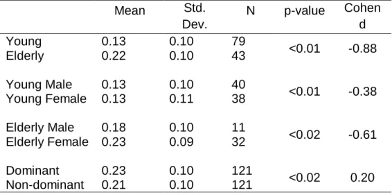
(20) used was the 3000/P1/0600T1/REG. The sensor matrix was distributed over 60 rows and 21 columns, with a sensor interval of 5.1 mm. Sampling frequency was set to 100 Hz. To reduce the effect of random noise, the data was filtered using a 4th order butter-worth filter with cutoff frequency of 4Hz.. 3.4 Results A total of 122 subjects (244 feet), which were separated into groups to be compared were analyzed: by age - young people (between 18 and 65 years) and elderly (over 65 years); by gender - men and women (in subgroups older men, older women, young men and young women); for dominance - the dominant and the non-dominant foot. The results showed differences statistically significant in all comparisons of means, except at the comparison of means in the young sub-group on gender. When calculatin the Cohen d effect size, it presented a large magnitude effect between the Age groups – young X elderly (-0.88) and Gender – for the Elderly (-0.61), medium magnitude effect between female X male (-0.38) and small magnitude effect between dominant foot X non-dominant foot (0.20) – table 3.1. Table 3.1: Arch Index’s means and standard deviations for the groups of interest and comparisons between groups.. Young Elderly. 0.13 0.22. Std. Dev. 0.10 0.10. Young Male Young Female. 0.13 0.13. 0.10 0.11. 40 38. <0.01. -0.38. Elderly Male Elderly Female. 0.18 0.23. 0.10 0.09. 11 32. <0.02. -0.61. Dominant Non-dominant. 0.23 0.21. 0.10 0.10. 121 121. <0.02. 0.20. Mean. 15. p-value. Cohen d. 79 43. <0.01. -0.88. N.
(21) Considering age, the elderly group had a mean arch index of 0.22 (± 0.10), an average that classifies the group as normal arch, while the youth group had a mean index of 0.13 (± 0.10), which classifies the group as high arch (cavus foot). The groups showed a statisticaly significant difference with large magnitude of effect (-0.88) - table 3.1. Comparing genders, young females and males had a very similar average of arch index (0.13), however elderly male = 0.18 (± 0.10) and elderly female 0.23 (± 0.09), showed a statistic significant difference (p≤0.02) – table 3.1. Analyzing dominance, the mean arc index of the dominant foot was 0.23 (± 0.10) - tab. 3.1, close to non-dominant foot mean 0.21 (± 0.10) - tab. 3.1, classifying both groups as normal feet, although the difference between the mean of the groups is statistically significant (p <0.02) - table 3.1.. 3.5 Discussion The aim of this study was to characterize the foot arch in elderly people in relation to gender and foot dominance and also to compare with a younger group. Whereas foot type is related to foot function (H. J. Hillstrom et al., 2013; R. Mootanah et al., 2013), it is important to perceive the difference of foot arch between ages and genders, to help prevent risk of injuries (Riskowski et al., 2013). The results of this study indicate that during standing the elderly group has flatter feet comparing to the young group, showing higher arch index mean. It corroborates with a previous study of Janchai et al. (2008) which found that feet changes with ageing in multiple aspects including arch height, although the author says that only women tend to develop lower arches, men tend to maintain a normal or high arch. Paiva et al. (2011) found that the women’s feet were proportionally wider than the men’s, whose feet had proportionally larger values for height of the dorsal foot but the Arch Index did not reveal significant differences between genders.. 16.
(22) Fukano & Fukubayashi (2012) found that the foot was more flexible in the females than in the males, although they suggest that there is no sexual dimorphism in the longitudinal arch morphology of the foot under no-load conditions. On the other hand, in the present study, elderly women showed flatter feet than elderly men (0.23 against 0.18 of men’s arch index), while the AI mean of young women and young men was very similar (0.13). It reveals that somehow the foot have tended to turn flatter with aging and even flatter if it would be an elderly woman foot. Regarding foot dominance, results about dominant and non-dominant foot shows an statistical significant asymmetry, which despite a lack of studies about it among elderly people, is supported by Hillstrom et al. (2014) that observed about a quarter of their participants demonstrating an asymmetrical foot type, and suggest that it is important to assess both feet independently . There are some factors that this study did not encompass like plantar muscle’s stiffness or behavior of physical activity, which are relevant factors on the foot arch transformation through aging. Further investigations over physical activity level on elderly people and foot type can provide better understanding about the aging process of the foot, also a comparison between static and dynamic measuraments could provide a wider vision over the importance and the influence of the foot arch on the lower limb welfare.. 3.6 Conclusions This study showed that the foot arch change with aging, turning into a flatter foot. The differences between men and women on their physical activity behaviors or footwears through their aging, turn the women’s foot arch lower than the men’s. Regarding to foot dominance, the dominant foot of the elderly showed lower arch than the non-dominant. These findings showed the importance of taking care about the particularly of each elderly’s footwear. As we know, unless they are custom maded, the elderly’s shoes do not come out. 17.
(23) of the factory differing from male to female aspects or dominant to nondominant foot aspects.. 18.
(24) References Atamturk, Derya. (2009). Relationship of flatfoot and high arch with main anthropometric variables. Acta Orthop Traumatol Turc, 43(3), 254-259. doi: 10.3944/AOTT.2009.254 Aydog, S. T., Ozcakar, L., Tetik, O., Demirel, H. A., Hascelik, Z., & Doral, M. N. (2005). Relation between foot arch index and ankle strength in elite gymnasts: a preliminary study. Br J Sports Med, 39(3), e13. doi: 10.1136/bjsm.2004.011627 Burns, Joshua, Crosbie, Jack, Hunt, Adrienne, & Ouvrier, Robert. (2005). The effect of pes cavus on foot pain and plantar pressure. Clinical Biomechanics, 20(9), 877882. doi: http://dx.doi.org/10.1016/j.clinbiomech.2005.03.006 Caselli, Antonella, Pham, Hau, Giurini, John M., Armstrong, David G., & Veves, Aristidis. (2002). The Forefoot-to-Rearfoot Plantar Pressure Ratio Is Increased in Severe Diabetic Neuropathy and Can Predict Foot Ulceration. Diabetes Care, 25(6), 1066-1071. doi: 10.2337/diacare.25.6.1066 Cavanagh, P. R., Morag, E., Boulton, A. J. M., Young, M. J., Deffner, K. T., & Pammer, S. E. (1997). The relationship of static foot structure to dynamic foot function. Journal of Biomechanics, 30(3), 243-250. doi: http://dx.doi.org/10.1016/S00219290(96)00136-4 Cavanagh, PR, & Rodgers, MM. (1987). The arch index: a useful measure from footprints. J Biomech, 20, 547 - 551. Chiu, Min-Chi, Wu, Hsin-Chieh, & Chang, Li-Yu. (2013). Gait speed and gender effects on center of pressure progression during normal walking. Gait & Posture, 37(1), 43-48. doi: http://dx.doi.org/10.1016/j.gaitpost.2012.05.030 Chiu, Min-Chi, Wu, Hsin-Chieh, Chang, Li-Yu, & Wu, Min-Huan. (2013). Center of pressure progression characteristics under the plantar region for elderly adults. Gait & Posture, 37(3), 408-412. doi: http://dx.doi.org/10.1016/j.gaitpost.2012.08.010 de Castro, Marcelo P., Abreu, Sofia C., Sousa, Helena, Machado, Leandro, Santos, Rubim, & Vilas-Boas, João Paulo. (2014). In-Shoe Plantar Pressures and Ground Reaction Forces During Overweight Adults' Overground Walking. Research Quarterly for Exercise and Sport, 85(2), 188-197. doi: 10.1080/02701367.2014.893055 Fernández-Seguín, Lourdes M., Diaz Mancha, Juan Antonio, Sánchez Rodríguez, Raquel, Escamilla Martínez, Elena, Gómez Martín, Beatriz, & Ramos Ortega, Javier. (2014). Comparison of plantar pressures and contact area between normal and cavus foot. Gait & Posture, 39(2), 789-792. doi: http://dx.doi.org/10.1016/j.gaitpost.2013.10.018 Franettovich, Melinda M., McPoil, Thomas G., Russell, Trevor, Skardoon, Gillian, & Vicenzino, Bill. (2007). The Ability to Predict Dynamic Foot Posture from Static Measurements. Journal of the American Podiatric Medical Association, 97(2), 115-120. doi: 10.7547/0970115 Fukano, M., & Fukubayashi, T. (2012). Gender-based differences in the functional deformation of the foot longitudinal arch. Foot (Edinb), 22(1), 6-9. doi: 10.1016/j.foot.2011.08.002 Fukano, Mako, & Fukubayashi, Toru. (2012). Gender-based differences in the functional deformation of the foot longitudinal arch. The Foot, 22(1), 6-9. Hillstrom, H. J., Song, J., Kraszewski, A. P., Hafer, J. F., Mootanah, R., Dufour, A. B., . . . Deland, J. T., 3rd. (2013). Foot type biomechanics part 1: structure and function of the asymptomatic foot. Gait Posture, 37(3), 445-451. doi: 10.1016/j.gaitpost.2012.09.007 Hillstrom, Howard, Song, Jinsup, Neary, Michael, Brechue, William, Zifchock, Rebecca A., Svoboda, Steven, & Hannan, Marian T. (2014). Foot type symmetry and. 19.
(25) change of foot structures from sitting to standing conditions. Journal of Foot and Ankle Research, 7(Suppl 1), A34. doi: 10.1186/1757-1146-7-s1-a34 Imaizumi, K., Iwakami, Y., & Yamashita, K. (2011, Aug. 30 2011-Sept. 3 2011). Analysis of foot pressure distribution data for the evaluation of foot arch type. Paper presented at the Engineering in Medicine and Biology Society,EMBC, 2011 Annual International Conference of the IEEE. Imaizumi, K., Iwakami, Y., Yamashita, K., & Hiejima, Y. (2012, Aug. 28 2012-Sept. 1 2012). Development of an evaluation system for foot arch types in the elderly using foot pressure distribution data. Paper presented at the Engineering in Medicine and Biology Society (EMBC), 2012 Annual International Conference of the IEEE. Janchai, Siriporn , Chaiwanichsiri, Dootchai , Silpipat, Nutsulee , & Tiamprasitt, Jirayoo (2008). Ageing feet and plantar arch characteristics of the Thai elderly. Asian Biomedicine, 2(4), 297-303. Jonely, H., Brismee, J. M., Sizer, P. S., Jr., & James, C. R. (2011). Relationships between clinical measures of static foot posture and plantar pressure during static standing and walking. Clin Biomech (Bristol, Avon), 26(8), 873-879. doi: 10.1016/j.clinbiomech.2011.04.008 Kwan, Rachel Lai-Chu, Zheng, Yong-Ping, & Cheing, Gladys Lai-Ying. (2010). The effect of aging on the biomechanical properties of plantar soft tissues. Clinical Biomechanics, 25(6), 601-605. doi: http://dx.doi.org/10.1016/j.clinbiomech.2010.04.003 Lane, Tamara J., Landorf, Karl B., Bonanno, Daniel R., Raspovic, Anita, & Menz, Hylton B. (2014). Effects of shoe sole hardness on plantar pressure and comfort in older people with forefoot pain. Gait & Posture, 39(1), 247-251. doi: http://dx.doi.org/10.1016/j.gaitpost.2013.07.116 Lin, C. H., Chen, J. J., Wu, C. H., Lee, H. Y., & Liu, Y. H. (2004). Image analysis system for acquiring three-dimensional contour of foot arch during balanced standing. Comput Methods Programs Biomed, 75(2), 147-157. doi: 10.1016/j.cmpb.2003.12.003 McCrory, JL, Young, MJ, Boulton, AJM, & Cavanagh, PR. (1997). Arch index as a predictor of arch height. Foot, 7, 79 - 81. Menz, H. B., & Morris, M. E. (2005). Footwear Characteristics and Foot Problems in Older People. Gerontology, 51(5), 346-351. Menz, Hylton B., & Morris, Meg E. (2006). Clinical determinants of plantar forces and pressures during walking in older people. Gait & Posture, 24(2), 229-236. doi: http://dx.doi.org/10.1016/j.gaitpost.2005.09.002 Mootanah, R., Song, J., Lenhoff, M. W., Hafer, J. F., Backus, S. I., Gagnon, D., . . . Hillstrom, H. J. (2013). Foot Type Biomechanics Part 2: are structure and anthropometrics related to function? Gait Posture, 37(3), 452-456. doi: 10.1016/j.gaitpost.2012.09.008 Mootanah, Rajshree, Song, Jinsup, Lenhoff, Mark W., Hafer, Jocelyn F., Backus, Sherry I., Gagnon, David, . . . Hillstrom, Howard J. (2013). Foot Type Biomechanics Part 2: Are structure and anthropometrics related to function? Gait & Posture, 37(3), 452-456. doi: http://dx.doi.org/10.1016/j.gaitpost.2012.09.008 Nilsson, M. K., Friis, R., Michaelsen, M. S., Jakobsen, P. A., & Nielsen, R. O. (2012). Classification of the height and flexibility of the medial longitudinal arch of the foot. J Foot Ankle Res, 5, 3. doi: 10.1186/1757-1146-5-3 Ozer, Cenk Murat , & Barut, Cagatay (2012). Evaluation of the sole morphology of professional football players. International SportMed Journal, 13(1), 8-17.. 20.
(26) Paiva de Castro, A , Rebelatto, JR, & Aurichio, TR. (2011). The Effect of Gender on Foot Anthropometrics in Older People. Journal Of Sport Rehabilitation, 20(03), 10. Periyasamy, R., & Anand, S. (2013). The effect of foot arch on plantar pressure distribution during standing. J Med Eng Technol, 37(5), 342-347. doi: 10.3109/03091902.2013.810788 Periyasamy, R., Mishra, A., Anand, Sneh, & Ammini, A. C. (2011). Preliminary investigation of foot pressure distribution variation in men and women adults while standing. The Foot, 21(3), 142-148. doi: http://dx.doi.org/10.1016/j.foot.2011.03.001 Putti, A. B., Arnold, G. P., & Abboud, R. J. (2010). Foot pressure differences in men and women. Foot and Ankle Surgery, 16(1), 21-24. doi: http://dx.doi.org/10.1016/j.fas.2009.03.005 Razeghi, Mohsen, & Batt, Mark Edward. (2002). Foot type classification: a critical review of current methods. Gait & Posture, 15(3), 282-291. doi: http://dx.doi.org/10.1016/S0966-6362(01)00151-5 Riskowski, J. L., Dufour, A. B., Hagedorn, T. J., Hillstrom, H. J., Casey, V. A., & Hannan, M. T. (2013). Associations of foot posture and function to lower extremity pain: results from a population-based foot study. Arthritis Care Res (Hoboken), 65(11), 1804-1812. doi: 10.1002/acr.22049 Scott, G., Menz, H. B., & Newcombe, L. (2007). Age-related differences in foot structure and function. Gait Posture, 26(1), 68-75. doi: 10.1016/j.gaitpost.2006.07.009 Teyhen, Deydre S, Stoltenberg, Brian E, Collinsworth, Keith M, Giesel, Crystal L, Williams, Drew G, Kardouni, Cryus H, . . . McPoil, Thomas. (2009). Dynamic plantar pressure parameters associated with static arch height index during gait. Clinical Biomechanics, 24(4), 391-396. Wong, C. K., Weil, R., & de Boer, E. (2012). Standardizing foot-type classification using arch index values. Physiother Can, 64(3), 280-283. doi: 10.3138/ptc.2011-40 Wozniacka, R., Bac, A., Matusik, S., Szczygiel, E., & Ciszek, E. (2013). Body weight and the medial longitudinal foot arch: high-arched foot, a hidden problem? Eur J Pediatr, 172(5), 683-691. doi: 10.1007/s00431-013-1943-5 Xiong, Shuping, Goonetilleke, Ravindra S., & Jiang, Zuhua. (2011). Pressure thresholds of the human foot: measurement reliability and effects of stimulus characteristics. Ergonomics, 54(3), 282-293. doi: 10.1080/00140139.2011.552736 Zifchock, Rebecca A., Davis, Irene, Hillstrom, Howard, & Song, Jinsup. (2006). The Effect of Gender, Age, and Lateral Dominance on Arch Height and Arch Stiffness. Foot & Ankle International, 27(5), 367-372. doi: 10.1177/107110070602700509. 21.
(27) 4. Article 2. Influence of arch index on plantar pressure in elderly people during standing.. Michel Bertani1,2, Denise Soares1,2, Leandro Machado1,2.. 1. University of Porto, Faculty of Sports, Rua Dr Placido Costa, 91-4200-450 Porto, Portugal 2. LABIOMEP - Porto Biomechanics Laboratory, Rua Dr. Plácido Costa, 91, 4200 450 Porto, Portugal. 22.
(28) 4.1 Abstract Background - The foot is an important and complex structure that provides support, balance and propulsion to locomotion, thus, its proper care can help to have a better life quality avoiding pain. The medial longitudinal arch is an important structure whose deformities are related to injury risks, also foot type classified by arch index shows significant association with maximum force and peak pressure at some foot regions. The purpose of this study was to analyze the arch index and plantar pressure distribution in elderly people during standing considering gender and foot-dominance, and compare the results to the younger group. Methods – The sample was composed by a total of 122 subjects, 79 healthy young subjects (38 women and 41 men) and 43 healthy elderly subjects (32 women and 11 men). Ten seconds of standing barefoot plantar pressure were measured through Tekscan F-Scan device, and the data processing, filtering, and arch index (AI) calculation were performed using MATLAB™ 7.0. Findings – Young group AI mean (0.13) appears higher than the elderly group mean (0.22), also the peak pressure: maximum, in the toes, forefoot and hindfoot regions are lower for the elderly group but lower in the middlefoot. The male group presented higher AI than the female group; the pressure peak in the regions of the forefoot and hind foot, toes and also the maximum pressure peak, were greater in the male than in the female. Dominant feet showed lower AI than non-dominant, and it also presented lower plantar pressure in the halux, central forefoot and hindfoot regions. Interpretation – The foot arch has showed higher plantar pressure and peak preassure in comparisons where the AI was lower, contrary the middlefoot area, where low arched feet showed higher pressure peak values. Keywords: Arch Index; Elderly; Gender; Foot Dominance.. 23.
(29) 4.2 Introduction The foot is a complex structure composed by 26 bones and more than 30 joints, and its fundamental functions are support, shock absorption, and weight bearing (Wozniacka et al., 2013). The foot represents the interface to the exterior for maintaining or shifting one’s center of gravity and, during motion, the foot also plays the role of adapting to irregular surfaces and generating propulsion force to locomotion (Chiu, Wu, & Chang, 2013; Imaizumi et al., 2011; Imaizumi et al., 2012). According to Imaizumi et al. (2012), the foot has three anatomical arch structures: medial longitudinal arch (MLA), lateral longitudinal arch (MLA), and forefoot transverse arch. The MLA, in particular, plays an important role with regard to shock absorption and the action of walking. Foot type categorizes feet as planus - low arched with a valgus hindfoot and/or varus forefoot; rectus - well aligned hindfoot and forefoot; and cavus - high arched with a varus hindfoot and/or valgus forefoot (H. J. Hillstrom et al., 2013). Some pathologies normally appear associated with certain type of foot, but is not clear in the literature why this happens or even why some subjects with planus or cavus foot type do not show any pathology. There are many methods used to classify foot type, using: visual inspection, footprint analysis or radiographic evaluation (Burns et al., 2005), and these kinds of methods can be classified as direct - somatometric measurements, clinical. assessment,. radiographic. evaluation,. and. ultrasonography. quantification; or indirect methods - footprints and photographic techniques; (Wozniacka et al., 2013). The footprint technique can be static or dynamic but both was evaluated to be reliable methods to classify foot type and to compare with foot function (P. R. Cavanagh et al., 1997; P. Cavanagh & Rodgers, 1987; Imaizumi et al., 2012; McCrory et al., 1997). Abnormal plantar pressure patterns are thought to contribute to the development of a wide variety of foot conditions, including forefoot pain, plantar neuropathic ulceration and metatarsal stress fractures (Hylton B. Menz &. 24.
(30) Morris, 2006). Also, the foot type classified by arch index (P. Cavanagh & Rodgers, 1987) shows significantly associations with maximum force and peak pressure at some foot regions (Hylton B. Menz & Morris, 2006). As observed by Periyasamy & Anand (2013), foot arch type could influence the plantar pressure distribution and can have an impact on the biomechanics of an individual posture and gait. Aging may cause musculoskeletal degeneration and influence the medial longitudinal arch structure. Compared to youngsters, elderly subjects exhibited more flatter and more pronated feet, and also display a longer contact time under the midfoot during gait as obtained from plantar pressure assessment (Chiu, Wu, Chang, et al., 2013). Due to a growing concern about health quality in elderly people, and keeping in mind that foot problems can carry to serious mobility impairments, resulting in a decrease of their autonomy, the aim of this study was to analyze the arch index and plantar pressure distribution in elderly people during standing, and compare the results with young adults while considering gender and foot-dominance.. 4.3 Methodology Sample: The young group was composed by a total of 79 healthy subjects (38 women - age of 24.24 ± 5.73 years old, weight 60.59 ± 12.11 kg, height 1.61 ± 0.06 m; 41 men - age of 26.00 ± 5,71 years old, weight 78.03 ± 16.75 kg, height 1.75 ± 0.06m). They went voluntarily to the Biomechanics Laboratory for the collection of data, where they freely signed an informed consent accordingly to the Helsinky protocol. The elderly group was formed after a contact with the manager of the Elderly Day Care of the Social Center enrolled, who allowed inviting the elders subscribed on the social center program, to voluntarily participate in this study. A total of 43 healthy elderly subjects (32 women - age of 73.28 ± 7.73 years old, weight 72.68 ± 11.87 kg, height 1.52 ± 0.06 m; 11 men - age of 73.91 ± 7.01. 25.
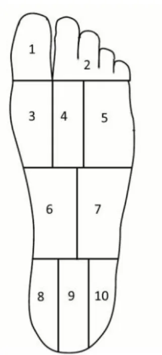
(31) years old, weight 76.35 ± 10.95 kg, height 1.65 ± 0.07m), without previous comorbidities related to autonomous capability of gait enrolled the study. All volunteers freely signed an informed consent accordingly to the Helsinky protocol. The project was approved by the ethical committee of the institution involved in the Study.. Protocol:. The. procedure. of. data. collection. followed. the. sequence:. anthropometric measurements (height and weight), questionnaire related to comorbidities, equipage of slipper with Tekscan insole, calibration of weight into the Tekscan® software, adaptation to the standing position, and when subject is judged stable, 10-s recording of footprint and plantar pressure were performed. In the process of adapting to the position, subjects were asked to stand in both feet in the anatomic position with eyes open looking forward to the horizon. Both feet of all participants were analyzed. The contact area of the foot, excluding toes, was divided into three equal parts: the forefoot, midfoot and hindfoot. After calculating the active area of each part, the arch index was calculated dividing the area of the midfoot by the sum of the three areas (midfoot / [forefoot + midfoot + hindfoot]). Arch indices ≥0.260 were considered low-arched; arch indices between 0.210 and 0.260 were considered normal; and arch indices ≤0.210 were considered high-arched (P. R. Cavanagh et al., 1997). To analyze the plantar pressure, the foot was divided into 10 regions (figure 1) (de Castro et al., 2014) and the values of peak plantar pressure, time when the peak pressure occurs and contact area in each region were calculated (tables 1 to 3). The peak pressure provides information about the maximum load received by the foot during support, being one of the variables most used by research groups to verify the performance of shoes. The data processing, filtering, and arch index calculation were performed using MATLAB 7.0 (MathWorks, Massachusetts, USA).. 26.
(32) Figure 1: the 10 regions of foot-sole. Instruments and devices: The device used to measure foot pressure distribution was F-SCAN (TEKSCAN inc., Boston, MA, USA). The insole model used was the 3000/P1/0600T1/REG. The sensor matrix was distributed over 60 rows and 21 columns, with a sensor interval of 5.1 mm. Sampling frequency was set at 100 Hz. To reduce the effect of random noise, the data was filtered using a 4th order butter-worth filter with cutoff frequency of 4Hz.. 4.4 Results Comparisons between age, gender and foot dominance were processed. The arch index of each group was calculated and compared between them, also the peak pressure of the regions previously stipulated were calculated and compared. The data are presented in the tables 4.1, 4.2 and 4.3. The results of table 4.1 show a significant difference between young and elderly groups (irrespective of gender and foot dominance) on plantar arch index (0.13 ± 0.10 / 0.22 ± 0.10 – p<0.01), indicating that the young group classify as cavus foot (high arch) and the elderly group as rectus foot (normal arch). The groups also present significant differences in maximum peak pressure (270.38 ±. 27.
(33) 165.34 / 195.95 ± 79.02 - p<0.01), and in region 1 (42.39 ± 36.39 / 22.33 ± 30.81 - p<0.01), region 2 (43.67 ± 45.85 / 18.57 ± 23.25 - p<0.01), region 4 (94.61 ± 35.54 / 84.41 ± 38.00 - p<0.05), region 5 (100,85 ± 53,05 / 82,93 ± 43,36 - p<0,01), region 6 (36,85 ± 25,68 / 62.13 ± 33.25 - p<0.01), region 7 (53.59 ± 36.78 / 79.69 ± 38.86 - p<0.01), where, probably due to a smaller contact area of the foot with the ground, young people have showed higher peak pressure than the elderly group in all foot area but in the middle region. In the middle foot, the elderly group presents a higher peak pressure in the medial middle-foot (almost double than the young group) and in the lateral middle-foot. The pressure discomfort threshold is up to 200 kPa and the pressure pain threshold is up to 300kPa (Xiong, Goonetilleke, & Jiang, 2011), values comparable to some of the peak pressures found in this work. In a study with diabetic people, was related that above of the threshold of 550 kPa during 30 months, 78% of the subjects had ulcers (Caselli, Pham, Giurini, Armstrong, & Veves, 2002). Table 4.1: Comparison between young and elderly (irrespective of gender and foot dominance) – Arch Index (AI), Maximum Peak pressure (MPP) and mean pressure on regions: 1- Hálux (HLX), 2- Other Toes (OT), 3- Forefoot Medial (FM), 4- Forefoot Central (FC), 5- Forefoot Lateral (FL), 6- Midfoot Medial (MM), 7- Midfoot Lateral (ML), 8- Hindfoot Medial (HM), 9- Hindfoot Central (HC), 10- Hindfoot Lateral (HL). Region Young Elderly Sig. 0.13 ± 0.10 0.22 ± 0,10 * 0.00 AI 270.38 ± 165.34 195,95 ± 79,02 * 0.00 MPP 1 42.39 ± 36.39 22,33 ± 30,81 * 0.00 HLX 2 43.67 ± 45.85 18.57 ± 23.25 * 0.00 OT 3 88.50 ± 46.29 84.28 ± 54.74 0.54 FM 4 94.61 ± 35.54 84.41 ± 38.00 * 0.05 FC 5 100.85 ± 53.05 82.93 ± 43.36 * 0.01 FL 6 36.85 ± 25.68 62.13 ± 33.25 * 0.00 MM 7 53.59 ± 36.78 79.69 ± 38.86 * 0.00 ML 8 163.21 ± 69.39 146.89 ± 56.22 0.07 HM 9 162.85 ± 66.55 155.42 ± 64.35 0.41 HC 10 54.03 ± 36.50 61.08 ± 30.93 0.13 HL Values expressed in kilopascal (KPa), except for the AI. * Statistical significant differences between Young and elderly people. 28.
(34) The results in table 4.2 show a significant difference between males and females (irrespective of age and foot dominance) in the arch index (0.18 ± 0.11 / 0.14 ± 0.10 - p<0.01), although the averages of both sexes classify cavus foot, the results show that the feet of the male group have higher arch than the female. Also, significant differences were found in maximum peak pressure (216.67 ± 114.09 / 279.86 ± 172.12 - p<0.01), and in region 1 (29.46 ± 33.073 / 43.13 ± 37.92 - p<0.01), region 4 (84.02 ± 35.64 / 101.72 ± 36.23 - p<0.01), region 5 (77.88 ± 39.96 / 118.81 ± 54.46 - p<0.01), region 6 (50.95 ± 32.90 / 38.39 ± 26.48 - p<0.01), region 8 (150.16 ± 63.67 / 167.71 ± 66.63 - p<0.05), region 10 (61.7 ± 33.62 / 49.69 ± 35.07 - p<0.01). In summary, the male group presented higher mean peak pressure in regions 1, 4, 5, 8 and lower in regions 6 and 10. Table 4.2: Comparison between gender female and male (irrespective of age and foot dominance) – Arch Index (AI), Max Peak pressure (MPP) and mean pressure on regions: 1- Hálux (HLX), 2- Other Toes (OT), 3- Forefoot Medial (FM), 4- Forefoot Central (FC), 5- Forefoot Lateral (FL), 6- Midfoot Medial (MM), 7- Midfoot Lateral (ML), 8- Hindfoot Medial (HM), 9- Hindfoot Central (HC), 10- Hindfoot Lateral (HL). Region Female Male Sig. * 0.18 ± 0.11 0.14 ± 0.10 0.00 AI - MPP 216.67 ± 114.09 279.86 ± 172.12 * 0.00 1 HLX * 29.46 ± 33.07 43.13 ± 37.92 0.00 2 31.22 ± 43.03 39.96 ± 37.96 0.11 OT 3 82.59 ± 46.62 93.49 ± 52.74 0.10 FM 4 * 84.02 ± 35.64 101.72 ± 36.23 0.00 FC 5 * 77.88 ± 39.96 118.81 ± 54.46 0.00 FL 6 MM * 50.95 ± 32.90 38.39 ± 26.48 0.00 7 65.41 ± 38.27 60.20 ± 41.3 0.32 ML 8 HM * 150.16 ± 63.67 167.71 ± 66.63 0.04 9 158.61 ± 65.72 162.50 ± 66.03 0.67 HC 10 HL * 61.7 ± 33.62 49.69 ± 35.07 0.01 Values expressed in kilopascal (KPa), except for the AI. * Statistical significant differences between gender female and male.. 29.
(35) Regarding the foot dominance in the elderly group, the results on table 4.3 show a significant difference between the dominant and non-dominant feet of the elderly in the arch index (0.23 ± 0.10 / 0.21 ± 0.10 – p < 0.02), although both averages classify as rectus foot (normal arch index), the results show that the non-dominant feet have higher arch than the dominant feet. Also, were found significant differences in regions 1 (15.54 ± 28.06 / 29.13 ± 32.24 – p < 0.01), 4 (101.81 ± 62.44 / 77.83 ± 34.39 – p < 0.01), 9 (175.15 ± 90.37 / 144.99 ± 58.41 – p < 0.03) and 10 (55.99 ± 24.78 / 66.17 ± 35.63 – p < 0.04), where, the dominant feet showed higher peak pressure in regions 4 and 9 and lower in regions 1 and 10.. Table 4.3: Comparrison between dominant and non-dominant foot of the elderly individuals (irrespective of gender) – Arch Index (AI), Max Peak pressure (MPP) and mean pressure on regions: 1- Hálux (HLX), 2- Other Toes (OT), 3- Forefoot Medial (FM), 4- Forefoot Central (FC), 5- Forefoot Lateral (FL), 6- Midfoot Medial (MM), 7- Midfoot Lateral (ML), 8- Hindfoot Medial (HM), 9- Hindfoot Central (HC), 10- Hindfoot Lateral (HL). Region Dominant Non-dominant Sig. * 0.02 AI 0.23 ± 0.10 0.21 ± 0.10 MPP 218.23 ± 110.32 192.85 ± 89.18 0.15 1 HLX 15.54 ± 28.06 29.13 ± 32.24 * 0.01 2 OT 25.67 ± 47.46 26.22 ± 45.98 0.95 3 FM 99.47 ± 93.36 95.66 ± 69.12 0.65 4 FC 101.81 ± 62.44 77.83 ± 34.39 * 0.00 5 FL 94.94 ± 59.35 87.73 ± 64.59 0.49 6 MM 66.82 ± 33.58 80.14 ± 96.99 0.36 7 ML 99.17 ± 95.78 73.86 ± 40.15 0.12 8 HM 161.84 ± 74.00 139.25 ± 53.93 0.05 9 HC 175.15 ± 90.37 144.99 ± 58.41 * 0.02 10 HL 55.99 ± 24.78 66.17 ± 35.63 * 0.04 Values expressed in kilopascal (KPa), except for the AI. * Statistical significant differences between dominant and nondominant foot of the elderly individuals.. 30.
(36) 4.5 Discussion The present study investigated the arch index and plantar pressure distribution in elderly people during standing, and compared the results between age, gender and foot-dominance. Regarding to age differences, the results showed a lower arch in the elderly group, corroborating with previous studies that indicate flatter foot in the elderly population. In relation to the maximum peak pressure, these values are generaly statistically higher in the young group. This can be explaned by their higher arch foot, which presents less contact area than the normal foot, distributing all weight in a smaller area, and increasing pressure. FernándezSeguín et al. (2014) report that 60% of the population that have cavus feet suffer from pain, which is related to excessive plantar pressure. The foot of young group seems to be supinated in the forefoot area, showing growing peak pressure values from medial to lateral areas. In the middle-foot the lateral region also has higher peak pressure. Although the hind-foot regions did not show significant differences, the peak pressure in the hind-foot of the young group (high arch index) showed a trend to be pronated, the peak pressure growing from lateral area to medial area. In relation to the foot type, more specificly its inclination, the elderly group showed a similar values of peak pressure in the forefoot region, being the higher value in the central forefoot, looking like a neutral foot. Also, the hindfoot presented the higher value in the central hindfoot region. This condraticts the results from Chiu et al. (2013), who found that the elderly exhibit more heel eversion. The elderly group also showed half values of peak pressure in the halux and other toes than the young group, it might be because the foot of the elderly group is more neutral, so don’t need to force the hallux and other toes to keep the balance. The results also show a very similar value of peak pressure between the regions of the forefoot area and a trend of high peak pressure in the central hindfoot. As supported by other studies, the middlefoot area has higher pressure values in the elderly group (flatter foot) than the young group. Scott et al. (2007) indicated that elderly people have a stronger tendency to. 31.
(37) exhibit more pronated feet, which in a certain way was found in the present study, the elderly foot appears neutral and normal arched but more pronated than the young group. These findings are also supported by Chiu et al. (2013), displaying in their study that elderly has flatter and more pronated feet than younger adults, and also a decline in the magnitude of pressures under the heel, lateral forefoot and hallux. Despite some studies referring that there is no significant differences in the longitudinal foot arch between men and women (M. Fukano & T. Fukubayashi, 2012; Paiva de Castro et al., 2011), gender comparisons in the present study revealed that the female group has a lower arch than male (although both have arch index of cavus foot), leading to a higher maximum peak pressure for the male group, usually related with cavus foot. Similarly to the age groups comparisons, the male group (group that presented higher arch) presented supinated forefoot, lower middle foot peak pressure, and pronated hind foot. While the female group, that presented an arch index close to the normal foot arch index, presents a neutral forefoot with higher pressure region at the central forefoot, and more neutral hindfoot than the male group. Putti et al. (2010) divided the foot into 10 regions and found there was no difference in peak pressure between genders in any of the regions. However, Periyasamy et al. (2011) found that men have greater pressure under the hind foot compared to women. The rationale for considering the foot dominance was that impairments on subjects balance, measured through differences on arch index and plantar pressure, could carry to the development of problems in the ankle and knee joints, but also in the hip and spine. In this study we found significant differences between dominant and non-dominant feet in the arch index, where both groups were classified as normal foot, but the dominant feet presented a flatter medial longitudinal arch. As normal foot, the trend to be a neutral foot is confirmed in both groups, although the non-dominant group (high arched) shows greater halux peak pressure as all high arched groups in previous comparisons. For being flatter than non-dominant group, it was expected that. 32.
(38) the pressure was well distributed and, as a consequence, lower in the forefoot and in the hindfoot. Against these assumptions, the dominant group showed higher pressure in these two areas, showing that elderly people at standing position load predominantly the dominant foot. According to Periyasamy & Anand (2013), during standing, the contact area and mean pressure value of both left and right feet are not similar in all the foot type groups. The lack of an analysis of cinemetry and a statistical analysis of correlation between the increase of pressure and the type of arch are the limitations of this study. Further investigations would be interesting concerning correlation of the hindfoot with the forefoot in each type of arch, since in the present study while the forefoot has showed greater lateral peak pressure, the hindfoot showed greater medial peak pressure.. 4.6 Conclusions This study has showed higher plantar pressure and peak preassure in all analysed regions of the foot where the arch index was lower (cavus foot), except in the middlefoot area, where low arched feet (planus foot) showed higher peak pressure values. Also, the lower arches in the comparisons showed growing values of peak pressure from medial forefoot to the lateral forefoot but reverse of this on the hindfoot. These findings show the importance of analyzing peak pressure and foot arch to fit proper footwear to the elderly person. Well fitted shoes can avoid discomfort, pain, caluses and even ulcers in the worst scenario.. 33.
(39) References Burns, Joshua, Crosbie, Jack, Hunt, Adrienne, & Ouvrier, Robert. (2005). The effect of pes cavus on foot pain and plantar pressure. Clinical Biomechanics, 20(9), 877882. doi: http://dx.doi.org/10.1016/j.clinbiomech.2005.03.006 Cavanagh, P. R., Morag, E., Boulton, A. J. M., Young, M. J., Deffner, K. T., & Pammer, S. E. (1997). The relationship of static foot structure to dynamic foot function. Journal of Biomechanics, 30(3), 243-250. doi: http://dx.doi.org/10.1016/S00219290(96)00136-4 Cavanagh, PR, & Rodgers, MM. (1987). The arch index: a useful measure from footprints. J Biomech, 20, 547 - 551. Caselli, Antonella, Pham, Hau, Giurini, John M., Armstrong, David G., & Veves, Aristidis. (2002). The Forefoot-to-Rearfoot Plantar Pressure Ratio Is Increased in Severe Diabetic Neuropathy and Can Predict Foot Ulceration. Diabetes Care, 25(6), 1066-1071. doi: 10.2337/diacare.25.6.1066 Chiu, Min-Chi, Wu, Hsin-Chieh, & Chang, Li-Yu. (2013). Gait speed and gender effects on center of pressure progression during normal walking. Gait & Posture, 37(1), 43-48. doi: http://dx.doi.org/10.1016/j.gaitpost.2012.05.030 Chiu, Min-Chi, Wu, Hsin-Chieh, Chang, Li-Yu, & Wu, Min-Huan. (2013). Center of pressure progression characteristics under the plantar region for elderly adults. Gait & Posture, 37(3), 408-412. doi: http://dx.doi.org/10.1016/j.gaitpost.2012.08.010 de Castro, Marcelo P., Abreu, Sofia C., Sousa, Helena, Machado, Leandro, Santos, Rubim, & Vilas-Boas, João Paulo. (2014). In-Shoe Plantar Pressures and Ground Reaction Forces During Overweight Adults' Overground Walking. Research Quarterly for Exercise and Sport, 85(2), 188-197. doi: 10.1080/02701367.2014.893055 Fernández-Seguín, Lourdes M., Diaz Mancha, Juan Antonio, Sánchez Rodríguez, Raquel, Escamilla Martínez, Elena, Gómez Martín, Beatriz, & Ramos Ortega, Javier. (2014). Comparison of plantar pressures and contact area between normal and cavus foot. Gait & Posture, 39(2), 789-792. doi: http://dx.doi.org/10.1016/j.gaitpost.2013.10.018 Fukano, M., & Fukubayashi, T. (2012). Gender-based differences in the functional deformation of the foot longitudinal arch. Foot (Edinb), 22(1), 6-9. doi: 10.1016/j.foot.2011.08.002 Hillstrom, H. J., Song, J., Kraszewski, A. P., Hafer, J. F., Mootanah, R., Dufour, A. B., . . . Deland, J. T., 3rd. (2013). Foot type biomechanics part 1: structure and function of the asymptomatic foot. Gait Posture, 37(3), 445-451. doi: 10.1016/j.gaitpost.2012.09.007 Imaizumi, K., Iwakami, Y., & Yamashita, K. (2011, Aug. 30 2011-Sept. 3 2011). Analysis of foot pressure distribution data for the evaluation of foot arch type. Paper presented at the Engineering in Medicine and Biology Society,EMBC, 2011 Annual International Conference of the IEEE. Imaizumi, K., Iwakami, Y., Yamashita, K., & Hiejima, Y. (2012, Aug. 28 2012-Sept. 1 2012). Development of an evaluation system for foot arch types in the elderly using foot pressure distribution data. Paper presented at the Engineering in. 34.
(40) Medicine and Biology Society (EMBC), 2012 Annual International Conference of the IEEE. McCrory, JL, Young, MJ, Boulton, AJM, & Cavanagh, PR. (1997). Arch index as a predictor of arch height. Foot, 7, 79 - 81. Menz, Hylton B., & Morris, Meg E. (2006). Clinical determinants of plantar forces and pressures during walking in older people. Gait & Posture, 24(2), 229-236. doi: http://dx.doi.org/10.1016/j.gaitpost.2005.09.002 Paiva de Castro, A , Rebelatto, JR, & Aurichio, TR. (2011). The Effect of Gender on Foot Anthropometrics in Older People. Journal Of Sport Rehabilitation, 20(03), 10. Periyasamy, R., & Anand, S. (2013). The effect of foot arch on plantar pressure distribution during standing. J Med Eng Technol, 37(5), 342-347. doi: 10.3109/03091902.2013.810788 Periyasamy, R., Mishra, A., Anand, Sneh, & Ammini, A. C. (2011). Preliminary investigation of foot pressure distribution variation in men and women adults while standing. The Foot, 21(3), 142-148. doi: http://dx.doi.org/10.1016/j.foot.2011.03.001 Putti, A. B., Arnold, G. P., & Abboud, R. J. (2010). Foot pressure differences in men and women. Foot and Ankle Surgery, 16(1), 21-24. doi: http://dx.doi.org/10.1016/j.fas.2009.03.005 Scott, G., Menz, H. B., & Newcombe, L. (2007). Age-related differences in foot structure and function. Gait Posture, 26(1), 68-75. doi: 10.1016/j.gaitpost.2006.07.009 Xiong, Shuping, Goonetilleke, Ravindra S., & Jiang, Zuhua. (2011). Pressure thresholds of the human foot: measurement reliability and effects of stimulus characteristics. Ergonomics, 54(3), 282-293. doi: 10.1080/00140139.2011.552736 Wozniacka, R., Bac, A., Matusik, S., Szczygiel, E., & Ciszek, E. (2013). Body weight and the medial longitudinal foot arch: high-arched foot, a hidden problem? Eur J Pediatr, 172(5), 683-691. doi: 10.1007/s00431-013-1943-5. 35.
Imagem


+3


Documentos relacionados