UNIVERSIDADE
DE
LISBOA
FACULDADE
DE
FARMÁCIA
MOLECULAR
MECHANISMS
OF
MICROGLIA
REACTIVITY
TO
BILIRUBIN:
EVALUATION
OF
POTENTIAL
NEUROLOGICAL
EFFECTS
Sandra Isabel Leitão da Silva
DOUTORAMENTO EM FARMÁCIA
(Biologia Celular e Molecular)
UNIVERSIDADE
DE
LISBOA
FACULDADE
DE
FARMÁCIA
MOLECULAR
MECHANISMS
OF
MICROGLIA
REACTIVITY
TO
BILIRUBIN:
EVALUATION
OF
POTENTIAL
NEUROLOGICAL
EFFECTS
Sandra Isabel Leitão da Silva
Research advisors: Dora Maria Tuna de Oliveira Brites, PhD.
Rui Fernando Marques da Silva, PhD.
DOUTORAMENTO EM FARMÁCIA
(Biologia Celular e Molecular)
MOLECULAR
MECHANISMS
OF
MICROGLIA
REACTIVITY
TO
BILIRUBIN:
EVALUATION
OF
POTENTIAL
NEUROLOGICAL
EFFECTS
MECANISMOS
MOLECULARES
DE
REACTIVIDADE
DA
MICROGLIA
À
BILIRRUBINA:
AVALIAÇÃO
DE
POTENCIAIS
EFEITOS
NEUROLÓGICOS
Dissertação apresentada à Faculdade de Farmácia da Universidade de Lisboa
para obtenção do grau de Doutor em Farmácia (Biologia Celular e Molecular)
Sandra Isabel Leitão da Silva
2010
Para a elaboração da presente tese de doutoramento foram usados integralmente como capítulos, artigos científicos publicados, ou submetidos para publicação, em revistas científicas internacionais indexadas. Estes trabalhos foram realizados em colaboração com os seguintes autores: Ana Rita Vaz, Catarina Osório, Andreia Barateiro, Adelaide Fernandes, Ana Sofia Falcão, Maria José Diógenes, Maria Alexandra Brito, Nico van Rooijen, Ana Sebastião, Rui Silva e Dora Brites.
De acordo com o disposto no ponto 1 do artigo nº41 do Regulamento de Estudos Pós-Graduados da Universidade de Lisboa, deliberação nº 93/2006, publicada em Diário da República – II Série nº 153 – 5 de Julho de 2003, a Autora desta dissertação declara que participou na concepção e execução do trabalho experimental, na interpretação dos resultados obtidos e na redacção dos manuscritos.
Os estudos apresentados nesta dissertação foram realizados no grupo de investigação “Neuron Glia Biology in Health & Disease”, Research Institute for Medicines and Pharmaceutical Sciences (iMed.UL), Faculdade de Farmácia da Universidade de Lisboa. Parte do trabalho foi também realizado no Departamento de Investigação Cellular Neuroscience do Max-Delbrück Centrum em Berlim, Alemanha, sob a orientação do Professor Doutor Helmut Kettenmann.
O trabalho foi subsidiado pelos projectos POCTI/SAU/MMO/55955/2004 e FCT-PTDC/SAU-NEU/64385/2006 concedidos à Professora Doutora Dora Brites pela Fundação para a Ciência e Tecnologia (FCT), sendo que a Autora usufruiu de uma bolsa de Doutoramento (SFRH/BD/30326/2006) concedida pela FCT, Lisboa, Portugal.
Acknowledgements/Agradecimentos
As minhas primeiras palavras de agradecimento vão naturalmente para a Professora Doutora Dora Brites, orientadora deste doutoramento. Gostaria de lhe agradecer o facto de me ter aberto as portas do mundo da ciência e por tão bem me ter guiado nos seus trilhos. Gostaria de salientar as suas notáveis qualidades científicas e o seu enorme espírito crítico, bem como o seu sentido de justiça e igualdade. Os seus elevados padrões de rigor científico e o seu constante encorajamento ao longo deste caminho fizeram-me acreditar que podia chegar ao fim e, mais importante do que isso, que era sempre possível fazer mais e melhor. Como já lhe tenho dito muitas vezes, o seu olhar consegue sempre ver mais além! Os seus ensinamentos ser-me-ão úteis ao longo da vida académica e pessoal e não esquecerei o tempo que passei a trabalhar ao seu lado nem o facto de ter depositado em mim a confiança para desempenhar esta tarefa. Só espero ter estado à altura das suas expectativas!
Ao Professor Doutor Rui Silva, co-orientador desta tese, quero agradecer a constante ajuda e encorajamento para superar as dificuldades que foram surgindo ao longo deste percurso académico. O seu olhar crítico sobre as questões essenciais foram extremamente valiosas para a concretização deste trabalho. Quero também agradecer a preocupação e carinho que sempre demonstrou para comigo e a forma como sempre me conseguia fazer sorrir mesmo quando tudo corria mal!
À Professora Doutora Alexandra Brito gostaria de agradecer por, apesar de não ter uma responsabilidade directa sobre o meu trabalho, ter sempre demonstrado interesse e disponibilidade para me ajudar. Os seus conselhos científicos foram pertinentes, assertivos e de extrema validade. Gostaria também de lhe agradecer a amizade que desenvolvemos ao longo dos anos.
I would also like to thank Professor Helmut Kettenmann and his lab, in the Research Department Cellular Neuroscience from the Max-Delbrück Centrum in Berlin, Germany for welcoming me in Berlin during my training period and for providing me with so much valuable knowledge.
Gostaria também de agradecer às Professoras Doutoras Ana Sebastião e Maria José Diógenes pela imensa disponibilidade que demonstraram desde o primeiro dia da colaboração que encetámos e pelo seu contributo científico para este trabalho.
À Liliana Bernardino quero endereçar um agradecimento sentido pela ajuda tão preciosa que me prestou na implementação das culturas organotípicas de cortes de hipocampo. A tua enorme disponibilidade e simpatia, bem como a fé que sempre tiveste que tudo ia correr bem, fizeram-me acreditar que seria possível completar esta tarefa hercúlea!
À Adelaide tenho prometido um merecido agradecimento desde o primeiro dia em que entrei no CPM! Foste tu quem me encorajou a enveredar pelo mundo da ciência e foste também tu quem me deu a mão nas vezes em que me perdi pelo caminho… Por todos os momentos em que dispensaste o teu tempo para me ajudares, pelo enorme contributo intelectual e científico que deste a esta tese e pela forma como me fizeste sentir acarinhada neste grupo, um enorme obrigado! Sem a tua ajuda tenho a certeza que este trabalho não seria o mesmo!
À Sofia tenho que agradecer os ensinamentos que me transmitiu durante este período e a forma atenciosa e carinhosa como me tratou desde o início, fazendo-me sentir em casa e parte integrante desta equipa! As nossas conversas à hora do lanche foram uma fonte de motivação e de força para continuar, bem como o teu exemplo de perseverança apesar das dificuldades!
Um agradecimento muito especial à minha companheira de jornada, Rita. Fizemos esta travessia quase de mãos dadas e estivemos presentes uma para a outra nos teus e nos meus momentos bons e maus. A tua amizade foi um factor determinante para conseguir chegar até aqui e só espero ter correspondido na mesma medida! Mereces toda a felicidade do mundo e eu tenho a certeza que a encontrarás e que alcançarás o sucesso tanto a nível profissional como pessoal.
Quero agradecer também à Andreia, colega e amiga deste laboratório que se tornou a nossa casa ao longo destes anos. Muito obrigado pela tua boa-disposição e por toda a ajuda que me prestaste. Só tenho pena que não estejas aqui neste momento mas esperamos o teu regresso!
Não posso deixar de agradecer às meninas do nosso laboratório e futuras doutoras: à Inês, com quem tanto me identifico quer a nível pessoal quer a nível profissional, muito obrigado por me ouvires tantas vezes e por me encorajares quando desanimei. Tenho a certeza que também tu alcançarás este momento e todo o sucesso que mereces! À pequena Ema que tanto cresceu desde que cá chegou! És um doce de pessoa, sempre pronta para ouvir e ajudar e tão interessada nos outros e no mundo que a rodeia. Tens crescido tanto nos últimos tempos que vamos ter que deixar de te chamar “Pequena”! À Filipa quero agradecer todo o apoio que me ofereceste, o que deste e o que não deste uma vez que é característica tua estar sempre pronta para ajudar em qualquer circunstância! Desejo-te as maiores felicidades no teu percurso académico e pessoal.
Não posso deixar de fora a Cibelle, a nossa Belli, que me ajudou tanto com o seu conhecimento infinito sobre cortes e imunohistoquímica! Gostei muito de partilhar contigo o mesmo espaço e de desenvolvermos esta amizade!
Gostaria ainda de agradecer à Eduarda, que por cá passou e avançou para outros voos, por todas as conversas inspiradoras que tivemos e que me fizeram ter mais força para superar esta prova! Mesmo não estando cá, a tua ajuda foi preciosa. Agradeço também à Catarina Osório, que fez parte dos trabalhos aqui apresentados e que sempre se mostrou disponível para me acompanhar neste percurso.
Um agradecimento especial para a Inês Milagre e para a Maria João Nunes que perderam tanto tempo com as minhas dúvidas e questões e que nunca hesitaram em me ajudar quando eu precisei! Gostaria também de agradecer à Professora Elsa, que sempre se preocupou tanto comigo e que ao longo deste caminho me foi dando alento e motivação para continuar! Cheguei ao fim e estou viva e de saúde! ☺
Quero agradecer a todos os inquilinos deste pequeno T0 que partilhamos aqui na cave! Os momentos de diversão e as conversas animadas contribuíram e muito para fazer deste sítio um local de trabalho muito agradável!
Quero agradecer especialmente às minhas colegas de faculdade que também seguiram o caminho da ciência, Maria João e Cati, e que tanto me influenciaram nesta escolha!
Quero agradecer também aos meus queridos amigos Rita, Cláudia e Badalo, João e Ju e a todos os restantes membros da nossa grande família! Muito obrigado por estarem sempre atentos e dispostos a ajudar e por me encorajarem constantemente! Continuo a achar o condomínio fechado uma ideia vencedora! ☺
Aos meus sogros Fernanda e Guedes e aos meus cunhados Carla e Zé obrigado por me fazerem sentir parte desta família e pelo interesse que demonstraram no meu trabalho.
Aos meus manos Ana e Nuno quero agradecer do fundo do coração todo o amor e carinho que têm por mim e que me deu força neste caminho. Saber-vos por perto e saber que acreditam em mim e nas minhas capacidades tem sido o meu alento, a minha motivação e só espero que se possam orgulhar de mim neste momento!
Aos meus sobrinhos Jaime e Vasco, Gui, Miguel e Francisco quero agradecer por trazerem tanta luz e sorrisos para a minha vida! São vocês que fazem a vida ter mais cor!
À minha mãe, um agradecimento sentido a quem sempre acreditou em mim e me ensinou a acreditar em mim mesma. És a pessoa mais corajosa que conheço à face da terra e o teu exemplo de força e coragem para superar a prova mais difícil de todas
Um agradecimento do fundo do meu coração e da minha alma vai para uma das pessoas mais importantes da minha vida: o meu Pai. Dedico-te esta tese porque sempre me ensinaste a ser perseverante e a não desistir nunca e por isso prometi a mim mesma que completaria esta tarefa e que seria em tua honra. Espero que, estejas onde estiveres, possas ter orgulho em mim…
Por fim, um agradecimento muito especial ao homem da minha vida, o Tiago. Muito obrigado por todos os momentos que passámos juntos e também por entenderes as minhas ausências ao longo deste caminho. Nunca, nem por um segundo, duvidaste da minha capacidade de empreender esta tarefa e foi isso que me impulsionou a conclui-la! Adoro-te!
Contents
Abbreviations ... xxi
Abstract ... xxv
Resumo ... xxvii
Chapter I - General Introduction ... 1
1. The brain – an integrated network of interactive neurons and glia ... 3
1.1. Neurons ... 4
1.2. Oligodendrocytes ... 5
1.3. Astrocytes ... 5
1.4. Microglia ... 6
1.4.1. Heterogeneity of microglial phenotypes ... 6
1.5. Extracellular matrix ... 8
2. Role of microglia in different brain development stages ... 9
2.1. Embryonic period ... 9
2.2. Neonatal period ... 10
2.3. Microglia in the aging brain ... 10
3. Functional roles of microglia – activation and overactivation ... 11
3.1. Surveillance functions and role in synaptic plasticity ... 12
3.2. Role of microglia in innate and adaptive responses ... 14
3.2.1. Phagocytosis ... 14
3.2.2. Production of mediators in neuroprotection and neurodegeneration ... 15
4. Microglial reactivity and modulation by cell interplay ... 19
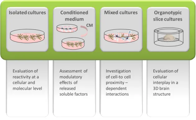
4.1. Models for the evaluation of microglial reactivity ... 19
4.2. Reciprocal reactivity modulation by interplay of microglia with neighboring cells 22
5. Involvement of microglia in the progression of neurological diseases ... 24
5.1. Acute and chronic neurological diseases ... 24
5.2. Neonatal hyperbilirubinemia ... 27
5.2.1. Molecular mechanisms of bilirubin-induced CNS injury ... 30
6. Global aims of the thesis ... 33
7. References ... 34
Chapter II - Features of bilirubin-induced reactive microglia: from phagocytosis to inflammation ... 47
2. Material and Methods ... 52
2.1. Chemicals ... 52
2.2. Primary culture of microglia ... 53
2.3. Cell treatment ... 54
2.4. Measurement of cytokine release ... 54
2.5. Western blot assay ... 54
2.6. Detection of NF-κB activation ... 55
2.7. Morphological Analysis ... 55
2.8. Assessment of microglial phagocytic properties ... 56
2.9. Gelatin zymography ... 56
2.10. Evaluation of microglial cell death ... 56
2.11. Statistical analysis ... 57
3. Results ... 57
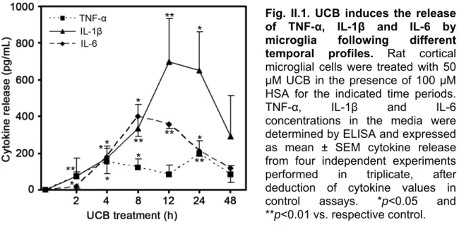
3.1. UCB triggers IL-1β, TNF-α and IL-6 secretion following different temporal profiles ... 57
3.2. p38 and ERK1/2 phosphorylation is elicited by UCB in microglia at an early time point 58 3.3. NF-κB signalling pathway is triggered in UCB-activated microglia ... 60
3.4. UCB-induced NF-κB translocation depends on both ERK1/2 and p38 ... 61
3.5. Microglia depict morphological changes upon UCB stimulation ... 63
3.6. UCB differently modulates microglial phagocytosis depending on exposure time 64 3.7. Release of active MMPs is enhanced upon UCB stimulation of microglia ... 65
3.8. UCB-stimulated microglia evidence enhanced COX-2 expression... 66
3.9. UCB reduces microglial viability leading to loss of membrane integrity and increased caspase activity ... 67
4. Discussion ... 69
5. References ... 74
Chapter III - Dynamics of neuron-glia interplay upon exposure to unconjugated bilirubin ... 79
1. Introduction ... 82
2. Material and Methods ... 83
2.1. Chemicals ... 83
2.2. Primary culture of microglia ... 84
2.4. Primary neuronal cell cultures ... 85
2.5. Neuron-microglia mixed cultures ... 85
2.6. Cell treatment ... 85
2.7. Measurement of cytokine release ... 86
2.8. Gelatin zymography ... 86
2.9. Assessment of microglial phagocytic properties ... 86
2.10. Quantification of nitrite levels ... 86
2.11. Evaluation of cell death ... 87
2.12. Neurite Extension and Ramification ... 87
2.13. Statistical Analysis ... 88
3. Results ... 88
3.1. Conditioned media from UCB-treated astrocytes and neurons modulate microglial secretion of cytokines, as compared to UCB-activated microglia ... 88
3.2. Conditioned media from UCB-treated astrocytes and neurons have opposing effects on microglial MMP-9 activity, as compared to UCB-activated microglia ... 90
3.3. Nitric oxide (NO) release by microglia is elicited by UCB and is higher by naïve microglia exposed to conditioned medium from UCB-treated neurons ... 90
3.4. Loss of viability in UCB-stimulated microglia is less in naïve microglia exposed to conditioned medium from UCB-treated astrocytes (ACM), but higher if treated with medium from neurons incubated with UCB (NCM) ... 92
3.5. Microglial phagocytic properties enhanced by UCB further increase by medium collected from neurons exposed to UCB ... 93
3.6. UCB-induced neuronal network impairment is prevented by microglia environment ... 95
3.7. Neuronal cell death triggered by UCB is diminished in the presence of microglia 97 4. Discussion ... 98
5. References ... 101
Chapter IV - Unconjugated bilirubin neurotoxicity is modulated by microglia and prevented by glycoursodeoxycholic acid and interleukin-10 ... 105
1. Introduction ... 108
2. Material and Methods ... 110
2.1. Chemicals ... 110
2.2. Organotypic-cultured hippocampal slices ... 110
2.3. Preparation of microglia-depleted organotypic-cultured hippocampal slices 111 2.4. Organotypic-cultured hippocampal slices treatment ... 111
2.5. Assessment of cell death in organotypic-cultured hippocampal slices ... 112
2.6. Quantification of nitrite levels ... 112
2.7. Primary neuronal cell cultures ... 112
2.8. Cell treatment ... 113
2.9. Neurite extension and ramification ... 113
2.10. Measurement of glutamate ... 113
2.11. Evaluation of cell death ... 114
2.12. Western blot assay ... 114
2.13. Statistical Analysis ... 114
3. Results ... 115
3.1. Microglia modulate UCB-induced glutamate release and NO production in organotypic-cultured hippocampal slices ... 115
3.2. NMDA receptors and NO are implicated in UCB-induced neurite impairment and in cell death ... 116
3.3. UCB-elicited accumulation of extracellular glutamate is reduced by both GUDCA and IL-10, but not abolished ... 119
3.4. GUDCA or IL-10 pre-treatment counteracts impairment of neurite outgrowth and ramification, as well as cell death in UCB-treated neurons ... 119
3.5. UCB decreases the expression of pre-synaptic proteins and this event is abrogated by GUDCA, but not by IL-10 ... 121
3.6. Hampering of UCB-induced NO and glutamate production, as well as cell death by GUDCA was reproduced in hippocampal slices ... 123
4. Discussion ... 124
5. References ... 129
Chapter V -Final considerations ... 135
1. Concluding remarks and perspectives ... 137
2. References ... 143
Abbreviations
ACM Astrocyte conditioned medium
AD Alzheimer’s disease
AGUDC Ácido glicoursodesoxicólico
ALS Amyotrophic lateral sclerosis
AMPA α-amino-3-hydroxyl-5-methyl-4-isoxazole-propionate
AP-1 Activator protein-1
ATP Adenosine triphosphate
Aβ Amyloid β
BBB Blood-brain barrier
BDNF Brain-derived neurotrophic factor
BIND Bilirubin-induced neurological dysfunction
BNC Bilirrubina não conjugada
CNS Central nervous system
COX-2 Cyclooxigenase-2
CR3 Complement receptor 3
CSF Colony stimulating factor
DIV Days in vitro
ECM Extracellular matrix
ERK1/2 Extracellular signal regulated kinases 1 and 2
GABA γ-aminobutyric acid
GDNF Glial cell-line derived neurotrophic factor
GFAP Glial fibrillary acidic protein
GLT-1 Glutamate transporter-1
GTP Guanosine triphosphate
GUDCA Glycoursodeoxycholic acid
HIV Human immunodeficiency virus
HSA Human serum albumin
Iba1 Ionized calcium-binding adaptor molecule 1
Ig Immunoglobulin
IKK Inhibitor of nuclear factor-κB kinase complex
IL Interleukin
IL-1R Interleukin-1 receptor
IRAK 4 IL-1R-associated kinase 4
IκB Inhibitor of nuclear factor-κB
JNK1/2 c-Jun N-terminal kinases 1 and 2
LDH Lactate dehydrogenase
L-NAME N-ω-nitro-L-arginine methyl ester
LPS Lipopolysaccharide
LTP Long term potentiation
Mac 1 Macrophage receptor 1
MAP-2 Microtubule associated protein-2
MAPK Mitogen-activated protein kinase
MCAO Middle cerebral artery occlusion
MEKK MAPK kinase kinase
mGluR Metabotropic glutamate receptor
MHC Major histocompatibility complex
MK-801
[(+)-5-methyl-10,11-dihydro-5Hdibenzo[a,d]cyclohepten-5,10-imine maleate)]
MMP Matrix metalloproteinase
MRI Magnetic resonance imaging
Mrp1 Multidrug resistance associated protein 1
MS Multiple sclerosis
MyD88 Myeloid differentiation factor 88
NCM Neuron conditioned medium
NF-κB Nuclear factor-κB
NGF Nerve growth factor
NMDA N-methyl-D-aspartate
nNOS Neuronal nitric oxide synthase
NO Nitric oxide
NT Neurotrophin
OPC Oligodendrocyte precursor cell
OSC Organotypic slice culture
OX Orexin
PAMP Pathogen-associated molecular pattern
PARP Poly-ADP ribose polymerase
PD Parkinson’s disease
PGE2 Prostaglandin E2
ROS Reactive oxygen species
SNAP-25 Synaptosomal-associated protein-25
STAT Signal transducer and activator of transcription
TACM Conditioned medium from UCB-treated astrocytes
TF Transcription factors
TGF-β Transforming growth factor-β
TLR Toll-like receptor
TNCM Conditioned medium from UCB-treated neurons
TNFR Tumour necrosis factor receptor
TNF-α Tumour necrosis factor-α
TRAF TNFR-associated factor
TREM-2 Triggering receptor expressed on myeloid cells-2
UCB Unconjugated bilirubin
UDCA Ursodeoxycholic acid
Abstract
Microglia are active sensors in the brain that rapidly engage adequate functional activity states in response to injury to restore homeostasis. During the neonatal period, the brain is more vulnerable to several injury conditions such as the one induced by hyperbilirubinemia, a common situation observed in the newborn, where excessive levels of unconjugated bilirubin (UCB) can reach and damage the brain. Although UCB-induced neuronal and astrocytic toxicity have already been approached, the role of microglia in this condition remains unclear. Thus, this thesis intended to investigate microglial reactivity to UCB and to characterize the intervention of other brain cells in the modulation of their response.
Isolated microglial cells showed to acquire a phagocytic phenotype upon UCB exposure that preceded the release of pro-inflammatory cytokines. This release showed to involve activation of upstream signalling pathways such as mitogen-activated protein kinases (MAPKs) and nuclear factor-κB (NF-κB). We next investigated whether and how the microenvironment influenced microglial response to UCB. Our findings revealed that soluble factors released by UCB-stimulated astrocytes refrained microglial activation while neuron-microglia interaction, evaluated using conditioned media and mixed culture models, signalled microglial clearance functions but also enhanced its inflammatory potential, ultimately leading to microglia demise. Finally, we evaluated microglial neuroprotective or neurotoxic effects in a cell-to-cell concerted action in response to UCB. Microglia revealed to participate in glutamate homeostasis, and to induce the release of this neurotransmitter and of nitric oxide (NO) in UCB-treated organotypic-cultured hippocampal slices, molecules that showed to be key players in UCB-induced neurotoxicity. Moreover, our results point to interleukin (IL)-10 and glycoursodeoxycholic acid (GUDCA) as promising therapies in neonatal hyperbilirubinemia.
In conclusion, microglia displays a dual activation profile in response to UCB stimulation which is tailored by the influence of neighbouring cells. Collectively, these data contribute for the understanding of microglia’s role in hyperbilirubinemia and reinforce their remarkable functional plasticity.
Keywords: Astrocytes; Hyperbilirubinemia; Inflammation; Intracellular signalling; Microglia; Neurons; Neuron-glia dynamics; Phagocytosis.
Resumo
As células da micróglia são sensores activos de dano no cérebro e rapidamente adquirem estados funcionais de activação adequados à restauração da homeostase. Durante o período neonatal o cérebro está mais vulnerável a certas lesões tais como as induzidas pela hiperbilirrubinémia, uma condição comum no recém-nascido, na qual níveis excessivos de bilirrubina não conjugada (BNC) podem atingir e lesar o cérebro. A toxicidade desencadeada pela BNC em neurónios e astrócitos já foi alvo de estudos prévios, no entanto o papel da micróglia nesta condição ainda não está completamente esclarecido. Assim, foi objectivo desta tese investigar a reactividade da micróglia à BNC e caracterizar a intervenção das restantes células do cérebro na modulação da sua resposta.
As células de micróglia isoladas reagem à exposição à BNC pela aquisição de um fenótipo fagocítico que precede a libertação de citocinas pró-inflamatórias. Esta libertação parece envolver a activação a montante de vias de sinalização tais como as cinases proteicas activadas por mitogénios (mitogen-activated protein kinases, MAPKs) e o factor nuclear-κB (nuclear factor-κB, NF-κB). Em seguida investigámos a influência do micro-ambiente celular na resposta da micróglia desencadeada pela BNC. Verificámos então que astrócitos expostos à BNC libertam factores solúveis que contêm a activação da micróglia. Por outro lado, a interacção entre neurónios e micróglia, avaliada através do modelo dos meios condicionados e do das culturas mistas, parece sinalizar a micróglia a empreender funções de eliminação de restos celulares, ao mesmo tempo que aumenta o potencial inflamatório desta célula levando, em última análise, à degeneração celular. Por fim, estudámos os efeitos neuroprotectores ou neurotóxicos da micróglia face à BNC num modelo onde as interacções celulares são preservadas. Os resultados obtidos em culturas organotípicas de fatias de hipocampo revelam a participação das células da micróglia na homeostase do glutamato para além de libertarem este neurotransmissor em conjunto com o óxido nítrico (nitric oxide, NO). Os dados obtidos demonstram ainda que o glutamato e o NO são peças chave na neurotoxicidade provocada pela BNC. Por fim, a interleucina-10 e o ácido glicoursodesoxicólico (AGUDC) salientam-se como promissores agentes terapêuticos na hiperbilirrubinémia neonatal.
Em conclusão, a micróglia apresenta um duplo perfil de activação em resposta à BNC que é modulaado pela influência das células adjacentes. Em conjunto, estes dados contribuem para uma melhor clarificação do papel da micróglia na hiperbilirrubinémia e corroboram a sua notável plasticidade funcional.
Palavras chave: Astrócitos; Fagocitose; Hiperbilirrubinémia; Inflamação; Interacção neurónio-glia; Micróglia; Neurónios.
Chapter I
GENERAL INTRODUCTION
1. The brain – an integrated network of interactive neurons and glia
Neurons are the functioning unit of the central nervous system (CNS) and are responsible for information processing. Glia make up 90% of the cells in the human brain, and although their name derives from the greek word “glue”, research in the past 20 years has proven that these cells not only provide physical support to neurons but make important contributions in the formation and operation of the neural circuitry and play a key role in the brain’s immune functions (Fig.I.1) (Allen and Barres, 2009; Miller, 2005).
Fig. I.1. The brain as an integrated network. Glial cells (i.e. astrocytes, microglia and
oligodendrocytes) are intimately related to neurons and blood vessels, contributing for their support and participating in various key brain functions. Adapted from Allen and Barres (2009).
The major difference between glia and neurons is that the latter ones fire action potentials that underlie sensation, movement and thought, while glial cells lack this capacity. Nevertheless, emerging research suggests that glial cells participate in information processing and interact with synapses (Wake et al., 2009). In fact, the
Microglia
Astrocyte Neuron
Oligodendrocyte
acknowledgement of neuron-glia crosstalk during synaptic transmission has progressively challenged the classical view of the brain as a network of neuronal contacts but rather as an integrated circuit of interactive neurons and glia (Bezzi and Volterra, 2001). Hence glia is emerging as critical participants in every aspect of brain development, function, and disease (Barres, 2008).
1.1. Neurons
Neurons are highly specialized nerve cells responsible for communicating information in both chemical and electrical forms throughout the body (Bloom, 2003). Their structure comprises a cell body, or soma, from which an elaborate arborisation tree emerges. Neuronal processes give neurons the regionalization of their functions, their polarity and capacity to connect to other neurons, to sensory cells and to effector cells (Hammond, 2001). Information is received and processed through the dendritic branches, whose major structural components are the actin and microtubule cytoskeleton together with microtubule-associated proteins (MAPs), responsible for stabilization (Chen and Ghosh, 2005). The neuronal axon is a long cytoplasmic process that culminates in a highly specialized structure, the pre-synaptic terminal, which, together with the post-synaptic terminal of the adjacent neuron forms the synapse (Bloom, 2003). The high molecular weight MAP-2 protein is more common in dendrites than in axons and for this reason, MAP-2A and MAP-2B antibodies coupled to fluorescent molecules are useful tools for labelling dendrites, allowing morphometric analysis in cell cultures (Hammond, 2001).
Electrical signalling in the nervous system involves the movement of ions across the neuronal plasma membrane through specific transmembrane proteins called ion channels. These ionic currents evoke transient changes of membrane potential (action potentials) which fire neuronal communication (Hammond, 2001). In the synapse, neurotransmitters are released and establish communication between adjacent neurons or between neurons and effector or sensory cells (Bloom, 2003). Several molecules serve as neurotransmitters, such as acetylcholine, amino acids like glutamate, γ-aminobutyric acid (GABA) and glycine and neuropeptides and their release at the pre-synaptic terminal is induced by calcium-regulated vesicle exocytosis. Neurotransmitters released into the synaptic cleft bind their specific receptors in the post-synaptic terminal of the adjacent neuron and propagate action potential (Hammond, 2001).
Glutamate is the major excitatory neurotransmitter in the CNS and activates two main types of post-synaptic receptors: ionotropic glutamate receptors that are ligand-gated ion channels and are divided into N-methyl-D-aspartate (NMDA), α-amino-3-hydroxyl-5-methyl-4-isoxazole-propionate (AMPA) and kainate receptors, and
metabotropic glutamate receptors (mGluRs) that are receptors coupled to guanosine triphosphate (GTP)-binding proteins. Glutamate’s concentration in the synaptic cleft must be kept low as high or sustained glutamate receptor activation may induce neuronal death via intracellular calcium and/or sodium deregulation, a mechanism called excitotoxicity (Gras et al., 2006).
1.2. Oligodendrocytes
Oligodendrocytes are the myelinating cells of the CNS and constitute about 5 to 10 % of the total glial population. Their processes enwrap neuron axons and form myelin, an insulating lipid-rich membrane sheath that speeds the conduction of electrical impulses. Therefore, saltatory propagation of action potentials occurs at the nodes of Ranvier between oligodendrocyte myelin sheaths (Bradl and Lassmann, 2010). Oligodendrocytes are vulnerable to cytotoxicity induced by dysfunctional astrocytes (Sharma et al., 2010) or activated microglia as oligodendroglial cell death can be initiated by increased levels of cytokines and reactive oxygen species (ROS) (Sherwin and Fern, 2005). Demyelination due to damage to oligodendrocytes leads to various diseases from neurodevelopment to neurodegeneration such as periventricular leukomalacia (Volpe, 2009) and multiple sclerosis (MS) (Allen and Barres, 2009), respectively. Moreover, the existence of direct chemical synapses between pyramidal neurons and oligodendrocyte precursor cells (OPCs) in the mammalian hippocampus has been demonstrated, underscoring the active role of glial cells in information processing (Bergles et al., 2000).
1.3. Astrocytes
Astrocytes comprise about 85% of the glial population and are star-shaped cells. They exert structural functions, are required for neuronal survival, neurite formation and angiogenesis and maintain CNS homeostasis by regulating pH, ionic concentrations and osmolarity (Montgomery, 1994). Astrocytes also provide metabolic support to neurons by fueling their activity with energy and substrates and remove excess neurotransmitter molecules from the extracellular space (Tsacopoulos and Magistretti, 1996). These cells take part in the neurovascular unit of the blood-brain barrier (BBB) since their end-feet ensheathe endothelial cells in micro-vessels (Cardoso et al., 2010). By signalling blood vessels to expand or narrow, astrocytes regulate local blood flow to provide oxygen and nutrients to neurons in need. Moreover, astrocytes were shown to induce tight junctions in endothelial cells, thus participating actively in the establishment of the CNS boundaries (Yamagata et al., 1997). More recently, bidirectional communication between neurons and astrocytes was demonstrated. This fact altered the long-lasting paradigm of the bipartite synapse, established between a pre- and a post-synaptic neuronal element,
leading to the novel concept of the tripartite synapse, where astrocytic participation in the regulation of synaptic activity (Araque et al., 1999; Perea et al., 2009) and plasticity (Barker and Ullian, 2010) is acknowledged. Moreover, astrocytes can communicate to each other through the generation of calcium waves (Cornell-Bell et al., 1990) and to neurons by the activation of calcium-based signalling cascades triggered by neurotransmitters released by neurons which feedback the production of neuroactive substances (Araque et al., 1999). Furthermore, Alvarez-Maubecin et al. (2000) demonstrated gap-junctional electrical coupling between neurons and astrocytes and also that selective changes in the membrane potential of glia modulate neuronal excitability.
Considering these interesting concepts, a new role has been established for glial cells, particularly astrocytes, in the so-called neuron-exclusive functions such as information processing.
1.4. Microglia
Microglia are small glial cells that reside within the CNS parenchyma and constitute about 10 to 20 % of the total glial cell population (Chew et al., 2006; Vilhardt, 2005). They are ubiquitously distributed in the brain, being present in both grey and white matter and exhibiting a higher density within the hippocampus, basal ganglia and substantia nigra (Walter and Neumann, 2009).
Microglial cells share many properties of macrophages as they belong to the mononuclear phagocyte lineage (Vilhardt, 2005). The notion that microglial cells are the resident immune cells of the CNS has altered the concept of the brain as an immunologically privileged organ supported by the existence of the BBB. Although microglial immunocompetent functions are not on the same scale as that of peripheral leucocytes, it is well established that they act as the brain’s innate immune system (Aloisi, 2001; Streit, 2002).
1.4.1. Heterogeneity of microglial phenotypes
The traditional classification of microglial subtypes described a resting and an activated state. In the resting state microglia would display a ramified morphology, with small bipolar or rod-shaped cell bodies bearing multiple branching processes (Chew et al., 2006; Kim and de Vellis, 2005), while in the activated state, microglia would undergo morphological changes acquiring an amoeboid appearance, up-regulate several cell surface markers and produce a plethora of bioactive substances (Ladeby et al., 2005).
However, emerging research has updated the concept of microglial activation redefining it as a shift in activity states rather than an activation process in itself (Fig. I.2).
Fig. I.2. Microglial activation is a shift between activity states. Microglia in its
“resting” state plays a very important role in surveying the brain parenchyma (1) and can be prompted into an activated state (2) due to the presence of activating signals (such as inflammatory mediators and neurotransmitters) or the absence of constitutive calming signals, such as chemokines or neurotrophins that trigger a transition to an alerted state. During the activation process, microglia commit to distinct response phenotypes which can be altered throughout the pathological process (3, 4 and 4.1) by the influence of other nerve and immune cells (illustrated as feedback signals). At the end of this process, microglia can either return to a resting state (1), be eliminated (5) or step into a post-activated state (6), in which the cells acquire a kind of “memory” (indicated in the figure by a floppy disk icon). Adapted from Hanisch (2008).
Therefore, microglial activation should no longer be considered an all-or-none event but an adaptive response to microenvironmental changes. (Hanisch and Kettenmann, 2007). Indeed, Kreutzberg (1996) had already dubbed microglia as a sensor of pathology.
The discoveries made by Nimmerjahn et al. (2005) and Davalos et al. (2005) demonstrated that resting microglia showed highly motile extension and retraction of processes which enabled them to constantly scan the neural parenchyma. These novel
1. Surveying microglia 2. Alerted microglia Activating signals Calming signals 3. Reactive phenotype 4.1. Reactive phenotype 4. Reactive phenotype Feedback signals 5. Elimination 6. Post‐activated microglia
findings, that we will further explore below, produced a dramatic change in the concept of resting microglia since this phenotype could no longer be considered dormant or inactive but rather a surveying state with an important role in the normal and healthy brain (Hanisch and Kettenmann, 2007; Raivich, 2005). Hence, microglia function as local sentinels that can detect microdamages in the brain and rapidly repair such minute insults without even being noticed. However, stronger or more prolonged insults may trigger more drastic changes in microglial functional phenotype (Hanisch and Kettenmann, 2007). Some authors now define activated microglia as a cell working to restore CNS parenchymal homeostasis, in accordance with the new concept of microglial activation phenotypes (Streit and Xue, 2009). The traditional classification of microglial subtypes based on morphological criteria is no longer suitable, being replaced by the definition of functional activation states (Schwartz et al., 2006). Furthermore, microglia do not constitute a single and uniform cell population but comprise a family of different cell phenotypes with ultimate beneficial or destructive outcomes, being microglial responses tailored in regional and insult-specific manners (Carson et al., 2007).
1.5. Extracellular matrix
The extracellular matrix (ECM) accounts for about 20% of the adult brain volume and is composed of collagen, proteoglycans, hyaluronan, fibronectin, elastins, laminins, vitronectin, thrombospondins, tenascins, among other proteins and carbohydrates (Zimmermann and Dours-Zimmermann, 2008). Mounting evidence demonstrates that ECM is not merely mechanical scaffolding for nervous cells. ECM components are involved in neural migration, proliferation, axon growth and guidance, or synapse formation, processes that are essential during brain development. Furthermore, several cell matrix receptors, like integrins, and cadherins have been shown to affect long-term potentiation (LTP), revealing the implication of ECM in synaptic plasticity, as reviewed in Pavlov et al. (2004). Interactions with ECM can also regulate immune cell action in both a positive and negative fashion. For instance, ECM breakdown, from either microbe colonization or resulting from the action of matrix metalloproteinases (MMPs) released during inflammation, can produce fragments of specific ECM proteins that may act as pro-inflammatory agents. In contrast, ECM proteins can also contribute to the down-regulation of inflammation since decorin and biglycan were reported to inhibit the classical pathway of complement activation (Morwood and Nicholson, 2006). Moreover, the interaction of microglial receptors with ECM components such as cell adhesion molecules contributes to microglial chemotaxis (Hristova et al., 2010; Raivich, 2005).
2. Role of microglia in different brain development stages
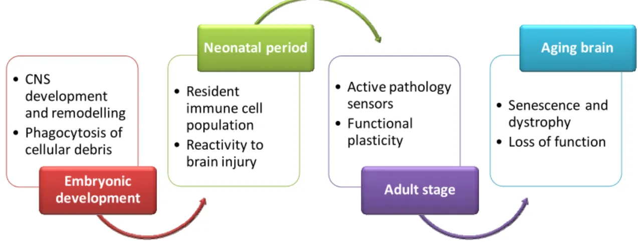
The function of microglia depends on the age of the cell or organism (Walter and Neumann, 2009). Therefore, we will next address the involvement of microglia in different development stages of the brain (Fig. I.3).
Fig. I.3. Microglia along brain development. Summary of the role of microglia in
different brain development stages, from embryonic period to the aging brain.
2.1. Embryonic period
The origin of microglia has been a very contentious issue over the years. However current view concerning microglial ontogenesis comprises a mesodermal origin for microglia corroborating the original observations made by Pio del Rio-Hortega that “microglia arise from polyblasts or embryonic cells of the meninges” (Cuadros and Navascues, 1998). In fact, microglia are descendants of mesodermal fetal macrophages which appear early in development and derive from primitive macrophages of the yolk sac. The entry of fetal macrophages into the neuroepithelium and invasion through the CNS occurs later in embryonic development, at about mid-gestation or earlier in rodents, and late during the first trimester in humans (Alliot et al., 1999; Chan et al., 2007; Streit, 2001). Additionally, it is now accepted that microglial cells can be acutely derived from blood-borne monocytes in the adult upon certain pathologic conditions (Graeber and Streit, 2009; Kaur et al., 2001).
After their entry in the CNS, microglial precursors proliferate and spread through the CNS by tangential and radial migration. Finally, mesodermal precursors originate resident microglia that display an amoeboid-globoid appearance, with an enlarged cell body and retracted processes (Chew et al., 2006; Cuadros and Navascues, 1998). Microglia play an active role in CNS development and remodelling since they phagocyte dead cells, cellular debris and aberrant axons (Kim and de Vellis, 2005; Streit, 2001). Later in development microglia also participate in neuritogenesis, axonal growth and
• CNS development and remodelling • Phagocytosis of cellular debris Embryonic development • Resident immune cell population • Reactivity to brain injury Neonatal period • Active pathology sensors • Functional plasticity Adult stage • Senescence and dystrophy • Loss of function Aging brain
guidance, as well as on the subsequent synaptogenesis and even in vasculogenesis. The production of trophic factors by microglia also contributes for the development, proliferation and growth of neurons and glia (Cuadros and Navascues, 1998; Nakajima and Kohsaka, 2004).
2.2. Neonatal period
In the neonatal period microglial cells transform into a more ramified morphology constituting the resident immune cell population and presenting a slow turnover rate (Cuadros and Navascues, 1998). An interesting study suggests that resident microglia, in contrast to immune cells in other tissues, are not terminally differentiated along the myeloid lineage. This immature state of microglial cells could explain their graded response to activation as well as their enormous plasticity and capacity to assume different phenotypes upon brain injury (Santambrogio et al., 2001).
Birth is not a deadline for the end of brain maturity since several key processes such as synaptogenesis and gliogenesis take place postnatally rendering the brain highly vulnerable to injury during the neonatal period. (Rice and Barone, 2000).
Interestingly, microglia has been implicated in neonatal pathologic conditions such as hypoxic-ischemic (Vexler and Yenari, 2009) or excitotoxic brain injury (Dommergues et al., 2003). In these cases, activated microglial cells rapidly accumulate in the injured neonatal brain displaying up-regulation of cell surface markers such as major histocompatibility complex I and II (MHC I and II) or complement receptors and producing inflammatory mediators (Doverhag et al., 2010) and ROS. The inflammatory response, in conjunction with excitotoxic and oxidative responses, is the major contributor to ischemic injury in the immature brain (Vexler and Yenari, 2009).
2.3. Microglia in the aging brain
An additional phenotype has recently been added to the concept of microglial functional plasticity: a dystrophic or senescent phenotype (Streit et al., 2004). The prevalence of degenerating microglia increases dramatically in Alzheimer’s disease (AD), co-localizing with neurofibrillary degeneration (Graeber and Streit, 2009). Moreover, fragmentation of cytoplasm (cytorrhexis) has been pointed to be indicative of widespread microglial degeneration in amyotrophic lateral sclerosis (ALS) models (Fendrick et al., 2007) and beading and clusters of fragmented twigs have also been demonstrated in the aged brain (Hasegawa-Ishii et al., 2010). Such degenerative changes may underlie a loss of microglial functionality and support, leading to pathological outcomes like neurodegenerative changes and synapse loss (Streit and
essentially beneficial, becoming destructive only when they escape from the strict control imposed on them (Schwartz et al., 2006).
3. Functional roles of microglia – activation and overactivation
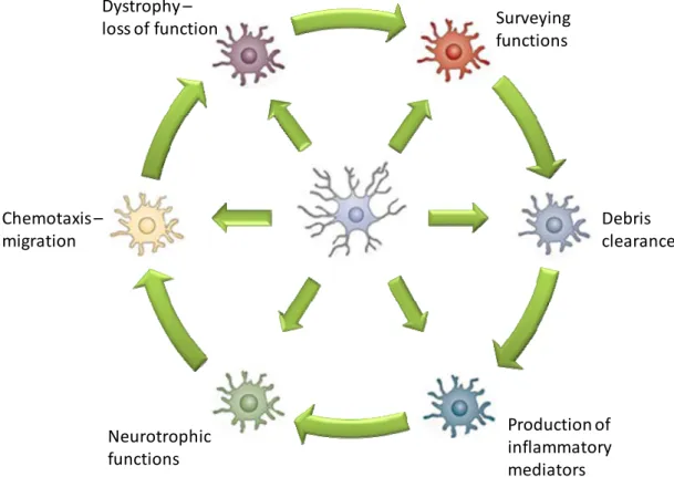
The definition of microglia’s function in the adult brain is not simple since it encompasses the ability to respond to environmental changes and toxic insults (Fig. I.4).
Fig. I.4. Functional activation states of microglia. Resident microglia can become
activated to adopt one of many diverse phenotypes depending on several circumstantial variables and these reactive phenotypes may be alternated along disease progression. Adapted from Perry et al. (2010).
Perhaps the most comprehensive term is “pathology sensor” coined by Kreutzberg (1996). In fact, microglia assume an activated state and exhibit immunological functions in response to danger signals. Nevertheless, several evidences have demonstrated that overactivation might lead to microglial cell death, possibly as a safety mechanism to prevent further deleterious effects (Liu et al., 2001; Polazzi and Contestabile, 2006). In the following sections we will approach the several functions performed by microglia in the healthy and injured brain.
fg
fg
hv
Surveyingfunctions Debris clearance Production of inflammatory mediators Neurotrophic functions Chemotaxis – migration Dystrophy – loss of function3.1. Surveillance functions and role in synaptic plasticity
As stated above, resident and “so-called resting” microglia are not functionally silent cells. In vivo two-photon microscopy demonstrated that microglial processes are remarkably motile presenting high velocities of extension and retraction interspersed with brief static periods. By continuously sampling the environment with their highly motile protrusions, microglia exert an important surveillance function and completely scan the brain parenchyma every few hours (Nimmerjahn et al., 2005). Interestingly, adenosine triphosphate (ATP) released from damaged tissue regulates microglial branch dynamics in the intact brain and mediates a rapid microglial response towards injury (Davalos et al., 2005).
Recently microglia have been shown to directly interact with synapses by establishing intimate but transient connections with pre-synaptic and post-synaptic elements. The frequency of these connections in the healthy brain corresponded with the static periods observed by Nimmerjahn et al. (2005), while this connectivity increased significantly during ischemic lesion (Wake et al., 2009) or after excitotoxic brain injury (Hasegawa et al., 2007). Such observations led to the assumption that microglia monitor the synapse functional status making brief contacts with the healthy ones and prolonged contacts with the injured or pathological synapses. Additionally, pre-synaptic buttons disappear after prolonged contact with microglia in ischemic conditions, suggesting that microglia might produce synaptic stripping in a similar fashion as the one observed after facial nerve axotomy (Wake et al., 2009). In this latter case, microglia become activated and promote the detachment of afferent synaptic terminals from the surface of motoneurons in an attempt to remove the excitatory input and limit further deleterious outcomes (Kreutzberg, 1996; Moran and Graeber, 2004). These findings underscore microglia’s function as sensors of the integrity of the CNS circuitry and may render them a novel epithet as the CNS electrician (Graeber and Streit, 2009).
Moreover, mounting evidence of microglial interaction with synaptic elements contributes for the concept that microglial cells also participate in synaptic plasticity, a process connoted as a hallmark of neurons (Ben Achour and Pascual, 2010). Synaptic plasticity is the basis of learning and memory and consists in a modification of synaptic strength (Ben Achour and Pascual, 2010). Evidence of communication between microglia, astrocytes and neurons relies on the expression of matching sets of transmitters and receptors. Indeed, microglia express a variety of neurotransmitter receptors such as AMPA, mGluRs, GABA receptor, as well as purinergic, cholinergic, adrenergic, cannabinoid, dopaminergic and opioid receptors. Activation of those receptors may trigger the release of several neuroactive substances like glutamate, ATP, nitric oxide (NO), cytokines and chemokines (Pocock and Kettenmann, 2007).
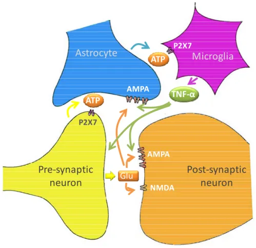
The high density of purinergic P2X7 receptors in microglia mediates microglial participation in the synapse and incited the emergence of quadpartite synapse concept (Fig. I.5) (Bennett, 2007).
Fig. I.5. Microglia are vital members of the “quadpartite synapse”, which also includes synaptic and post-synaptic neurons, and astrocytes. Input to the
pre-synaptic terminal induces glutamate (Glu) release which binds to α-amino-3-hydroxyl-5-methyl-4-isoxazole-propionate (AMPA) and N-methyl-D-aspartate (NMDA) receptors on the post-synaptic neuron and also to AMPA receptors on the astrocytes end-feet, leading to the release of adenosine triphosphate (ATP) from the astrocyte. This acts as a chemoattractant driving microglia into intimate contact with synapses. ATP binds to P2X7 receptors on the pre-synaptic terminal, potentiating glutamate release from the terminal and acts on P2X7 receptors on microglia, leading to the release of tumour necrosis factor (TNF)-α which increases the effectiveness of the AMPA receptors on both the neuron and astrocytes. Adapted from Bennett et al. (2009).
ATP released from astrocyte processes at the synapse acts as a chemoattractant on microglia driving them into intimate contact with synapses. Once microglia processes are interlaced at the synapse they can themselves release ATP due to nerve terminal-derived glutamate. ATP may also act on synaptic microglia P2X7 receptors to evoke the release of cytokines such as tumour necrosis factor-α (TNF-α), which can then increase AMPA receptors in the post-synaptic membrane processes. Thus the P2X7 receptor
ATP
P2X7ATP
P2X7TNF‐α
AMPA AMPA NMDAGlu
Astrocyte
Microglia
Pre‐synaptic
neuron
Post‐synaptic
neuron
mediates mechanisms that give rise to significant changes in the efficacy of synapses (Bennett et al., 2009).
3.2. Role of microglia in innate and adaptive responses
Microglia constitute the first line of defence against invading microorganisms (Town et al., 2005) and are intricately implicated in the initiation and propagation of the inflammatory response (Chew et al., 2006). Activation of microglia can result in different functional phenotypes entailing several characteristic features such as up-regulation of cell surface markers, production of pro-inflammatory mediators and phagocytosis (Hanisch and Kettenmann, 2007). Some authors refer to microglial activation as a continuum where the innate or phagocytic response is at one end and the adaptive response (comprising antigen presentation functions) is at the other (Town et al., 2005). Therefore, the duration and type of stimulation received by microglia will determine the activation features displayed by these cells reinforcing their remarkable plasticity in response to injury (Town et al., 2005).
3.2.1. Phagocytosis
Phagocytosis is defined as the process by which large particles (>0,5 µm) are recognized, internalized and digested by phagocytic cells involving actin-dependent mechanisms. This process is critical for the removal of infectious agents or senescent cells and is performed by immune cells of the body such as macrophages and microglia. Interaction of specific receptors expressed by phagocytic cells with ligands in the surface of the particle is the first step for particle internalization, followed by actin polymerization at the site of ingestion and internalization culminating in the formation of a phagolysosome. Recognition mechanisms can be cell-mediated by cellular receptors like integrins or mannose and scavenger receptors or humoral by the opsonisation of the invading pathogen by antibodies or complement proteins which are then recognized by specific receptors (Aderem and Underhill, 1999).
Activation of microglial phagocytic receptors can be associated with the production of inflammatory products, as occurs in the case of Toll-like receptors (TLRs) or Fc- receptors’ interaction with microbes (Neumann et al., 2009). On the other hand, phagocytosis may occur in the absence of inflammation by the engagement of several receptors such as complement, scavenger, phosphatidylserine (Neumann et al., 2009) or purinergic (Koizumi et al., 2007) receptors as well as triggering receptor expressed on myeloid cells-2 (TREM-2) (Takahashi et al., 2005). In fact, removal of apoptotic cells by phagocytosis is described as a rather silent process which is essential during brain development as well as in some pathologic conditions. Studies in hippocampal slices
show that microglia rapidly respond to neuronal death leading to phagocytic clearance (Petersen and Dailey, 2004). Several putative “eat-me” signals have been established for apoptotic cells such as phosphatidylserine externalization, interaction with vitronectin receptors and changes in the pattern of glycosilation of surface proteins allowing microglial recognition of apoptotic cells by lectin receptors (Ravichandran, 2003; Witting et al., 2000).
As described previously, the phagocytic role of microglia in CNS development and remodelling is widely established as these cells promote a clearance of dead or dying cells and debris. In addition, microglial phagocytic activity is not limited to clearing the corpses but it is also implicated in promoting axon pruning and cell death (Mallat et al., 2005) as demonstrated by the studies of Marin-Teva et al. (2004) demonstrating that microglia provoke the death of developing Purkinje cells by producing high levels of superoxide. A putative hypothesis for the recognition of these Purkinje cells as prey for phagocytic microglia involves the activation of caspase-3 which can lead to the production of chemotactic agents like lysophosphatidylcholine (Lauber et al., 2003). A role for microglia in the removal of unwanted synapses tagged by complement has also been demonstrated (Stevens et al., 2007).
In the context of brain pathology, phagocytosis performed by microglia also plays an essential role in the clearance of tissue debris and, therefore, in the generation of a pro-regenerative environment (Neumann et al., 2009). In fact, microglia is able to phagocyte myelin debris upon a demyelinating insult (Glezer et al., 2007; Takahashi et al., 2007) and amyloid-β (Aβ) from senile plaques (Kakimura et al., 2002), thus triggering repair. Nonetheless, an inadequate regenerative response may prevail if insufficient clearance by microglia occurs (leading to pathology) or when microglial phagocytic properties decline with aging (Zhao et al., 2006).
3.2.2. Production of mediators in neuroprotection and neurodegeneration
Activated microglia release a plethora of soluble factors, which may be pro-inflammatory and cytotoxic or, in contrast, exert neuroprotective or neurotrophic functions (Block and Hong, 2005; Kim and de Vellis, 2005).
In fact, microglia are recognized producers of neurotrophic molecules such as neurotrophins (NT), nerve growth factor (NGF), brain-derived neurotrophic factor (BDNF), basic fibroblast growth factor, hepatocyte growth factor, glial cell-line derived neurotrophic factor (GDNF), plasminogen, platelet-derived growth factor, among several others (Nakajima et al., 2001a; Nakajima and Kohsaka, 2004). This panoply of molecules have been reported to exert important roles in the development of the CNS given their implication in the survival, maturation and differentiation of neurons,
astrocytes and oligodendrocytes as well as their involvement in neurite outgrowth and synapse homeostasis (Kim and de Vellis, 2005; Nakajima and Kohsaka, 2004). Furthermore, upon exposure to toxic insults, microglia also proved to secrete molecules with a neuroprotective potential. For instance, lipopolysaccharide (LPS) stimulation of microglia led to increased production of BDNF, NT-4/5 and NGF suggesting the participation of these cells in neuronal regeneration (Miwa et al., 1997; Nakajima et al., 2001a). After middle cerebral artery occlusion (MCAO), microglia secretes increased amounts of insulin-like growth factor-1 that can promote neural precursors proliferation, survival and differentiation, thus rendering microglia a pro-neurogenic role upon stroke (Thored et al., 2009). In addition, glutamate may signal microglia to release neurotrophic factors like BDNF, GDNF and NGF as an attempt to rescue neurons from excitotoxic damage (Liang et al., 2010).
Microglia can also play additional neuroprotective actions as glutamate scavengers by the expression of glutamate transporter-1 (GLT-1) (Nakajima et al., 2001b). Moreover, GLT-1 expression by microglia is up-regulated upon motoneuron injury in axotomized rat facial nucleus (Lopez-Redondo et al., 2000) or in response to TNF-α production (Persson et al., 2005). Glutamine synthetase, the enzyme responsible for converting glutamate into the less toxic glutamine, is also expressed in activated microglia (Chretien et al., 2002; Rimaniol et al., 2000). Furthermore, the concept of protective autoimmunity, developed by Schwartz et al. (2003) illustrates an interaction between activated microglia and the adaptive immune system (namely T-cells) which ultimately contributes for the orchestration of glutamate homeostasis (Schwartz et al., 2003).
On the other hand, activation of microglia is also associated with the release of a myriad of potentially cytotoxic substances such as ROS, proteases, pro-inflammatory cytokines, chemokines, glutamate, among others (Aloisi, 2001; Kim and de Vellis, 2005). In fact, microglia is one of the principal sources of cytokines in the brain, such as IL-1β, TNF-α and IL-6 (Hanisch, 2002). Cytokines are low molecular weight proteins which participate in a wide range of biological responses such as development and modulation of inflammation (Acarin et al., 2001; Gebicke-Haerter, 2001). Interleukin (IL)-1β and TNF-α are two important cytokines with pleiotropic functions (Fig. I.6). Both cytokines are implicated in CNS inflammation given their ability to induce the expression of adhesion molecules and chemokines leading to BBB breakdown and facilitating leukocyte recruitment into the CNS (Allan and Rothwell, 2003; Hanisch, 2002; Lee and Benveniste, 1999).
Fig. I.6. Overview of the signaling pathways following cytokine receptor activation.
Tumour necrosis factor (TNF)-α interacts with its specific receptor TNFR-1 initiating intracellular cascades through the recruitment of TNFR-associated factor (TRAF) 2. This event may lead to TNF-α-induced phosphorylation of members of the mitogen-activated protein kinases (MAPKs) family such as MAPK kinase kinases (MEKKs) and subsequently of the MAPKs [namely p38, extracellular signal regulated kinases 1 and 2 (ERK1/2) and c-Jun N-terminal kinases 1 and 2 (JNK1/2)]. MAPKs activation may then induce the activation of transcription factors (TF) such as nuclear factor-kB (NF-kB). Stimulation of the NF-kB pathway may be initiated by the phosphorylation of the inhibitor of NF-κB (IκB) kinase complex (IKK) which is crucial for the degradation of IκB allowing NF-κB to translocate into the nucleus and promote gene transcription. Additionally, Interleukin (IL)-1 may also trigger NF-κB and MAPKs activation by the engagement of its membrane receptor IL-1R and consequent recruitment of myeloid differentiation factor 88 (MyD88). This latter adaptor protein associates with IL-1R-associated kinase 4 (IRAK 4) and subsequently activates TRAF6. These pathways may consequently result in modification of the inflammatory response and even cell death.
IL-1β has been broadly implicated in acute neuronal injuries such as the ones resulting from ischemic or excitotoxic challenges (Hagberg et al., 1996). In fact, studies where the action of IL-1β was inhibited culminated in marked reduction of the extent of cell death elicited by ischemic, traumatic or excitotoxic brain injury (Allan and Rothwell,
2003). TNF-α was reported to promote neuronal (Venters et al., 2000) and oligodendroglial cell death and is also involved in demyelination (Akassoglou et al., 1998). Nevertheless, the neuroprotective or neurotoxic outcomes induced by TNF-α seem to depend on the existence of two distinguishable signalling pathways mediated through the TNF receptor (TNFR) 1 and 2 (Arnett et al., 2001). Indeed, a recent report demonstrates that microglia up-regulates TNFR2 expression in response to an inflammatory stimulus and that this event leads to the induction of anti-inflammatory pathways (Veroni et al., 2010). In addition, IL-6 can have both anti- and pro-inflammatory actions since it can significantly reduce ischemic brain damage after MCAO (Loddick et al., 1998) or, in contrast, contribute to the pathophysiology of many diseases (Gadient and Otten, 1997). Microglial cells express surface receptors for this multitude of cytokines, thus propagating the inflammatory response (Aloisi, 2001). Among the upstream signalling pathways involved in the production of cytokines by microglia are mitogen-activated protein kinases (MAPKs), responsible for the phosphorylation of several transcription factors such as nuclear factor-κB (NF-κB) or activator protein-1 (AP-1) (Koj, 1996; Roux and Blenis, 2004). The nuclear translocation of these factors may culminate in the production of the above described cytokines as well as chemokines, complement factors and others (Nakajima et al., 2006).
A correlation between Notch-1 and NF-κB was recently confirmed in LPS-stimulated microglia demonstrating that Notch signalling may amplify the pro-inflammatory response of microglia by enhancing NF-κB signalling (Cao et al., 2010). The signal transducers and activators of transcription (STAT) pathway might also be activated in microglia upon ischemic lesion and lead to enhanced production of inflammatory products (Justicia et al., 2000; Planas et al., 1997).
Cytokine release can also raise the production of other inflammatory mediators such as NO (Chao et al., 1995). Microglial cells are reported to generate a burst of NO in response to injury, which may lead to neuronal dysfunction and cell death (Bal-Price and Brown, 2001; Cassina et al., 2002; Kawase et al., 1996; Sunico et al., 2010). Interestingly, oxidative stress may lead to excessive glutamate production in microglia, culminating in excitotoxic brain damage (Barger et al., 2007; Golde et al., 2002). The release of glutamate in microglia is conveyed by the Xc antiporter, an heterodimeric protein complex which exchanges extracellular cystine for intracellular glutamate (Barger and Basile, 2001; Domercq et al., 2007). Glutamate is an excitatory neurotransmitter but when present in the synaptic space above a certain threshold will lead to excitotoxicity, that is, to the excessive activation of its receptors (namely NMDA, AMPA or kainate
receptors) with subsequent increase in intracellular calcium influx that culminates in neuronal death (Johnston, 2005).
The extensive list of bioactive substances released by microglia in response to brain injury encompasses molecules with chemoattractant properties such as chemokines and MMPs (involved in the degradation of ECM components facilitating leukocyte recruitment); molecules implicated in the initiation and propagation of inflammation like prostanoids produced by cyclooxigenase-2 (COX-2) and complement factors; substances involved in endoplasmic reticulum stress such as cathepsins; anti-inflammatory cytokines like IL-10 and transforming growth factor (TGF)-β; and others (Aloisi, 2001; Kim and de Vellis, 2005; Nakajima and Kohsaka, 2004).
The inflammatory response engaged by microglia has also been implied in the inhibition of neurogenesis (Ekdahl et al., 2003; Monje et al., 2003) adding on the notion that inflammation equals toxicity. Nevertheless, inflammation is a normal response of the organism to infection, injury and trauma and some authors envisage this response as primarily beneficial (Amor et al., 2010; Harry and Kraft, 2008) and as an internal mechanism for repair (Schwartz et al., 2003). Yet, deregulation of the inflammatory response and chronic immune activation can lead to deleterious effects and ultimately cell death (Graeber and Streit, 2009; Harry and Kraft, 2008). It is, therefore, important to recognize the beneficial aspects of the immune response and harness them, in order to contribute to damage resolution instead of aggravating injury.
4. Microglial reactivity and modulation by cell interplay
4.1. Models for the evaluation of microglial reactivity
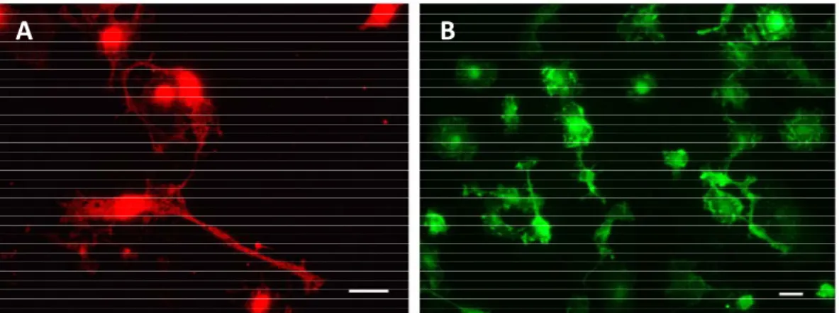
Activation of microglia comprises the up-regulation of several surface markers, which in the “resting” or “surveying” state are expressed at low or moderate levels (Aloisi, 2001; Guillemin and Brew, 2004). Some of these markers have been used to identify microglia in tissue sections and cell cultures (Fig. I.7) but, the existence of a single specific molecular marker for microglia is still unknown (Graeber and Streit, 2009). This is explained by the fact that microglia share several antigens with macrophages, endothelial cells, lymphocytes, among other cells (Guillemin and Brew, 2004). Therefore, the known microglia markers are cell-specific in the sense that they do not label other glial cells or neurons (Graeber and Streit, 2009). Some of the most commonly used surface markers are lectins (carbohydrate binding proteins) such as wheat germ agglutinin, mistletoe lectin, Ricinus communis agglutinin-1, Griffonia simplicifolia B4 isolectin and Lycopersicon esculentum (tomato) lectin, which present an increased
affinity for microglial cell but also bind neurons and myelin nodes and membranes (Acarin et al., 1994). Other very commonly used markers are β2-integrins (CD11a, CD11b and CD11c); the ligand for CD11a is intercellular adhesion molecule-1 whereas the ligands for CD11b and CD11c are complement proteins (Kim and de Vellis, 2005). CD11b is also commonly known as complement rerceptor-3 (CR3), and the antibodies aimed at its recognition are orexin (OX) 42 or macrophage receptor 1 (Mac 1) depending on the rat or mouse origin of the protein, respectively. Additional antibodies have been proposed for the detection of activated microglia such as ED-1, also called CD-68, which is expressed on the membranes of cytoplasmic granules such as phagolysosomes and has been shown to correlate with phagocytic activity (Damoiseaux et al., 1994); or OX-6 that recognizes MHC class II antigens (Guillemin and Brew, 2004). One of the most versatile immunocytochemical marker for microglia is ionized calcium-binding adaptor molecule 1 (Iba1), an EF-hand protein which is up-regulated in activated microglia (Imai et al., 1996).
Fig. I.7. Surface markers used for microglial labelling. Rat primary cortical microglia
are labelled with ionized calcium-binding adaptor molecule 1 (Iba1) (A) or with orexin (OX) 42, an antibody raised against rat complement receptor 3 (B). Scale bar represents 20 µm.
However, the expression of these surface markers depends on the activation state of microglial cells, as demonstrated by Cristovão and colleagues (2010), and its presence is not always constant in all activation processes. For instance, activated microglia accumulated in the facial nucleus after axotomy do not express ED-1 (Graeber et al., 1998), and MHC class II can be also found in ramified microglia besides being present in the activated state (Streit et al., 2004). These observations further corroborate the functional plasticity exhibited by microglia. Additionally, different behaviors have been