FACULDADE DE MEDICINA
Molecular mechanisms controlling the survival
and differentiation of human γδ thymocytes
SÉRGIO TIAGO DE FREITAS RIBEIRO
Tese especialmente elaborada para obtenção do grau de Doutor em Ciências Biomédicas na especialidade de Imunologia Orientador: Professor Doutor Bruno Miguel de Carvalho e Silva Santos
2017
2017
UNIVERSIDADE DE LISBOA FACULDADE DE MEDICINA
Molecular mechanisms controlling the survival and differentiation of human γδ thymocytes
SÉRGIO TIAGO DE FREITAS RIBEIRO
Tese especialmente elaborada para obtenção do grau de Doutor em Ciências Biomédicas na especialidade de Imunologia
Orientador: Professor Doutor Bruno Miguel de Carvalho e Silva Santos Juri:
Presidente:
• Doutor José Luís Bliebernicht Ducla Soares Vogais:
• Professor Paul Coffer, Full Professor do University Medical Center, Utrecht, Holanda;
• Doutor Peter Jordan, Investigador Principal, Instituto Nacional de Saúde Doutor Ricardo Jorge; • Doutor Luís Ricardo Simões da Silva Graça, Professor Associado com Agregação da Faculdade
de Medicina da Universidade de Lisboa;
• Doutora Ana Cristina Gomas Espada de Sousa, Investigadora Principal, Professora Associada Convidada com Agregação da Faculdade de Medicina da Universidade de Lisboa;
• Doutor João Pedro Monteiro e Louro Machado de Simas, Professor Associado da Faculdade de Medicina da Universidade de Lisboa;
• Doutor João Pedro Taborda Barata, Professor Associado Convidado da Faculdade de Medicina da Universidade de Lisboa
• Doutor Bruno Miguel de Carvalho e Silva Santos, Professor Associado com Agregação da Faculdade de Medicina da Universidade de Lisboa; (orientador)
Instituição Financiadora: Fundação para a Ciência e Tecnologia, IP (SFRH/BD/84123/2012)
2017
PREFACE
This thesis describes the research work under the scope of my PhD project developed between 2013 and 2016 at the Instituto de Medicina Molecular (Lisbon, Portugal) at Silva-Santos lab (T cell Differentiation & Tumor Targeting unit) under the supervision of Professor Bruno Silva Santos, PhD.
Sérgio T Ribeiro was financially supported by a scholarship from “Programa SFRH” (SFRH/BD/84123/2012), Fundação para a Ciência e Tecnologia, Portugal, and is a PhD candidate from Programa Doutoral do Centro Académico de Medicina de Lisboa from Universidade de Lisboa.
This thesis is organized in five chapters, which are preceded of an abstract briefly describing the work developed and a summary written in Portuguese. In chapter I an introductory review, the state of the art and the aims of the work are provided. The chapter II comprises material and methods used to obtain the original results presented in chapter III. The chapter IV comprises the conclusions and the biological implications of the results. The future perspectives are highlighted in chapter V.
JC Ribot and DV Correia developed part of the results presented in section 1 of chapter III as disclosed in Ribot et al JI 2014. M Tesio and JC Ribot helped the development of the results presented in section 2 of chapter III as disclosed in Ribeiro et al Leukemia 2016
The statements expressed in this thesis are from the exclusive
responsibility of the author.
The impression of this thesis was approved by Conselho Científico
from Faculdade de Medicina da Universidade de Lisboa at 22 of
November 2016.
The following agencies financially support the development of the work presented in this thesis:
TABLE OF CONTENTS Figures index ... 9 Table index ... 11 Acknowledgments ... 12 List of abbreviations ... 13 Abstract ... 17 Resumo (Portuguese) ... 19
I. GENERAL INTRODUCTION ... 24
1. The immune system ... 24
1.1. Thymus and T cell development ... 25
1.1.1. γδ T cell development ... 28
1.1.2. Mouse γδ T cell subsets ... 30
2. Human γδ T cells ... 31
2.1. Human γδ T cell subsets ... 32
2.1.2. Vδ1 T cells ... 33
2.1.3. Vδ2 T cells ... 35
2.2. Five layers of receptor signaling in γδ T-cell differentiation and activation ... 39
2.2.1. Signal 1: T cell receptor ... 39
2.2.2. Signal 2: costimulatory receptors ... 44
2.2.3. Signal 3: cytokine receptors ... 46
2.2.4. Signal 4: natural killer receptors ... 48
2.2.5. Signal 5: inhibitory receptors ... 50
3. T cell malignancy ... 53
3.2. Aberrant signaling in T-ALL ... 55
4. Protein kinase CK2 ... 58
4.1. Molecular features of CK2 ... 58
4.2. Biological functions of CK2 ... 59
4.3. CK2 inhibitors ... 61
5. Main objectives of the study ... 63
II. MATERIAL AND METHODS ... 66
Statement of Ethics ... 66
Isolation and in vitro cell culture ... 67
Viral transduction of PEER cell line ... 67
Chemicals and Antibodies ... 68
Cell surface phenotype analysis, cell viability, cell cycle and proliferation analysis by flow cytometry ... 68
In vitro tumor-killing assays ... 69
CK2 kinase activity assay ... 69
Immunobloting ... 70
RNA isolation, cDNA synthesis and quantative real time-PCR ... 70
In vivo mouse experiments ... 71
Statistical analysis ... 72
III. RESULTS ... 74
1. Human γδ thymocytes are functionally immature and differentiate into cytotoxic type 1 effector T cells upon IL-2/IL-15 signaling ... 74
Introductory background: ... 74
1.1 Human γδ thymocytes are devoid of cytotoxicity and IFN-γ production 75 1.2 IL-2 and IL-15 signals drive human γδ cytotoxic type 1 cell differentiation ... 77
1.3 Vδ1 and Vδ2 T cell subsets follow similar rules of functional differentiation ... 79
MAPK/ERK pathway ... 81
2. Casein Kinase 2 controls the survival of normal thymic and leukemic γδ T cells via promotion of AKT signaling ... 83
Introductory background: ... 83
2.1 Human γδ thymocytes have enhanced CK2 activity and are highly sensitive to its inhibition ... 84
2.2 CK2 activity in γδ thymocytes is modulated by TCR stimulation and promotes AKT signaling ... 86
2.3 CD27-dependent upregulation of CK2 activity and downstream AKT signaling in γδ T-ALL ... 88
2.4 γδ T-ALL cells are highly sensitive to CK2 inhibition in vitro and in vivo 91 IV. DISCUSSION and CONCLUSIONS ... 96
Human γδ thymocytes are functionally immature and differentiate into cytotoxic type 1 effector T cells upon IL-2/IL-15 signaling ... 96
Protein Kinase CK2 controls the survival of normal thymic and leukemic γδ T cells via promotion of AKT signaling ... 100
V. FUTURE PERSPECTIVES ... 110
VI. REFERENCES ... 116
VII. ADDITIONAL FILES ... 148
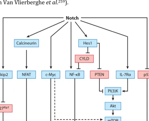
Figures index Figure 1: Stages in T cell development. ... 26 Figure 2: Five layers of cell membrane receptors involved in differentiation, activation and functions of γδ T cells ... 39 Figure 3: Downstream signaling pathways and molecules initiated in Notch-mediated acute T cell lymphoblastic leukemia (T-ALL). ... 56 Figure 4: Schematic representation of CK2α, α’ and β subunit tetrameric complexes. ... 59 Figure 5: Human γδ thymocytes are devoid of IFN-γ production and cytotoxic functions. ... 76 Figure 6: IL-2 and IL-15 signals differentiate γδ thymocytes into cytotoxic type 1 effector T cells. ... 78 Figure 7: Vδ1 and Vδ2 subsets of γδ T cells follow similar rules of functional differentiation. ... 80 Figure 8: The MAPK/ERK signaling pathway is required for IL-2–mediated type 1 differentiation of human γδ T cells. ... 82 Figure 9: Human γδ thymocytes have enhanced CK2 activity and are highly sensitive to CX-4945 ... 85 Figure 10: CK2 activity in γδ thymocytes is modulated by TCR stimulation and activates AKT signaling ... 87 Figure 11: γδ T-ALL cells display higher CK2 activity than αβ counterparts ... 88 Figure 12: CK2 activity in γδ T-ALL cells is potentiated by CD27 costimulation and promotes AKT signaling ... 89
Figure 13: γδ T-ALL cells are more susceptible than αβ T-ALL to apoptosis induced by CX-4945 ... 91 Figure 14: CX-4945 treatment inhibits γδ T-ALL growth in vivo ... 93 Figure 15: TCR and NKR receptor-ligand interactions mediating tumor cell recognition by human γδ T cells. ... 111
Table index Table 1: Distribution and repertoire of human γδ T cells. ... 33 Table 2: Co-receptors of γδ T cells – extracellular ligands and intracellular signaling pathways. ... 43 Table 3: Cancer cell types whose survival has been demonstrated to rely on CK2. 60 Table 4: List of primers used in this study for RT-qPCR analysis. ... 71
Acknowledgments
Quero expressar um especial agradecimento conjunto às pessoas que de variadas formas ajudaram não só no trabalho desenvolvido nestes últimos 4 anos, mas sobretudo pela transmissão de conhecimentos, inspiração e apoio: Bruno Silva-Santos Afonso Almeida Agostinho Freitas Alice Melão Ana Água Doce Ana Espada Sousa Ana Pamplona Ana Sílvia Gonçalves Ana Soares Ana Veríssimo André Simões Andreia Carneiro Anita Gomes Bethania Cassani Biagio Di Lorenzo Bruno F Ribeiro BSS lab members Daniel Correia Daniel Ribeiro David Guéniot Diogo Pereira Elizabeth Macintyre Elvira Leite Eva Rolo Fernando M Ribeiro Francisco Caiado Gisela Gordino Guiomar Rosa Hakan Norell Hélder Ribeiro Helena Brigas Helena Cabaço Helena Soares Henrique Girão Herwig Turk Hiroshi Kubo Hugo Gonçalves Joana Lapa Joana Martins Joana Palha João Ferreira João T Barata Joaquim Freitas Jorge Freitas José Ramalho Julie C Ribot Karine Serre Luís Graça Margarida Rei Maria M Freitas Melania Tesio Miguel A Ribeiro Miguel Muñoz Ruiz Miguel Ribeiro Natacha Sousa Nina Schmolka Noel de Keyzer Patrícia Almeida Paula Romero Paulo Pereira Pedro Oliveira Pedro Papotto Pedro Providência Rita Domingues Saulė Kiaunytė Sílvia Madeira Sofia Mensurado Telma Lança Tiago Amado Vanda Póvoa muito obrigado, Sérgio
List of abbreviations 7-AAD 7-aminoactinomycin D ADCC antibody-dependent cellular cytotoxicity Ags antigens AICD activation-induced cell death AKT Protein kinase B ALCL Anaplastic large cell lymphoma ALK Anaplastic lymphoma kinase ALL Acute lymphoblastic leukemia AML Acute myeloid leukemia AP-1 Activator protein 1 APCs Antigen presenting cell(s) ATP Adenosine triphosphate B-CLL B-cell chronic lymphocytic leukemia BAFT basic leucine zipper transcription factor ATF-like BAT3 B-associated transcript 3 Bcl-2 B-Cell Lymphoma 2 BCR B cell receptor BM bone marrow BTLA B and T lymphocyte attenuator BTN3A Butyrophilin-3A (CD277) CARs chimeric antigen receptors CCL3 Chemokine (C-C motif) ligand 3 (or MIP-1α) CCL4 Chemokine (C-C motif) ligand 4 (or MIP-1β) CCL5 Chemokine (C-C motif) ligand 5 (or RANTES) CCT6A Chaperonin Containing T-complex protein 1 subunit zeta CD Cluster of differentiation Cdc25 cell division cycle 25 CDR3δ complementarity-determining regions 3δ CFSE Carboxyfluorescein succinimidyl ester CK2 Casein kinase II CLL Chronic lymphocytic leukemia CML chronic myeloid leukemia CMV Cytomegalovirus CNS Central nervous system Csk C-terminal Src kinase cTEC cortical thymic epithelial cells CTLA-4 Cytotoxic T lymphocyte antigen 4 CXCL13 C-X-C Motif Chemokine Ligand 13 CXCR5 C-X-C chemokine receptor type 5 DAP10 DNAX-activating protein of 10 kDa DAP12 DNAX-activating protein of 12 kDa DC Dendritic cell DETCs Dendritic epidermal T cells
DLBCL Diffuse large B-cell lymphoma DN double-negative DNA Deoxyribonucleic Acid DNAM-1 DNAX accessory molecule-1 DOT Vδ1+ T cell DP double-positive DUSPs dual-specificity phosphatases E# embryonic day # EDTA Ethylenediaminetetraacetic acid eGFP enhanced green fluorescence protein EGIL European group for immunological characterization of leukemias EGR Early Growth Response ERK extracellular signal-regulated kinases ETPs Early T cell precursors FACS Fluorescence-activated cell sorting FAF-1 Fas-associated factor-1 FBS Fetal bovine serum FGF-2 fibroblast growth factor-2 FITC Fluorescein FPPS farnesyl pyrophosphate synthase GM-CSF granulocyte–macrophage colony-stimulating factor Grb2 Growth factor receptor-bound protein 2 GSK3β glycogen synthase kinase-3β HIV human immunodeficiency virus HMBPP (E)-4-hydroxy-3-methyl-but-2-enyl pyrophosphate HRP horseradish peroxidase ICAM-1 Intercellular Adhesion Molecule 1 ICOS Inducible T-cell co-stimulator Id3 Inhibitor of DNA binding 3 IFN-γ Interferon-γ Ig Immunoglobulin IkB Inhibitor of κB IKK IkB kinase IL-2 Interleukin-2 IL-2R Interleukin-2 Receptor IP3 Inositoltriphosphate IPP Isopentenyl pyrophosphate IRES internal ribosomal entry site ITAM Immunoreceptor tyrosine-based activation motif ITIM Immunoreceptor tyrosine-based inhibitory motif ITK interleukin-2-inducible T cell kinase ITSM Immunoreceptor tyrosine-based inhibitory switch motif Jak Janus kinase JNK c-Jun N-terminal kinase LAT Linker of activated T cells LCK Lymphocyte-specific protein tyrosine kinase
Lef1 Lymphoid enhancer binding factor 1 LFA-1 Lymphocyte function-associated antigen 1 LMO1/2 Rhombotin-1/2, LPC lysophosphatidylcholine LTβR lymphotoxin-β receptor mAb Monoclonal antibody MACS Magnetic-activated cell sorting MAPK mitogen-activated protein kinase MCL mantle cell lymphoma MDSCs myeloid-derived suppressor cells MFI Median fluorescence intensity MHC Major Histocompatibility Complex MICA MHC class I polypeptide-related sequence A MICB MHC class I polypeptide-related sequence B MM multiple myeloma MPM mannosyl-β1-phosphomycoketide mRNA messenger RNA MTD maximum tolerated dose mTEC medullary thymic epithelial cells mTOR mammalian target of rapamycin mTORC1 mTOR complex I MULT1 mouse UL16-binding protein-like transcript 1 NCR Natural cytotoxicity receptor NF-κB Nuclear factor kappa B NFAT Nuclear Factor of Activated T Cells NK Natural killer NKR Natural killer cell–associated receptor NKT Natural killer T cell NOD Non-obese diabetic NPM Nucleophosmin NRGS NOD-Rag1null IL-2Rγnull
NSG NOD-SCID IL-2Rγnull PAGE polyacrylamide gel electrophoresis PBL Peripheral blood lymphocyte PBMC Peripheral blood mononuclear cell PBS Phosphate Buffered Saline PCR Polymerase Chain Reaction PD-1 Programmed cell death protein 1 PE Phycoerythrin Pen/Strep Penicillin Streptomycin PHA Phytohemagglutinin PI3K Phosphatidylinositol 3-kinase PKA protein kinase A PKC protein kinase C PLC phospholipase PMA Phorbol 12-myristate 13-acetate
PRR Pattern recognition receptors PTEN Phosphatase and tensin homolog RAG recombination activating gene RNA Ribonucleic Acid ROR retinoic acid-related orphan receptor RPMI Roswell Park Memorial Institute cell culture medium RT-qPCR Real-time–quantitative polymerase chain reaction S6K 40S ribosomal protein S6 kinase SCID Severe combined immunodeficiency SDS sodium dodecyl sulfate SFKs Src family kinase SFKs Src-family kinases SH2 Src homology region 2 SHP-1/2 SH2 domain-containing phosphatase-1/2 SKG Sakaguchi (or Zap70m1Saka) Skip2 S-phase kinase interacting protein 2 SLP-76 Lymphocyte cytosolic protein 2 SOCS3 Suppressor of cytokine signaling 3 SOX13 sex-determining region Y-box 13 SP Single positive STAT Signal transducer and activator of transcription Syk Spleen tyrosine kinase TAL1 T-cell acute lymphocytic leukemia protein 1 TCF1 T cell factor TCM Central memory T cell TCR T cell receptor TEM Effector memory T cell TGF-β Transforming growth factor β Th1 T helper cell type 1 (Interferon γ-producing cell) Th17 T helper cell type 17 (IL-17-producing cell) TIL Tumor infiltrating lymphocyte TLR Toll-like receptor TLX1/3 T-cell leukemia homeobox 1/3 TNF Tumor necrosis factor TNFR Tumor necrosis factor receptor TRAF TNF receptor associated factors TRAV14 TCRα variable region 14 TSC2 tuberous sclerosis complex 2 ULBP UL16 binding protein UV Ultraviolet ZAP70 zeta-chain (TCR) associated protein kinase 70kDa β2m β2-microglobulin γc Common cytokine receptor γ chain
Abstract
Among the various leukocyte populations that build up the immune defense against infections and tumors, γδ T lymphocytes constitute an enigmatic lineage whose molecular mechanisms of differentiation and activation are still poorly understood. The key roles played by γδ T cells in immunity critically depend on their survival, activation and differentiation into effectors capable of secreting cytokines and killing infected or transformed cells. These processes are controlled, at the molecular level, by surface receptors that capture key extracellular cues and convey downstream intracellular signals that regulate both lymphocyte physiology and pathology.
In this PhD thesis we evaluated the contribution of cell receptor signaling pathways to human γδ T cell differentiation and activation. Firstly, we showed that human γδ thymocytes are functionally immature and their differentiation program requires additional IL-2 or IL-15 signals to drive their differentiation into IFN-γ and TNF-α producers endowed with potent cytotoxicity against leukemia targets. We further elucidated the signals involved in normal/ healthy γδ T cell maintenance as well as in γδ T cell tumorigenesis. We identified that the protein kinase CK2 (Casein Kinase 2) is overactivated in the γδ T cell lineage compared to αβ counterparts. We further showed that the clinical grade-inhibitor of CK2, CX-4945, impairs γδ T cell survival by inhibiting the CK2/AKT/mTOR/GSK3β signaling pathway. Moreover, we showed that CK2 is hyperactivated in γδ T acute lymphoblastic leukemia (T-ALL) samples, compared to both normal γδ T cells and αβ T-ALL. Importantly, we demonstrated a high sensitivity of γδ T-ALL cells to CX-4945 treatment in vitro and in vivo, thus supporting the use of CK2 inhibitors as a putative therapy for γδ T-ALL. Overall, the data presented in this thesis provided new evidences indicating that: (i) human γδ thymocytes are functionally immature and require IL-2 or IL-15 to differentiate into type 1 cytotoxic effector lymphocytes; (ii) the protein kinase CK2
is a novel determinant of both healthy and leukemic γδ T cell survival; and (iii) the CX-4945 chemical inhibitor is a promising therapeutic approach for γδ T-ALL.
Keywords: γδ T lymphocytes; T cell differentiation; acute lymphoblastic leukemia;
Resumo (Portuguese)
O sistema imunitário consiste num vasto e complexo conjunto de moléculas, células, tecidos e órgãos responsáveis pela proteção de um organismo contra agentes externos ou células transformadas do próprio organismo. Estes mecanismos de proteção são conceptualizados em dois tipos de resposta imunitária, a imunidade inata e a imunidade adquirida ou adaptativa. Estes mecanismos são, em larga media, baseados no principio básico da distinção entre o “próprio” e o “não próprio”. Os linfócitos T, ou células T, são responsáveis pela imunidade adaptativa celular, desempenhando um papel fundamental no reconhecimento de organismos invasores ou moléculas “estranhas” ou associadas a stress celular. As células T derivam das células estaminais hematopoiéticas da medula óssea e maturam no timo onde ocorre a recombinação somática V(D)J, que contribui para a diversidade dos seus receptores de células T (TCR). As células T do timo (timócitos), através de sucessivos estádios de seleção, dão origem a células T αβ ou T γδ, que se caracterizam respectivamente pela expressão de TCRαβ ou TCRγδ.
As células T γδ são caracterizadas como células T não convencionais, apresentando características quer das células do sistema inato quer do sistema adquirido. Por exemplo, as células T γδ são independentes do complexo principal de histocompatibilidade (MHC) aquando do reconhecimento do antigénio pelo TCR. As células T γδ têm a capacidade de reconhecer e responder a vários estímulos incluindo bactérias, vírus e células tumorais, tendo recentemente sido as células imunitárias associadas a um prognóstico mais favorável em doentes oncológicos. No homem, existem maioritariamente dois subtipos de células T γδ, Vδ1 e Vδ2, que reconhecem ligandos distintos e apresentam diferentes tropismos. As células Vδ1 são o subtipo maioritário no timo e tecidos epiteliais, ao passo que as células Vδ2 se encontram maioritariamente no sangue (e gânglios linfáticos). As funções das células T γδ resultam, predominantemente, da sua ativação e diferenciação em
células capazes de produzirem citocinas e induzirem a morte das células alvo. Estas funções são iniciadas por sinais extracelulares, captados pelas células e que desencadeiam diversas vias de sinalização no interior da célula. Em última instância, estas vias de sinalização determinam a resposta das células T ao estímulo, assegurando uma função imunológica normal e uma resposta adequada em situações de stress. Porém, estes mecanismos são amplamente desconhecidos no caso da linhagem T γδ.
Nesta tese de doutoramento, analisamos a contribuição de diferentes vias de sinalização mediadas por diferentes citocinas na diferenciação de timócitos γδ humanos. São ainda analisados, com especial detalhe, os mecanismos moleculares envolvidos na sobrevivência das células T γδ normais ou tumorais. A compreensão e modulação destes mecanismos moleculares é especialmente importante na medida em que estes podem constituir potenciais alvos terapêuticos em contexto de imunoterapia envolvendo células T γδ. Estas vias podem, por exemplo, ser moduladas com o objectivo de promover as funções citotóxicas das células γδ contra células tumorais; ou constituir alvos terapêuticos em situações de leucemia linfoblástica aguda com origem nas células T γδ.
Os resultados descritos nesta tese mostram que os timócitos γδ, ao contrario do que se observa nos linfócitos T γδ periféricos, são funcionalmente imaturos. Isto é, os timócitos γδ (maioritariamente do subtipo Vδ1) não têm a capacidade de produzir citocinas nem a capacidade de induzir a morte celular de células tumorais, o que mostra que o programa de diferenciação das células T γδ humanas não é finalizado no timo. Tal constitui uma diferença importante em relação aos timócitos γδ de murganho. No entanto, a estimulação dos timócitos γδ humanos com interleucina-2 (IL-2) ou IL-15, mas não com IL-4 ou IL-7, promove a diferenciação celular, através da via de sinalização da proteína cinase activada por mitogénios (MAPK)/ proteína cinase activada por sinais extracelulares (ERK), em células citotóxicas produtoras de interferão-γ (IFN-γ) e factor de necrose tumoral-
α (TNF-α). Esta diferenciação dos timócitos necessita apenas das citocinas (IL-2 ou IL-15), mas não de sinais derivados do TCR. Por outro lado, os nossos resultados mostram que a sobrevivência das células T γδ humanas é dependente da proteína cinase CK2, que tem uma atividade aumentada quando comparada com a das células T αβ. A CK2 é uma enzima pleiotrópica que fosforila resíduos de serina e treonina. Esta cinase apresenta atividade constitutiva que, frequentemente, se encontra hiperativada em células tumorais. Nesta tese, mostramos que a ativação do TCR nas células T γδ leva a um aumento da atividade de CK2 e à ativação das vias de sinalização AKT/ receptor mamífero de rapamicina (mTOR), promovendo a sobrevivência celular. Por outro lado, a inibição especifica de CK2, através do inibidor CX-4945, leva a um bloqueio da via AKT e a um aumenta da morte celular por apoptose das células T γδ, mas não das T αβ saudáveis. Verificamos, ainda, que a hiperativação de CK2 é uma característica adquirida por células malignas de leucemia linfoblástica aguda (LLA) com origem na linhagem T γδ. Os resultados obtidos em amostras provenientes de doentes mostram que as células transformadas são do subtipo Vδ1, e que o tratamento com o inibidor CX-4945 leva à apoptose destas células LLA-T γδ primárias. O conjunto dos resultados obtidos quer in vitro e in vivo (modelo xenotransplante em murganhos imunodeficientes) mostram que as células LLA-T γδ são sensíveis ao inibidor da CK2, CX-4945, abrindo novas oportunidades à sua utilização no tratamento de doentes com este tipo de leucemia.
Globalmente, os resultados apresentados e discutidos nesta tese, demonstram que as células T γδ provenientes do timo humano são funcionalmente imaturas, necessitando de um último estádio de diferenciação dependente de IL-2 ou IL-15, de modo a adquirirem capacidades de produção de IFN-γ e TNF-α e citotoxicidade anti-tumoral. Por outro lado, mostramos que as células T γδ, em comparação com as células T αβ, apresentam uma maior atividade da proteína cinase CK2 e maior suscetibilidade à morte celular após inibição de CK2. As funções de CK2 podem ser exacerbadas por estímulos externos como TCR, quer em células normais quer em
células transformadas. Dada a hiperativação da CK2 em células transformadas LLA-T γδ, e a sua susceptibilidade ao inibidor CX-4945, propomos que a CK2 constitui um importante alvo terapêutico em doentes diagnosticados com este subtipo de leucemia. Palavras-chave: linfócitos T γδ; diferenciação de células T; leucemia linfoblástica aguda; vias de sinalização; proteína cinase CK2.
Chapter I
General Introduction
I. GENERAL INTRODUCTION
1. The immune system
The immune system is responsible for discriminating the cells and tissues that are a legitimate part of the body, from foreign molecules and organisms (“nonself”) that might be present in the organism. By complex cellular and molecular events mediated by innate (antigen-nonspecific) and adaptive (antigen-specific) immune response, the immune system eliminates those nonself invaders, which are often dangerous bacteria or viruses. In addition, the immune system can recognize, and usually eliminate, “altered self”–cells or tissues that otherwise could originate
malignancies1.
Leukocytes of the innate immune system serve as sentinels for detecting general signs of danger. These cells are usually equipped with pattern recognition receptors (PRR) leading to a fast and unspecific activity against groups of pathogens. Thus, these innate myeloid cells (Phagocytes, Mast cells, Basophils, Eosinophils) and innate-like lymphocytes (NK cells, NKT cells and γδ T cells) constitute the first line of defense against pathogens and/or transformed cells. Innate responses can also modulate the adaptive branch of the immune system. In particular, dendritic cells act as critical antigen-presenting cells (APCs) to start αβ cell responses.
Additionally, the adaptive system is composed of B cells and αβ T lymphocytes, which react specifically to diverse pathogenic conditions. Adaptive immunity is triggered later and usually generates memory cells that persist in the organism and can be reactivated upon a second encounter with the same antigen. The cells of the adaptive immune system express a comprehensive repertoire of cell surface antigen-specific immunoglobulin receptors - B cell receptors (BCR) for B cells and T cell receptors (TCR) for T cells - that can collectively recognize millions of distinct antigens. These receptors are generated by somatic recombination
mediated by recombination activating gene (RAG) enzymes, an extraordinary process common to all jawed vertebrates which developed primary (like the
thymus) and secondary (like spleen and lymph nodes) lymphoid organs1.
1.1. Thymus and T cell development
The anatomic sites where the major steps in lymphocyte development occur are called primary or generative lymphoid organs. These include the bone marrow, where the precursors of all lymphocytes arise and B cells mature, and the thymus, where T cells mature. Following maturation, the lymphocytes enter the circulation and peripheral lymphoid organs (e.g. the spleen and lymph nodes) where they
survey for invading pathogens and/or tumor cells2.
The thymus is present in all jawed vertebrates, present similar structure and undergoes the same shrinkage with age and plays the same immunological function as in human beings. The lymphocytes in the thymus, also called thymocytes, are T lymphocytes at various stages of maturation. The most immature cells enter the thymus, and their maturation begins in the cortex where thymic cortical epithelial cells (cTEC) produce interleukin-7 (IL-7), a critical factor for survival and differentiation. During maturation, through different steps of cell selection, thymocytes migrate toward the medulla where medullary thymic epithelial cells (mTEC) play a special role in presenting self-antigens to developing T cells and causing their ablation (negative selection). This is one mechanism of ensuring that the immune system remains tolerant to self-antigens, avoiding autoimmunity. It is only after maturation, that positively-selected naïve T cells exit the thymus and enter the blood and peripheral lymphoid tissues.
Due a large extend to the remarkable technical advances, mainly by the creation of genetically altered animals, T cell development is better characterized in mice than in human. However, human T cell development appears to depend on similar mechanisms (as in mice), and the key factors in T cell development may be broadly conserved among jawed vertebrates. Of note, the congenital absence of the
thymus, as occurs in DiGeorge syndrome in humans or in the nude mouse strain (athymic), is characterized by very low numbers of mature T cells in the circulation and peripheral lymphoid tissues and severe deficiencies in T cell–
mediated immunity1.
It is well established that T-cell precursors that arrives in the thymus contain TCR genes in their germline configuration and do not express TCR, CD3, ζ chains, CD4, or CD8 receptors. These double-negative (DN) thymocytes are highly immature progenitors, comprising about 1–2% of thymocytes, that in both murine and human undergo successive stages of cell selection. T cell commitment corresponds, at the molecular level, to productive TCR rearrangements. These occurs at four gene loci, TCRα, β, γ and δ, present in all jawed vertebrates. The protein products of somatic recombination in T cells can pair in two stable complexes, TCRαβ or TCRγδ, which are mutually exclusive and thus define two T cell lineage, αβ and γδ T cells, which diverge in T cell development3–5. Figure 1: Stages in T cell development. Early T cell precursors (ETPs) differentiate from double negative (DN) to double positive (DP) to single positive (SP) stages. Arrows indicate cell differentiation. Note that ETP and DN2 thymocytes contain non-T-cell options. β- and γδ- selection occurs during the accumulation of the DN3 T cells (adapted from Peng Li et al6).
Immature murine DN thymocytes can be separated into four populations (DN 1-4)
based on the expression of CD44 and CD25 (Ref. 7). However, immature human
thymocytes do not express the same surface markers and are characterized by the
differential expression of CD34, CD38, and CD1a4.
During T cell maturation, there is a precise order in which TCR genes are rearranged and expressed. In the mouse, surface expression of the TCRγδ occurs 3 to 4 days after precursor cells arrived the thymus, and the TCRαβ is expressed 2 or 3 days later. In human fetal thymuses, TCRγδ expression begins at about 9 weeks
of gestation, followed by expression of the TCRαβ at 10 weeks1.
These differentiation steps are controlled by a complex system of several
transcriptional factors3. The transcription factors Notch-1 and GATA-3 are
responsible to commit developing lymphocytes to the T cell lineage. Firstly, the cell surface molecules of Notch family are proteolytically cleaved following interaction with specific ligands on neighboring cells in the thymus. In mammals, two families
of Notch ligands, Delta-like (DL) and Jagged, have been identified8. The activated
intracellular domain of Notch proteins migrate to the nucleus and modulate the
expression of specific target genes1,9. T cell development depend on several
transcription factors, including: GATA-3, c-Myc, members of the Runx family,
members of the E2A/HEB family, and members of the Ikaros family10,11. In addition
the Wnt/TCF signaling cascade has an important role in proliferation coupled with differentiation of T cells11.
Somatic rearrangement of the genes encoding the TCRβ, TCRγ and TCRδ chains, mediated by Rag-1 and Rag-2 proteins, is essential for TCR expression and diversity. The rearrangement of these TCR genes begins in DN2 cells and is mostly completed during the DN3 stage. If a cell succeeds in productively rearranging its TCRγ as well as its TCRδ loci before it makes a productive TCRβ rearrangement, it is selected into the γδ T cell lineage. Otherwise, when a TCRβ chain is expressed from a productively rearranged Tcrβ locus (V-D-J recombination) the cells progress to a αβ T cell lineage. The TCRβ pairing with the invariant pre-TCRα chain
forms a pre-TCR12–15. Pre-TCR signaling, coupled to signals from cytokine and
possibly other receptors, promotes cell survival, proliferation and differentiation.
Following β-selection DN4 cells subsequently become DP CD4+CD8+ population
that comprises 75–88% of thymocytes. DP cells complete Tcrα gene rearrangement (V-J recombination) and present TCRαβ heterodimers at cell surface. The expression of functional TCRαβ provides the substrate for
MHC-mediated positive selection into SP CD4+ or CD8+ populations followed by negative
selection that eliminates autoreactive αβ thymocytes. Finally, these mature thymocytes proliferate shortly immediately before migrating to the periphery to
function as mature αβ T cells16. However this conventional model of T cell
development does not apply to MHC-unrestricted γδ T cells.
1.1.1. γδ T cell development
Early during thymocyte differentiation γδ T cells diverge from αβ T cells and continue along different developmental paths. The bifurcation of αβ and γδ lineage-commitment is thought to be complete at DN3 and these decisions are influenced by different site-specific signals, derived from cytokines, chemokines,
TCRs, Wnt signaling, Notch ligands, and others thymic microenvironment stimuli17.
Successful in-frame rearrangement of the TCRγ and δ genes results in the
expression of a TCRγδ complex and favos differentiation along the γδ lineage17–19.
In both mouse and humans, TCRδ rearranges first, followed closely by TCRγ
rearrangement17.
This recombination joins any one of several variable (V) gene segments with any one of several joining (J) segments and, in TCRδ, also with diversity (D) gene segments. The somatic recombination of multiple gene segments contributes to the diversity of receptor-structure. Following gene recombination, only cells that
make productive TCRs are selected to survive20.
Factors such as IL-7R expression21 or Notch signaling22–25 are described to be
TCRγ locus by regulating Rag accessibility27. Moreover, murine thymocytes with
abundant IL-7R are more likely to give rise to γδ T cell lineage than thymocytes
lacking IL-7R28. In addition, immature thymocytes with high levels of IL-7R express
more of the transcription factor SOX13 (sex-determining region Y-box 13) protein. SOX13 is a putative T cell lineage regulator that promotes the γδ lineage and impairs αβ development. Mice deficient in SOX13 expression can still produce
mature αβ T cells, whereas the development of γδ T cells is impaired17,29.
Notch signaling is also thought to be involved in αβ versus γδ T cell lineage commitment. In mouse, high levels of Notch signaling are required for αβ T cell development compared to γδ T cells. Reduced levels of Notch1 gene in vivo favors
γδ T cell development over αβ T cells30,31. However γδ T cell commitment may
require different ligands of Notch32. In humans, the involvement of Notch signaling
in T cell development is less clear and paradoxical to the mouse data. Toribio and Plum’s groups showed that increased Notch activation results in increased γδ T
cell development at the cost of αβ T cells33–35. Thus, the involvement of Notch in γδ
T cell commitment remains somewhat controversial.
The γδ T cell lineage fate is not simply instructed by the type of rearranged TCR complex that is found on the surface of a given cell. Instead, the “strength” of the
signal that is delivered by the TCR complex seems to be critical36,37. Thus, TCRγδ,
which appears to signal relatively strongly, directs cells towards the γδ lineage, whereas the preTCR, which generates a weaker signal (than TCRγδ), promotes the
development of αβ lineage cells38–41. This model suggests that a strong TCRγδ
signal would result in higher activation of the extracellular signal-regulated kinases/ mitogen-activated protein kinases (ERK-MAPK) pathway, leading to a higher induction of the Early Growth Response (EGR1, EGR3) transcription factors
and their target Inhibitor of DNA binding 3 (Id3) in a mouse model36,42,43. Id
proteins are direct inhibitors of E2A, a helix-loop-helix protein, which activates the
pre-TCRα promoter44. The higher accumulation of Id3 results in stronger
weaker signals through the Notch receptors and pre-TCR would result in more modest accumulation of Id3, and hence weaker inhibition of E2A leading to further
development along the αβ lineage17,41. By analogy, as a consequence it is also likely
that the signals downstream of each TCR results in differential regulation of transcription factors that are essential for the functional maturation of different
effector subsets41. Thus, the TCR signal strength required for the development of
multiple subsets of γδ T cells is likely heterogeneous. Consistent with this hypothesis, in the host lab, Muñoz-Ruiz et al elegantly showed that TCR signal strength controls the differentiation of specific subsets of mouse γδ T cells during
thymic ontogeny45.
1.1.2. Mouse γδ T cell subsets
γδ T cells are conserved throughout evolution and across vertebrate species. However, γδ T cell subsets are highly heterogeneous. The different subsets of γδ T cells are typically grouped by the variable (V) segments encoded by rearranged Vγ and/or Vδ genes, with Vγ being the most significant in the mouse, while Vδ is the
most relevant in humans20,46–48.
Murine γδ T cells are generated in the thymus in “developmental waves” that
sequentially populate different tissues during embryonic development20.
Following Vγ gene nomenclature of Heilig and Tonegawa19 for murine γδ T cells,
thymocytes bearing an invariant canonical Vγ5Vδ1 TCR at embryonic day E15-17 are the first to leave the foetal thymus, giving rise to skin-associated dendritic epidermal T cells (DETCs). Thymocytes bearing a Vγ6Jγ1Cγ1 TCR at E16-18 give rise to the γδ T cells in the tongue and reproductive tract, whereas peri-and postnatal thymocytes bearing Vγ1Cγ1 and Vγ4Cγ1 TCRs give rise to systemic γδ T cells (that predominate in lymph nodes and spleen).
Many of the studies elucidating the physiological roles of γδ T cells have been performed in murine models, where the identification of pro-inflammatory subsets
The segregation of the two functional γδ T cell subsets has been greatly facilitated
by the identification of cell surface markers: CD27, CD122 and NK1.1 mark IFN-γ-producing γδ cells, whereas their IL-17-expressing counterparts display a CD27-
CCR6+ phenotype49–51. Moreover, the two subsets show distinct Vγ chain usage in
their TCR repertoires, with a bias towards Vγ1 among IFN-γ-producing γδ cells,
and an enrichment in Vγ4 and Vγ6 in IL-17-producing γδ cells52. Moreover, the
transcription factors Sox13 and RORγt (Rorc) are essential for IL-17 producing γδ
T-cells, including Vγ6+ and some Vγ4+ T cells, while Eomes and Tbet are hallmarks
of IFN-γ producing subtypes, such as Vγ5+ dendritic epidermal T-cells41. The
diversity of mouse γδ T cell subsets and associated functions is reviewed elsewhere20,39,41,48,52–55.
2. Human γδ T cells
Human γδ T cells, like their murine counterparts, are a minor population (1–10% of nucleated cells) in peripheral blood but are abundant in tissues, especially in
epithelial layers46. For identification purposes, they are usually sub-divided based
on the variable regions of TCRδ. Using Lefranc and Rabbits numenclarure56, the
human γδ T cell subsets comprise the Vδ1, Vδ2 and, the less studied, Vδ3 subset. Vδ1 γδ T cells are the predominant subset found at mucosal surfaces (Table 1). By
contrast, Vδ2 γδ T cells (that are almost exclusively Vγ9+) largely dominate in the
peripheral blood18,57,58. Indeed, Vδ2 T cells can represent more than 50% of blood
leucocytes after certain bacterial or parasitic infections59. However the ligands,
signaling mechanisms of cell activation and functional development as well as the cell receptors involved are poorly described.
γδ T cells may be considered a component of the adaptive immune system as they somatically rearrange their TCR genes to generate great diversity; and can selective expand particular subpopulations upon infection. But, on the other hand, γδ cells endow the T cell compartment with a rapid, innate-like reaction to insults, which places them in the afferent phase of the immune response. γδ T cells are
responsible for “lymphoid stress surveillance”, i.e., sensing and responding immediately to infections or non-microbial stress without the need of clonal expansion or de novo differentiation, in synchrony with prototypic innate immune
responses60. Critically, this implicates γδ T cells in inflammation61, autoimmunity62,
infectious diseases63,64, and tumor surveillance65–67. This section 2 describes the
main characteristic of human γδ T cells including their subsets, functions as well as the mechanisms involved in their differentiation and activation.
2.1. Human γδ T cell subsets
In humans, γδ T cells have a small repertoire of V gene segments to select from when undergoing TCR chain rearrangement in comparison with those available for
Vα (43–45 (Ref.68)) and Vβ (40–48 (Ref.69)). Three main Vδ gene segments, Vδ1,
Vδ2 and Vδ3 (Table 1), are most frequently used in rearrangement of the δ chain; less commonly used are the five V segments that have both Vδ and Vα designation
(Vδ4/TRAV14, Vδ5/TRAV29, Vδ6/TRAV23, Vδ7/TRAV36 and Vδ8/TRAV38)70).
Seven functional Vγ gene segments, Vγ2, Vγ3, Vγ4, Vγ5, Vγ8, Vγ9 and Vγ11, located within the γ locus on chromosome 7 in humans, are used for rearrangement of the
γ chain71. Despite the restricted repertoire of Vδ and Vγ gene segments available
for rearrangement, the complementarity-determining regions 3δ (CDR3δ) allows the incorporation of multiple Dδ segments (in forward and reverse direction) and
theoretically results in the most diverse of all the rearranged receptors71,
representing about 1018 different repertoire possibilities compared to only 1016 of
αβ T cells1. The genetic evolution of TCRγδ associated genes and ligand recognition
from non-human primates to humans is reviewed elsewhere71.
Table 1: Distribution and repertoire of human γδ T cells.
(Adapted from Silva-Santos et al.; Bonneville et al. and Chien et al.47,54,72)
Subset distribution Body Most common VγVδ pairs V(D)J diversity TCR-ligand/ reactivity
Vδ1 Thymus, spleen, liver, gut epithelium, dermis variable High CD1d-sulfatide/ α-GalCer; CD1c-sulfatide/ LPC/ MPM73; MICA; CMV-infected cells; Vδ2 Peripheral blood Vγ9Vδ2 Intermediate Phosphoantigens; ULBP4; F1-ATPase53; CCT6A74
Vδ3 epithelium Liver, gut variable High Not defined
2.1.1. 2.1.2. Vδ1 T cells Vδ1-expressing γδ T cells represent only 10-30% of all γδ T cells in the peripheral blood but is very abundant in healthy epithelial tissues and comprises the major T
cell population in the gut epithelium75. The Vδ1 T-cell population expands upon
infections with bacteria (Mycobacterium tuberculosis, Listeria monocytogenes and
Borrelia burgdorferi) and virus (HIV and cytomegalovirus (CMV))76–78.
Importantly, γδ tumor infiltrating lymphocytes (TILs) were described as being the immune population that gives the most favorable prognostic in several cancer
types79 and the abundance of Vδ1 TILs usually correlate with increased survival80,
with some notable exceptions81.
γδ TILs isolated from various types of cancer, including colorectal, breast, prostate,
ovarian and melanoma82–84, recognize and kill both the autologous tumor and a
broad range of related tumors, presumably via the recognition of shared
observation that Vδ1 T cells infiltrate and respond to solid tumors, supports the
use of Vδ1 T cells as a promising cancer immunotherapeutic approach72.
Several studies attempted to improve Vδ1 T cell efficacy to be used in cancer treatments. In the host lab, Correia and collaborators formulated an in vitro protocol that induces the expression of natural cytotoxicity receptors (NCRs; e.g. NKp30, NKp44 and NKp46), on Vδ1 T cells isolated from peripheral blood. These
de novo NCRs, which had been previously regarded as NK-specific markers,
enhance tumor targeting by Vδ1 T cells86. Although neither Vδ1+ nor Vδ2+ cells
express NCRs constitutively, these can be selectively upregulated in Vδ1+ cells by
AKT-dependent signals provided synergistically by γc cytokines (IL-2 or IL-15)
and TCR stimulation86,87. However, the intracellular signaling mechanisms
involved in γδ T cell activation and expansion are still poorly understood. The insufficient γδ T cell numbers was an additional limitation to the use of Vδ1 T cells in the clinic. This limitation was recently overcome by a robust protocol for the
selective expansion and differentiation of cytotoxic Vδ1 T cells88. These cells
selectively target leukemic cells via the combined action of TCR and NCRs and
production of IFN-γ and TNF-α, in vitro and in vivo, but not IL-1788, which has been
implicated in the promotion of tumor cell growth72,89.
In contrast to Vδ2 T cells, the Vδ1 T cell population is not prone to activation-induced cell death (AICD). As a result, tumor-reactive Vδ1 T cells can persist in the
circulation for many years90–92. The therapeutic potential of Vδ1 T cells is largely
determined by the nature of ligands that bind and activate their receptors. Vδ1 T cells are activated by stress-induced self-antigens that are oſten constitutively
expressed by solid tumors, leukemias and lymphomas92. In particular, Vδ1 T cells
recognize MIC-A/B93,94 induced by oxidative stress95, and oſten upregulated on
malignant cells96. The MIC-A/B recognition by NKG2D receptor on Vδ1 (but also on
Vδ2) T cells is critical for recognition and subsequent killing of several target
cancers via perforin and granzymes92,97. Vδ1 T cells have been reported to
receptors53. Vδ1 T cells are able to sense glycolipids presented by CD1c73,98–100; or
CD1d71,101–103 . Until now, the identified ligands of Vδ1 T cells are cancer and virus-associated molecules, which highlight the tissue-immunosurveillance functions of these cells54. 2.1.3. Vδ2 T cells γδ T cells make up approximately 4% of the peripheral blood lymphocytes (PBL) T cells in healthy human adults but can expand up to 60% of blood leucocytes during a variety of infectious diseases. Most of the expanded γδ T cells express Vγ9 and Vδ2 TCR chains, suggesting that some of the specificities within this population may be important in responding to pathogenic challenges. Vδ2 T cells are found only in humans and higher primates and constitute the best studied γδ T cell subset.
Vδ2 T cells are often sub-divided on the basis of surface expression of two receptors: CD45RA and CD27. However, CD27 does not identify a human γδ T
subset comparable to the CD27+ γδ T subset in mouse (i.e. pre-committed to robust
IFN-γ secretion)50
. CD27 is a member of the TNF receptor family with known co-stimulatory activity that is expressed by a major proportion of human Vδ2 T
cells104. CD27 and CD45RA identify four Vδ2 T subsets: (i) ‘Naive’ (Tnaive) CD45RA+
CD27+ Vγ9Vδ2+ cells are highly proliferative, do not secrete IFN-γ, and generally
comprise 10–20% of those in peripheral blood (but the major population in lymph
nodes)105. (ii) Tnaive cells become largely CD45RA- CD27+ (and CD45RO+) after TCR
stimulation. In healthy individuals, these ‘Central Memory’ (TCM) cells represent
25% and 50% of Vγ9Vδ2 cells in lymph nodes and peripheral blood, respectively.
TCM cells appear to proliferate less than Tnaive but can secrete low levels of IFN-γ106.
(iii) Following additional TCR stimulation, TCM cells generate CD45RA- CD27- (and
CD45RO+) ‘Effector Memory’ (TEM) cells that lose CCR7 and CD62L, but acquired
the tissue-associated chemokine receptors CCR2, CCR5, CCR6 and CXCR3 (Ref.106).
tumor necrosis factor-α (TNF-α) and are present in higher percentages in blood
and inflammatory sites. (iv) TCM cells also appear to generate a CD45RA+ (CD27-,
CCR7-) effector memory (TEMRA) population when activated with IL-15 (Ref.107),
however these cells are virtually absent from blood. TEMRA cells express abundant
perforin, granulysin and display robust cytolytic activity (but little production of
IFN-γ). Additionally, TEMRA cells are unresponsive to further TCR stimulation and
have little proliferative capacity, a phenotype consistent with a terminally differentiated state108.
Vδ2 T cells are unique in their recognition of low-molecular-weight non-peptidic and phospho-containing compounds collectively called phosphoantigens (PAgs). Vδ2 T cells responds robustly to isopentenyl pyrophosphate (IPP), an intermediate in the human mevalonate pathway and to
(E)-4-hydroxy-3-methyl-but-2-enyl-pyrophosphate (HMBPP), a microbial isoprenoid intermediate109. Importantly,
several microbial and plant PAgs produced through the deoxyxylulose pathway, such as HMBPP, show 10,000-fold higher activity than those from mammalian
PAgs54. Nanomolar concentrations of HMB-PP lead to rapid TCR-dependent
activation of Vγ9Vδ2 T cells, enabling them to respond to a diverse range of
pathogens, including Mycobacterium tuberculosis110,111 and Plasmodium
falciparum112. Indeed, in vitro incubation of PBL with mycobacterial lysates
induces an expansion of Vγ9Vδ2-expressing γδ T cells, and the stimulatory
components are protease resistant and phosphatase sensitive54,113. Importantly,
the recognition of PAgs is restricted mainly to higher primates, with mice and
other rodents not possessing any T cell subsets that respond to PAgs114.
The ability to respond to prenyl pyrophosphates has been used to redirect Vδ2 T cells to tumors by manipulating isoprenoid metabolism in the cancer cells. Such manipulation can be achieved using aminobisphosphonates (e.g. zoledronate, pamidronate, risedronate) which inhibit farnesyl pyrophosphate synthase (FPPS) in the mevalonate pathway leading to the accumulation of prenyl pyrophosphate
PAgs, they can respond robustly to naturally occurring primary alkylamines, such as iso-butylamine that are secreted by bacteria and found in certain edible
plants117. Collectively, the reactivity of these Vγ9Vδ2 γδ T cells could allow them to perceive microbial products in diverse infectious contexts. The mode of PAg recognition has been amply studied. Early findings indicated that recognition of PAgs by human Vγ9Vδ2 T cells required cell surface presentation by species-specific molecules118. These have been recently identified as butyrophilin molecules, which are encoded within the MHC class I locus and act as PAg-binding
molecules and activate Vγ9Vδ2 T cells119. Butyrophilin-3A (BTN3A/CD277) is
present in humans in three isoforms (BTN3A1, BTN3A2 and BTN3A3) structurally
homologous to the B7 superfamily of proteins120. Recent studies have shown that
antibodies specific for BTN3A1 could either mimic PAg-mediated activation of the
TCR (antibody 20.1) or abrogate this stimulatory effect (antibody 103.2)121. These
findings raised a series of new questions associated with the occurrence of both stimulatory and inhibitory anti-BTN mAbs that was better understood after the
characterization of the 3D structure of BTN3A1 (Ref.122). Currently findings
indicates that while the inhibitory antibody bounds to the distal part of the V-like domain, the stimulatory antibody bounds to a more membrane proximal region of the V-like domain, preventing or facilitating the BTN3 homodimerization
respectively123. However, the precise role of BTN3A molecules in PAg induced
activation of Vγ9Vδ2 T cells has been controversial and remains to be fully elucidated. Different molecular mechanisms have been proposed in order to clarify
the Vγ9Vδ2 T cell activation process by BTN3A1. While Vavassori et al.119 suggest
that BTN3A1 as antigen presenting molecule triggers Vγ9Vδ2 T cell activation,
data from Sandstrom et al.124 and Harly et al.121 support an inside-out signaling
mechanism for BTN3A1, where phosphonatigens bind to its intracellular region and in which model immobilization of BTN3A1 at the cell surface may contribute to an extracellular cue for recognition by Vγ9Vδ2 TCRs. This could be through several means that work individually or in concert to initiate TCR recognition: (i)
immobilization/clustering of BTN3A that increases the avidity for the TCR, (ii) a conformational change of the BTN3A extracellular domains from non-stimulatory to stimulatory, or (iii) the two previous situations resulting in the recruitment of an additional factor that directly engages the Vγ9Vδ2 TCR. The models (i) and (ii) invoke a direct interaction between Vγ9Vδ2 TCRs and the extracellular domains of BTN3A supported by several modulatory molecules (e.g. RhoB125) whereas model
(iii) involves an unknown accessory protein that is the true Vγ9Vδ2 TCR ligand71,124–128.
Several approaches have been developed in order to redirect Vδ2 T cells to target different tumors. Zheng et al. combined the extracellular domains of TCR from
Vγ9Vδ2 TILs and conjugated them with Fc domain of human IgG1 (Ref.129). This
bispecific construct mediated the killing of a range of ovarian cancer cells via antibody-dependent cellular cytotoxicity (ADCC). ADCC can be mediated by binding of CD16 (FccRIII) to the Fc region of IgGs. A similar approach involves transducing Vδ2 T cells with chimeric antigen receptors (CARs) that recognizes conformational epitopes independently of their TCR (recently reviewed in Maus et
al.130
). Recently, Deniger et al. have transduced polyclonal γδ T cells with a CD19-specific CAR, demonstrating their efficacy in killing CD19+ leukemia cells131.
Additionally, the transduction of TCRαβ into γδ T cells132 or the transduction of a
specific TCRγδ into αβ T cells133 have been showed to efficiently target specific
tumors114.
Altogether the presence an accumulation of γδ T cells in areas of disease relevance (tissues prone to cancers and/or infections) makes them an obvious target for immunotherapy. Understanding the antigens to which they respond, how they respond to them, as well as the molecular mechanism involved in γδ T-cell differentiation and activation, will provide the first steps in effective management of these cells in the clinic71.
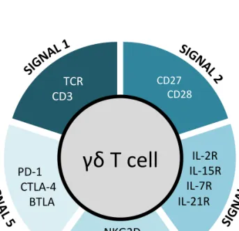
2.2. Five layers of receptor signaling in γδ T-cell differentiation and activation
γδ T cells functional responses are initiated upon recognition of antigens that are likely induced by stress signals and sensed by either T-cell or natural killer receptors. Some γδ T cell populations are also particularly responsive to cytokines
or innate toll-like receptor (TLR) agonists47,54. Following proliferation and effector
responses, the return to homeostasis is controlled by inhibitory receptors. Altogether, the various layers of T (TCR and costimulatory/inhibitory receptors), NK, and cytokine receptor signals synergistically regulate the activation and differentiation of effector γδ T cell populations (Figure 2).
Figure 2: Five layers of cell membrane receptors involved in differentiation, activation and functions of γδ T cells
2.2.1. Signal 1: T cell receptor
The TCRγδ complex is composed by the TCRγδ itself and various CD3 chains
following the stoichiometry: TCRγδCD3ε2γδζ2 in humans, and TCRγδCD3ε2γ2ζ2 in
mice134. The assembly of a TCRγδ complex in thymic progenitors has immediate
γδ T cell
TCR CD3 CD27 CD28 IL-2R IL-15R IL-7R IL-21R NKG2D NKp30 NKp44 PD-1 CTLA-4 BTLAconsequences for γδ T cell development. The “strong” signals stemming from the TCRγδ (when compared to the “weaker” pre-TCR signalling) drive γδ/αβ common
precursors into the γδ lineage36,37. These “stronger” TCRγδ signals associate with
increased phosphorylation of ERK1/2, abundant calcium release and induction of
early growth response (Egr) transcription factors135,136.
The TCR complex does not present intrinsic kinase activity but the intracellular signalling is initiated after phosphorylation of immunoreceptor tyrosine-based activation motifs (ITAMs) in the CD3 cytoplasmic domains by the Src-family
kinases (SFKs) Lck and Fyn137. The recruitment of these SFKs to the TCR complex
in γδ T cells remains obscure since these cells do not express the CD4 or CD8 coreceptors, that have been shown, in αβ T cells, to be responsible for recruiting
SFKs upon TCRαβ ligation137. Nonetheless, the importance of SFKs in γδ T cells is
underscored by the substantial phosphorylation of ERK upon inhibition of Csk, a
potent inhibitor of SFKs138.
SFK-mediated phosphorylation of the ITAMs on CD3 chains allows the recruitment, phospholylation and activation of Zap70 that facilitates phosphorylation of the scaffolding proteins SLP-76 and LAT. This lead to the formation of a supramolecular signalosome that recruits the phospholipase PLCγ1 resulting on
propagation of downstream signalling events136. Here again, γδ T cell signalling is
different from αβ T cells, since mutations on the binding site of PLCγ1 on LAT resulted in a severe block in murine αβ thymocyte development while γδ T cell numbers were only modestly reduced in the thymus, intestine and liver, and remained normal in the skin. Unexpectedly, a population of γδ T cells in the secondary lymphoid organs in these mice underwent uncontrolled expansion and caused autoimmune pathology, suggesting distinct functions for
LAT/PLCγ1-mediated signalling in subpopulations of γδ T cells135,139.
In humans, the major γδ T cell subset in the peripheral blood, Vγ9Vδ2 T cells, are uniquely and specifically reactive to self- and foreign non-peptidic phosphorylated
intermediates of isoprenoid synthesis – “phosphoantigens’’ or “phosphoagonists”
(P-Ags)109,111,140. These P-Ags were shown to trigger bona fide Vγ9Vδ2 TCR
signalling in various studies. Cipriani and colleagues showed that the activation of Vγ9Vδ2 T cells with the P-Ag isopentenyl pyrophosphate (IPP), induced rapid and persistent PKC-dependent phosphorylation of ERK1/2, p38 MAPK, and JNK, resulting in NF-κB and AP-1 activation as well as the release of CCL-3, CCL-4, IFN-γ
and TNF-α 141. Moreover, P-Ag stimulation and CD3-crosslinking produced
identical phosphorylation of the signalling proteins Zap70, PI3K, LAT, ERK1/2 and p38 MAPK142,143; and induced highly sustained calcium signalling in Vγ9Vδ2 T cells 144. Importantly, activation by P-Ags is the basis of current cancer immunotherapy strategies involving Vγ9Vδ2 T cells145. Recent work has produced some puzzling results on the role of the TCRγδ in the development of effector subsets of murine γδ T cells146–148, namely CD27+ CD122+
γδ T cells producing IFN-γ or CD27- CCR6+ γδ T cells making IL-1749,149. First, Chien
and co-workers showed that T10/T22-specific γδ T cells required thymic expression of their TCR ligands to differentiate into IFN-γ producers, in contrast
with “ligand naïve” IL-17 producers49. Consistent with this, TCR-dependent thymic
selection was also shown to set the functional potential of DETC progenitors away
from IL-17 production150. Furthermore, peripheral IL-17-producing CD27- CCR6+
γδ T cells were shown to expand and produce IL-17 independently of TCR
activation151. However, a subsequent study by Chien and collaborators
demonstrated that a subset of phycoerythrin (PE)-specific γδ T cells produced IL-17 specifically upon TCR ligation152. Moreover, a recent study by Hayday and
colleagues suggested that an impairment in Zap70 signalling (in SKG mice) mostly affected the development of IL-17+ rather than IFN-γ+ γδ T cells153. The authors
further proposed that “innate-like” γδ T cell populations, including IL-17 producers and some subsets of IFN-γ producers, receive strong TCR signals during thymic development to become hyporesponsive to TCR stimulation in the
manipulating distinct TCRγδ signalling pathways and their downstream (transcriptional and post-transcriptional) mechanisms on discrete γδ T cell subsets.
Table 2: Co-receptors of γδ T cells – extracellular ligands and intracellular signaling pathways. Receptor Ligands Intracellular signalling initiators/ adaptors Downstream signalling pathway Target molecules Reference CD28 B7.1 (CD80) B7.2 (CD86) PI3K ITK Grb2 PI3K/ AKT Grb2/ MEK/ ERK IL-2, NF-κB, AP-1, Bcl-xL, NFAT 154–157 CD27 CD70 TRAF2 TRAF5 Siva IKK/ NF-κB JNK NF-κB, Ca 2+, cyclinD2, Bcl2a1, Bcl-xL 158–161
CD30 CD30L TRAF2 TRAF5 TRAF/ IKK/ IkB Ca2+
NF-κB, IL-4, IFN-γ, IL-8, CC chemokines 158,162,163 4-1BB (CD137) CD137L TRAF2 NF-κB, IFN-γ 164–166 IL-2R
IL-15R IL-2 IL-15 Jak1 Jak3
PI3K/ AKT Jak/ STAT4/ STAT5 MEK/ ERK STAT1 IFN-γ, TNF-α, T-bet, Eomesodermin 167–170
IL-7R IL-7 Jak1 Jak3 STAT3 IL-17, SOCS3 171
IL-21R IL-21 Jak1 Jak3 STAT3 CXCL13, CXCR5 172
NKG2D MIC(A-B) ULBP(1-6) H60 MULT1 RAE1 DAP10 PI3K/ AKT Grb2/ VAV1/ SOS1 PKCθ/ Ca2+ NF-κB, RelB, Bcl-xL, Bcl-2 144,158,173–175
NKp30 B7-H6 BAT3 CD3ζ cAMP/ PKA CC-chemokines: CCL3, CCL4, CCL5 78,176–178
NKp44 NKp44L DAP12 Zap70/ Syk 177,179–181 DNAM-1 (CD226) Nectin- like-5 Nectin-2 PKC LFA-1 Fyn SLP-76/ VAV1/ ERK 182,183 PD-1 PD-L1 (B7-H1) PD-L2 (B7-DC) SHP-1 SHP-2 CK2/ PTEN/ PI3K/ AKT MEK/ ERK GSK-3, Bcl-xL SMAD3, Cdc25A, IFN-γ, IL-2 184–187
BTLA HVEM SHP-1 SHP-2 Zap70/ ERK IL-17, TNF, IL-2 188–190