INSTITUTO DE INVESTIGAÇÃO E FORMAÇÃO AVANÇADA ÉVORA, JANEIRO 2016
ORIENTADORES: Prof.a Doutora Elisa Maria Varela Bettencourt
Prof.a Doutora Maria Manuela Melo Oliveira Prof. Doutor Olivier M. Lepage
Tese apresentada à Universidade de Évora
para obtenção do Grau de Doutor em Ciências Veterinárias
Susana Oliveira Serrano Monteiro
DISTAL LIMB OSTEOARTHRITIS
INSTITUTO DE INVESTIGAÇÃO E FORMAÇÃO AVANÇADA ÉVORA, JANEIRO 2016
Tese apresentada à Universidade de Évora
para obtenção do Grau de Doutor em Ciências Veterinárias
Susana Oliveira Serrano Monteiro
DISTAL LIMB OSTEOARTHRITIS
IN THE HORSE
ORIENTADORES: Profª Doutora Elisa Maria Varela Bettencourt
Profª Doutora Maria Manuela Melo Oliveira Prof. Doutor Olivier M Lepage
i
INDEX
Abstract ... iv
!
Resumo ... v
!
Dedication ... vi
!
Funding acknowledgments ... vii
!
Personal acknowledgments ... viii
!
Tables index ... ix
!
Figures index ... x
!
List of abbreviations ... xii
!
INTRODUCTION ... 1
!
CHAPTER I – Review of literature ... 5
!
1.
!
Articular biology of equine distal limb joints ... 5!
2.
!
Pathogenesis of equine OA ... 6!
3.!
Diagnosis of equine OA ... 7!
3.1!
Lameness evaluation ... 7!
3.2!
Imaging techniques ... 9!
3.3!
Cartilage Biomarkers ... 11!
4.!
Treatment of equine OA ... 12!
4.1
!
Intra-articular synthetic drugs ... 13!
4.2
!
Intra-articular biologic strategies ... 14!
5.
!
References ... 16!
CHAPTER II – Comprehensive research work ... 25
!
1.
!
Survey to Portuguese equine veterinarians ... 25!
1.1
!
Materials & Methods ... 25!
1.2
!
Results ... 25!
1.3
!
Discussion ... 26!
2.
!
Effects of allogeneic ASCs pre-activated by INF-γ
on experimental OA in vitro ... 27!
2.1
!
Introduction ... 27!
2.2
!
Material & Methods ... 28!
2.3
!
Results ... 30!
2.4
!
Discussion ... 32!
3.
!
References ... 32!
CHAPTER III – Effects of allogeneic ACSs pre-activated by IFN-γ on equine groove model of metacarpo-phalangeal osteoarthritis ... 35
!
1.
!
Introduction ... 36!
2.
!
Materials & Methods ... 37!
2.1.
!
Isolation and characterization of ASCs ... 38!
2.2.
!
ASCs pre-activation with IFN-γ ... 38!
2.3.
!
Groove model of metacarpophalangeal osteoarthritis ... 38!
2.4.
!
Intra-articular treatment ... 39!
2.5.
!
Arthroscopic evaluation ... 39!
2.6.
!
Lameness evaluation ... 39!
2.7.
!
Radiographic Examination ... 40!
2.8.
!
Synovial fluid analysis ... 40!
2.9.
!
Postmortem Examination ... 40!
2.10.
!
Statistical Analysis ... 42!
3.
!
Results ... 42!
3.1.
!
Arthroscopic evaluation ... 42!
ii
3.3.
!
Radiographic examination ... 44!
3.4.
!
Synovial fluid ... 45!
3.5.
!
Macroscopic and microscopic evaluation ... 46!
4.
!
Discussion ... 49!
5.
!
References ... 52!
CHAPTER IV – Relationship between serum biomarkers of cartilage and osteoarticular disease in Lusitano horses ... 55
!
1.
!
Introduction ... 56!
2.
!
Materials & Methods ... 57!
2.1.
!
Examined horse population, age and exercise groups ... 57!
2.2.
!
Lameness examination ... 58!
2.3.
!
Radiographic examination and classification ... 58!
2.4.
!
Osteoarticular disease ... 59!
2.5.
!
Blood samples ... 60!
2.6.
!
Coll2-1, Coll2-1NO2 and Fib3-2 assays ... 60!
2.7.
!
Statistical analysis ... 60!
3.
!
Results ... 61!
3.1.
!
Effects of sex, age and exercise on serum biomarkers ... 61!
3.2.
!
Correlation between biomarkers ... 61!
3.3.
!
Relationship between Coll2-1, Coll2-1NO2 and Fib3-2 and clinical findings ... 61!
4.
!
Discussion ... 64!
5.
!
References ... 66!
CHAPTER V – Relationship between subjective and objective assessment of distal limb flexion test in Lusitano horses with osteoarticular disease ... 69
!
1.
!
Introduction ... 70!
2.
!
Materials & Methods ... 71!
2.1.
!
Sample population ... 71!
2.2.
!
Lameness examination ... 71!
2.3.
!
Objective lameness measures ... 72!
2.4.
!
Radiographic examination and classification ... 73!
2.5.
!
Statistical Analysis ... 73!
3.
!
Results ... 73!
3.1.
!
Relationship between subjective and objective evaluations ... 74!
3.2.
!
Correlation between flexion response and radiographic scores ... 74!
3.3.
!
Forelimb radiographic scores and responses to flexion ... 74!
3.4.
!
Hind limb radiographic scores and responses to flexion ... 75!
4.
!
Discussion ... 76!
5.
!
References ... 78!
CHAPTER VI - Effects of a Single Intra-articular Injection of High Concentrated and High Molecular Hyaluronic Acid Treatment on Distal Limb Osteoarthritis – a pilot study ... 81
!
1.
!
Introduction ... 82!
2.
!
Materials & Methods ... 83!
2.1.
!
Animals ... 84!
2.2.
!
Lameness evaluation ... 84!
2.3.
!
Synovial fluid analysis ... 85!
2.4.
!
Treatment protocol ... 85!
2.5.
!
Serum biomarkers analysis ... 85!
2.6.
!
Statistical analysis ... 85!
3.
!
Results ... 86!
iii
3.2.
!
Lameness examination ... 86!
3.3.
!
Synovial fluid analysis ... 88!
3.4.
!
Biomarkers analysis ... 88!
4.
!
Discussion ... 89!
5.
!
References ... 91!
GENERAL CONCLUSION ... 95
!
Appendix A – Proceedings of the 24th ECVS Annual Scientific Meeting ... I!
Appendix B – Review article ... II!
Appendix C – Questionnaire ... III!
iv
Abstract
The aim of this thesis was to study two objective methods of osteoarthritis (OA) diagnosis in horses and use them on the assessment of new intra-articular treatments. The studied methods were a new inertial-sensor based system of lameness detection and cartilage biomarkers in serum. It was found that distal limb flexion is significantly correlated to the presence of metacarpo-phalangeal OA in hind limbs and that inertial-sensors are sensitive in detecting asymmetry in these cases. A positive and significant correlation was observed between Coll2-1 concentration in serum and the presence of joint disease in males and young horses. Fib3-2 measurement has good potential to be used since it is not influenced by sex or age. Using an experimental model of OA, adipose stem cells pre-activated with interferon-gamma decreased joint inflammation and radiographic lesions. In clinical cases, a single injection of high-concentrated and high-molecular weight hyaluronic-acid decreased joint inflammation and biomarkers’ concentration.
v OSTEOARTRITE DO MEMBRO DISTAL NO CAVALO
Resumo
A finalidade desta tese foi estudar dois métodos de diagnóstico objetivo de osteoartrite (OA) em equinos e aplicá-los na avaliação de novas terapias intra-articulares. Utilizou-se um sistema de sensores de movimento e foi avaliada a concentração de biomarcadores de cartilagem no soro. Concluiu-se que a flexão distal positiva está correlacionada com OA na articulação metacarpo-falângica nos membros posteriores e que os sensores são sensíveis na detecção de assimetria nestes casos. Existe uma correlação positiva e significativa entre as concentrações de Coll2-1 e a presença de doença articular, sobretudo em machos e jovens. A dosagem de Fib3-2 tem utilidade por não ser influenciada pelo sexo nem idade. Num modelo experimental da doença, a terapia à base de células estaminais reduziu a inflamação articular e as lesões radiográficas. Em casos clínicos, o tratamento com ácido-hialurónico de alta concentração e peso molecular provoca uma diminuição da inflamação articular e dos biomarcadores no soro.
vi
Dedication
vii
Funding acknowledgments
• Instituto de Ciências Agrárias e Ambientais Mediterrânicas (ICAAM) supported the 3 months externship in the VetAgro Sup – Veterinary Campus of Lyon (France).
• This work was funded by Luso-French integrated actions (LF-IA) 2013/2014 and 2014/2015: - Travels and Lameness locator transportation between Lyon and Lisbon was funded by LF-IA: JLS/2013.210 and CT R08 (Vetagro Sup – Veterinary Campus of Lyon).
- Participation in the 24th Annual Meeting of ECVS (Berlin 2015) was funded by LF-IA: TC nº14/14 (Universidade de Évora).
- Participation in the First European Stem Cell Symposium for vets (Maastricht 2015) was funded by LF-IA: 2014-C17 (Universidade de Évora)
• Part of this work (biomarkers analysis) was funded by FEDER Funds through the Operational Programme for Competitiveness Factors - COMPETE and National Funds through FCT - Foundation for Science and Technology under the Strategic Projects PEst-C/AGR/UI0115/2011, PEst-OE/AGR/UI0115/2014 and through the Programa Operacional Regional do Alentejo (InAlentejo) Operation ALENT-07-0262-FEDER-001871/ Laboratório de Biotecnologia Aplicada e Tecnologias Agro-Ambientais.
• The lameness locator and the study about the use of allogeneic pre-activated mesenchymal stemcell was funded by the GREMERES (Groupe de Recherche en Médecine Et Réeducation des Equidés de Sport), VetAgro Sup – Veterinary Campus of Lyon (France).
• ARTIALIS Laboratory collaborated in biomarkers’ analysis study.
viii
Personal acknowledgments
To Dr. Elisa Bettencourt for being my supervisor, she is the main responsible for this part of my academic life, always transmitting energy and optimism, essential for this work.
To Dr. Olivier M. Lepage for accepting to be my co-supervisor, adding intellectual and scientific stimulus, sharing the innovative techniques I have learned and used during this work. I would also like to thank all other colleagues and staff of the VetAgro Sup - Veterinary Campus of Lyon and Sanofi-aventis, Montpetlier, for the multidisciplinary work we did together.
To Dr. Manuela Oliveira for being my co-supervisor and for having taught me everything I know about statistics and adding criticism to the data collection and analysis performed, which was very important to this research.
To all my colleagues and students at Universidade de Évora for their assistance, a special thanks to Dr. Liliane Damásio, Dr Luís Antunes and Dr Sandra Branco.
To Dr. Maria José Correia and Eng. António Saraiva for sharing the Unidade Clínica of Fundação Alter Real, Alter do Chão, and to be willing to contribute with the genetic background of these animals.
To Dr. Rui Martelo, Dr Ricardo Romão, Dr Joana Rua and staff of the Vetal Veterinary Clinic, Portalegre, for sharing their patients and be willing to participate in this project.
To Dr. João Borges, Dr. Gonçalo Paixão and the Hospital Militar de Equinos – Escola de Armas, Mafra, for sharing their patients, knowledge and interest in equine lameness evaluation and osteoarthritis treatment.
To Dr Kevin Keegan for assistance in interpretation of lameness locator results. To António Bettencourt and Graciete Rabaça for language assistance.
To Dr. Dulce Gomes for assistance in part of the statistical analysis.
To my family, for believing and allowing me to follow my dreams, nothing would be possible without them and in particular to my mother for being such an extraordinary person.
To my husband Pedro for his support at all moments and assistance in the graphic part of the work and my daughter Marisa that was born and grew together with this project, both have brought love and a different meaning to my life.
ix
Tables index
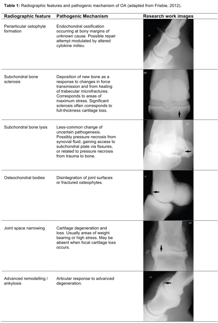
Table 1: Radiographic features and pathogenic mechanism of OA (adapted from Frisbie, 2012). ... 10
!
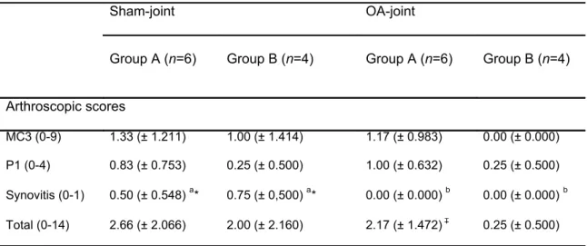
Table 2: Mean arthroscopic scores (± SD) from dorsal MCP joint at week 0. ... 43!
Table 3: Median A1/A2 ratio (± MAD) for asymmetry of front limbs trotting at straight line and afterdistal limb flexion. ... 44
!
Table 4: Median macroscopic and microscopic scores (± MAD) at week 10. ... 47
!
Table 5: Radiographic scoring system (adapted from Kellgren and Lawrence, 1957 and de Grauw etal., 2006). ... 59
!
Table 6: Distribution of objective lameness, disease and mean (± SEM) blood biomarkers’ values
n=51. ... 62
!
Table 7: Distribution of age classes and mean (± SEM) serum biomarkers’ values for horses with or
without osteoarticular disease n = 51. ... 63
!
Table 8: Distribution of sex classes and mean (± SEM) serum biomarkers’ values for horses with or
without osteoarticular disease n = 51. ... 63
!
Table 9: Distribution of exercise classes and mean (± SEM) serum biomarkers’ values for horses with or
without osteoarticular disease n = 51. ... 63
!
Table 10: Distribution of osteoarticular disease and mean (± SEM) serum biomarkers’ values n = 51.
x
Figures index
Figure 1: Horse instrumented with 3 inertial sensors during a lameness exam. There is a uni-axial
accelerometer attached to the head bumper; one uni-axial accelerometer placed between the tubera sacrale on the midline of the most dorsal aspect of the pelvis with tape and a third uni-axial gyroscope wrapped to the dorsal aspect of the right forelimb pastern (VetAgro Sup - Veterinary Campus of Lyon). ... 8
!
Figure 2: Illustration of data used for detection and quantification of lameness in horses using inertial
sensor system (Keegan 2004). A: There is less downward movement of the head during the stance phase of the right forelimb (righ forelimb lameness) and B: more downward movement of the pelvis during the stance phase of the right hind limb (left hind limb lameness). ... 8
!
Figure 3: Mean (± SD) glycosaminoglycan release speed over 12 days of culture (n = 10). Explant
model - culture medium controls compared to OA condition. Significantly different results are represented by *** P ˂ 0.001. ... 30
!
Figure 4: Mean (± SD) glycosaminoglycan release speed over 12 days culture (n = 10). Coculture -
ASCs low (25x103) and high dose (100x103) ASCs, with or without IFN-γ pre-treatment, compared to OA condition alone. Significantly different results are represented by * P ˂ 0.05 ** P ˂ 0.01 and *** P ˂ 0.001. ... 31
!
Figure 5: Macroscopic view of the articular surfaces of the metacarpo-phalangeal joint, (A) is the
distal aspect of the third metacarpal bone, (B) is the proximal aspect of the first phalanx and (C) is dorsal aspect of lateral and medial proximal sesamoid bones. Black arrows represent the transverse section and grey arrows represent the articular capsule with synovial membrane used for microscopic analysis. Rectangular areas in grey were excluded from examination. L = lateral and M = medial. ... 41
!
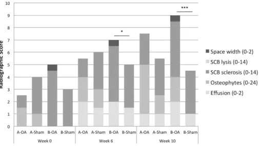
Figure 6: Radiographic scores at week 0, 6 and 10. Median radiographic total scores (0-56) of
evaluated features in ASCs-γ treated group (Group A, n = 6), vehicle treated group (Group B, n = 4) and respective sham-joints. Significantly different results between OA and sham-joints are represented by * P ˂ 0.05 and *** P ˂ 0.001. ... 44
!
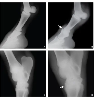
Figure 7: Radiographic views at week 10, the first row (A and B) are 45-degree
dorsolateral-palmaromedial oblique views of metacarpo-phalangeal joint from an individual of group A (ASCs-γ) and the second row (C and D) are from an individual of group B (vehicle). The first column (A and C) are the OA-joints and the second column (B and D) are the sham-joints. The arrows show osteophyte formation (grade 2) and the asterisk show the synovial effusion (grade 2) on the OA-joint of the individual of group B. ... 45
!
Figure 8: Synovial fluid. (A) Mean (± SD) Total Proteins and (B) Mean (± SD) Prostaglandin E2 values
in synovial fluid of the ASCs-γ treated group (Group A, n = 6) and the vehicle treated group (Group B,
n = 4) compared to respective sham-joints. Significantly different results are represented by * P ˂ 0.05,
** P ˂ 0.01 and *** P ˂ 0.001 (black asterisks: Group A; grey asterisks: Group B). ... 46
!
Figure 9: Light micrographs of grooved areas of the dorsal MC3 condylar cartilage (hematoxylin and
xi mesenchymal-like cell focus found in loose matrix. (B) ASCs-γ treated horse with microscopic lesions adjacent to groove and intra-groove chondrocyte-like cell focus in fibrous matrix. ... 48
!
Figure 10: Light micrographs of non-grooved areas of the dorsal MC3 condylar cartilage (hematoxylin
and eosin stained). (A) Sham-joint of ASCs-γ treated horse showing a swollen (raised) area of non-calcified cartilage of the palmar MC3. (B) Sham-joint of ASCs-γ treated horse showing a superficial/deep fissuring in non-calcified cartilage of the palmar MC3. ... 49
!
Figure 11: Latero-medial radiographs of equine distal limb region (A, B) and tarsal region (C, D)
illustrating grade 0-normal (A, C) and grade 4-severe OA lesions (arrows) of the proximal inter-phalangeal joint (B) and distal inter-tarsal and tarso-metatarsal joints (D). ... 59
!
Figure 12: Box plot of distal limb radiographic mean score for positive and negative flexion tests
assessed (A) subjectively and (B) objectively, head asymmetry change (ΔVS). There is no significant difference in radiographic scores for positive and negative subjective or objective flexion-test. ... 75
!
Figure 13: Box plot of distal limb radiographic mean score for positive and negative flexion tests
assessed A: subjectively and objectively for B: maximum pelvic height change (ΔPDMax) and for C: minimum pelvic height change (ΔPDMin). There was significant difference in radiographic scores between negative and positive response for ΔPDmin (* P = 0.013). ... 76
!
Figure 14: Box plot of objective lameness evaluations in fore limbs (A): head asymmetry (VS) at
straight line and (B): head asymmetry difference before and after flexion (ΔVS). There were no significant differences. ... 87
!
Figure 15: Box plot of objective lameness evaluations in hind limbs (A): maximum pelvic asymmetry
(PDmax) at straight line and (B): difference between before and after flexion asymmetry (ΔPDmax). Significant differences between ΔPDmax at week 0 and at week 8 are represented by * P < 0.05. ... 87
!
Figure 16: Box plot of objective lameness evaluations (A): minimum pelvic asymmetry (PDmin) at
straight line and (B): difference between before and after flexion asymmetry (ΔPDmin). There were no significant differences. ... 88
!
Figure 17: Box plot of serum biomarkers’ concentration (A): Coll2-1 (B): Coll2-1NO2 and (C): Fib3-2
xii
List of abbreviations
Abbreviation Unabbreviated Abbreviation Unabbreviated
AAEP american association of equine practicioners Hmin difference in head minimum height between left and right
ADAMTS
a desintegrin and metalloproteinase with thrombospondin motifs
HMW high molecular weight
ANOVA analysis of variance IA intra-articular
APMVE portuguese association of
equine veterinarians ICAAM
mediterranean agricultural and environmental sciences institute
ASCs adipose tissue- derived mesenchymal stem cells IFN-γ interferon-gamma ASCs-γ ASC activated with interferon-gamma IL interleukin
AUC area under the curve IM intramuscular
CD clusters of differentiation IQR interquartile range cDNA complementary deoxyribonucleic acid IV intravenous
cm centimeters κ kappa agreement
Coll2-1 type II collagen peptide
(108HRGYPGLDG116) kDa kilodalton
Coll2-1NO2 nitrated form of Coll2-1 kg kilogramme
CO2 carbon dioxide L liter
COX ciclooxygenase MAD median absolute deviation
Da daltons MC3 third metacarpal bone
DIP distal interphalangeal MCP metacarpophalangeal
DMEM dulbecco’s modified eagle’s medium µg microgramme
ECM extracellular matrix µL microliter
ECVDI european college of veterinary diagnostics and imaging mg miligramme ECVP european college of veterinary
pathology min minutes
ECVS european college of veterinary
surgeons mL mililiter
EDTA ethylenediamine tetraacetic
acid mm milimiter
ELISA enzyme like immunosorbent
assay mM milimolar
Fib3-1 fibulin 3 fragment 1 MSCs mesenchymal stem cells
Fib3-2 fibulin 3 fragment 2 MPA methylphrednisolone acetate
g gramme MMP metalloproteinase
ga gravitational force mRNA messenger ribonucleic acid
G gauge n number
GAG glicosaminoglycan ng nanogramme
GLM generalized linear model nM nanoMolar
HA hyaluronic acid NSAIDs non-steroidal anti-inflammatory drugs Hmax difference in head maximum
xiii OARSI osteoarthritis research society
international qRT-PCR
quantitative real-time polymerase chain reaction OCD osteochondrritis dissecans r pearson’s correlation coefficient
OSM oncostantin-M r2 coefficient of determination
P calculated probability RNA ribonucleic acid
P1 first phalanx ROC receiving operating characteristic curve PBS phosphate buffered saline rpm rotation per minute PDmax difference in pelvis maximum
height between left and right SAS statistical analysis software ΔPDmax PDmax change before and after flexion SCB subchondral bone
PDmin difference in pelvis minimum height between left and right SD standard deviation
ΔPDmin PD min change before and after flexion SEM standard error of the mean
pg picogramme SF synovial fluid
PGE2 prostaglandin E2 SPSS statistical package for the
social sciences
PIP proximal interphalangeal TNF-α tumor necrosis factor alpha
pM picomolar TA triamcinolone acetonide
PRP platelet rich plasma TP total proteins
PSB proximal sesamoid bones VS vector sum (total head
asymmetry)
1
INTRODUCTION
Lameness, and particularly osteoarthritis (OA), is still one of the main reasons for early retirement of horses and economic losses in the equine industry. Articular cartilage is the main component of a joint and is responsible, together with synovial fluid (SF), for the frictionless movement. OA is characterized by degeneration and loss of cartilage, w h i c h occurs when catabolic pathway overcomes the anabolic process (Frisbie, 2012). The general objective of this thesis was to advance further the diagnosis of OA and at treatment knowledge in horses. We used two objective diagnostic methodologies of OA: lameness detection with intertial sensors technology and analysis of serum cartilage biomarkers in combination with traditional subjective lameness assessment and radiographic examination. We used these methods to assess two novel intra-articular (IA) treatments: adipose tissue-derived allogeneic mesenchymal stem cells (ASCs) pre-activated with interferon-gamma (IFN-γ) on experimental induced OA and a high concentrated and high molecular weight (HMW) hyaluronic acid (HA) on naturally occurring disease.
Osteoarticular pain is the key of OA disease assessment but it is difficult to quantify in horses. A recent method appeared in order to objectively determine the amount of asymmetry of the four limbs. This system uses wireless transmission of data from small inertial sensors attached to the horse’s body. Keegan et al. first introduced this technology in 2002 and its repeatability and sensitivity has been demonstrated (Keegan et al., 2011; McCracken et al., 2012). In this research we have objectively evaluated lameness and response to IA treatments using this technology, owned by the VetAgro Sup – Veterinary Campus of Lyon. The PhD student spent 3 months in this institution, learning how to use this technology while testing pre-activated ASCs with IFN-γ (ASCs-γ) IA treatment.
Mesenchymal stem cells (MSCs) have reparative effects, due to their differentiation properties and to paracrine actions, but they need to be activated by inflammatory mediators released from immune cells. Pre-activation with IFN-γ enhances the therapeutic activity of MSCs in animal models of colitis and graft versus host disease (Polchert et al., 2008; Duijvestein et al., 2011). Therefore, pre-activation of MSCs could lead to a more efficient therapeutic activity than unstimulated cells, initiating regeneration and repair of hyaline cartilage. Murine collagenase induced OA models showed that a single injection of autologous ASCs reduced synovitis, enthesophyte formation and decreased score of cartilage lesions (ter Huurne et al., 2012). Furthermore, ASCs have recently shown a protective effect in human cartilage in vitro by preventing chondrocyte apoptosis and fibrosis (Maumus et al., 2013). Our objective was to study the effects of INF-γ pre-activated or “primed” allogeneic ASCs in an experimental OA model in horses. The preliminary results of this experiment were orally presented at the I congresso da Associação Portuguesa de Médicos Veterinários de Equinos (APMVE), Lisboa 2014, in the 24th Annual Meeting of the European College of Veterinary Surgeons (ECVS), Berlin 2015 (Appendix A: abstract) and at the 1rst European Stem Cell Symposium for vets, Maastricht 2015.
To achieve early recognition of disease, equine research, as a valid model for human OA, has focused on biomarker analysis. Verwilghen et al. (2011) correlated higher OA advanced degenerative
2
lesions with increased type II collagen 108HRGYPGLDG116 peptide (Coll2-1) in SF and severity of radiological lesions with increased Coll2-1 nitrated form (Coll2-1NO2)in horse blood (Verwilghen et al.,
2009). In humans, Fibulin 3 peptide fragment 1 (Fib3-1) and fragment 2 (Fib3-2) have recently shown to be elevated in serum of OA patients and differentiate between OA and normal populations (Henrotin
et al., 2012) but have never been studied in horses. In this project we have studied 1,
Coll2-1NO2 and Fib3-2 in naturally occuring OA in 51 Lusitano horses. The abstract have been accepted at
the poster session of the 25th Annual Meeting of the ECVS (Lisboa): Susana Monteiro, Olivier M Lepage, Luís Antunes, Liliane Damásio, Sandra Branco, Manuela Oliveira, Elisa Bettencourt. Relationship between biomarkers of cartilage in serum and degenerative joint disease in Lusitano horses. Using the same Lusitano horse population, subjective lameness assessment was compared to objective lameness detection and related to radiographic distal limb OA findings. The results have been presented at the V Congreso Annual de La Asociación de Veterinarios Especialistas en Équidos de España (AVEE), WEVA Intermadiate meeting España-Portugal (Madrid): Cómo están relacionados los hallazgos radiográficos de la extremidad distal con la evaluación objectiva de cojera.
Finally, a group of eight horses presenting clinical OA were treated with a single injection of HMW-HA and objectively assessed for lameness and serum biomarkers’ concentration. Hyluronic acid is injected due to viscosupplementation, light anti-infllammatory properties (Takahashi et al., 1999) and disease-modifying effects (Marshall et al., 2000). On the other hand, Coll2-1 and Coll2-1NO2
biomarker have been shown to be significantly decreased in serum of OA human patients after HA treatment (Henrotin et al., 2013). High molecular weight HA is reported to have superior cartilage-sparing properties in OA models (Asari et al., 1998), to enhance in vitro lubricant function (Antonacci
et al. 2012) as well as to induce better clinical improvement (Philips, 1989) when compared to the
lower molecular weight.
This thesis is divided in seven chapters. Chapter I contains a brief review on distal limb OA pathogenesis, diagnosis and conventional OA treatment. An extensive review of IA biologic therapies was published: Monteiro SO, Bettencourt EV, Lepage OM. Biologic strategies for intra-articular treatment and cartilage repair. J Eq Vet Sci 2015; 35:175-190 (Appendix B). Chapter II includes the comprehensive research work, which presents the results of a survey to Portuguese equine veterinarians about OA (Appendix C: questionnaire) and the preliminary results of the effects of equine allogeneic ASCs-γ in vitro (a collaborative study). The fundamental research work is presented in the following four chapters and consisted in testing ASCs-γ treatment on experimental OA in vivo (Chapter III), evaluating cartilage biomarkers in serum (Chapter IV) and studying lameness detection (Chapter V) in a Lusitano horse population and finally assessing the effect of a single injection of HA treatment on clinical cases of OA using both objective methodologies (Chapter VI). A general conclusion is presented at the end of the thesis.
We believe that veterinarian researchers should focus their work on objective methodologies and that biologic strategies will play an important role in equine OA therapy. This PhD thesis is just a piece of the puzzle.
3
References:
Asari A, Miyauchi S, Matsuzaka S, Ito T, Kominami E, Uchiyama Y. Molecular weight-dependent effects of hyaluronate on the arthritic synovium. Archives of Histology and Citology 1998; 61(2):125:135.
Duijvestein M, Wildenberg ME, Welling MM, Hennink S, Molendjik I, van Zuylen VL, Bosse T, Vos AC, de Jonge-Muller ES, Roelofs H, van der Weerd L, Verspaget HW, Fibbe WE, te Velde AA, van den Brink GR, Hommes DW. Pretreatment with Interferon-gamma enhances the therapeutic acivity of mesenchymal stromal cells in animal models of colitis. Stem Cells 2011; 29(10):1549-1558.
Frisbie D. Synovial Joint Biology and Pathobiology, In: Auer J and Stick J. Equine Surgery (4th
ed.) St Louis: Elsevier-Saunders; 2012: pp.1096-1113.
Henrontin Y, Gharbi M, Mazzucchelli G, Dubuc JE, De Pauw E, Deberq M. Fibulin 3 peptides Fib3-1 and Fib3-2 are potential biomarkers of osteoarthritis. Arthritis & Rheumatism 2012; 64(7):2260-2267.
Henrotin Y, Chevalier X, Deberg M, Balblanc JC, Richette P, Mulleman D, Maillet B, Rannou F, Piroth C, Mathieu P, Conrozier T. Early decrease of serum biomarkers of type II collagen degradation (Coll2-1) and joint inflammation (Coll2-1NO2) by hyaluronic acid intra-articular injections in patients with knee
osteoarthritis: a research study part of the biovisco study. Journal of Orthopedic Research 2013; 31(6):901-907.
Keegan KG, Kramer J, Yonezawa Y, Maki H, Pai PF, Dent EV, Kellerman TE, Wilson DA, Reed SK. Assessment of repeatability of wireless, inertial sensor-based lameness evaluation system for horses. American journal of veterinary research 2011; 72(9):1156-1163.
Keegan KG, Yonezawa Y, Pai PF, Wilson DA. Telemeterized accelerometer-based system for the detection of lameness in horses. Biomedical Sciences Instrumentation 2002; 38:107-112.
Marshall KW, Manolopoulos V, Mancer K, Staples J, Damyanovich A. Amelioration of disease severity by intraarticular Hylan therapy in bilateral canine osteoarhtirits. Journal of Orthopedic Research 2000; 18(3):416-425.
Maumus M, Manferdini C, Toupet K, Peyrafitte JA, Ferreira R, Facchini A, Gabusi E, Bourin P, Jorgensen C, Lisignoli G, Noel D. Adipose mesenchymal stem cells protect chondrocytes from degeneration associated with osteoarthritis. Stem cell research 2013; 11(2):834-844.
4
McCracken MJ, Kramer J, Keegan KG, Lopes M, Wilson DA, Reed SK, Lacarrubba A, Rasch M. Comparison of an inertial sensory system of lameness quantification with subjective lameness evaluation. Equine veterinary journal 2012; 44(6):652-656.
Philips MW. Clinical trial comparison of intra-articular sodium hyaluronate products in the horse. Journal Equine Veterinary Science 1989; 9(1):39-40.
Polchert D, Sobinsky J, Douglas G, Kidd M, Moadsiri A, Reina E, Genrich K, Mehotra S, Setty S, Smith B, Bartholomew A. INF-gamma activation of mesenchymal stem cells for treatment and prevention of graft versus host disease. European Journal of Immunology 2008; 38(6):1745-1755.
Takahashi K, Goomer RS, Harwood F, Kubo T, Hirasawa Y, Amiel D. The effects of hyaluronan on matrix metalloproteinases-3 (MMP-3, interleukin-1beta (IL-1beta), and tissue inhibitor of metalloproteinase-1 (TIMP-1) gene expression during the development of osteoarthritis. Osteoarthritis and Cartilage 1999; 7(2):182-190.
ter Huurne M, Schelbergen R, Blattes R, Blom A, de Munter W, Grevers LC, Jeanson J, Noel D, Casteilla L, Jorgensen C, van den Berg W, van Lent PL. Antiinflammatory and chondroprotective effects of intraarticular injection of adipose-derived stem cells in experimental osteoarthritis. Arthritis & Rheumatology 2012; 64(11):3604-3613.
Verwilghen D, Busoni V, Gangl M, Franck T, Lejeune JP, Vanderheyden L, Detilleux J, Grulke S, Deberg M, Henrotin T, Serteyn D. Relationship between biochemical markers and radiographic scores in the evaluation of the osteoarticular status of warmblood stallions. Research Veterinary Science 2009; 87(2): 319-328.
Verwilghen DR, Enzerink E, Martens A, Franck T, Balligand M, Henrotin Y, Detilleux J, Serteyn D. Relationship between arthroscopic joint evaluation and the levels of Coll2-1, Coll2-1NO(2), and myeloperoxidase in the blood and synovial fluid of horses affected with osteochondrosis of the tarsocrural joint. Osteoarthritits and Cartilage 2011; 19(11):1323-1329.
5
CHAPTER I – Review of literature
1. Articular biology of equine distal limb joints
Joint health is of major importance for horses’ performance and survival. The equine distal limb is made up of diarthrodial joints that allow smooth articulation of 2 bone ends. The joint capsule supports the synovial structures and is made up of a fibrous tissue surrounded by tendons and colateral ligaments. Below the capsule there is a thin synovial membrane with 3 types of cells: synovial type A cells that have a phagocytic function; type B cells that produce hyaluronic acid (HA) and an intermediate celular type (C cells). The gaps between synoviocytes, the absence of a basal membrane and the presence of subsynovial blood vessels allow exchange of molecules under 10 kilodalton (kDa) between plasma and sinovyal fluid (SF) (Todhunter, 1996b). Synoviocytes are also able to produce inflammatory mediators, making the synovial membrane an important structure during joint inflammation (Briston et al., 2010).
Hyaline cartilage covers the bone extremities and is made up of a small population of chondrocytes distributed in the extracelular matrix (ECM). This avascular connective tissue is especially developed for shock absorption and weight bearing (Palmer & Bertone, 1996), being highly dependent on the macromolecular arrangement (Kempson, 1980). The collagen molecules make a cross-linked network together with HA molecules. HA is connected by link proteins to proteoglycan aggrecans, which are negatively charged by their polyanionic glycosaminoglycan (GAG) sidechains: keratan and chondrotin sulphate (Ogston, 1970). This allows an osmotic swelling pressure, attracting water and expanding the collagen network, which results in a highly stiff but resilient tissue (Ogston, 1970; Eyre & Wu, 1995; van Weeren & Brama, 2001). The articular cartilage has a non-calcified section, which is divided in a superfitial tagential zone, where chondrocytes and collagen fibrils are horizontally oriented, an intermediate and deep zone, where they are vertically oriented and consequently less resistant. The underlying calcified section is separated by a tidemark from the subchondral bone (SCB) that consists of a dense cortical bone plate followed by a trabecular bone. SCB provides a structural support to the joint and vascular supply to the immature cartilage.
Viscosity of SF is due to high HA concentration, acting like a lubricant that reduces friction, improves joint motion and plays a role in joint homeostasis. This joint component is excellent for joint health assessment both in clinical and research settings; moreover, the correlation between concentration of small molecules in SF and plasma encourages research based on the evaluation of these molecules in the blood (van Weeren & Firth, 2008).
Equine distal limb has 3 synovial joints, 2 high-motion: metacarpo/tarsal-phalangeal (fetlock) and distal interphalangeal (DIP) joint and 1 low-motion: proximal interphalangeal (PIP) joint (Sisson, 1986).
6
2. Pathogenesis of equine OA
OA is the most frequently used term for joint disease and is specifically defined as degeneration and loss of articular cartilage, it is also known as osteoarthrosis or degenerative joint disease. The terminology debate is present because an inflammatory feature is not always present. However, inflammation is considered to be the key of OA pathogeny and the term osteoarthritis will be consistently used in this text. Equine degenerative arthritis was first reported in 1938, after comparing human and equine cartilage (Callender & Kelser, 1938). Although not life-threatening, OA is a disabling disease representing a very important economic burden.
The joint is a complex organ and there are several ways in which traumatic damage can occur, eventually resulting in cartilage degradation. Nowadays, it is recognised that OA can be the consequence of numerous disorders affecting not only the cartilage (Brandt et al., 2006) but other joint-associated tissues, like synovial membrane and subchondral bone (SCB). OA can also be defined as a “failed repair of damage that has been caused by excessive mechanical stress (defined as force/unit area) on joint tissue” (Brandt et al., 2009). This disease can be triggered by several factors, including genetics, molecular and biochemical changes of cells and ECM, or be secondary to conditions like osteochondritis dissecans (OCD), synovitis/capsulitis, desmitis and subchondral bone disease. Cartilage can also be disrupted by intra-articular (IA) fractures. Direct trauma can cause ulcerative lesions and cyclic fatigue damage can be responsible for collagen network injury. It is currently accepted that synovitis and capsulitis contribute to the degenerative process through the release of enzymes, inflammatory mediators and cytokines (Mcllwraith & Van Sickle, 1981; Briston et
al., 2010). Subchondral bone disease can also lead to secondary cartilage damage due to the loss of
support and the release of cytokines or the decrease of shock absorption, in case of bone sclerosis (Lories & Luyten, 2011; Pan et al., 2012).
OA is primarly trigerred by interleukin-1 beta (IL-1β) and tumor necrosis factor alpha (TNFα), identified as the major pro-inflammatory cytokines in joints (Smith et al., 1997; Kappor et al., 2011). Secretion of inflammatory mediators like substance P and prostaglandin E2 (PGE2) will induce further release of other pro-inflammatory cytokines (IL-6, IL-17, IL-18), chemokines and other inflammatory mediators such as nitric oxide, oncostatin-M (OSM) and leukemia inhibitory factor (Goldring, 2000). Trauma can, in fact, initiate the degradation process or cause a direct physical defect. This will initiate both IL-1β and (TNFα) release, up-regulating metaloproteinase (MMP) production by synoviocytes. MMP1 interstitial collagenase, MMP3 stromelysin and MMP13 (collagenase 3), disintegrin-like and metalloproteinase with thombospondin type 1 motifs 4 and 5 (ADAMTS 4 and 5) are the most important catabolic MMPs that are responsible for ECM degradation (Lefebvre et al., 1990; Mengshol
et al., 2000; Tortorella et al., 2001). Together with down-regulation of synthesis, articular cartilage
matrix components, like collagen II, aggregan and small proteoglycan are destroyed (Chadjichristos et
7
3. Diagnosis of equine OA
Cartilage lesions can be characterized by local splitting and fragmentation (fibrillation) of cartilage and are usually accompanied by synovitis and joint effusion (Jaffe, 1972). Degenerative lesions were at first not well correlated with the evidence of pain but cartilage wear lines were commonly seen (Nilsson, 1973). In horses, any joint can be affected, but, OA is more commonly found in distal limb joints (interphalangeal joints and metacarpo/tarsophalangeal joints), carpus, tarsus and stifle. A single joint can be affected but frequently several joints are affected at the same time (Kidd et al., 2001).
As previously mentioned, the equine distal limb has three synovial joints and there are differences between high and low-motion joints. High-motion joints frequently exhibit synovitis, cartilage erosion, subchondral sclerosis and capsular fibrosis. Low-motion joints usually show little evidence of synovitis but frequently present full thickness cartilage necrosis with little erosion but subchondral lysis, due to resorption of the subchondral bone by osteoclasts, generally progreding towards ankylosis (Pool, 1996).
Despite OA affecting most prominently the cartilage, clinical signs are essentially related to joint inflammation and pain. The inflammation role is controversial but it is accepted to be present at least in the initial stages of the disease. The most commonly complaints presented by the owner are slowly progressive lameness, often bilateral, and poor performance. Besides clinical history, it is essential to perform a thorough physical examination. It is common to find variable joint effusion and, at advanced stages of the disease, bone remodelling is sometimes associated to limited range of joint motion (Kidd
et al., 2001). To confirm the origin of pain veterinarians should perform a complete lameness
examination. The following section briefly describes some of the limitations of subjective assessment and introduces a new objective technology for lameness detection in horses.
3.1 Lameness evaluation
Lameness scales have been developed in order to improve communication among veterinarians, with their clients and to better assess and quantify the clinical improvement (Ross, 2003). Despite the routine application of these scales by equine practitioners, several confounding factors can influence their application in practice, namely the horse temperament and the veterinarians’ experience (Keegan, 2007). In recent years, subjective evaluations have been assessed and compared to new objective techniques. It has been found that subjective agreement for mild lameness is low, so, detection and evaluation of the most lame limb should not be performed by more than one evaluator, when doing clinical trials or when studying other lameness evaluation methods. Moreover, agreement is not improved by lungeing and flexion test compared to straight line alone, nor by videotaped recordings compared to real time evaluation (Keegan et al., 2004).
For an objective detection of lameness, we can use two different approaches: the kinematics, that describes motion using a treadmill, stationary cameras and passive markers and the kinetics, that studies the action of forces using stationary force plates or force-measuring treadmills (Keegan, 2007). The wireless, inertial sensor-based system device was first described as a method of continuous
8
lameness quantification, that could be used in the field (Keegan et al., 2002). This device consists of 2 single-axis acceleration sensors, one attached to the head and another between the tubera sacrale, and 1 single-axis piezoelectric, gyroscopic sensor, attached to the right front pastern (Figure 1). Lameness is detected and quantified by analysing the patterns of vertical head (Figure 2A) and pelvic (Figure 2B) movement during trot movement (Keegan, 2004). Data is then transmitted wirelessly and analysed using commercially available software (Lameness Locator®, Equinosis).
Figure 1: Horse instrumented with 3 inertial sensors during a lameness exam. There is a uni-axial accelerometer
attached to the head bumper; one uni-axial accelerometer placed between the tubera sacrale on the midline of the most dorsal aspect of the pelvis with tape and a third uni-axial gyroscope wrapped to the dorsal aspect of the right forelimb pastern (VetAgro Sup - Veterinary Campus of Lyon).
Figure 2: Illustration of data used for detection and quantification of lameness in horses using inertial sensor
system (Keegan 2004). A: There is less downward movement of the head during the stance phase of the right forelimb (righ forelimb lameness) and B: more downward movement of the pelvis during the stance phase of the right hind limb (left hind limb lameness).
9 Its repeatability was assessed (Keegan et al., 2011), compared to a stationary force plate (Keegan
et al., 2012) and sensitivity compared to subjective evaluations (McCracken et al., 2012). In the latter
study the inertial sensors detected lameness sooner than the consensus of 3 experienced evaluators, regardless the induced pain location or fore/hind limb lameness in the straight line. Furthermore, this device has been evaluated for proximal hind limb response to flexion (Marshall et al., 2012). It was found that subjective positive response to flexion resulted in significant changes in objective measurements of pelvic asymmetry. However, this was never studied for distal limb flexion in forelimbs and hind limbs.
3.2 Imaging techniques
Several imaging techniques, like scintigraphy, can be useful particularly if there is multiple limb lameness or no response to local analgesia. Standing magnetic resonance imaging (MRI) is available for horses and may be an option for distal limb problems particularly if soft tissue structures are a concern (Schramme, 2015). However, both exams are expensive and not available in every country nor every equine hospital. Eventually, ultrasound can detect different degrees of synovitis and periarticular fibrosis, however it is not frequently used for OA diagnosis. The gold standard method to detect OA is the radiographic examination. This method, noninvasive, portable and cost-effective, is very useful when lameness examination is performed thoroughly and regional analgesia is able to localise the source of pain. Despite being indispensable to evaluate joint status, there is, however, a poor correlation between radiographic changes and clinical signs (Widmer & Blevins, 1994). Baccarin
et al. (2012) have more recently reported no correlation between radiographic scores and the
presence or absence of lameness.
Joint inflammation, arthritis, can be seen radiographically as evident capsule distension without apparent new bone formation. Osteoarthritis or osteoarthrosis are characterized by bone involvement with or without an inflammatory soft-tissue component, respectively. If a primary cause is known, such as in OCD or IA fracture, the term secondary joint disease can be used. Immune mediated joint disease should be considered if several joints are affected and infection can be excluded. Degenerative joint disease is used to refer to any condition that affects the joint and its supporting structures. It is more frequently encountered in adults but can be present in young horses sometimes due to poor conformation and/or hard use (Butler, 2008). Radiographic abnormalities associated to joint disease include several features that can be related to different pathogenic mechanisms (Table 1).
10
Table 1: Radiographic features and pathogenic mechanism of OA (adapted from Frisbie, 2012). Radiographic feature Pathogenic Mechanism Research work images
Periarticular ostophyte formation
Endochondral ossification occurring at bony margins of unknown cause. Possible repair attempt modulated by altered cytokine milieu.
Subchondral bone sclerosis
Deposition of new bone as a response to changes in force transmission and from healing of trabecular microfractures. Corresponds to areas of maximum stress. Significant sclerosis often corresponds to full-thickness cartilage loss.
Subchondral bone lysis Less-common change of uncertain pathogenesis. Possibly pressure necrosis from synovial fluid, gaining access to subchondral plate via fissures, or related to pressure necrosis from trauma to bone.
Osteochondral bodies Disintegration of joint surfaces or fractured osteophytes.
Joint space narrowing Cartilage degeneration and loss. Usually areas of weight bearing or high stress. May be absent when focal cartilage loss occurs.
Advanced remodelling / ankylosis
Articular response to advanced degeneration.
11 One or more features can be associated with joint disease. Periarticular osteophytes should be differentiated from entheseophyte formation (bone production at ligament insertion) and, as discussed before, are not necessarily synonymous of clinical signs. Conversely, absence of radiographic changes does not preclude the existence of cartilage degeneration.
In order to quantify and compare radiographic evaluations semi-quantitative scales were developed allowing radiographic lesions’ classification. In humans, the Kellgren & Lawrence (1957) scale (0-4) is commonly used. In horses, metacarpophalangeal lesions may be classified from 0-4 depending on the severity of osteophytes, subchondral sclerosis and narrowing of the joint space (de Grauw et al., 2006).
Radiographic lesions are frequently present bilaterally and may only be found in latter stages of OA. Therefore, in order to find a method for earlier recognition of the disease, other techniques have been studied like the measurement of cartilage biomarkers in SF and serum.
3.3 Cartilage Biomarkers
Intensive research has been developed on equine cartilage degradation and inflammation biomarkers, which are described as more dynamic and more suitable signal-to-noise ratio than imaging techniques (Ameye et al., 2007). Ideally, they should be able to predict disease, differentiate OA affected from non-affected joints, quantify the degree of cartilage degradation and evaluate response to therapy (Mcllwraith, 2005; de Grauw et al., 2006). Direct biomarkers from cartilage metabolism are breakdown products like type II collagen (e.g. Coll2-1, Coll2-1NO2) and proteoglycan
fragments (e.g. GAG), interesting for clinical purposes. Indirect biomarkers can be produced secondarly to stress or damage, including inflammatory mediators (e.g. PGE2) proteolytic enzymes (e.g. MMPs); link proteins (e.g. cartilage oligomeric matrix protein) and pro-inflammatory cytokines (e.g. IL-1), being more interesting for research purposes. These molecules have their epitopes identified by specific immunoassays, that are commercially available for humans and increasingly present in the market for horses.
There are additional factors that should be considered when choosing the type of sample to be analysed. Repeated SF samples can be difficult to obtain in equine athletes. Moreover they are considered responsible for the increase of GAG and PGE2 concentrations (Frisbie et al., 2008) and have associated infection risk. Blood samples are better tolerated and it has been described as having significant correlation with the concentration of the molecules in SF (Catteral et al., 2010). Several factors can influence biomarkers in serum and SF. Exercise alone can induce significant increase of collagen metabolism biomarkers in serum (Billinghurst et al., 2003) and GAG in SF (Frisbie et al., 2008). This was not the case for PGE2, which was significantly increased in SF of experimental OA-joints compared to exercise alone-OA-joints. This difference was not evident in the serum (Frisbie et al., 2008).
Collagen type II molecules (Coll2-1 and Coll2-1NO2) were studied as potential biochemical markers
for cartilage metabolism in humans (Deberg et al., 2005) and in mice (Ameye et al., 2007). Type II collagen peptide (Coll2-1) is located in the collagen triple helix, requiring multiple enzymatic digestion
12
of the collagen molecule. Coll2-1NO2 is the nitrated form of Coll2-1, requiring oxidative stress in
articular cartilage and may indicate the level of inflammation in the synovium, being higher in rheumatoid human patients compared to OA patients (Deberg et al., 2005). Coll2-1 and Coll2-1NO2
are expressed in the ECM around superficial lesions and in tendon insertion sites but absent from healthy and intact cartilage (Ameye et al., 2007). In this study mices were followed for 5 months, Coll2-1 was found to be constant but Coll2-1NO2 increased with age, suggesting different biologic
processes and providing complementary information. In humans, type II collagen biomarkers are also expressed in superficial cartilage lesions and Coll2-1 decresases to reference values 3 months after hip replacement (Deberg et al., 2008). Moreover, Coll2-1 biomarker is significantly lower in patients responding to viscosuplementation (Henrontin et al., 2013) and has been shown to be significantly decreased in serum of OA human patients after systemic curcumin treatment (Henrotin et al., 2014). In horses, Verwilghen et al. (2009) studied these two molecules as diagnostic tools and reported a tendency towards an increase in severity of radiological class lesions with increased Coll2-1NO2.
Significantly higher plasmatic levels of Coll2-1 were found in degenerative joint disease group (Verwilghen et al., 2009) and higher SF and plasmatic values were associated to more severe degenerative cartilage lesions of tarsocrural joints assessed by arthroscopy (Verwilghen et al. 2011).
Fibulin 3 is a member of extracellular matrix proteins that has been found to be elevated in OA cartilage (Wu et al., 2007). More recently, fibulin 3 fragments (Fib3-1 and Fib3-2) were shown to be elevated in urine and serum of severe OA patients compared to healthy controls (Henrotin et al., 2012). In particular, Fib3-2 was not modified by sex, aging or hormonal changes and has never been studied in horses. Also, there are no studies in equine medicine reporting the serum profile of these molecules after applying any type of treatment.
4. Treatment of equine OA
The articular cartilage is incapable of an adequate self-repair and full-thickness cartilage defects are replaced by less functional fibrocartilage, predominantly composed by type I instead of the original type II collagen. The depth, size and location of the defect and age of the patient affect endogenous repair (Frisbie et al., 2012). In OA treatment, the target is not only to abolish pain effectively, but also, to re-establish its function. The additional factor is the constant pressure on equine veterinarians to successfully treat performance-limiting joint problems, preferably with something inexpensive, effective and risk-free (Richardson & Loinaz, 2007).
OA treatment has not yet a gold standard therapy and despite all efforts it is still accepted that established OA cannot be cured. Nevertheless, clinicians have several options between clinical signs or disease modyfing drugs available on the market, the latter drugs are supposed to decrease pain but also revert the disease evolution. Both groups have systemic and IA administration options. The most commonly used systemic drugs of the first group are the non-steroidal anti-inflammatory drugs (NSAIDs) like the non-selective cyclooxygenase (COX) inhibitors: phenylbutazone, suxibutazone, flunixin meglumine and ketoprofen. They are inexpensive but frequently associated to secondary
13 effects, like gastric ulcers and renal failure if long-term therapies are applied (Erkert et al., 2005; Goodrich & Nixon, 2006). More recently, selective COX-2 inhibitors, like meloxicam and firocoxib have been studied with encouraging results in equine synovitis and OA (de Grauw et al., 2009; Orsini et al., 2012). Systemic disease-modyfing drugs are becoming increasingly common and consist of polisulphate glycosaminoglycan (PSGAG), HA and nutraceutical agents like glucosamine and chondroitin sulphate. These therapies have limited evidence of efficacy (Richardson & Loinaz, 2007; Vandeweerd et al., 2012) and were not under the scope of this thesis. We did a review on IA drugs used to manage lameness in the equine athlete and focused in the most recent biologic strategies (Monteiro et al., 2015).
4.1 Intra-articular synthetic drugs
Corticosteroids
The most commonly used groups of drugs in IA treatment are corticosteroids, HA and PSGAG. Among these medications the corticosteroids have the most potent anti-inflammatory and pain relief effect, by inhibition of phospholipase A2 and COX-2 expressions (Masferrer & Seibert, 1994). They are also capable of supressing the most important cartilage degradation mediators, IL-1 and TNF-α (Laufer et al., 2002), and have cartilage-sparing effects at low-doses, without marked effects on chondrocyte health (Richardson & Dodge, 2003). A recent study showed that IA corticosteroid treatment, associated to systemic NSAIDs, have better owner-reported return to performance, in particularly in the fetlock joint and negative prognostic if DIP joint was involved (Brommer et al., 2012). Adverse effects may follow repeated injections of high doses of corticosteroids in vigorously exercised horses. Methylprednisolone acetate (MPA), particularly, inhibits proteoglycan synthesis and unfavorably influences the structural organization of collagens within cartilage in vitro model (Todhunter et al., 1996a) and in vivo model (Frisbie et al., 1998). Negative effects were not observed in subsequent in vitro and in vivo studies about MPA (Kawcak et al., 1998; Murphy et al., 2000; Murray
et al., 2002; Raynauld et al., 2003). Neverthless, in a 2010 review made by Mcllwraith he described
that betamethasone esters have no deleterious side effects, triamcinolone acetonide (TA) is chondroprotective and MPA has deleterious effects and therefore should be used juditiously.
Hyaluronic acid (HA)
Hyaluronic acid is also frequently used as an IA drug. This glycosaminoglycan is an important component of articular cartilage and SF. Antonacci et al. (2012) reported that equine SF from acutely inflamed joints presented significantly lower HA concentration, but, when concerning chronic inflammation cases, there was no difference compared to controls. The same authors reported poor boundary lubrication in acute post-injury, probably due to diminished concentration and molecular weight of HA. In clinical practice exogenous HA is injected due to viscosuplementation properties but also for the anti-infllammatory (Takahashi et al., 1999) and disease-modifying effects (Marshall et al., 2000). High molecular weight (HMW) HA is reported to have superior cartilage-sparing properties in OA models (Asari et al., 1998), to restore in vitro lubricant function (Antonacci et al., 2012) as well as
14
better clinical improvement (Philips, 1989) when compared to the lower molecular weight. Also, higher concentrations (20mg and 40mg) have been described to better improve lameness after a single injection, when compared to lower doses (Richardson & Loinaz, 2007). In humans, clinical efficacy has been described to be higher in mild cases of OA (Wang et al., 2004). In horses, there is still a lack of scientific evidence on clinical efficacy. A study on experimental OA revealed that 3 injections of 22mg HA together with 125mg amikacin, didn’t improve lameness but reduced cartilage fibrillation (Frisbie et al., 2009a). Some other studies also suggest that the natural time course of lubrication is important (Antonacci et al., 2012) and this could explain disappointing results in chronic cases. Corticosteroids are frequently combined with HA in order to combine potent anti-infllammatory with chondroprotective action (Mcllwraith, 2010). However, it has been found that a single IA injection of TA-HA association compared to the steroid alone, had worst clinical results 3 weeks post treatment and the same outcome 3 months post-injection (de Grauw et al., 2014).
Polysulfated Glycosaminoglycan (PSGAG)
PSGAG is a semisynthetic preparation, from bovine trachea, composed mainly of chondroitin sulphate, a glycosaminoglycan found in articular cartilage. Its intra articular efficacy has been reported in horses (Hamm & Jones, 1988). However, due to risk of joint infection (Gustafson et al., 1989), injections should be accompanied by an antibiotic administration (e.g. amikacin, 125 mg). HA together with sodium chondroitin sulphate and N-acetyl-D-glucosamine was compared to IA saline and amikacin alone (placebo). In this study, there was 16% improvement in lameness scores, slight improvement on radiographic bone proliferation and less cartilage erosion compared to placebo treated joints (Frisbie et al., 2013).
In conclusion, controversy about efficacy and recommended doses and protocols for intra-articular HA and PSGAG still exist. Their application in OA prevention also needs further clarification. Conversely, these drugs do not present limitations in competing events and detection times, as doping, like for systemic NSAIDs and corticosteroids. Therefore, these therapies still play a role for the treatment of OA.
In the last decades, a different category of disease modifying drugs has gained popularity both in human and equine medicine. These are based on biological and natural organism ability to repair and try to amplify their effects, the so-called regenerative or biologic therapies.
4.2 Intra-articular biologic strategies
These therapies may be classified into two main groups: the cell and the blood derived therapies (Appendix B: Biologic strategies for intra articular treatment and cartilage repair). Few studies on the effect of biologic therapies on equine cartilage regeneration have been published, most being in vitro or on disease-induced protocols, and scarce studies were done on natural occurring disease. Blood derived therapies are widely available because there are no restrictions and few legislation regulating their use. They are based on autologous products, so readily available from the patient, and supposed to be safe for the same reason. There are two main products being used in equine patients,
15 autologous conditioned serum and platelet rich plasma (PRP). The main interest associated with these products is based on their anti-catabolic and anabolic effects. As performance enhancement has not been demonstrated, these therapies are not included in the prohibited list of doping controls and, consequently, competing horses can benefit from their effects. They have been adapted from human medicine, with little supporting investigation. Nevertheless, they are being widely applied to equine patients.
Mesenchymal stem cells
Stem cells are unspecialized cells, which have the ability to renew themselves indefinitely and, under appropriate conditions, give rise to a variety of mature cell types (Caplan, 1991). Multipotency means they can differentiate into two or more cell lines (Spencer et al., 2011). Autologous multipotent cells are normally used because they have good differentiation capacity and require simple procedures to be collected from the patient. Mesenchymal line, containing undifferentiated cells from bone marrow and adipose adult tissues are the two populations most frequently used for cartilage repair (Caplan, 2007). Some studies report a significant higher chondrogenic potential of bone marrow-derived stem cells, synovial membrane and peri-articular fat (Mochizuki et al., 2006; Vidal et
al., 2008; Vinardell et al., 2012). However, adipose subcutaneous tissue has gained popularity
because it is more readily available and allows obtaining a high number of cells in the initial yield, which means little need of expansion and culture time (Vidal et al., 2008).
In horses, two studies using a carpal osteochondral fragment model of osteoarthritis and focused on clinical assessment of pain besides radiographic lesions, SF and histologic evaluation, did not found significant improvement in pain score after MSCs therapy (Frisbie et al., 2009b; Mcllwraith et al., 2011). These studies in experimental osteoarthritis models give little support to the use of MSCs at the acute phase. Even so, a prospective multicenter study was designed on naturally occurring OA. Treated joints were mostly stifles with advanced fibrillation and loss of soft tissue structures and received bone marrow MSCs and HA. In an average of 21 months follow-up, authors reported 76% return to work, from which 38% of horses returned to their primary intended use. Authors also found a superior long-term outcome, when treatment was performed in at least one month after diagnosis (Ferris et al., 2009). More recently, this has been published in 33 clinical cases of stifle lesions using autologous bone marrow derived MSCs (Ferris et al., 2014) and allogeneic peripheral blood-MSCs added with PRP, in 91 clinical cases of OA in different joints (Broeks et al., 2014). Both studies reported high rates of horses returning to work, 75% at 24 months and 78% at 6 months follow-up, respectively. Based on these reports, IA administration of stem cells seems an interesting and straightforward therapy.
16
5. References
Ameye LG; Deberg M, Oliveira M, Labasse A, Aeschliman JM, Henrotin Y. The chemical biomarkers C2C, Coll2-1, and Coll2-1NO2 provide complementary information on type II collagen catabolism in
healthy and osteoarthritic Mice. Arthritis & Rheumatism 2007; 56(10):3336-3346.
Antonacci JM, Schmidt TA, Serventi LA, Cai MZ, Shu Y, Schumacher BL, Mcllwraith CW, Sah RL. Effects of equine joint injury on boundary lubrication of articular cartilage by synovial fluid: role of hyaluronan. Arthritis & Rheumatism 2012; 64(9):2917-2926.
Baccarin R, Moraes A, Veiga A, Fernandes R, Amaku M, Silva L, Hagen S. Relationship between clinical and radiographic examination for equine osteoarthritis diagnosis. Brazilian Journal of Veterinary Research in Animal Science 2012; 49(1):73-81.
Billinghurst RC, Brama PAJ, van Weeren PR, Knowlton MS, Mcllwraith CW. Significant exercise-related changes in serum levels of two biomarkers of collagen metabolism in young horses. Osteoarthritis and Cartilage 2003; 11(10):760-769.
Brandt KD, Dieppe P, Radin E. Etiopathogenesis of osteoarthritits. Medical Clinics of North America 2009; 93(1):1-24.
Brandt KD, Radin EL, Dieppe PA, van de Putte L. Yet more evidence that osteoarthritis is not a cartilage disease. Annals of the rheumatic diseases 2006; 65(10):1261-1264.
Briston L, Dudhia J, Lees P. Age-related differences in prostaglandin E2 synthesis by equine cartilage explants and synoviocytes. Journal of veterinary pharmacology and therapeutics 2010; 33(3):268-276.
Broeckx S, Suls M, Beerts C, Vandenberghe A, Seys B, Wuertz-Kozak K, Duchateau L, Spaas JH. Allogenic mesenchymal stem cells as a treatment for equine degenerative joint disease: a pilot study. Current stem cell research & therapy 2014; 9(6):497-503.
Brommer H, Schipper P, Barneveld A, van Weeren PR. Systemic or intrasynovial medication as singular or combination treatment in horses with (peri-) synovial pain. Veterinary Record 2012; 171(21):527. doi:10.1136/vr.100811.
Butler JA. General principles. In: Butler JA, Colles CM, Dyson SJ, Kold SE, Poulos PW. Clinical radiology of the Horse (3rd ed) West Sussex: Wiley- Blackwell; 2008: pp.34-35.
Callender GR, Kelser RA. Degenerative arthritis: a comparison of the pathological changes in man and equines. The American Journal of Pathology 1938; 14(3):253.