FAC U LD A D E D E FA R M ÁC IA R u te I sa b el M ar tin s. D EVE LO PM ENT AN D AP PL IC A TO N O F A H IG H -TH ROUG H PU T M ETH O D FO R D ET ER M IN A T IO N O F Z IN C I N P ET F O O D
D
E
V
E
L
O
P
M
E
N
T A
N
D A
P
P
L
IC
A
T
IO
N O
F
A H
IG
H
-T
H
R
O
U
G
H
P
U
T M
ET
H
O
D F
O
R
D
ET
E
R
M
IN
A
T
IO
N O
F Z
IN
C I
N P
ET F
O
O
D
Ru
te I
sa
be
l C
or
rie
a M
ar
tin
s
DEVELOPMENT AND APPLICATION
OF A HIGH-THROUGHPUT METHOD
FOR DETERMINATION OF ZINC IN PET
FOOD
Rute Isabel Correia Martins
M
2018
M.
FFUP
2018
MESTRADO EM CONTROLO DE QUALIDADE ESPECIALIDADE DE ÁGUA E ALIMENTOS
Departamento de Ciências Químicas Laboratório de Química Aplicada
DEVELOPMENT AND APPLICATION OF A HIGH-THROUGHPUT
METHOD FOR DETERMINATION OF ZINC IN PET FOOD
Dissertação do 2.º Ciclo de Estudos Conducente ao Grau de Mestre
em Controlo de Qualidade – Especialidade de Água e Alimentos
Rute Isabel Correia Martins
Trabalho realizado sob a orientação de:
Orientadora:
Professora Doutora Marcela Alves Segundo
(Laboratório de Química Aplicada da Faculdade de Farmácia da Universidade do Porto)Coorientadora:
Doutora Luisa Maria Ribeiro da Silva Barreiros
(Laboratório de Química Aplicada da Faculdade de Farmácia da Universidade do Porto)DE ACORDO COM A LEGISLAÇÃO EM
VIGOR, NÃO É PERMITIDA A REPRODUÇÃO
DE QUALQUER PARTE DESTA
DISSERTAÇÃO.
Acknowledgements
To Faculty of Pharmacy of the University of Porto and Professor Doctor Beatriz Oliveira for receiving me as a Master’s student.
To Soja de Portugal and to the European Regional Development Fund (FEDER) through the Operational Competitiveness Program (COMPETE) - Portugal 2020 for supporting and funding my dissertation through Project MinDog - reference number 017616.
To Professor Doctor Marcela Segundo for having chosen me as her Master's student, sympathy, supervision and for the excellent research ideas which made my work attractive. To Doctor Luisa Barreiros for sympathy, support and companionship.
To Ana Margarida Pereira for performing the digested samples and ICP-MS analysis. To my laboratory colleagues for the help and friendship.
To my friends, especially Filipa and Sónia, for the friendship.
To my lovely parents and grandmother, for an unconditional love and support, to whom I dedicate this dissertation.
“A scientist in his laboratory is not a mere technician: he is also a child confronting natural phenomena that impress him as though they were fairy tales”
Abstract
Nowadays, a balanced and nutrient-rich diet is as important to humans as to animals. However, in the case of animals, more care is needed, as it is essential that their carers play a protective role as well as provide hygiene and food. In general, it is interesting and necessary to be aware of the composition of the food that is provided to animals, so that they have eating habits appropriate to their age and size.
In this dissertation, the central theme was the determination of zinc in pet food. Zinc is an essential trace element for animals in several biological processes, particularly in energy production and it is acquired from food ingestion. It should be noted that zinc is an almost non-toxic substance, but it can sometimes become toxic when it interacts with other nutrients in the animal's body.
Therefore, a simple, fast and low-cost methodology for determination of zinc(II) in pet food is proposed by fluorimetric determination with fluorescent probes, namely
potassium
5-((4-(bis(pyridin-2-ylmethyl)amino)phenyl)carbamoyl)-2-(2-chloro-7-fluoro-6-oxido-3-oxo-3H-xanthen-9-yl)benzoate (Newport Green DCF) and 2,2'-((4-(2,7-difluoro-3,6-dihydroxy-4aH-xanthen-9-yl)-3-methoxyphenyl)azanediyl)diacetic acid (FluoZin-1) in microplate format. Several aspects were studied, namely probe fluorescence stability over time, probe concentration, and pH of reaction media.
The developed methodology provided a LOD of 13 µg/L and a LOQ of 39 µg/L in sample acid digests, corresponding to a LOD of 5 mg Zn/kg in pet food samples. Intra- and inter-day repeatability were assessed for 100 µg/L, providing relative standard deviation values < 3.7 and 7.1%, respectively.
Resumo
Atualmente, uma dieta equilibrada e rica em nutrientes é tão importante para os seres humanos quanto para os animais. No entanto, no caso dos animais, é necessário mais cuidado, pois é essencial que os seus cuidadores exerçam proteção, além de providenciar higiene e alimentação. Em geral, é interessante e necessário estar ciente da composição do alimento que é fornecido aos animais, para que estes tenham hábitos alimentares adequados à sua idade e porte.
Nesta dissertação, o tema central foi a determinação de zinco em alimento composto para animais de companhia. O zinco é um oligoelemento essencial para os animais em vários processos biológicos, particularmente na produção de energia e é adquirido a partir da ingestão de alimentos. Deve-se notar que o zinco é uma substância quase não tóxica, mas por vezes pode-se tornar tóxica quando interage com outros nutrientes no corpo do animal.
Portanto, uma metodologia simples, rápida e de baixo custo para determinação de zinco(II) em alimento composto para animais de companhia foi proposta por determinação fluorimétrica com o uso de sondas fluorescentes, nomeadamente: 5-((4-(bis(piridin-2-ilmetil)amino)fenil)carbamoil)-2-(2-cloro-7-fluoro-6-oxido-3-oxo-3H-xanten-9-il)benzoato de potássio (Newport Green DCF) e ácido 2,2'-((4-(2,7-difluoro-3,6-di-hidroxi-4aH-xanten-9-il)-3-metoxifenil)azanodiil)diacético (FluoZin-1), em formato de microplaca. Vários aspetos foram estudados, nomeadamente, a estabilidade da fluorescência da sonda ao longo do tempo, a concentração da sonda e o pH do meio reacional.
A metodologia desenvolvida forneceu um limite de deteção de 13 µg/L e um limite de quantificação de 39 µg/L em amostras de digestão ácida, correspondendo a um limite de deteção de 5 mg Zn/kg em amostras alimentares para animais de companhia. A repetibilidade intra e inter-dia foi avaliada para 100 µg/L, fornecendo valores de desvio padrão relativo < 3,7 e 7,1 %, respetivamente.
Palavras-chave: zinco(II); alimento para animais de companhia; FluoZin-1; Newport Green DCF; ensaios em microplaca
Index
List of Figures ... ix
List of Tables ... x
List of Abbreviations ... xi
CHAPTER 1 – Introduction ... 2
1.1 Pet food ... 21.1.1 Pet Food Categories ... 2
1.1.2 Evolution of pet food ... 4
1.1.3 Diet formulation and feed processing ... 5
1.1.4 Nutrient content and regulation of pet food safety ... 6
1.2 Zinc in pet food and animal nutrition ... 7
1.2.1 Absorption and bioavailability of dietary zinc in dogs ... 8
1.2.2 Dog pathologies related to zinc ... 8
1.2.3 Regulation of zinc in pet food ... 9
1.3 Analytical methods for determination of Zn(II) in pet food ... 11
1.3.1 Fluorimetry Theory ... 11
1.3.2 Molecular Fluorescence Techniques ... 12
1.3.3 Instrument Design ... 13
1.3.4 Fluorescent probes to detect zinc in food ... 14
Objective ... 21
CHAPTER 2 – Materials and Methods ... 23
2.1 Reagents and solutions ... 23
2.2 Samples ... 24
2.3 Apparatus ... 25
2.4 Preliminary procedure to evaluate the response of the probes under study .. 25
2.5 Determination of zinc in pet food ... 27
CHAPTER 3 – Results and Discussion ... 29
3.1 Results using Newport Green DCF probe ... 29
3.2 Results using FluoZin-1 probe ... 33
3.2.1 Fluorescence stability over time ... 35
3.2.2 Fluorescence intensity at different pH ... 36
3.3 Application to pet food sample digests ... 37
3.4 Figures of merit ... 41
CHAPTER 4 – Conclusions and Future Work ... 43
List of Figures
Figure 1.1. Pet food categories ... 3
Figure 1.2. Lethal acrodermatitis ... 9
Figure 1.3. Schematic illustration of photosynthesis ... 11
Figure 1.4. Schematic representation of fluorescence ... 12
Figure 1.5. Schematic representation of phosphorescence ... 12
Figure 1.6. Pyrene structure ... 13
Figure 1.7. Fluorimeter design ... 14
Figure 1.8. FluoZin-1 structure ... 15
Figure 1.9. Sensibility of metal ions with FluoZin-1 using the same concentration of probe and two levels for ion concentration ... 15
Figure 1.10. Newport Green DCF structure ... 17
Figure 1.11. Sensibility of metal ions with Newport Green DCF using the same concentration of probe and two levels for ion concentration ... 17
Figure 1.12. Newport Green PDX structure ... 18
Figure 1.13. Sensibility of metal ions with Newport Green PDX using the same concentration of probe and two levels for ion concentration ... 19
Figure 1.14. FuraZin-1 structure ... 20
Figure 1.15. Sensibility of metal ions with FuraZin-1 using the same concentration of probe and two levels for ion concentration ... 20
Figure 2.1. Schematic representation of the experimental procedure ... 27
Figure 2.2. Schematic representation of the determination of zinc(II) in pet food .... 27
Figure 3.1. Fluorescence intensity of Newport Green DCF probe in presence of increasing concentrations of zinc(II) concentrations in buffer solution (pH 4.0) ... 32
Figure 3.2. Fluorescence intensity of Newport Green DCF probe in presence of increasing concentrations of zinc(II) concentrations in buffer solution (pH 7.0) ... 32
Figure 3.3. Excitation and emission spectra of FluoZin-1 in buffer solution 5 mM at pH 7.0 using a conventional spectrofluorimeter ... 33
Figure 3.4. FluoZin-1 probe (1.25 μM) excitation and emission spectrum in microplate with different concentrations of zinc(II) ... 34
Figure 3.5. Calibration curve of zinc(II) using FluoZin-1 at 2.5 µM ... 35
Figure 3.6. Fluorescence of FluoZin-1 along time ... 35
List of Tables
Table 3.1. Fluorescence intensity using different concentrations of Newport Green
DCF in buffer solution at pH 7.0 ... 29
Table 3.2. Fluorescence intensity results at pH 4.0 and pH 10.0, using 1.5 µM of Newport Green DCF ... 30
Table 3.3. Fluorescence intensity of Newport Green probe at pH 4.0, using zinc(II) stock solution in 5 mM buffer ... 30
Table 3.4. Fluorescence intensity of Newport Green DCF probe at pH 4.0, using zinc(II) stock solution in 10 mM HNO3 ... 31
Table 3.5. Fluorescence intensity (n=3) obtained for different concentrations of FluoZin-1 probe and calibration parameters calculated from these results ... 34
Table 3.6. Values of Zn(II) concentrations in pet food digest and Zn concentrations in pet food in different samples ... 37
Table 3.7. Intensity of fluorescence probe at 1:200 and 1:400 dilutions solutions concentrations ... 38
Table 3.8. Intensity of fluorescence probe at new different dilutions solutions concentrations ... 38
Table 3.9. Intensity of fluorescence probe at dilutions solutions concentrations after fortification at 200 µg/L ... 39
Table 3.10. Intensity of fluorescence probe at 1:800 dilutions solutions concentrations after fortification at 200 µg/L ... 39
Table 3.11. Intensity of fluorescence probe at 1:200 dilutions at pH 2.0 ... 40
Table 3.12. Intensity of fluorescence probe at 1:200 dilutions at pH 7.0 ... 40
Table 3.13. Intensity of fluorescence probe at 1:200 dilutions at pH 4.0 ... 41
List of Abbreviations
AAFCO - American Association of Food Control Officers BW - Body weight
Cys - Cysteine
EC - European Commission EU - European Union
DMSO - Dimethyl sulfoxide
FDA - Food and Drug Administration
FEDIAF - European Pet Food Industry Federation FRET - Förster resonance energy transfer
FLD - Fluorescence detector
ICP-MS - Inductively coupled plasma mass spectrometry LOD - Limit of detection
LOQ - Limit of quantification NRC - National Research Council UV - Ultraviolet
CHAPTER 1
CHAPTER 1 – Introduction
1.1 Pet food
Dogs differ from cats in that they are not strict carnivores, falling more in the omnivorous category. This fact allows more choices in ingredient selection and formulation Therefore, it is entirely feasible to formulate an adequate dog diet using no animal tissue-based ingredients. This fact and the remarkable adaptability of the dog have led to the successful use of commercial diets with significant differences in terms of ingredients, texture and form (1).
1.1.1 Pet Food Categories
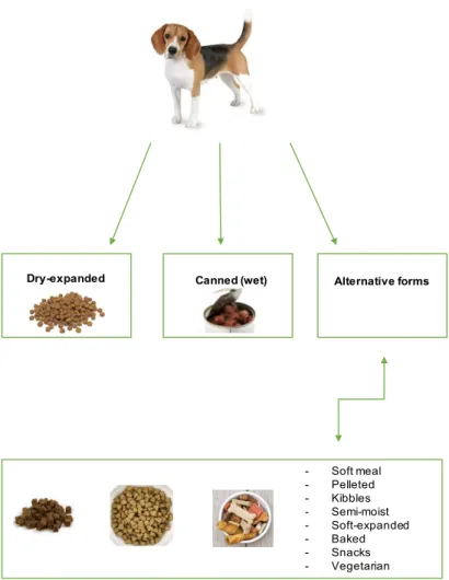
Pet food can be found in either dried or wet forms. Dry-expanded pet food typically contain 10-12% final moisture content and are formulated using cereal grains, cereal grain by-products, soybean products, animal by-products (including milk by-products), fats and oils, vitamins, and micro- and macro-minerals. Dry dog foods may be marketed as meal, pellets, kibbles, extruded, or baked products (Figure 1.1) (2).
The majority of dry pet food marketed is extrusion processed. The extrusion process typically results in moderate to high levels of gelatinization of dietary starch. The gelatinizing of the starch makes the hydrolysis (digestion) occurs in the upper gut, resulting in the more effective use of starch by the animal and reducing digestive upsets (2).
Dry dog foods typically contain 5-12.5% of fat material, with the quantity of the fat being applied post-processing. These fat levels are accomplished by spraying a fraction of the liquefied fat into the extruded product. Also, it is common to spray-coat the pet food with various protein digests and/or flavours to increase its acceptance by the pet (1).
Canned foods (wet) for dogs represents an important market share, with high annual consumption. Many of the ingredients used in canned (wet) pet foods are also used in dry-expanded and semi-moist pet foods. Although, this formulations not contain the same level of ingredients, because canned (wet) pet foods are high in moisture (usually 74-78%) given that contain higher levels of fresh or frozen meat, poultry, or fish products and animal by-products (1).
Canned (wet) pet food processing is rather complex. Usually, the meat and fat ingredients are combined with some amount of water and then appropriate amounts of dry ingredients, such as vitamins, minerals, and amino acids, are added. The mixture may be paled to begin the cooking process, or it may be added directly to the can through a mixture
device. After filling, the package is sealed with a double-seam cap and retorted. The process is essentially a heat and pressure-cooking, associated with sterilization process that ensures destruction of food-borne microorganisms (1).
Commercial pet foods have created a wide variety of pet food choices and textures and these foods are produced from basic ingredients including fish of various species, poultry, beef, and so on. It is common, for quality control, to have the main ingredient of the meat identified as part of the label (chicken, salmon, turkey giblets, for instance) (2).
Figure 1.1. Pet food categories (adapted from (1))
According to Regulation (EC) Nº 767/2009 of the European Parliament and of the Council of 13 July 2009 concerning the placing on the market and use of feeding stuffs, labelling is intended to legislation, traceability and control. Besides that, it shall provide the necessary information to purchasers, allowing them to make the best choice by taking account their needs and should be consistent, coherent, transparent and intelligible.
Dry-expanded Canned (wet) Alternative forms
- Soft meal - Pelleted - Kibbles - Semi-moist - Soft-expanded - Baked - Snacks - Vegetarian
1.1.2 Evolution of pet food
The evolution and the large variety of pet foods make the selection of a proper diet a complex and confusing process. Many pet foods are intended to provide the primary source of nutrition for a pet. So, it is extremely important to select a product able of providing optimal nutrition and promoting long-term health (1).
Corn forms, meal, pelleted, and kibbled pet foods represent a small part of the pet food industry. However, some pet owners prefer one of these forms, and they often provide a lower-cost alternative dry foods (Figure 1.1) (1).
So, new alternatives can be found in the market, such as the soft meal, aims to in a set of ingredients which has been ground or otherwise reduced in particle size. Thus, this category is comparable to an almost simple mixture of dry ingredients together with fats or oils and, eventually, a flavouring agent (1). Pelleted food is in the form of a pill and is made using conventional feed convert technology. The grain in a pelleted diet is heat prepared, allowing better starch availability because the steam conditioning process prior to pelleting does not result in significant starch gelatinization. The dry ingredients are weighed, uniformly mixed, steam conditioned, pelleted, cooled, and then coated with tallow, oils, and/or flavours prior to packaging (1).
Kibbles are often blended with raw or cooked meat to constitute the compound feed but, if properly formulated, can be a complete and balanced diet. This pet food is processed in a way similar to baked products (1).
Semi-moist pet foods usually have an intermediate moisture level of 25-35%. These products are generally cooked through an extrusion process that is similar to the process used for dry-expanded pet food (1). Semi-moist products often enclose many of the same basic ingredients as dry-expanded products. However, they usually contain meat by-product slurries incorporated prior to or during extrusion (1).
Soft-expanded (soft dry) pet foods are comparable to semi-moist in that they often contain almost high level of meat by-products and have higher levels of fats and oils than dry-expanded foods. They alter from semi-moist with respect to taking on an expanded appearance after extrusion (1).
Baked pet foods products are normally given the form of bones or some other conform shape to attract consumers. Baked pet foods are produced using a classic step of dough formation. The essence of the process contain large levels of cereal flours (> 50%) and have limited capacity to incorporate wet meat by-products (1).
A number of plant-based dry and wet foods that carry the complete and balanced label application are available for dogs and have shown to be nutritionally acceptable (1).
1.1.3 Diet formulation and feed processing
Pets can be quite independent in their feeding habits and often exhibit food preferences that may have been conditioned by previous dietary experiences (1). These preferences can lead to the selection of certain form (dry, semi-moist, canned), texture (mixed, soft-moist, dry) or flavour of food, e.g. fish. It is sometimes difficult to change a pet's feeding behaviour, even though the new feed may be nutritionally adequate. A conditioned diet preference should not be confused with nutritional requirements (1).
The most important consideration in choosing a commercial pet food for a dog is its nutrient content. This refers not only to the exact levels of nutrients in the food but also to the digestibility and availability of all essential nutrients. Nutrients can be supplied in commercial pet food through a large number of different ingredients, as discussed below (2).
The ingredients of pet food vary in form and quality, and it is this diversity that can make the selection of a suitable dog food a challenging task (2). The ingredient list is an important item that consumers examine when choosing a pet food. The terms used for ingredients in pet foods are limited to those assigned by the American Association of Food Control Officers (AAFCO) (3). This process includes oversight by the Food and Drug Administration (FDA), when applicable. The Guidelines of AAFCO and the National Research Council (NRC) of the National Academy of Sciences in the United States of America are used by industry as a basis of work and the industry is contributing to the research studies published. In addition, the industry has internal Nutritional Guidelines for cats, dogs, and rabbits (3).
As mentioned before, there are several characteristics necessary for companion animals to have complete feeding stuffs, i.e. (i) a minimum amount of nutrients necessary for energy; (ii) balanced composition, because the nutrients that are present in the ingredients must be in correct nutritional proportions; (iii) at the level of digestion, since the animals have to be able to digest the food and subsequently absorb the nutrients; (iv) quality in appearance and taste, as well as to be free from any additive that would harm the health of the animal (4).
The ingredient list of pet food may be composed by:
• Meat and animal derivatives – meats are a good sources of protein, and protein, increase the palatability of a product and presents a high digestibility; • Fish – a good source of high quality protein (the oily fish contains vitamin A,
vitamin D and omega 3);
• Dairy products and eggs – provide high quality and digestible protein with calcium and vitamins;
• Vegetables – provide a good source of vitamins, minerals and fibre; • Cereals and cereal by-products – provide an essential source of energy; • Fats and oils – provide a supply of energy and important fatty acids;
• Various sugars – sucrose, fructose and glucose, all of these are naturally present in fruit, vegetables and cereals;
• Vitamins and minerals supplements – added to ensure that pets are receiving the required daily dietary intake;
• Additives – vitamins, flavours, preservatives, antioxidants and colouring agents (4).
Minerals have different profiles of bioavailability, and many factors can affect your availability in the diet. It is important not only to have adequate amounts of each mineral relative to the animal's requirement but also to consider the relationship between minerals and the overall balance of the compound feed. Higher levels of any mineral can affect the ability of the body to absorb other minerals in the diet. For example, higher levels of cadmium, copper, and possibly vitamin D can inhibit the absorption of zinc in dogs (5). Moreover, dog needs a careful balance of calcium, phosphorus and vitamin D for strong, health bones and teeth; as well, the protein is required to maintain the body muscles.
Over the years, the food industry has been growing at the level of food technology, and the knowledge of the owners of the pets themselves, at the level of the growing will of knowledge about animal nutrition has transformed the lives of pets, as animals live longer and healthier lives (3). It should also be noted that these nutritional needs vary according to their size, age and level of physical activity (3).
1.1.4 Nutrient content and regulation of pet food safety
There are several methods for assessing the nutrient content, mainly require laboratory analysis or theoretical calculation. When pet food manufacturers formulate and produce pet foods, the level of nutrients present in the food can be determined using a conduct a laboratory analysis of the finished product (2). They include the proximate analysis, which is a commonly used panel of tests that provides information about a selected group of nutrients. The laboratory procedures involved in analysis provide the percentages of moisture, crude protein, crude fat, minerals, and fiber that are contained in the food (2).
The European Pet Food Industry Federation (FEDIAF) Nutritional Guidelines for Complete and Complementary pet food for dogs destine to confirm the basic nutrient levels under biochemical, bacteriological and organoleptic control required by dog food (3); to help
pet food manufacturers assess the nutritional value of practical pet foods for healthy animals (3); to act as the reference on pet nutrition in Europe and local authorities, consumer organizations, professionals and customers (3); to appreciate cooperation between pet food manufacturers, pet care professionals and competent authorities by providing accurately sound information on the formulation and assessment of pet foods (3); to complement FEDIAF’s Guide to Good Practice for the Manufacture of Safe Pet Foods and the FEDIAF’s Guide to Good Practice for Communication on Pet Food (3).
According to Regulation (EC) Nº 183/2005 on feed hygiene, acknowledging the importance of good hygiene practices and encouraging the development of EU and national Guides to good practice to ease the interpretation and implementation of the EU legal framework, FEDIAF created in 2017 a Guide to Good Practice allow good practices on the safety and hygiene of pet food processes and products. This document is meant to be a functional and helpful tool for pet food manufacturers to help them in grow a potent pet food safety management system and quit with safety and hygiene legal requirements as well minimum manufacturing conditions requirements with regards to facilities and equipment, personnel, production, quality control, storage, and register, which must be fulfilled by the pet food manufacturer (6).
1.2 Zinc in pet food and animal nutrition
Zinc is a chemical element of the group 12 of the Periodic Table, valence of +2, whose chemical symbol is represented by Zn, with atomic weight of 65.4 g/mol, isolated by Andreas Sigismund Marggraf, in 1746. At room temperature, Zn is a solid state with a melting point of 692.68 K and a boiling point of 1180 K. This metal is very important in the feeding of living beings, since as it interfere with the metabolism of proteins and nucleic acids, as well in the proper functioning of biological systems (7). It exists in the whole body, as zinc(II) or Zn2+, mainly as an intracellular constituent, and it is present in most tissues in relatively low concentrations (7).
Concerning its existence in dogs, Zn has been measured as total body Zn content of child puppies and young adult dogs as 23.1 and 9.5 mg kg/BW (body weight), respectively and it is acquired through the ingested food (1). As expected, the sources of zinc(II) in pet foods are quite diverse. Animal sources, particularly beef products and other types of red meat as well as whole grains, are fairly good sources of Zn(II). Despite that, most pet foods are supplemented with Zn(II) including zinc carbonate (52.1% Zn(II) in weight), zinc chloride (48% Zn(II) in weight), zinc oxide (78% Zn(II) in weight) and zinc sulphate monohydrate (36.4% Zn(II) in weight) (1). Animal sources of zinc, such as meat
and eggs, are generally more readily absorbed than are plant sources. Dietary compounds that inhibit zinc absorption include excess levels of calcium, iron, copper, fiber, and the presence of phytate (8).
1.2.1 Absorption and bioavailability of dietary zinc in dogs
The absorption of dietary zinc is generally a function of other substances in the diet that adjust its bioavailability. Most animal products and seafood are almost free of constituents that interfere with zinc absorption and amino acids derived from meat digestion may actually improve the absorption of zinc. Vegetable products are more likely to contain chemicals that interfere with zinc absorption, the most notable of these being phytate – present in many plants sources including cereals such as corn, wheat, and rice and also in oilseed meals such as soy, peanut, and sesame. Dietary phytate reduce the absorption of zinc, and this effect is aggravated by high concentrations of edible calcium (2).
Magnesium in high concentrations may also enhance the effect of phytate on zinc bioavailability, but this effect is not as important as that of calcium (2). Phytate is known to make complexes of zinc more strongly than it does with copper or manganese. High content of copper and iron can reduce the absorption and/or bioavailability of zinc, but the amounts required to do this would be unlikely to be found in standard pet food. In fact, zinc absorption in puppies’ stomach is around of 25-34% (2).
1.2.2 Dog pathologies related to zinc
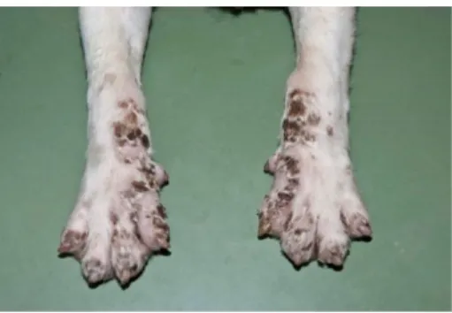
Zinc deficiency is commonly correlated with growth retardation in young animals. Other clinical signs include anorexia, testicular atrophy, impaired reproductive performance, immune system dysfunction, conjunctivitis, and the development of skin lesions. In dogs and cats, hair coat advance is usually the first clinical signs of zinc deficiency (8).
A few breeds of dogs are affected by zinc malabsorption, and variable levels of this
disorder have been reported. The most severe zinc-related pathology is lethal
acrodermatitis (Figure 1.2) (8).
This genetic disease is inherited as an autosomal recessive gene and results in an inability to absorb dietary zinc, even when high levels of the mineral are combined to the diet. At birth, affected puppies have lighter pigmentation than normal for the breed (8).
Figure 1.2. Lethal acrodermatitis (9)
Subsequently, a few reports of experimentally induced zinc deficiency in puppies and dogs are given. In one study that puppies (age not stated) fed a standard diet containing 33 mg Zn/kgand 11 g Ca/kg developed signs of Zn deficiency. Signs of zinc deficiency were not seen in dogs fed diets containing 3.0 g Ca/kg and 133 mg Zn/kg. More frequently, zinc deficiency has been reported in growing dogs being fed cereal-based, and low quality pet foods consist of high concentrations of substances that masks Zn (1).
The clinical and biochemical findings in such reports have been basically identical to those described in experimental cases of Zn deficiency; and, in all cases, affected animals have recovered when Zn supplementation was performed or a change to the diet was made with a source promoting higher bioavailability of Zn (2).
Zinc is an almost non-toxic substance, but it can sometimes become toxic when it interacts with other nutrients in the animal's body.
A few cases of inadvertent over consumption of zinc by dogs, namely by puppies, have been reported, and the clinical signs detected included acute gastroenteritis and anemia (2).
1.2.3 Regulation of zinc in pet food
Zinc is essential element in the survival of live beings, but can be harmful, in the form of oxide or sulphide. In Portugal, there are several regulations on the consumption of zinc in animals, going on to cite: “ by Commission Implementing Regulation (EU) 2016/1095 of 6 July 2016 concerning the authorization of zinc acetate dihydrate, anhydrous zinc chloride, zinc oxide, zinc sulphate heptahydrate, zinc sulphate monohydrate, zinc chelates and amino acids in hydrated form, zinc chelates and protein hydrolysates, zinc chelate with glycine in hydrated (solid) form and zinc chelate with glycine in the hydrated (liquid) form as feed additives of all the species and amending Regulations (EC) Nº 1334/2003, (EC) Nº 479/2006, (EU) Nº 335/2010 and Implementing Regulations (EU) Nº 991/2012 and (EU) Nº 636/2013, according to the European Food Safety Authority, the above-mentioned
compounds have no adverse effects on animal health or human health, and no safety concerns have arisen for users provided adequate protective measures have been missed and sources of zinc. It should also be noted that the European Authority considers that it is not necessary to establish specific post-marketing monitoring requirements. Accordingly, this Regulation states that a number of zinc compounds are considered as nutritional additives for animal feed, where the provisions on the total zinc content in compound feeding stuffs for additives authorized by this Regulation, which relate environmental impact of zinc supplementation of feeding stuffs, it is appropriate to align the maximum zinc levels in Regulation (EU) Nº 335/2010 and in Implementing Regulations (EU) Nº 991/2012 and (EU) Nº 636/2013 with the provisions of this Regulation as regards the zinc content in compound feeding stuffs.
According to Regulation (EU) 2016/1416 of 24 August 2016 of the European Parliament and of the Council, zinc can be found both in food and in the packaging itself where food is placed. The European Authority has adopted a scientific opinion on the use of the additive zinc oxide, nanoparticles, uncoated (MCA nº 1050) and zinc oxide, nanoparticles coated with [3-(methacryloxy) propyl] trimethoxysilane (MCA nº 1046). The European Authority has concluded that these additives do not migrate in nanoforms from polyolefins. In another opinion, the European Authority extended this conclusion to the migration of nanoparticles from zinc oxide to non-plasticized polymers.
It therefore stated that its safety assessment concerned the migration of soluble ionic zinc, which must comply with the specific migration limit for zinc specified in Annex II to the Regulation. For the coated form of zinc oxide, nanoparticles, migration levels of [3-(methacryloxy) propyl] trimethoxysilane should remain within the specific migration limits in force for this substance, ie 0.05 mg/kg. Therefore, the two additives should be included in the Union list.”
In Annex I of the EU Register of Feed Additives pursuant to Regulation (EC) Nº 1831/2003 feed additives including trace elements were authorized and specific EU directives that set their maximum content in feed were established.
According to Directive 2004/10/EC on the principles of good laboratory practice and the verification of their applications for tests on chemical substances, laboratories have to carry out tests on chemical products in accordance to this Directive (6).
Therefore, Regulation (EC) Nº 152/2009 laying down the methods of sampling and analysis for the official control of feed demonstrates as well specifies the method for sampling to determine constituents, additives and undesirable substances (6).
1.3 Analytical methods for determination of Zn(II) in pet food
1.3.1 Fluorimetry Theory
Fluorimetry is a technique of quantitative chemical analysis well suited to analyte determination in complex samples. This technique of optical measurement is sensitive, specificity, versatile, simple and relatively inexpensive (10). These instruments are distinguished not only by their reliability but also by the care of the accompanying operational instructions and supporting scientific reference material (11).
Thus, there are some concepts that can be approached together with this technique, such as absorption and "instantaneous" re-emission of radiant energy from a molecule or atom accompanied by a change in wavelength. This phenomenon is known as fluorescence.
When a part of light is absorbed by a molecule, the molecule is raised to an excited state. There are a variety of ways in which the excited molecule can manifest or dissipate this energy. First, if the energy absorbed exceeds the energy of the chemical bonds that hold the molecule together, the molecule may be torn apart. This is called photolysis.
Second, if an excited state is produced, the molecule is in an unstable condition and can achieve a more stable state by converting this excitation to vibrational energy, which in turn is dissipated as heat to the surroundings. Third, an excited molecule can cause the chemical transformation to occur (11). An important example of this is photosynthesis (Figure 1.3) (12).
Figure 1.3. Schematic illustration of photosynthesis (adapted from (12))
In addition to these "radiation-less" processes, there are a number of light-scattering processes in which an incident photon at one frequency is absorbed resulting in re-emission at a lower frequency. When the absorption and re-emission occur as a single transition, the phenomenon is called the Raman effect. The intensity of scattered light in the Raman effect is much lower than that observed in fluorescence (11). Finally, a molecule or atom raised
Photosynthesis
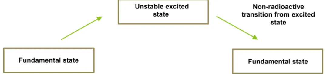
Solar energy
to an excited state by the absorption of a quantum (photon) can lose energy in separate steps, first to a lower excited state and then from the intermediate state to the ground state. If the decay and emission of radiation take place from this intermediate level in a microsecond or less, the phenomena are called fluorescence (Figure 1.4) (13).
Figure 1.4. Schematic representation of fluorescence (adapted by (13))
If the re-emission shows a persistence that lasts seconds or longer, it is termed phosphorescence. This phenomenon is characterized by the emission wavelength, where the excited molecules transit to a metastable state of lesser extent that may last longer (seconds or hours). In the remaining transition from the metastable state to the ground state, energy is released, in the form of light (11) (Figure 1.5).
Figure 1.5. Schematic representation of phosphorescence (adapted by (13))
1.3.2 Molecular Fluorescence Techniques
Molecular fluorescence involves the emission of radiation as excited electrons return to the ground state. The wavelengths of radiation emitted are different from those absorbed and are useful in the identification of a molecule. The intensity of the emitted radiation can be used in quantitative methods and the wavelength of maximum emission can be used qualitatively (14).
A considerable number of compounds demonstrate fluorescence and it provides the basis of very sensitive methods of quantitation. Fluorescent compounds often contain
Fundamental state Unstable excited state Fundamental state Unstable excited state Fundamental state Non-radioactive transition from excited
state
multiple conjugated bond systems with the associated delocalized π electrons, and the presence of electron-donating groups, such as amine and hydroxyl, increases the possibility of fluorescence. Most molecules that present fluorescence have rigid, planar structures, such as pyrene (Figure 1.6) (14).
Figure 1.6. Pyrene structure
1.3.3 Instrument Design
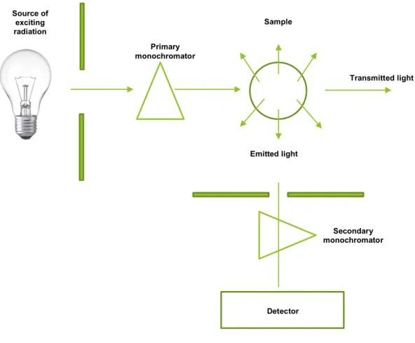
Fluorescent radiation is emitted in all directions and this feature is adequate to avoid interferences that may be caused by the transmission of incident radiation by the sample (14). In fact, by positioning the detection system at a right angle in relation to the excitation light source, only fluoresce radiation is detected. Some instruments are designed to measure "front face" fluorescence, i.e. the radiation that is emitted along a light path at an acute angle to the incident radiation. Such fluoresce measurements are comparable to reflectance measurements (14).
Despite the measurement of the emitted radiation by these means, it is still possible for scattered or reflected incident radiation to reach the detector. To prevent this, fluorimeters require a filter or a second monochromatic system between the sample and the detector. Many simple fluorimeters use filters as both primary and second wavelength selectors and other instruments (such as spectrofluorometers) use true optical monochromators for both components (14).
Other instruments incorporate a simple cut-off filter system for the emitted radiation while retaining the optical monochromatic for the excitation radiation. Because the wavelengths of both excitation and emission are characteristic of the molecule, it is debatable which the excitation monochromator is the most important, it allows greater sensitivity and discrimination of the target molecule (Figure 1.7) (14).
Figure 1.7. Fluorimeter design (adapted from (14))
1.3.4 Fluorescent probes to detect zinc in food
In the large arsenal of fluorescent Zn2+ sensors, particularly within biological systems, there are probes in all three main classes: small-molecule probes, genetically encoded sensors, and hybrid probes. Small-molecule sensors constitute the largest class by far, and this group can be further subdivided into two categories: intensity-based probes, where Zn2+ binding induces an increase in fluorescence intensity, or ratiometric probes,
where Zn2+ binding shifts the excitation and/or emission wavelength (15).
There are multiple families of genetically encoded Zn2+ sensors based on the FRET (Förster resonance energy transfer) between two fluorescent proteins, and many of these have been targeted to different cellular locations. Finally, there are a handful of hybrid probes, which as the name suggests are a combination of the aforementioned classes and have both genetically encoded and exogenous elements. These hybrid probes include small molecules with targeting groups that interact with specific enzymes and genetically encoded proteins whose signal output is modulated by binding of a small-molecule fluorophore (15). There are several probes that allow analysts to detect zinc in various
Detector Sample Primary monochromator Source of exciting radiation Secondary monochromator Transmitted light Emitted light
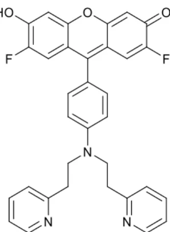
foods, such as FluoZin-1 (2,2'-((4-(2,7-difluoro-3,6-dihydroxy-4aH-xanthen-9-yl)-3-methoxyphenyl)azanediyl)diacetic acid), Newport Green DCF (potassium 5-((4-(bis(pyridin-2-ylmethyl)amino)phenyl)carbamoyl)-2-(2-chloro-7-fluoro-6-oxido-3-oxo-3H-xanthen-9-yl) benzoate), Newport Green PDX (9-(4-(bis(2-(pyridin-2-yl)ethyl)amino)phenyl)-2,7-difluoro-6-hydroxy-3H-xanthen-3-one), and FuraZin-1 (2-{6-[bis(carboxymethyl)amino]-5-methoxy-1-benzofuran-2-yl}-1,3-oxazole-5-carboxylic acid) (Figure 1.8).
Figure 1.8. FluoZin-1 structure
The FluoZin-1 can be used for quantitation of zinc with excitation and emission fluorescence at 495 nm and 515 nm, respectively (16).
This fluorescence probe is sensitive to ions at oxidation state +2, such as nickel, lead, cadmium, mercury and zinc and the constant of dissociation of its complex with Zn2+
in buffer pH 7.0 is 8 µM. The fluorescence obtained by the complex between these ions and FluoZin-1 is represented in Figure 1.9, showing its relevant sensitivity to Zn(II) and Cd(II) (16).
Figure 1.9. Sensibility of metal ions with FluoZin-1 using the same concentration of probe and two levels for ion concentration (adapted from (16))
0 50 100 150 200 250 300
Ni(II) Zn(II) Cd(II) Hg(II) Pb(II)
(F -F0 )/ F0 Metal ions 1 µM 100 µM
There are also some studies with FluoZin-1 probe concerning the determination of stability constants of Cu(I), Cd(II) and Zn(II) complexes with thiols using fluorescent probes. The authors of this study performed several fluorimetric competing-ligand titrations, used to measure stability constants of Zn(II), Cd(II) and Cu(I) complexes of cysteine and glutathione.
Cu(I) stability constants were also determined for the dipeptides Arginine-Cys and Glutamine-Cys which are produced by a kelp under copper tension. Fluorescent ionic
indicators FluoZin-1 and BTC
(N-[2-[(acetyloxy)methoxy]-2-oxoethyl]-N-[3-(2-benzothiazolyl)-6-[2-[2-[bis[2-[(acetyloxy)methoxy]-2-oxoethyl]amino]-5-methylphenoxy] ethoxy]-2-oxo-2H-1-benzopyran-7-yl]-glycine)were used as competing binders in titrations involving Zn(II) and Cd(II). The measured the stability constants of the Cd(II) and Zn(II) complexes were consistent with the previous work, validating the previous method and assumptions (17).
The applicability of the proposed competition assay was further demonstrated using FluoZin-1 in a binding study for Zn2+ and bovine serum albumin. This competitive fluorimetric assay provided a sensitive, simple and generic approach for affinity estimation of metal and biomolecule binding (18).
Another study was about a ratiometric displacement approach to Cu(II) sensing by fluorescence. This is a new design of Cu(II) sensor with a selective and sensitive methodology for allows for quantitative measurement of Cu(II) in aqueous solution, under physiologically relevant conditions. To prevent fluorescence quenching by Cu(II) and to achieve a ratiometric response to metal binding, the authors devised a sensor system that separates the sensing and the signalling events. Well, two different fluorogenic ligands capable of metal chelation are employed whereby one of the ligands binds Cu(II), while the other produces a fluorescent signal to report the binding event (19).
Other work described new fluorescent indicators with nanomolar to micromolar affinities for Zn2+ that have been synthesized in wavelengths from UV to the far red for
application in cells. Therefore, the UV light-excited and wavelength indicators are ratiometric and non-ratiometric, respectively. Furthermore, this indicators present large and pH-independent fluorescence increases with increasing zinc(II) concentrations. The probe was titrated with unbuffered Zn2+ solutions at pH 7.0; the zero Zn2+ measurements were
made in the presence of the Zn2+ chelator, N,N,N’,N'-tetra(2-picolyl) ethylenediamine. In
vitro, FuraZin-1 exhibits a fluorescence excitation wavelength shift from 378 to 330 nm in
the presence of increasing [Zn2+] (emission monitored at 510 nm), which allows for
ratiometric ion measurements (20).
The Newport Green DCF probe can be used for excitation and emission fluorescence of 505 nm and 535 nm, respectively (Figure 1.10) (16).
This probe is sensitive to certain ions at divalent state such as cadmium, zinc, cobalt, nickel, with the constant of dissociation of the complex with Zn2+ in buffer pH 7.0 as 1 µM (16).
Figure 1.10. Newport Green DCF structure
The fluorescence obtained by the complex between these ions and Newport Green DCF is represented in Figure 1.11, showing its sensitivity to Zn(II), but with possible interference from Ni(II) and Co(II) (16).
Figure 1.11. Sensibility of metal ions with Newport Green DCF using the same concentration of probe and two levels for ion concentration (adapted from (16))
There are some studies on application of Newport Green DCF in biological tissues, namely concerning zinc and lead accumulation characteristics and in vivo distribution of Zn2+ in the hyperaccumulator Noccaea caerulescens. This study demonstrated that the
0 2 4 6 8 10 12 14
Co(II) Ni(II) Zn(II) Cd(II)
(F -F0 )/ F0 Metal ions 1 µM 100 µM
younger plants were more tolerant to Zn2+ and Pb2+ than the older plants. The accumulation
of Zn2+ in shoots or roots was not significantly affected by treatment regime or plant age. Pb accumulated mainly in the roots (0.16 - 0.23 wt% dry mass), confirming substantial tolerance to Pb. The concentration of phosphorus in older plant shoots decreased ∼ 25% in the plants treated with Zn2+, but enhanced ∼ 26% in the plants treated with Zn2+ and Pb2+.
The high ratio of Zn to Pb in both fresh and dry leaves is suggestive of the formation of insoluble Zn-salts. The Zn2+ distribution in living cells was examined using the fluorescent
probe, showing that Zn2+ was mainly located in the apoplastic space of the leaf epidermal
cells. Hence, this probe in combination with laser confocal microscopy proved a useful tool for elucidating cellular and tissue-level distribution of Zn2+ in living plant cells at high resolution (21).
Similarly, to the probe refer above, Newport Green PDX can be used for excitation and emission fluorescence of 505 nm and 535 nm, respectively (Figure 1.12). Newport Green PDX is also sensitive to divalent ions such as nickel(II), mercury(II), cadmium(II), zinc(II), with the constant of dissociation of probe complex to Zn2+ in buffer pH 7.0 as 1 µM
(16).
Figure 1.12. Newport Green PDX structure
The fluorescence elicited by the complex between these ions and Newport Green PDX is represented in Figure 1.13, showing its selectivity to Zn(II), despite some response towards Hg(II) (16).
Figure 1.13. Sensibility of metal ions with Newport Green PDX using the same concentration of probe and two levels for ion concentration (adapted from (16))
It should be stressed that, in order to work with aqueous solutions, it is necessary to add a micellar structure because this probe is not soluble in water (16). There are some studies about this probe, such as determination of Islet viability using a zinc-specific fluorescent dye and a semi-automated assessment method in cells of human pancreatic. Newport Green PDX is a zinc-specific fluorescent dye that reacts with zinc rich β-cells. It was possible to verify the difference between the zinc bond in relation to the probe under study, as well as with fluorescein diacetate, because fluorescence intensities in response to zinc concentrations of 0 to 100 µmol/L demonstrated that the intensity of the probe increased with the increase of zinc concentrations mentioned above, but with fluorescein diacetate no differences in fluorescence intensity were observed (22).
Other work reports the combined action of zinc in neuronal morphology as this metal ion was highly enriched with the postsynaptic density. Zinc-sensitive ProSAP1/Shank2 or ProSAP2/Shank3 leads to increased synapse density. Although, all of them caused a decrease upon knockdown models. They are comparable the efficient of 2-[2-methyl-8-[(4-methylphenyl)sulfonylamino]quinolin-6-yl]oxyacetic acid (zinquin) and Newport Green PDX. The results show that the detected zinc signal localizes with postsynaptic markers. This probe was used to confirm the data with zinquin that postsynaptic structures contain zinc (23).
The FuraZin-1 can be used upon excitation and emission fluorescence of 505 nm and 535 nm, respectively (Figure 1.14). This probe is sensitive to certain ions such as lead, zinc, and cadmium, with the dissociation constant of the complex with Zn2+ in buffer pH 7.0
as 1 µM. 0 5 10 15 20 25 30
Hg(II) Ni(II) Zn(II) Cd(II)
(F -F0 )/ F0 Metal ions 1 µM 100 µM
Figure 1.14. FuraZin-1 structure
The fluorescence elicited by the complex between these ions and FuraZin-1 is represented in Figure 1.15, showing its selectivity to Zn(II) and Cd(II), also responding with less intensity towards Pb(II) (16).
Figure 1.15. Sensibility of metal ions with FuraZin-1 using the same concentration of probe and two levels for ion concentration (adapted from (16))
It was possible to observe zinc as a neurotoxin in neuronal cell culture, using FuraZin-1. Using ionosphere sodium of pyrithione (the zinc-specific ionosphere of zinc concentrations in cultured neurons), it was possible to observe that this probe is quite selective for these cells from a probe concentration of 3 M (24).
Another study using this probe is the increase in the expression of ZRC1 (protein of
Saccharomyces cerevisiae of fungi kingdom that has been implicated in the storage and
detoxification of excess zinc in the vacuole) expression in a novel mechanism of zinc homeostasis and stress tolerance. The results obtain show that ZRC1 and its induction in
0 0.2 0.4 0.6 0.8 1 1.2 1.4 1.6 1.8 2
Pb(II) Zn(II) Cd(II)
(F -F0 )/ F0 Metal ions 1 µM 100 µM
zinc-limited cells are required for resistance to this zinc shock. Therefore, FuraZin-1 was a suitable indicator of fluorescence of vascular zinc levels indicated that ZRC1 is required for the rapid transport of zinc in vacuole during zinc shock (25).
This probe was also applied to cultured cortical and hippocampal neurons when intracellular pH drops from 6.6 to 6.1, yet nuclear intracellular stores release micromolar amounts of zinc into the cytosol. Therefore, Zn2+ releasing stores were not mitochondria or acidic organelles but rather intracellular Zn2+ ligands. Recording, ligands chelated zinc and
upon acidification were releasing it into the medium. When pH was decrease from 6.6 to 6.1, only the zinc-cysteine complexes accelerated the rate of zinc release. These complex may represent the stores responsible for an acid-induced intracellular zinc release (25).
So, despite the successful examples presented upon using these fluorescent probes in biological media, no methods for quantification of zinc in pet foods using these probes was not reported.
Objective
The aim of this work was a development of a methodology for the determination of zinc in pet foods samples, after acid digestion, based on the conversion of all zinc forms to zinc(II).
CHAPTER 2
CHAPTER 2 – Materials and Methods
In this chapter, reagents, standards and samples that were used in all laboratory work are described, as well as the techniques used for the determination of zinc(II) in pet food samples with fluorescent probes. In addition, this chapter includes laboratory procedures as well as the description of studies performed to assess the effect of different parameters of the method.
2.1 Reagents and solutions
All chemicals used in this work were of analytical reagent grade with no further purification. For the preparation of all solutions, water from Milli-Q system resistivity > 18 MΩ cm (Gottingen, Germany) and dimethyl sulfoxide (DMSO) from Merck (Darmstadt, Germany) were used.
The fluorescent reagents under study were Newport Green DCF (potassium 5 -((4- (bis(pyridin-2-ylmethyl)amino)phenyl)carbamoyl)-2-(2-chloro-7-fluoro-6-oxido-3-oxo-3H-xanthen-9-yl)benzoate) dipotassium salt, cell impermeant (catalog number N7990) and FluoZin-1(2,2’-((4-(2,7-difluoro-3,6-dihydroxy-4aH-xanthen-9-yl)-3-methoxypjenyl)azanedi- yl)diacetic acid), tripotassium salt, cell impermeant (catalog number F24180) acquired from Invitrogen-Thermo Fisher Scientific (Massachusetts, USA). Probe stock standard solutions were prepared by dissolving the solid in 1 mL DMSO at 1 mg/mL (Newport Green DCF) and at 0.5 mg/mL (FluoZin-1).
The standard stock solution of Zn(II) (1.00 mg/mL) in 4% nitric acid was obtained by SCP Science (Baie-d’Urfé, Canada) (item 140-001-301, 125 mL, UN Code 3264).
Moreover, the stock solution of nitric acid 10 mM, used to prepare the zinc(II) standard working solutions, was prepared by diluting commercial nitric acid 70% (w/w) from Sigma-Aldrich (St. Louis, USA).
The working standard solutions were obtained by dilution of the stock solution using glass pipettes, micropipettes and volumetric flasks (class A) of different volumes. Micropipettes (models P20, P100, P1000 and P5000 with corresponding maximum capacities of 20, 100, 1000 and 5000 µL) and multichannel micropipette (model P200 with corresponding maximum capacity of 200 µL) were used.
Whenever necessary, pH of solutions was measured using a combined glass pH electrode from Crison (Barcelona, Spain) and millivoltmeter model RE357TX from EDT direction Electrochemistry Products (Dover, UK).
hydrogen carbonate/sodium carbonate (pH 10.0), prepared from reagents acquired from Sigma-Aldrich.
2.2 Samples
For the completion of this scientific work several tests were carried out with three pet food samples, namely samples A, B and C.
These samples were solubilized by microwave-assisted acid digestion using an MLS 1200 Mega high-performance microwave digestion unit (Milestone, Sorisole, Italy) equipped with an HPR-1000/10 S rotor. After weighing the sample using a plastic spatula, 3 mL of HNO3 and 1 mL of H2O2 were added to each polytetrafluoroethylene digestion vessel. The
samples were subsequently submitted to a microwave heating program of 250 W for 1 min, 0 W for 1 min, 250 W for 5 min, 400 W for 5 min and, finally, 650 W for 5 min. The vessels were then allowed to cool to room temperature. Thereafter, the content was transferred to 25 mL polypropylene volumetric flasks and water was added to bring up to total volume. This digest was analysed by the developed fluorimetric assay. A blank constituted by 500 µL of water was included in each digestion run. Each sample was digested in duplicate, after a digestion pre-treatment procedure, where the samples were oven dried at 65 °C to constant weight and then ground in a 1 mm sieve mill (26).
For comparison purposes, zinc present in sample digests was also determined by inductively coupled plasma mass spectrometry (ICP-MS) using an iCAP Q™ (Thermo Fisher Scientific, Schwerte, Germany) instrument, equipped with a MicroMist™ nebulizer, a Peltier cooled cyclonic spray chamber, a standard quartz torch and nickel skimmer and sampling cones. High purity (99.9997%) Ar (Gasin II, Leça da Palmeira, Portugal) was used as the nebulizer and plasma gas. The ICP-MS operated under the following conditions: RF power 1550 W; auxiliary Ar flow rate 0.80 L/min; nebulizer flow rate 1.08 L/min and plasma flow rate 14 L/min. Zinc was determined as 66Zn isotopes as described in detail elsewhere (26). The analysed samples contained 10.8 ± 0.5 mg/L (sample A), 7.48 ± 0.88 mg/L (sample B) and 7.54 ± 0.33 mg/L (sample C) of zinc(II) concentrations in pet food digest, corresponding to 526 ± 14 mg Zn/kg, 342 ± 32 mg Zn/kg, and 357 ± 15 mg Zn/kg dried pet food (26).
2.3 Apparatus
For the implementation of the methodology for determination of zinc(II) in pet food samples, a fluorimeter (Model FP-6500, Jasco, Easton, USA) was applied to analyse the excitation and emission spectra of the probes under study.
In the microplate reader (Model Cytation 3, Bio-Tek, Winooski, USA), through the
Gen 5 2.06 reader software program, different analysis protocols were performed,
depending on the emission and excitation wavelengths of the probes, with the objective of determining the fluorescence intensity of solutions placed in 96-well microplates, suitable for fluorescence measurements (black, opaque wells, ref. 10588885, Thermo Fisher Scientific, Massachusetts, USA).
2.4 Preliminary procedure to evaluate the response of the probes
under study
For the probes under study to be maintained in suitable storage conditions (fluorescence stability during six months), 100 µL portions were aliquoted, subsequently frozen at - 5 to - 20 ºC, and protected from light.
From the stock solution of Newport Green DCF (1 mg/mL, 1.26 mM) intermediate solutions of 30.0, 3.0, 1.5 and 0.75 µM were prepared. From the stock solution of FluoZin-1 (0.5 mg/mL, 0.83 mM), intermediate solutions of 30.0, 5.0, 2.5, and FluoZin-1.25 µM were also prepared. All intermediate solutions were prepared in buffer or ultrapure water, depending on the experimental work performed.
In order to prepare the acetic acid/acetate buffer solution (1000 mM, pH 3.9), 2.45 g of NaCH3COO, were weighed into a plastic cylindrical flask, dissolved with ca. 20 mL of
water and transferred to a 50 mL volumetric flask, where the volume was made up with water. The acetic acid solution was prepared by pipetting 2.9 mL of glacial acetic acid, into a 50 mL volumetric flask, making up the volume with water. Afterwards, the two solutions were mixed in a glass beaker and the pH was measured, providing a value of 4.0.
From this solution, the working buffer solution at 5 mM was prepared by 1:20 dilution,
i.e. by placing 0.5 mL of buffer solution in a plastic cylindrical flask and by adding 9.5 mL of
water.
For preparing buffer solutions at pH 7.0 and pH 10.0, the procedure was similar with the exception of the reagents used. In the case of pH 7.0, a 1000 mM K2HPO4 / KH2PO4
different plastic beakers, dissolved with ca. 20 mL of water and transferred to a 50 mL volumetric flask, where the volume was made up with water. The two solutions were combined (25 mL of K2HPO4 and 25 mL of KH2PO4 solution) in a 100 mL volumetric flask,
and the volume was also made up with water.
In the case of pH 10.0, a 1000 mM buffer solution was prepared by weighing 210 mg of NaHCO3 in plastic beaker and by adding 10.7 mL of 100 mM NaOH, and by
transferring it to a 100 mL volumetric flask, where the volume was made up with water. The 100 mM NaOH solution, was prepared by weighting 0.080 g NaOH 1 M in 20 mL of water. That cup was placed on ice, with water to the final volume of 20 mL.
For the study of the fluorescent probes several standard solutions of zinc(II) were prepared. For the preparation of the working solutions of zinc(II) (20 µg/L and 1 µg/L) a nitric acid solutions at 10 mM HNO3 was prepared. Thus, 1280 µL of 70% (w/w) nitric acid with a
concentration of 15.4 M was added to water aiming a total volume of 20 mL, providing a nitric acid solution with a concentration of 1 M. After this, from the 1 M solution of nitric acid, 200 µL is diluted to a total volume of 20 mL, thus leaving a solution of 10 mM nitric acid.
For the first zinc(II) intermediate solution at 20 µg/L, the initial standard solution of zinc(II) was diluted, in an eppendorf by placing 20 µL of standard zinc(II) solution and by adding 980 µL of 10 mM HNO3. For the second zinc(II) intermediate solution at 1 µg/L, the
intermediate solution 20 µg/L of zinc(II) was also diluted in an eppendorf, by placing 50 µL of standard zinc(II) solution and by adding 950 µL of 10 mM HNO3.
Subsequently, several dilutions were made from the second zinc(II) intermediate - 50, 200 and 500 µg/L.
Microplate analysis was perfomed by placing the solutions each well (in triplicate) as follows: 1) 160 µL of buffer 5 mM; 2) 80 µL of zinc(II) standard solution of according to the dilutions prepared; 3) 20 µL of intermediate solution probe, as can be seen in Figure 2.1.
It should be noted that three controls were performed in which 160 µL of 5 mM buffer solution, 80 µL of 1 µg/mL zinc(II) solution and 20 µL of 5 mM buffer solution were placed (in order to control the fluorescence intensity value of the zinc solution); 160 µL of 5 mM buffer solution, 80 µL of 10 mM HNO3 solution and 20 µL of 1.5 µM intermediate probe
solution (for the purpose of controlling the fluorescence intensity value of the probe); 160 µL of 5 mM buffer solution, 80 µL of 10 mM HNO3 solution and 20 µL of 5 mM buffer solution
(with the aim of controlling the fluorescence intensity value of the buffer solution); respectively.
After that, readings were taken, at 5, 15 and 30 minutes, depending on the study implemented.
It should be recalled that in the case of the Newport Green DCF probe the excitation wavelength used was 500 nm and the emission wavelength 535 nm. As for the FluoZin-1 probe, the excitation wavelength was 484 nm and the emission wavelength 520 nm.
Figure 2.1. Schematic representation of the experimental procedure
2.5 Determination of zinc in pet food
For a more detailed study of the FluoZin-1 probe several fluorescence intensity assays were performed as well as their application in pet food samples, subsequently digested, as well as assays for repeatability, reproducibility, pH stability and fluorescence intensity stability over time, namely four hours (Figure 2.2).
Figure 2.2. Schematic representation of the determination of zinc(II) in pet food
Zn(II) stock solution Zn(II) intermediate solution 1 Zn(II) intermediate solution 2 Zn(II) work solutions 10 mM HNO3 Probe stock solution 5 mM Buffer Probe intermediate solution 1 Probe intermediate solution 2 Blank
solution solution Sample
Adjust the pH of the solutions
20 mLvolumetric flask 1:200 dilution of blank and
sample
Direct measurement with
10 mM HNO3
Fortification with Zn(II) intermediate solution Probe intermediate solution 2 Fluorescence determination
CHAPTER 3
CHAPTER 3 – Results and Discussion
3.1 Results using Newport Green DCF probe
Potassium
5-((4-(bis(pyridin-2-ylmethyl)amino)phenyl)carbamoyl)-2-(2-chloro-7-fluoro-6-oxido-3-oxo-3H-xanthen-9-yl)benzoate (C39H24Cl2K2N4O6), also known as Newport
Green DCF, has been applied for assays, at pH 7.0, in several cells of the human body,
namely in neuronal cells.The probe concentration used in these studies ranged from 5 to
20 µM, prepared previously in water (27).
Therefore, the pH was set at 7.0 and the influence of the probe concentration was studied. For each probe concentration tested, the fluorescence value was similar regardless of the concentration of zinc(II) present. For the concentration of 0.75 μM, the mean fluorescence intensity was 786, increasing to 1191 and 2056 for the probe concentrations of 1.5 and 3.0 μM, respectively (Table 3.1). Actually, it has been found that there is an increase in signal with probe concentration (linear ratio y = 566 x + 353, where y is the fluorescence intensity and x is the probe concentration), but not for different concentrations of zinc(II), which could mean that the complexation reaction between fluorescent probe and Zn(II) was not taking place.
Table 3.1. Fluorescence intensity using different concentrations of Newport Green DCF in buffer solution at pH 7.0 [Newport Green DCF] (µM) [Zn(II)] (µg/L) 0.75 1.5 3.0 0 755 ± 21 1190 ± 29 1976 ± 131 50 848 ± 28 1187 ± 26 2070 ± 98 200 820 ± 12 1240 ± 68 2070 ± 47 500 716 ± 67 1195 ± 28 2228 ± 389 1000 792 ± 26 1145 ± 55 1938 ± 222
Since no difference was found for different zinc(II) concentrations, the pH of the reaction medium was changed by using other buffer solutions (acetic acid / acetate buffer at pH 4.0 and hydrogen carbonate / carbonate buffer at pH 10.0) and maintaining the concentration of probe at 1.5 μM. (Table 3.2).
Table 3.2. Fluorescence intensity results at pH 4.0 and pH 10.0, using 1.5 µM of Newport Green DCF pH [Zn(II)] (µg/L) 4.0 10.0 0 1306 ± 90 1407 ± 48 50 1336 ± 36 1506 ± 46 200 1331 ± 64 1477 ± 9 500 1380 ± 29 1486 ± 21 1000 1328 ± 20 1417 ± 19
Again, there was no change in fluorescence for increasing zinc(II) concentrations. Concerning the fluorescence mean value, an increase of about 9% at pH 4.0 and of about 22% at pH 10.0 was observed when compared to the results obtained at pH 7.0.
Next, it was hypothesized that no change in fluorescence occurred at different concentrations of zinc(II) due to lack of buffering in the standard solution, initially prepared in 10 mM nitric acid. Experiments were then performed with the preparation of the standards in acetic acid/acetate buffer at pH 4.0. There was an increase in the mean fluorescence intensity of 37% and 98%, depending on the amount of probe used (0.75 μM and 1.5 μM). However, the fluorescence signal was independent of the zinc(II) concentration (Tables 3.3 and 3.4).
Table 3.3. Fluorescence intensity of Newport Green probe at pH 4.0, using zinc(II) stock solution in 5 mM buffer [Newport Green DCF] (µM) [Zn(II)] (µg/L) 0.75 1.5 0 570 ± 9 1633 ± 40 50 1260 ± 19 1807 ± 40 200 1271 ± 16 1781 ± 67 500 1248 ± 66 1906 ± 30 1000 1185 ± 59 1818 ± 110