Universidade do Minho Escola de Ciências da Saúde
Joana Sofia da Cruz Pereira
T
HE EFFECTS OF ANTIPSYCHOTICS IN ASTROCYTIC PLASTICITY AND SOCIAL BEHAVIOR IN AN ANIMAL MODEL OF SCHIZOPHRENIADissertação de Mestrado Mestrado em Ciências da Saúde
Trabalho efetuado sob a orientação do:
Professor Doutor João Miguel Seiça Bessa Peixoto
iii
“If my mind can conceive it, and my heart can believe it – then I can achieve it. “ Muhammad Ali
v
A
CKNOWLEDGEMENTS/A
GRADECIMENTOSEsta tese representa dois anos não só de momentos de trabalho, concentração, dedicação, mas também de momentos de felicidade e até de nervosismo. Como tal, agradeço a todos aqueles que partilharam estes momentos comigo.
Ao meu orientador, João Bessa, por todo o conhecimento, pelas palavras, pela tranquilidade e sobretudo, pela confiança transmitida, por me fazer querer ser melhor.
À minha co-orientadora, Mónica Morais, por toda a ajuda, por todo o apoio, pela paciência, por tudo o que me ensinaste. Guardarei sempre com carinho as infinitas horas de confocal e biotério, e todas as peripécias que partilhamos. Agradeço ainda ao Carlos Portugal, pela preciosa ajuda estatística e ainda todas as gargalhadas.
À Sónia Gomes, pelo apoio, por tudo o que me ensinaste no biotério e pela tua amabilidade.
Às meninas fit do laboratório, Cátia, Cláudia, Francisca e Vanessa, por todos os momentos de
descontração.
Ao Master Gang, pelos almoços, pelos lanches, pelos cafés, pelas gargalhadas e pelas lágrimas. Um grande obrigada à Bárbara, ao Eduardo, à Leonor, à Margarida, ao Mendanha, à Sofia. Um obrigada especial à Ana, à Diana e à Sara, pelos vídeos, pelas imagens no projector, pelos desabafos partilhados, pela dança, pelas piadas e pelo heart and brain.
À Catarina e à Susana, por serem as amigas de sempre, pelo apoio apesar da distância. A nossa amizade já leva muitos anos, e espero acumular muitos mais.
Ao Miguel, o meu partner in crime, o meu melhor amigo, a minha criança favorita. Não há palavras suficientes para te agradecer todo o apoio que sempre me deste. Pelos “mesmo à Joana”. Por todas as aventuras que partilhamos e as que vêm aí.
À minha família, o meu pilar. Aos meus avôs, por terem contribuído para uma infância que não poderia ter sido mais feliz. Ao meu irmão, por ser um chato de primeira, que nunca me dá um elogio com as palavras que toda a gente usa. Por ser tudo aquilo que eu pedi durante nove anos e muito mais. Aos meus pais, pelo amor, pelo apoio incondicional. Uma vida não chega para retribuir tudo o que me dão. A vocês dedico tudo o que faço.
vii
A
BSTRACTThe effects of antipsychotics in astrocytic plasticity and social behavior in an animal model of schizophrenia
Schizophrenia is a debilitating psychiatric disorder that affects approximately 1% of the population and is characterized by psychotic events, as well as cognitive and negative symptoms, namely impairments in social interaction. The discovery of drugs that are able to reduce the psychotic symptoms of the disorder helped to shed some light on the mechanisms involved in the etiology of schizophrenia. However, the high complexity of the disease along with the adverse side effects associated with antipsychotic drugs seem to build a long road to the desired treatment of this severe disorder. Functional and structural studies have reported abnormalities in multiple brain areas, namely the hippocampus, one of the particular regions in which the proliferation of stem cells occurs in the adult brain. Importantly, impairments in adult neurogenesis have been described in the brains of schizophrenic patients. However, the possible role of newly formed glial cells and astrocytic plasticity in the etiology of this disorder and in the effects of antipsychotic drugs remains largely unknown.
In the present study, a neurodevelopmental animal model of schizophrenia based in the prenatal exposure to alkylating agent methylazoxymethanol (MAM) was used to evaluate the impact of different classes of antipsychotic (AP) drugs in gliogenesis, astrocytic remodeling and social behavior in rats. Animals exposed to MAM in the prenatal period (GD17) were treated in adulthood with a first generation AP (haloperidol), two second generation AP´s (clozapine and risperidone) and a third generation AP (aripiprazole). The results revealed significant impairments in social behavior, gliogenesis and astrocytic morphology in animals prenatally exposed to MAM. While chronic treatment with aripiprazole was able to revert the impairments in social behavior and restore the levels of gliogenesis, the first and second generation AP´s haloperidol, clozapine and risperidone revealed a specific effect in restoring the detrimental effects of MAM exposure in astrocytic morphology and complexity. However, no significant differences in the expression of astrocytic-related genes were observed in the hippocampus.
In conclusion, these results suggest that gliogenesis and astrocytic plasticity may play a key role in social behavior in the context of schizophrenia. Furthermore, the effects of different classes of AP drugs in these phenomena may pave a new way in the treatment of the negative symptoms of schizophrenia.
ix
R
ESUMOEfeitos de antipsicóticos em plasticidade astrocítica e comportamento social num modelo animal de esquizofrenia
A esquizofrenia é uma doença psiquiátrica debilitante, que afeta aproximadamente 1% da população e que é caracterizada por fenómenos psicóticos, bem como por sintomas cognitivos e negativos, nomeadamente distúrbios em interação social. A descoberta de fármacos que reduzem os sintomas psicóticos da doença contribuiu para o avanço no conhecimento de mecanismos afetados no contexto da esquizofrenia. No entanto, a alta complexidade da doença aliada aos efeitos secundários adversos dos antipsicóticos afastam a possibilidade de um tratamento eficaz para esta grave doença. Estudos funcionais e estruturais mostram que várias áreas estão afetadas nesta doença, nomeadamente o hipocampo, uma das poucas estruturas onde a proliferação de células estaminais ocorre no cérebro adulto. Curiosamente, já foi mostrado que a neurogénese adulta encontra-se afetada nos cérebros de pacientes com esquizofrenia. No entanto, o papel de células gliais recentemente formadas e plasticidade de astrócitos na origem desta doença bem como os efeitos de antipsicóticos a este nível é ainda altamente desconhecido.
Neste estudo, o modelo animal de esquizofrenia de injeção pré-natal do agente alquilante acetato de metilazoximetanol (MAM), foi usado para avaliar o impacto de antipsicóticos (AP) de diferentes classes em gliogénese, remodelação de astrócitos maduros e comportamento social em rato. Animais expostos a MAM durante o período pré-natal (GD17) foram tratados em idade adulta com um AP de primeira geração (haloperidol), dois de AP’s de segunda geração (clozapina e risperidona) e um AP de terceira geração (aripiprazole). Os resultados mostram distúrbios significativos ao nível do comportamento social, da gliogénese e da morfologia astrocítica em animais expostos a MAM durante o período pré-natal. Embora o tratamento crónico com aripiprazole tenha permitido reverter os distúrbios em comportamento social e recuperar os níveis gliogénese, os AP’s de primeira e segunda geração haloperidol, clozapina e risperidona mostraram um efeito específico relativamente à recuperação dos efeitos prejudiciais da exposição a MAM na morfologia e complexidade astrocítica. No entanto, não foram encontradas diferenças significativas relativamente à expressão de genes relacionados com astrócitos no hipocampo.
Em conclusão, estes resultados indicam que a gliogénese e plasticidade astrocítica parecem estar a ter um papel fundamental ao nível de comportamento social no contexto da esquizofrenia. Além disso, os efeitos de fármacos AP de diferentes classes nestes fenómenos podem ajudar a construir um novo caminho para o tratamento dos sintomas negativos da esquizofrenia.
xi
I
NDEX Acknowledgements/Agradecimentos ... v Abstract... vii Resumo... ix Index ………..xiList of Figures ... xiii
List of Tables ... xv Abbreviations ... xvii 1. Introduction ... 1 1.1. Schizophrenia ... 3 1.1.1.Risk Factors ... 3 1.2. Pathophysiology of Schizophrenia ... 4 1.2.1. Dopamine………4 1.2.2 Glutamate……… ... 5 1.2.3.Antipsychotic treatment ... 6
1.3. Structural and Functional Morphological Abnormalities in Schizophrenia ... 10
1.3.1.Structural abnormalities in frontotemporal and limbic areas ... 10
1.3.2.Functional Connectivity Abnormalities ... 11
1.4. Animal models of schizophrenia ... 12
1.5. Neural Stem Cells ... 13
1.5.1.Neurogenesis and Gliogenesis ... 13
1.5.2.Neurogenesis in Schizophrenia ... 15
1.6. Astrocytes ... 16
1.6.1.Astrocytes in psychiatric disorders ... 17
1.6.2.Evidences of astrocytic involvement in schizophrenia ... 17
1.7. Objectives ... 18
2. Materials and methods ... 19
2.1. Animals ... 21
2.2. Prenatal exposure to MAM ... 21
xii
2.4. Behavior Tests ... 22
2.4.1.Prepulse Inhibition Test ... 22
2.4.2.Tumble and Play ... 22
2.5. Brain Processing ... 23
2.6. Immunofluorescence ... 23
2.6.1.GFAP/Ki-67 Immunohistochemistry ... 23
2.6.2.GFAP/Ki-67………. 24
2.7. Molecular Analysis ... 25
2.7.1.RNA extraction, cDNA conversion and real-time PCR analysis ... 25
2.8. Experimental Design ... 26
2.9. Data Analysis ... 26
3. Results ... 27
3.1. Prepulse inhibition ... 29
3.2. Effects of Antipsychotic Treatment in Social Behavior ... 29
3.3. Gliagenesis ... 32 3.4. Astrocytic Remodelling ... 33 3.5. Gene Expression ... 35 4. Discussion ... 37 5. Conclusions ... 45 6. References ... 49
xiii
L
IST OFF
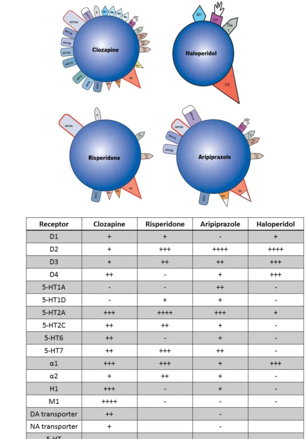
IGURESFigure 1 – Schematic representation of receptor targets (a) and affinity (b) of different antipsychotics.
... 9
Figure 2 - Schematic representation of adult neurogenesis in the adult rodent and human brain.. . 15 Figure 3 – Schematic representation of the experimental design. ... 26 Figure 4 – Percentage of inhibition of the startle reflex measured before and after chronic treatment
with antipsychotic drugs of different classes. ... 29
Figure 5 - Evaluation of playful behavior in control, MAM and treated animals. ... 31 Figure 6 - Analysis of proliferating glial cells in the subgranular zone of the dentate gyrus of the
hippocampus. ... 32
Figure 7 - Morphometric analysis of GFAP immunopositive astrocytes using Simple Neurite Tracer to
reconstruct astrocytes in the dentate gyrus. ... 34
Figure 8 - MAM insult did not affect hippocampal mRNA expression levels of Notch, S100B, GFAP,
xv
L
IST OFT
ABLESTable 1. Dilutions and specificities of the primary antibodies (AB) used in the immunofluorescence
protocol. ... 24
Table 2. Dilutions and specificities of the secondary antibodies (AB) used in the immunofluorescence
protocol. ... 24
Table 3 . Sequences of primers used for real time RT-PCR and the corresponding product size. .... 25 Table 4 - Prenatal exposure to MAM induced deficits in social behavior, gliogenesis and glial plasticity.
xvii
A
BBREVIATIONS % - Percentage µL – Microlitres 5-HT – Serotonin AB - AntibodyAMPA - α-amino-3-hydroxy-5-methyl-4-isoxazolepropionic acid receptor AP – Antipsychotic Arip – Aripiprazole Cloz – Clozapine Cm - Centimetres Ctrl – Control DAPI - 4',6-diamidino-2-phenylindole dB - Decibel
DISC1 - Disrupted in schizophrenia 1
DSM – Diagnostic and Statistical Manual of Mental Disorders EPS – Extrapyramidal Symptoms
fMRI – Functional magnetic resonance imaging GD 17 – Gestational Day 17
GFAP - Glial Fribilary Acidic Protein Hal – Haloperidol
Hip – Hippocampus IP - Intraperitoneal
MAM - Methylazoxymethanol acetate MK-801 – Dizocilpine
MRI – Magnetic resonance imaging Ms - Milliseconds
NMDA - N-methyl-D-aspartate receptor NRG – Neuregulin
NSC – Neural stem cell PCP – Phencyclidine
xviii
PFA – Paraformaldehyde PPI – Prepulse Inhibition PPI - Prepulse inhibition test Ris – Risperidone
RT-PCR - Real time polymerase chain reaction Schz – Schizophrenia
SEM - Standard error of the mean SGZ – Subgranular zone
SPECT - Single-photon emission computed tomography SVZ – Subventricular zone
1
1. INTRODUCTION
1
Introduction
3 1. INTRODUCTION
Mental disorders are estimated to contribute for about 13% of global disease, surpassing cancer and cardiovascular diseases (Collins et al., 2011). Therefore, these disorders are not only more frequent than previously thought but also account for a greater burden of disease than previously expected, becoming a major global health challenge of the 21st century (Wittchen et al., 2011).
Schizophrenia is one of the most debilitating and severe mental disorder, that typically starts in late adolescence or early adulthood and is characterized by psychotic episodes, seriously affecting the quality of life of individuals (WHO, 2016).
Even though the discovery of antipsychotic agents contributed to further extend the knowledge about the etiology of schizophrenia, these drugs are still far from being appropriate for the treatment of all symptoms and cause severe side effects which require a delicate balance in the treatment of schizophrenic patients.
1.1. S
CHIZOPHRENIAPsychotic disorders are severe psychiatric conditions characterized by specific psychopathological phenomena in the domains of thought and perception. According to the diagnostic and statistical manual of mental disorders, schizophrenia is the most common psychotic disorder (DSM V, 2013), with a lifetime prevalence of about 1% (McGrath et al., 2008). Furthermore, schizophrenia presents an earlier onset and more severe symptoms in men than in women (Os & Kapur, 2009). Life expectancy of patients with schizophrenia is 2-2.5 shorter than the general population (WHO, 2016), not only due to high suicide rates, but particularly related with premature cardiovascular disease as a result of lifestyle factors, such as smoking, unhealthy diet, poor exercise and obesity (Os & Kapur, 2009) and of treatment with antipsychotic drugs that can increase the risk of diabetes and obesity (Stahl & Muntner, 2013).
1.1.1.
R
ISKF
ACTORSBy definition, schizophrenia is a disorder that must last for six months or longer, including at least one month of positive symptoms, namely hallucinations (clear and vivid sensorial experiences that occur in the absence of an external stimulus), delusions (fixed beliefs that frequently concern the misinterpretation of experiences and perceptions), disorganized speech, catatonic or grossly disorganized behavior and negative symptoms (DSM, 2013; Stahl & Muntner, 2013). Although the positive symptoms of
4
schizophrenia are the most dramatic feature of the disorder, negative symptoms such as diminished communication, affective blunting, lack of pleasure from positive stimuli, reduced motivation and social withdrawal have a significant functional impact, determining whether patients have a poor outcome or are successfully reintegrated in their daily life activities (Stahl & Muntner, 2013). Additionally, seven cognitive domains were described as frequently impaired in schizophrenic patients, namely working memory, attention/vigilance, processing speed, reasoning and problem solving, verbal learning and memory, visual learning and memory and social cognition (Kern et al., 2004).
On the pursue of the etiology of schizophrenia multiple developmental, environmental, genetic and epigenetic risk factors have been identified (Millan et al., 2014). Environmental elements, namely the degree of urbanization at birth, family history of psychiatric disorders (Sorensen et al., 2014) and developmental disturbances such as viral infections during pregnancy and obstetric complications are strongly linked to the development of schizophrenia. Other factors such as paternal age and cannabis use have been associated with the onset of the disorder (Matheson et al., 2011). Additionally, genome-wide associations and copy number variant studies have identified several risk loci (Dinan et al., 2014) that contribute to the notion that schizophrenia is a polygenic disorder (Srinivasan et al., 2016); particularly Disrupted-in Schizophrenia 1 (DISC1) and Neuregulin 1 (NRG1) are two major candidate susceptibility genes that might be relevant in the pathogenesis of schizophrenia (He et al., 2016).
1.2. P
ATHOPHYSIOLOGY OFS
CHIZOPHRENIA1.2.1. Dopamine
For over 40 years, the leading theory regarding the pathogenesis of this disorder has been defined as the dopamine hypothesis of schizophrenia (Howes et al., 2016). This hypothesis rose from clinical studies that demonstrated that dopaminergic agonists and stimulants could worsen psychosis in schizophrenic patients and induce psychosis in healthy individuals (Angrist & Samuel, 1970; Connell, 1957) and also by the finding that antipsychotic drugs exert their effect by affecting the dopamine system (Carlsson & Lindqvist, 1963). This fact was later confirmed by the association of the efficacy of antipsychotics with their degree of affinity for dopamine D2 receptors (Seeman & Lee, 1975). Moreover, post-mortem studies revealed elevated levels of dopamine, its receptors and metabolites in the striatum of schizophrenic patients (Lee & Seeman, 1980; Seeman & Kapur, 2000).
Five main dopamine pathways have been identified in the brain: the mesolimbic dopamine pathway, the mesocortical dopamine pathway, the tuberoinfundibular pathway, the nigrostriatal pathway and also a pathway that involves the thalamus. The mesolimbic dopamine pathway concerns projections of
5
dopaminergic neurons from the ventral tegmental area of the brainstem to the limbic areas of the brain, particularly the nucleus accumbens in the ventral striatum (Meltzer & Stahl, 1976). This pathway is usually associated to motivation and reward (Salamone & Correa, 2012) has been implicated in amphetamine psychosis (Snyder, 1972) and also in the positive symptoms of schizophrenia (Meltzer & Stahl, 1976). Another key dopamine pathway is the mesocortical pathway that involves dopaminergic neurons that project from the ventral tegmental area to the prefrontal cortex, which is not only associated with executive functions and cognition (Floresco & Magyar, 2006), but is also known to regulate affect and emotions (Laviolette, 2007). Interestingly, an hypofunction of the dopamine system has been observed in this pathway: lesions of dopaminergic neurons in the prefrontal cortex culminated in increased levels of dopamine and D2 receptor density in the striatum (Pycock et al., 1980) whereas the use of dopamine agonists in the prefrontal cortex resulted in reduced levels of dopamine metabolites in the striatum (Scatton et al., 1982). Therefore, it has been hypothesized that the hypofunction of dopamine in this pathway may contribute to the cognitive, negative and affective symptoms of schizophrenia (Laruelle & Abi-Dargham, 2000). The nigrostriatal dopamine pathway comprises the dopaminergic neurons that project from the substantia nigra to the basal ganglia or striatum which control motor function (Engert & Pruessner, 2008). Hyperactivity in this pathway is associated to hyperkinetic movements as dyskinesias and tics (Korchounov et al., 2010), and elevated levels of striatal dopamine have been linked to prodromal symptoms of schizophrenia (Howes et al., 2009). The dopaminergic neurons that project from the hypothalamus to the anterior pituitary constitute the tuberoinfundibular dopamine pathway, which are responsible for the inhibition of prolactin release (Andrews & Grattan, 2002). Finally, a dopamine pathway that innervates the thalamus from several sites as the ventral mesencephalon, the periaqueductal grey matter, the lateral parabrachial nucleus and multiple hypothalamic nuclei has been described in primates (Sánchez-González et al., 2005). Although this pathway remains relatively unexplored, altered levels of dopamine have been reported in the thalamus of patients with schizophrenia (Sánchez-González et al., 2005).
1.2.2. Glutamate
In the brain, excitatory transmission is mainly glutamatergic, with glutamatergic neurons consuming between 60 and 80 percent of total brain metabolic activity (Rothman et al., 2003). Glutamatergic neurotransmission takes place through ionotropic and metabotropic glutamate receptors, and each are subdivided in 3 groups. Group I metabotropic glutamate receptors (mGluR1 and mGluR5) are mainly postsynaptic, while group II (mGluR2 and mGluR3) and group III (mGluR4, mGluR6, mGluR7, mGluR8) are mainly presynaptic and modulate the release of this neurotransmitter (Kew & Kemp, 2005). On the
6
other hand, ionotropic glutamate receptors are designated accordingly to the agonists initially found to selectively activate them: kainate, a-amino-3-hydroxy-5-methyl-4-isoazolepropionic acid (AMPA) and N-methyl-D-aspartate (NMDA) (Dingledine et al., 1999).
The abnormalities in the glutamatergic system were first evidenced by the finding that levels of glutamate were reduced in the CSF of patients with schizophrenia (Kim et al., 1980). However, other research groups were not able to replicate these results (Korpi et al., 1987; Perry, 1982). Nonetheless, the current predominant hypothesis is the dysfunction of the NMDA receptor (Howes et al., 2015). Post-mortem studies revealed that the density of NMDAR1 subunit is reduced in the superior temporal cortex (Humphries et al., 1996) and in the superior frontal cortex (Sokolov, 1998) in patients with schizophrenia. However, overall findings concerning the density of NMDA receptor have been inconsistent (Hammond et al., 2014). Still, current data suggests that alterations in the glutamatergic system may be principally related to aberrant glutamate receptor localization rather than a generalized deficit (Hammond et al., 2014).
The first line of evidence for the involvement of NMDA receptor dysfunction hypothesis of schizophrenia derived from the observation that non-competitive NMDA receptor antagonists, including ketamine, phencyclidine (PCP) and dizocilpine (MK-801), induce immediate psychological conditions that resemble both positive and negative symptoms observed in patients with schizophrenia (Javitt, 2007; Krystal et al., 1994; Morgan & Curran, 2006), which led to the adoption of NMDA receptor antagonists as an approach to model schizophrenia in animals (Howes et al., 2015).
1.2.3. Antipsychotic treatment
Ever since the accidental discovery of the first antipsychotic drugs in the 1950’s, the diversity of available treatments for schizophrenia has expanded significantly (Miyamoto et al., 2005; Stahl & Muntner, 2013). The first drug used as an antipsychotic was chlorpromazine, which has antihistaminic properties, even though its antipsychotic effect is not mediated by this feature (Stahl & Muntner, 2013). In fact, the common characteristic to all first generation (or classical) antipsychotics is the high affinity for dopamine D2 receptor (Stahl & Muntner, 2013) and their binding affinity for the D2 receptor is strongly correlated to the therapeutic doses of these drugs (Creese et al., 1976; Miyamoto et al., 2001; Seeman, 1987; Seeman et al., 1976). However, studies have shown that antipsychotic effects are related with a striatal D2 receptor occupancy of 65-70% (Farde et al., 1992; Kapur et al., 1996; Kapur et al., 2000; Nordström et al., 1993) and that dopamine D2 receptor occupancy above 80% dramatically increases the risk of extrapyramidal symptoms (EPS; such as dyskinesia and Parkinsonism) (Farde et al., 1992). Although a low dosage of haloperidol (2-5 mg/day) can induce an occupancy of 60-80% of the D2 receptor
7
(Kapur et al., 1996; Kapur et al., 1997), the dosage used in the clinical practice must be much higher due to the fact that long-term treatment with classical antipsychotics leads to the upregulation of D2 receptors in humans (Lee et al., 1978; Silvestri et al., 2000). Furthermore, the unspecific blockade of dopamine D2 receptors by classical antipsychotics like haloperidol interferes with the beneficial effects of dopamine. As previously referred, the mesolimbic dopamine system is associated with pleasure and reward, thus the blockade of this system might not only reduce the positive symptoms of the disease, but also block reward functionality, leading to the worsening of negative symptoms and apathy (Stahl & Muntner, 2013). Also, the blockade of dopamine receptors in the tuberoinfundibular pathway interferes with the inhibition of prolactin, resulting in an elevation of plasma concentration of prolactin, a condition designated as hyperprolactinemia (Stahl & Muntner, 2013). This side effect is associated with amenorrhea (i.e., irregular or lack of menstrual periods) and galactorrhea (i.e., breast secretions) thus interfering with fertility, particularly in women, and may also result in a more rapid demineralization of bones (Stahl & Muntner, 2013).
Notwithstanding, atypical or second-generation antipsychotics are drugs that are defined by their serotonin-dopamine antagonism, implicating that 5HT2A antagonism accompanies dopamine D2 receptor antagonism (Miyamoto et al., 2005; Stahl & Muntner, 2013). The stimulation of 5HT2A by serotonin theoretically blocks dopamine downstream release in the striatum, therefore 5HT2A antagonism may interfere with the obstruction of dopamine release by 5HT2A receptors thus reducing EPS and hyperprolactinemia (Stahl & Muntner, 2013) and also contribute to the mood-stabilizing properties of atypical antipsychotics (Brugue & Vieta, 2007).
Clozapine was the first antipsychotic to be described as atypical, considering the reduced EPS and hyperprolactinemia (Stahl & Muntner, 2013). Interestingly, this drug has higher affinity for 5HT2A receptors than it does for D2 receptors and is the only antipsychotic that seems to reduce the risk of suicide in schizophrenic patients (Baldessarini & Frankenburg, 1991; Meltzer et al., 2003), while being the “gold-standard” drug used in patients that present treatment resistance to classical antipsychotics (Chakos et al., 2001). Furthermore, clozapine might even reduce tardive dyskinesia severity in patients suffering from this problem, particularly over long treatment periods (Stahl & Muntner, 2013). However, clozapine is also associated with cardiometabolic complications (such as metabolic syndrome, diabetes mellitus, weight gain and obesity), presenting the highest risk for the development of these side effects compared with other atypical antipsychotic drugs (Zimbron et al., 2016). The administration of clozapine is also associated with agranulocytosis (Hazewinkel et al., 2013), seizures and myocarditis therefore being used only when the alternative treatment is ineffective, rather than as a first-line treatment (Stahl &
8
Muntner, 2013). Remarkably, another atypical antipsychotic – risperidone – exhibits a very different pharmacological profile, displaying a higher affinity for D2 receptors than other atypical antipsychotics, for example clozapine (Stahl & Muntner, 2013). In fact, risperidone can induce dose-dependent EPS, when administered at higher doses (Chouinard, 1995).
A more recent antipsychotic drug, aripiprazole, acts as a partial agonist of D2 and D3 dopamine receptors, meaning that despite the high occupancy of striatal D2 receptors (Gründer et al., 2009), this drugs binds to the D2 and D3 receptors in a way that allows the balance of dopamine release by not blocking completely the flow of this neurotransmitter while not acting as a stimulant (Stahl & Muntner, 2013). Despite not presenting 5HT2A antagonism at higher affinity than its affinity for dopamine D2 receptors, aripiprazole induces reduced hyperprolactinemia and EPS, unlike other atypical antipsychotic drugs (Stahl & Muntner, 2013).
9
Figure 1 – Schematic representation of receptor targets (a) and affinity (b) of different
antipsychotics. Each antipsychotic presents different targets and binding affinities. Adapted from Stahl & Muntner (2013) and Miyamoto et al. (2005).
10
1.3.
S
TRUCTURAL ANDF
UNCTIONALM
ORPHOLOGICALA
BNORMALITIES INS
CHIZOPHRENIASince Eugen Bleuler and Emil Kraepelin (Kraepelin, 1919) first described it over 100 years ago, schizophrenia has been considered a brain disease (Falkai et al., 2011). In 1976, the first computer assisted tomography investigation by Johnstone and colleagues revealed increased volumes of the lateral ventricles in schizophrenia (Johnstone et al., 1976). The technological advances resulted in the development of magnetic resonance imaging (MRI) that pushed the field forward, by allowing better morphological analyses (Falkai et al., 2011). This technique allows the spatial segmentation of brain regions into white and grey matter and cerebrospinal fluid. Studies using MRI have shown deficits in grey matter volume in various brain regions in patients with schizophrenia.
1.3.1. Structural abnormalities in frontotemporal and limbic areas
As previously referred, the prefrontal cortex (PFC) is involved in cognitive executive functions, which are affected in schizophrenia, thus being considered as a key area for the pathophysiology of this disorder (John, 2009; Kinney et al., 1998). Volumetric studies have reported reductions in the PFC (Cannon et al., 2002; Schlaepfer et al., 1994) and functional positron emission tomography (PET) studies reported metabolic hypofrontality (Carter et al., 1998; Schroder et al., 1996). Furthermore, the normal asymmetry verified in the prefrontal cortex (right>left) is reduced in schizophrenia (Bilder et al., 1994), which has been linked to disturbances in the neurodevelopment. Additionally, decreased levels of glucose uptake in the PFC have been associated with negative symptoms in schizophrenic patients (Corcoran & Frith, 1993; Schroder et al., 1996).
Post-mortem and structural MRI studies have reported volume loss in the medial temporal lobe, particularly in the hippocampus, representing one of the most consistent structural abnormalities (Heckers, 2001). Also, post-mortem studies revealed decreased volume of hippocampal subfields, that might be associated to the positive symptoms of schizophrenia (Bogerts, 1997; Bogerts et al., 1990; Bogerts et al., 1993), which was also observed in recent studies using functional magnetic resonance (fMRI) (Haukvik et al., 2015). The hippocampus is involved in emotional regulation and cognition, two affected features in schizophrenia (Heckers & Konradi, 2002). Anatomical and behavioral studies showed that the anterior hippocampus is associated with affect, emotion and stress, while the posterior part essentially involved in cognitive functions (Fanselow & Dong, 2010). Multiple studies have shown a volume reduction in the posterior hippocampus (Becker et al., 1996; Bogerts et al., 1993; Hirayasu et al., 1998; Narr et al., 2001; Rametti et al., 2007; Velakoulis et al., 2001; Yamasue et al., 2004) while other studies shown a decrease
11
in the volume of the anterior part of the hippocampus in schizophrenic patients (Pegues et al., 2003; Szeszko et al., 2003).
The thalamus transfers peripheral sensory inputs to the cortex, being critical in filtering sensory information, coordinating cognitive input to the cortex and mediating corticocortical connections between regions such as frontal and temporal areas, that are affected in schizophrenia. Structural MRI studies revealed a decrease in the thalamic volumes (Brickman et al., 2004; Gaser et al., 2004). Also, a study using PET and single-photon emission tomography (SPECT) showed decreased levels of metabolic activity in the thalamus associated with cognitive deficits and worsening of negative and positive symptoms (Min et al., 1999).
1.3.2. Functional Connectivity Abnormalities
Studies of white matter tracts using diffusion tensor imaging (DTI) of the hippocampus and the fornix body revealed reduced fractional anisotropy, which is a measure of the coherence along white matter tracts, supporting the hypothesis of functional disconnectivity (Kalus et al., 2004; Kuroki et al., 2006; White et al., 2007; Zhou et al., 2008). Furthermore, better performance in cognitive functions such as verbal declarative memory was associated to higher levels of fractional anisotropy of the hippocampus in schizophrenia (Lim et al., 2006). In the entorhinal cortex, a study using DTI and MRI showed a reduction in the volume of this area and a reduction of diffusional anisotropy, suggesting disturbed connectivity to the hippocampus (Kalus et al., 2005) . The neuronal fibers that cross limbic pathways from the posterior hippocampus are connected to the anterior thalamic complex, anterior cingulate cortex, to prefrontal areas and pathways associated with information processing and higher cognition (Fanselow & Dong, 2010; Goldman-Rakic et al., 1984). Therefore, disturbances of connectivity in prefronto-temporal neuronal networks might result in negative and cognitive symptoms (Harrison, 2004; Kuroki et al., 2006; Rajarethinam et al., 2001). Also, in a fMRI study investigating non-articulatory maintenance of phonological information, a subprocess of working memory, reduced connectivity of the prefrontal cortex with the hippocampus and the intraparietal cortex was found in schizophrenic patients (Henseler et al., 2010). A meta-analysis of fMRI studies of executive function has also shown decreased activation of the anterior cingulate cortex, the ventrolateral and dorsolateral prefrontal cortex, and thalamus (Minzenberg et al., 2009). Likewise, disturbances in the prefronto-parietal-thalamic network have been linked to working memory deficits in schizophrenia (Schneider et al., 2007). Therefore, fMRI and DTI studies confirm the hypothesis of dysfunction of the cortico-prefrontal-thalamo-temporo-limbic network in schizophrenia (Weinberg, 1996).
12
1.4.
A
NIMAL MODELS OF SCHIZOPHRENIADespite the fact that characteristics exclusive to humans such as thoughts cannot be evaluated in animals though representing core features of psychiatric disorders, animal models of complex neuropsychiatric disorders still represent remarkable preclinical tools to study the etiology of this disorder (Powell & Miyakawa, 2006).
Rats are highly sociable animals, that are organized in a structured social system and establish a hierarchy that highly influences their development (Jones et al., 2011). Therefore, social isolation of rat pups at the age of weaning impacts brain development, leading to behavioral impairments in adulthood (Fone & Porkess, 2008; Lapiz et al., 2003), which are not rescued by social re-integration at later stages (Pascual et al., 2006). Post-weaning social isolation rodents causes impairments in sensorimotor gating, augments responses to novelty (neophobia), induces spontaneous locomotor activity, cognitive deficits and increased anxiety, which model some characteristics observable in human patients with schizophrenia (Jones et al., 2011).
In the 1970’s, the genetic analysis of a Scottish family with an unusual high prevalence of schizophrenia led to the discovery of a mutation in DISC1 (disrupted in schizophrenia) gene that codes for a scaffolding protein that interacts with other proteins to promote development and growth (Cameron & Glover, 2015). This gene is primarily expressed in the hippocampus of the postnatal brain and its downregulation has been shown to affect cell proliferation in the DG of the adult hippocampus of mice (Mao et al., 2009). DISC1 has also been implicated in the guidance of the migration of new neurons in the DG, and in fact the knockdown of this protein leads to enhanced maturation and aberrant morphology of new neurons in the DG of the adult hippocampus in mice (Cameron & Glover, 2015). Furthermore, knockout mice display reduced brain volume and cortical thickness along with enlarged ventricles (Jaaro-Peled, 2009). Also, subtle impairments in PPI have been reported, which are reverted by treatment with both clozapine and haloperidol (Clapcote et al., 2007; Hikida et al., 2007).
As previously referred, exposure of the neonate to environmental insults during the developmental or perinatal period increases the risk of developing schizophrenia (Lewis & Levitt, 2002). Methylazoxymethanol (MAM) is a natural agent of seeds of cycad plants, that acts as an antimitotic agent that methylates DNA (Matsumot & Higa, 1966) and targets specifically neuroblasts proliferation in the CNS without inducing teratogenic effects in peripheral organs (Cattabeni & DiLuca, 1997). Treatment of pregnant rat dams with MAM selectively impacts neurodevelopment without affecting litter size or pup body weight (Balduini et al., 1991b; Flagstad et al., 2004). In fact, admnistration of pregnant rat dams impacts the brain regions that undergo the fastest development in the fetus, inducing long-lasting
13
behavioral and anatomical impairments in the offspring (Moore et al., 2006; Lodge and Grace, 2009) which are dependent on the gestational day of admnistration (Talamini et al., 1998; 2000; Fiore et al., 1999). Admnistration of MAM on gestational day (GD) 17, when cortical cell proliferation is considerably diminished leads to a more restricted preferential size reduction in neocortical and limbic areas, including medial prefrontal (PFC), occipital and entorhinal cortices and the hippocampus, and increased neuronal density in the perirhinal cortex (Moore et al., 2006; Matricon et al., 2010). Also, treatment with MAM at GD17 seems to have reasonable face validity for cognitive and positive symptoms, and also construct validity structural impairments and dopaminergic alterations (Jones et al., 2011).
1.5. N
EURALS
TEMC
ELLSNeural stem cells (NSCs) can be described as cells that present the capability of generating all the cell types in the brain, contrasting with neural progenitors (NPs) that present more restricted potential (Homem et al., 2015). Neural stem and progenitor cells (NSPCs) expand through symmetric self-renewing divisions in the beginning of development. Later, these cells divide asymmetrically, meaning that one of the daughter cells undergoes differentiation after one or several rounds of transit amplifying cells, while the other daughter cell continues a proliferating progenitor (Homem et al., 2015).
1.5.1. Neurogenesis and Gliogenesis
Both neurogenesis (the formation of new neuronal cells) and gliogenesis (generation of new glial cells) occur primarily in the subgranular zone of the hippocampus (SGZ) and in the subventricular zone, since the particular ontogeny of these brain regions allows the preserved capability of generating proliferating cells in the adult brain (Rusznak et al., 2016). These brain regions are capable of both neurogenesis and gliogenesis in the adult, particularly after stimulation or injury (Rusznak et al., 2016) .
In the SVZ, a particular type of NSCs – B cells –resemble mature astrocytes and express astrocytic molecular markers, including glial fibrillary acidic protein (GFAP) (Apple et al., 2016). These cells generate transit-amplifying progenitors – C cell – which in turn give rise to neuroblasts – A cells – that migrate along the rostral migratory stream until the olfactory bulb (OB) (Apple et al., 2016; Kronenberg et al., 2007). In rodents, neuronal progenitor cells generated in the SVZ travel through the rostral migratory stream, reaching the olfactory bulbs, where they differentiate into periglomerular cells, interneurons or granule cells (Alvarez-Buylla & Garcia-Verdugo, 2002; Gheusi et al., 2013; Lazarini et al., 2014; Sakamoto et al., 2014). Adult olfactory neurogenesis is required for specific forms of olfactory behavior (Wang et al., 2015) and other studies have shown that gliogenesis also occurs in the rodent olfactory bulb as well as in the SVZ of
14
patients with multiple sclerosis (Aguirre & Gallo, 2004; Nait-Oumesmar et al., 2008). Even though olfactory neurogenesis in humans is not the same as in rodents as a result of the variable relevance of olfactory stimuli in the survival of the species (Maresh et al., 2008), under some conditions human olfactory neurogenesis can be a prominent feature, for example in Parkinson’s disease and depression (Huisman et al., 2004; Maheu et al., 2015). Nevertheless, the number of new neurons generated in the human olfactory bulb is lower than the level of neurogenesis observed in the rodent olfactory bulb (Bergmann et al., 2012). However, the adult human SVZ retains active neurogenesis (Ernst & Frisén, 2015; Knoth et al., 2010; Sanai et al., 2011) which possibly reflects that if the cells generated in the SVZ are not migrating to the olfactory bulbs, then possibly these cells are migrating to the neighboring striatum, which might explain the pronounced striatal neurogenesis observed in the human brain (Ernst et al., 2014).
The main role of the SGZ is to generate new granule cells in the dentate gyrus of the hippocampus (Lepousez et al., 2015). Studies in rodents have shown that hippocampal-dependent behaviors activate more newly generated cells than older granule cells (Ramirez-Amaya et al., 2006; Snyder et al., 2009a; Snyder et al., 2009b). A study revealed three characteristics of adult hippocampal neurogenesis in humans: 1) a larger proportion of neurons in the hippocampus are replaced in the adult human brain; 2) the age-dependent decline of neurogenesis in the hippocampus is less marked in human brains; and 3) in rodents, hippocampal neurogenesis leads to a net increase in the number of neurons in the dentate gyrus, whereas the continuous neurogenesis in the hippocampus contributes to a pool of neurons with specific functional properties in humans (Spalding et al., 2013). Therefore, these results indicate that hippocampal neurogenesis in humans is essential in maintaining hippocampal functions (Cameron & Glover, 2015; Lepousez et al., 2015). Likewise, the generation of astrocytic cells also occurs in the hippocampus of the adult human brain (Eriksson et al., 1998).
15
Figure 2 - Schematic representation of adult neurogenesis in the adult rodent and human brain. New neurons are represented in green. a) In the rodent brain, neuroblasts that are generated in the subventricular zone (SVZ) travel through the lateral ventricle (LV) until the olfactory bulbs (OB). b) In humans, neuroblasts are also present in the subventricular zone, however the new neurons are integrated in the striatum. In the dentate gyrus (DG) of rodents (a) and humans (b) new neurons are also continuously generated. Nevertheless, the generation of new neurons in the DG is less significant in the adult rodent brain c), compared to the adult human brain d). Adapted from Ernst & Frisén (2015).
1.5.2. Neurogenesis in Schizophrenia
Impairments in neurogenesis have already been reported in different disorders, namely altered hippocampal neurogenesis in Alzheimer’s disease (Martinez-Canabal, 2014; Vivar, 2015; Winner & Winkler, 2015), Parkinson’s disease (Huisman et al., 2004), stroke and depression (Duman et al., 1999; Madsen et al., 2000; Sahay & Hen, 2007; Sapolsky, 2000).
16
In 2006, Reif and colleagues studied the proliferation of neural stem cells in the hippocampus in post-mortem brains of patients with unipolar major depression, bipolar disorder and schizophrenia, using Ki-67, an endogenous proliferation marker. Interestingly, this study showed that the proliferation of stem cells is not reduced in patients with unipolar depression, in contrast with a significant reduction of proliferation of neural stem cells in the hippocampus of schizophrenic patients, thus suggesting an involvement of adult neurogenesis and gliogenesis in the etiology of schizophrenia (Reif et al., 2006).
1.6.
A
STROCYTESAstrocytes are the most common type of cell in the brain (Molofsky & Deneen, 2015), performing various key functions essential to the homeostasis of the central nervous system (CNS) such as the modulation of several neurotransmitters, glucose metabolism, glutamate neurotransmission, blood-brain barrier formation and maintenance and synaptogenesis (Rajkowska & Miguel-Hidalgo, 2007). Moreover, astrocytes are also responsible for the regulation of the cortical cerebral blood flow (Takano et al., 2006) and also act in response to central nervous system insults such as ischemia, trauma, infection and neurodegenerative disease through a process designated as reactive astrogliosis, which involves modifications in their morphology and molecular expression (Sofroniew, 2009).
In humans, cortical astrocytes can be distinguished in three different cell types – protoplasmic, interlaminar and varicose projection astrocytes. In summary, protoplasmic astrocytes are the most common cortical astrocytes that are immunostained by glial fibrillary acidic protein, which is an intermediate filament protein (GFAP), revealing a characteristic “bushy” structure. These cells communicate intrinsically through gap junctions via calcium signaling, which ultimately leads to the release of transmitters in the extracellular space that can happen as a result of the neuron-astrocyte communication or transmission with other astrocytes (Mohn & Koob, 2015). Interestingly, these astrocytes are arranged in nonoverlapping territories with independent signaling domains (Oberheim et al., 2009). Interlaminar astrocytes are a particular type of cell described in primates, present in layer I of the cortex (Mohn & Koob, 2015). These astrocytes extend a process into layers III and IV of the cortical molecular layers ending there and contacting with blood vessels and other cells, not following the pattern of independent domains observed in the protoplasmic populations (Mohn & Koob, 2015). Finally, varicose projection astrocytes are present in the layers V-VI and extend processes to layers III, IV and V creating contacts with other cells and blood vessel projections.
17 1.6.1. Astrocytes in psychiatric disorders
Interestingly, glial pathology has been found in various psychiatric disorders. A study showed decreased glial cell density in layers 3 and 5 of the dorsolateral prefrontal cortex and in deeper layers of the caudal orbitofrontal cortex in major depressive disorder (Rajkowska et al., 1999). Similarly, reductions of glial cell density were also found in the dorsolateral prefrontal cortex in patients with bipolar disorder (Rajkowska, 2000). Likewise, Onguer and collegues reported unaltered neuronal density and neuronal volume in addition to reduced glial cell density in the subgenual part of the anterior cingulate cortex in major depressive disorder, which was also observed in bipolar disorder patients (Öngür et al., 1998). Another study confirmed this observations, showing reduced levels of GFAP in major depressive disorder and bipolar disorder using proteomics (Johnston-Wilson et al., 2000). Furthermore, elevated levels of GFAP in the parietal, superior frontal and cerebellar cortices (Laurence & Fatemi, 2005) have been observed in the brains and cerebrospinal fluid (Ahlsén et al., 1993) of autism spectrum disorder patients implicating a role of glial cells in this pathology.
1.6.2. Evidences of astrocytic involvement in schizophrenia
Initially, several studies using the histochemical Holzer staining approach reported increased densities of astrocytes in the hippocampus, the periaqueductal grey matter and in multiple cortical areas of schizophrenic patients (Casanova et al., 1990; Stevens et al., 1988; Stevens, 1982). However, when other techniques were used such as the Nissl staining or GFAP immunostaining, no evidence for astrogliosis in schizophrenia was found (Damadzic et al., 2001; Falkai et al., 1999; Garey, 2010; Rajkowska et al., 2002; Schmitt et al., 2009; Williams et al., 2013a; Williams et al., 2013b). Instead, some studies have shown loss of astrocytes in diverse cortical and subcortical areas in schizophrenia (Rajkowska et al., 2002; Steffek et al., 2008; Williams et al., 2013a), which is particularly pronounced in the white matter (Williams et al., 2013a). Likewise, reduced levels of GFAP mRNA have been found in white matter regions of brains of schizophrenic patients (Webster et al., 2005). However another study failed to find altered densities of GFAP in frontal or temporal association cortices (Garey, 2010). Moreover, increased density of S100B immunoreactive astrocytes was reported in post-mortem brains of paranoid but not residual schizophrenic patients (Steiner et al., 2008) as well as increased levels of S100B in bodily fluids of schizophrenic patients (O’Connell et al., 2013; Rothermundt et al., 2004). However, other studies have failed to replicate these findings in serum levels of S100B in schizophrenia (Uzbay et al., 2013; van der Leeuw et al., 2013). Finally, a polymorphism of the excitatory amino acid transporter 2 (EAAT2), an important glutamate transporter primarily expressed in astrocytes, was associated to impairments in executive functions and working memory in schizophrenia (Spangaro et al., 2012). In summary, taking in account the wide range of
18
functions of astrocytes in the CNS as well as clues provided by studies in patients, it is plausible to suggest that astrocytes may be involved in the pathophysiology of schizophrenia.
1.7.
O
BJECTIVESSchizophrenia is a highly complex, debilitating psychiatric disorder that is characterized by positive, negative and cognitive impairments that impact the quality of life not only of patients, but also of their caretakers. Over the last decades, adult neurogenesis has been proposed to be involved in the pathophysiology of several neuropsychiatric disorders. However, glial pathology has been suggested as a relevant factor to understand the etiology of schizophrenia. Therefore, using a well-established neurodevelopmental model of schizophrenia this work aimed to target the modulation of antipsychotics with different pharmacological profiles in the formation and morphology of astrocytes. More specifically, this study aimed to:
1. Evaluate the effect of MAM and the possible modulation of different antipsychotics in behavioral traits that are impaired in patients with schizophrenia;
2. Investigate possible impairments in gliogenesis and astrocyte morphology induced by MAM and the effects of antipsychotics in these measures;
3. Assess the expression of astrocytic-related genes in the hippocampus of MAM and antipsychotic-treated animals.
19
2.
MATERIALS AND METHODS
2
21 2. MATERIALS AND METHODS
2.1. A
NIMALSExperiments were carried out in male Wistar rats (Charles River Laboratories, Barcelona, Spain), weighing 300–400 g, aged four months. These animals were housed (three per cage) under standard laboratory conditions (12h light/ dark cycle, at 22 ºC, relative humidity of 55%, food and water provided ad libitum). All experiments have been approved by the Institutional Ethical Commission and were performed in accordance with the European Community Council Directive 86/609/EEC and 2010/63/EU regarding the use of animals for scientific purposes. The experiments were designed to reduce animal suffering, as well as the number of animals used. Prior to the behavioral experiments, all animals were daily handled (for 10 minutes) and also habituated to experimental room at least 30 minutes before the performance of the tests.
2.2. P
RENATAL EXPOSURE TOMAM
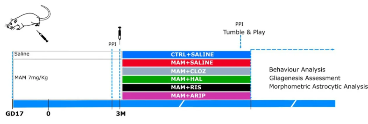
To evaluate the impact of different antipsychotics in the methylazoxymethanol (MAM) model of schizophrenia, animals were assigned to two different experimental groups: controls (CTRL) and MAM, in which the offspring of pregnant Wistar rats were injected subcutaneously with saline (1 ml/kg) or MAM (20 mg/kg; National Cancer Institute, Midwest Research Institute, Kansas City, MO, USA) at gestational day 17 (GD17). Subsequently, the MAM group was subdivided into five experimental groups treated at the age of three months for five weeks with vehicle, clozapine, haloperidol, risperidone or aripiprazole.
2.3. D
RUGSIn order to evaluate the effects of different antipsychotic drugs in the MAM model of schizophrenia, the offspring of female rats injected with MAM were daily injected intraperitoneally (i.p.; 1 ml/kg) for five weeks with clozapine (CLOZ, atypical antipsychotic; 2,5 mg/kg in ultra-pure water with HCl (2M; Panreac, Barcelona, Spain); Kemprotec, Middlesborough, UK), haloperidol (HAL, classical antipsychotic; 0,05 mg/kg in ultra-pure water; Sigma-Aldrich, St Louis, MO, USA), risperidone (RIS, atypical antipsychotic; 0,25 mg/kg in ultra-pure water and glacial acetic acid (Carlo Erba, Barcelona, Spain); Kemprotec, Middlesborough, UK), aripiprazole (ARIP, atypical antipsychotic; 1 mg/kg in ultra-pure water and Tween 80 (1%, Barcelona, Spain); Kemprotec, Middlesborough, UK).
22
2.4. B
EHAVIORT
ESTS2.4.1. Prepulse Inhibition Test
Prepulse inhibition test (PPI) consists in exposing the animal to a non-startle auditory stimulus before subjecting the individual to a strong startling stimulus. This test measures the acoustic startle reflex, reflecting the sensorimotor gating, as the exposure to the pre-stimulus inhibits the startle response to a strong auditory stimulus. To perform this test, the animals were placed in Plexiglas cylinders with 16 cm length and a diameter of 9 cm. The cylinders were set onto a horizontal plate equipped with a transducer that allows the detection of startle response in a sound attenuated chamber. After an acclimatization period of five minutes with white noise [70dB(A)], five startle trials of 120 dB bursts of white noise were delivered, during 40 ms. The session consisted in the presentation of ten startle trials of 120 dB, followed by prepulse intensities of 2,4,8 and 16 dB(A) above background level, respectively PP72, PP74, PP78 and PP86 with a duration of 20 ms. The startle amplitude was measured as the mean of ten startle trials applied, considering the prepulse intensity of 74 dB, that represents the only prepulse intensity in which another animal model of schizophrenia, DISC1 transgenic mice, present deficits in prepulse inhibition (Hikida et al., 2007). PPI (in percentage) was calculated as follows: 100 − (Mean of all startle amplitudes on prepulse trials/Basal startle amplitude) x 100.
2.4.2. Tumble and Play
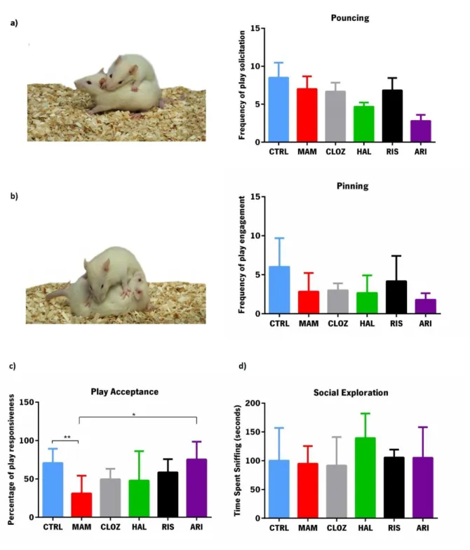
Tumble and play is a behavioral test that allows the evaluation of social play behavior, which was assessed according to the protocol previously described by Borges and colleagues (Borges et al., 2013). Briefly, two strange rats from the same experimental group were placed in an unfamiliar housing cage for 10 minutes after 3,5 hours of social isolation. The behaviors of the animals were recorded using a video camera and subsequently analyzed by a blind observer. The performance of the animals was scored per pair of animals by investigating the frequency of pouncing, which is an indicator of play solicitation, as it is an attempt of an animal to rub or nose the back of the neck of the other animal, and the frequency of pinning, that is the response of the to the solicitation, in which the solicited animal fully rotates on its dorsal surface, with the other animal standing on top of it and also the amount of time spent in social exploration, which is considered as the time spent sniffing any part of the body of the test partner, including the anogenital area.
23
2.5. B
RAINP
ROCESSINGAnimals were anaesthetized by an i.p injection of sodium pentobarbital (Eutasil, 60 mg/Kg, i.p.; Ceva Saúde Animal, Portugal). For molecular analysis (RT-PCR) the animals were transcardially perfused with 0,9% saline and the brains were excised and macrodissected. For the analysis of the astrocytic morphology (immunofluorescence) the animals were transcardially perfused with 0,9% saline and 4%
PFA. Brains were collected and embedded inO.C.T (Tissue-Tek O.C.T. compound, Sakura Finetek Europe,
Netherlands)and posteriorly kept at -20 ºC.Serial coronal sections (20 µm) of the prefrontal cortex (PFC;
Bregma 3.20) and covering the length of the hippocampus (Hipp; Bregma -5.080) were sliced in a cryostat and collected to slides that were kept at -20 Cº.
2.6. I
MMUNOFLUORESCENCE2.6.1. GFAP/Ki-67 Immunohistochemistry
In order to assess astrocytic morphology and astrogliogenesis, an immunofluorescence protocol using antibodies against GFAP and Ki-67 (Table 1) was used in the hippocampus. GFAP is an intermediate filament protein highly present in astrocytes; therefore, an immunostaining against this protein allows the specific labelling of astrocytes (Hol & Pekny, 2015). Also, Ki-67 is a protein expressed during the cell cycle except in G0, representing an endogenous proliferation marker (Kokoeva et al., 2007). Therefore, the combination of antibodies against GFAP and Ki-67 will allow the tracking of new astrocytes.
After selection of the slides (Bregma: -3.24) and posterior defrost, the samples were permeabilized for 10 minutes in a PBS-T (0.5%) solution, in order to potentiate the binding of the antibody to the antigen. Then, samples were washed with PBS and submerged in boiling citrate buffer (10mM, Sigma Aldrich, St Louis, MO, USA) and held at sub-boiling for 20 minutes for antigen retrieval. After cooling at room temperature (RT) for 15 minutes, slides were washed three times with PBS and 4% bovine serum albumin (BSA, Sigma Aldrich, St Louis, MO, USA) was added for 30 minutes at RT to decrease unspecific antibody bounds. The samples were incubated overnight with primary antibodies (Table 1) diluted in PBS, in a humid chamber at 4ºC. On the consecutive day, the slides were washed with PBS and incubated with the corresponding secondary antibodies (Table 2) diluted in PBS, at RT for 2 hours in humid chamber. Subsequently, samples were washed again and incubated with DAPI (1:1000; Invitrogen, California, USA) for 10 minutes. After being washed, the slides were mounted with Immu-mount (ThermoFisher Scientific, USA) and stored at 4ºC.
24
Table 1. Dilutions and specificities of the primary antibodies (AB) used in the immunofluorescence protocol.
Primary AB Specie Dilution Company
anti-GFAP Mouse 1:200 Sigma, Missouri, USA
anti-Ki-67 Rabbit 1:300 AbCam, Cambridge,
UK
Table 2. Dilutions and specificities of the secondary antibodies (AB) used in the immunofluorescence protocol.
Secondary AB Specie Dilution Company
Alexa Fluor® 594
anti-mouse Goat 1:1000
ThermoFisher Scientific, USA Alexa Fluor® 488
anti-rabbit Goat 1:1000
ThermoFisher Scientific, USA
2.6.2. GFAP/Ki-67
Gliagenesis was measured as a quotient of double positive GFAP/Ki-67 positive cells and the total number of Ki-67-positive cells in the subgranular zone of the dentate gyrus (SGZ; defined as a 3-cell layer-thick region on the inner side if the granule layer of the dentate gyrus), by counting the corresponding cell numbers using FV10-ASW 2.0 Viewer software (Olympus, Germany) to visualize the images obtained in the confocal microscope (600x magnification (oil), Olympus FV1000, Germany). The proliferation density was evaluated in the SGZ, and estimated as a quotient between the total number of Ki-67-positive cells and the area of the SGZ, which was determined on a motorized Olympus BX51 (Olympus, Germany) and Stereo Investigator 10 Software® (MicroBrightField, Williston, VT, USA).
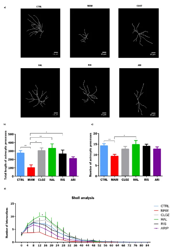
Using the images obtained in the confocal microscope, astrocytic morphology was evaluated with the help of Simple Neurite Tracer software which allowed the 3D reconstruction of astrocytes. For each animal (n=5 per group), six astrocytes were selected in the DG and reconstructed. Total length of the astrocytic processes, total number of ramifications and also Sholl analysis was determined for each astrocyte. The measurements from single astrocytes were averaged from each animal and compared among experimental groups.
25
2.7. M
OLECULARA
NALYSIS2.7.1. RNA extraction, cDNA conversion and real-time PCR analysis
Total RNA was isolated from the macrodissected hippocampal tissue using the Direct-zol RNA miniPrep kit (Zymo Research, Irvine, CA, USA). Briefly, tissue was mechanically homogenized using a 20 G needle and a syringe in 600 µl of Qiazol Reagent (Qiagen, Valencia, CA, USA) and then total RNA was prepared according to the instructions of the manufacturer. Total RNA (500 ng) was reverse transcribed into cDNA using qScript™ cDNA SuperMix (Quanta Biosciences™, Gaithersburg, Md, USA).
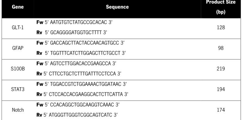
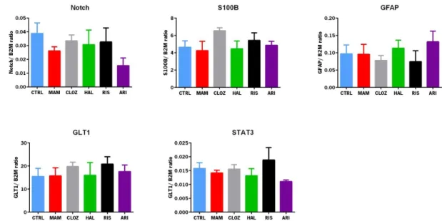
Using PrimerBlast software (NCBI, USA), primers for Glutamate transporter-1 isoform-b (GLT-1), glial fibrillary acidic protein (GFAP), S100 calcium binding protein B (S100-B), signal transducer and activator of transcription 3 (STAT3) and Notch were designed (Table 3). RT-PCR plates were analyzed in an Applied Biosystems 7500 Fast Real-Time PCR System (Applied Biosystems, LLC, CA, USA) using 5x HOT FIREPol® EvaGreen® qPCR Mix Plus (ROX; Solis Biodyne, Tartu, Estonia). Considering the expression levels of the housekeeping gene Beta-2-Microglobulin (B2M), the expression levels target genes were normalized and the relative expression was calculated applying the ΔΔCt method. Results are expressed as fold-change of mRNA levels between the corresponding experimental groups after normalization to B2M expression levels.
Table 3 . Sequences of primers used for real time RT-PCR and the corresponding product
size.
Gene Sequence Product Size
(bp) GLT-1 Fw 5’ AATGTGTCTATGCCGCACAC 3’ Rv 5’ GCAGGGGATGGTGCTTTT 3’ 128 GFAP Fw 5’ GACCAGCTTACTACCAACAGTGCC 3’ Rv 5’ TGGTTTCATCTTGGAGCTTCTGCCT 3’ 98 S100B Fw 5’ AGTCCTTGGACACCGAAGCCA 3’ Rv 5’ CTTCCTGCTCTTTGATTTCCTCCA 3’ 219 STAT3 Fw 5’ TGGACCGTCTGGAAAACTGGATAAC 3’ Rv 5’ CTCCACCACGAAGGCACTCTTCATTA 3’ 194 Notch Fw 5’ CCACAGGCTGGCAAGGTCAAAC 3’ Rv 5’ ATGGGTTGGGTCGGCAGTCATC 3’ 174
26
2.8.
E
XPERIMENTALD
ESIGNFigure 3– Schematic representation of the experimental design. Pregnant rats were injected either with saline or MAM at gestational day 17. One week preceding the beginning of the antipsychotic treatment, and a week before the sacrifice, the prepulse inhibition test (PPI) was performed to assess sensorimotor gating. At the age of three months, animals were injected with saline or with an antipsychotic, for five weeks. One week before the sacrifice the animals performed the tumble and play test, to investigate social behavior.
2.9. D
ATAA
NALYSISStatistical analysis was performed using SPSS (IBM, New York, USA). To investigate differences in the Sholl analysis of the astrocytes a repeated measures ANOVA was used and One-way ANOVA was used to analyze the remaining results. Differences between groups were measured by Tukey’s honestly significant difference test (Tukey HSD) post hoc analysis. Statistical significance was accepted for P< 0.05. Results are expressed as mean± standard error of the mean (SEM).
27
3.
RESULTS
3
Results
29 3. RESULTS
3.1.
P
REPULSE INHIBITIONImpaired prepulse inhibition (PPI) of the acoustic startle reflex illustrates a deficit in sensorimotor gating, which is a minor neurological symptom in schizophrenic patients. Since this reflex is highly conserved across species, PPI is widely used in rodent models of schizophrenia (Jones et al., 2011). PPI was evaluated one week before the beginning of antipsychotic treatment and after five weeks of antipsychotic treatment. The percentage of prepulse inhibition before the beginning of the treatment was not different
between CTRL and MAM animals (F1,15 = 0.661, p=0.429) or between MAM animals treated with saline,
clozapine, haloperidol, risperidone or aripiprazole (F4,45 =0.066, p=0.992) (Figure 4). Likewise, prenatal
exposure to the cytostatic agent MAM did not induce alterations in PPI at the end of the experiment
(F1,16=1.754, p=0.204) and this effect was not altered by treatment with antipsychotics with different
pharmacological profiles (F4,46=0.202, p=0.936).
Figure 4– Percentage of inhibition of the startle reflex measured before and after chronic treatment with antipsychotic drugs of different classes. The results are represented as mean ± SEM. N=9-14 animals per group.
3.2. E
FFECTS OFA
NTIPSYCHOTICT
REATMENT INS
OCIALB
EHAVIORTo determine if MAM animals present deficits in playful behavior and if treatment with antipsychotics exerted any effect in this dimension, the tumble & play test was performed. As shown in figure 5a)), no
differences were found in the request of play solicitation between control and MAM animals (F1,10=0.335,
30
was not different between control and MAM animals (F1,10=3.107, p=0.108) or between MAM and any of
the antipsychotic treated animals (F4,24=0.847, p=0.509; figure 5b)). However, the probability of an animal
responding to play solicitation (Figure 5c)) is significantly higher in control than MAM animals
(F1,10=10.703, p=0.008) and also significantly different between MAM and treated animals (F4,24=2.360,
p=0.05). Post hoc tests indicated that aripiprazole was the only group in which play acceptance was significantly different compared with MAM animals (p=0.044). Finally, no differences were found
regarding social exploration between control and MAM groups (F1,10=0.078, p=0.786) or between MAM
31
Figure 5- Evaluation of playful behavior in control, MAM and treated animals. a) Number of
solicitation of playful behavior; b) Frequency of engagement in playful behavior, as a response to the play solicitation; c) Percentage of responses to play solicitation. d) Time spent in social exploration, namely sniffing the body of the test partner. Data represented as mean±SEM. *p≤0.05, p≤0.01. n= 5 animals per group. Behavior examples adapted from Trezza et al. (2010).
32
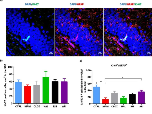
3.3. G
LIAGENESISTo investigate the possible role of the formation of glial cells in the context of schizophrenia, the evaluation of recently formed astrocytes in this animal model was performed (in animals treated and untreated with antipsychotics of different classes) using an immunofluorescence protocol to mark proliferating cells with Ki-67, an endogenous proliferation marker, and GFAP that is a classical glial cell marker.
The density of proliferating cells in the SGZ was unaltered in MAM animals (F1,8=2.358, p=0.164; Figure
6b)), and antipsychotic treatment did not induce any effects (F4,19=0.590, p=0.674). However, the
percentage of ki-67-positive cells that co-labelled with GFAP-immunopositive cells was significantly lower
in MAM animals (F1,8=23.018, p=0.001; Figure 6c)). Moreover, treatment with antipsychotics reverted the
insult implemented by exposition to MAM (F4,20=4.184, p=0.013). Specifically, post hoc tests revealed a
tendency of clozapine to revert this effect (p=0.06) and that aripiprazole had a significant effect in this parameter (p=0.02).
Figure 6 - Analysis of proliferating glial cells in the subgranular zone of the dentate gyrus of the hippocampus. a) Image of a niche of recently formed glial cells in the SGZ obtained by confocal