Universidade do Minho
Escola de Ciências da Saúde
Joana Sofia da Silva Correia
Exploring the Astrocytic Neuroprotective
Functions in a Chronic Mild Stress Model
of Depression
Dissertação de Mestrado
Mestrado em Ciências da Saúde
Trabalho realizado sob orientação do:
Doutor João Filipe Oliveira
e co-orientação da:
Doutora Luísa Pinto
DECLARAÇÃO
Nome: Joana Sofia da Silva Correia
Endereço electrónico: [email protected] Número do Bilhete de Identidade: 13538069
Título da dissertação:
Exploring the Astrocytic Neuroprotective Functions in a Chronic Mild Stress Model of Depression Orientador:
João Filipe Oliveira Co-orientador: Luísa Pinto
Ano de conclusão: 2014
Designação Ramo de Conhecimento do Mestrado: Ciências da Saúde
É AUTORIZADA A REPRODUÇÃO INTEGRAL DESTA TESE/TRABALHO APENAS PARA EFEITOS DE INVESTIGAÇÃO, MEDIANTE DECLARAÇÃO ESCRITA DO INTERESSADO, QUE A TAL SE COMPROMETE;
Universidade do Minho, 31 de Outubro de 2014
Your vision of where and who you want to be is the greatest asset you have. Without having a goal is difficult to score.
-ACKNOWLEDGMENTS/AGRADECIMENTOS
Ao Doutor João Oliveira agradeço, profundamente, todo o conhecimento transmitido e orientação, todos os desafios lançados e a constante insistência pela organização, e pelos momentos de descontração apesar dos problemas travados, sempre proporcionando uma visão mais ligeira dos problemas encontrados. Agradeço ainda toda a visão crítica do trabalho e o entusiasmo no desenvolvimento desta tese.
À Doutora Luísa Pinto agradeço o entusiasmo e a boa disposição, todos os desafios propostos, a grande disponibilidade e a visão critica sempre presente sobre o meu trabalho.
À Mestre Patrícia Patrício agradeço todo o apoio prestado, a importante ajuda no laboratório e partilha de conhecimento, toda a amizade e todos os momentos de descontração e desabafo.
Ao Mestre Dinis Alves agradeço toda a ajuda no laboratório, planeamento experimental e análises de dados. A partilha de informação, de música e boa disposição.
À Mestre Mónica Morais agradeço a ajuda no biotério, toda a disponibilidade para esclarecimento de dúvidas e a amizade.
Ao Mestre António Pinheiro agradeço a boa disposição, a partilha de conhecimento, as brincadeiras e a amizade.
À Mestre Ana Rita agradeço a boa disposição e a ajuda e, sobretudo a alegria num simples “bom dia!” e todas as gargalhadas que tão bem me fazem.
Às Mestre Vanessa Sardinha e Mestre Sónia Gomes e restantes astrochicks agradeço as sugestões, toda a ajuda no biotério e na bancada, todas as loucuras e alegria.
Ao Doutor João Bessa e ao Professor Doutor Nuno Sousa agradeço a oportunidade de integrar uma óptima equipa e de desenvolver um trabalho que me permitiu crescer.
Aos NeRDs agradeço todo o carinho, as sugestões, as críticas construtivas a este trabalho. Às minhas colegas de laboratório Mafalda Santiago, Cláudia Miranda, Rita Silva e Ana Maria agradeço os jantares, as brincadeiras, o carinho e toda a amizade.
Aos meus queridos amigos que deixei no Porto, ao grupo Anabela, agradeço a constante presença, a perspicácia, a musicalidade que vibra em vocês, as brincadeiras e os desabafos.
À minha amiga Ana Carvas, agradeço a amizade forte que cresceu num ano mas que com certeza se prolongará por muito tempo. És como uma irmã para mim.
À minha família agradeço a compreensão, o apoio, o carinho, os ensinamentos, a paciência, a firmeza, a ajuda na tomada de decisões, os puxões de orelhas, que por vezes são bem precisos, e toda a boa disposição.
ABSTRACT
Depression is a multidimensional psychiatric disorder that affects millions of people worldwide. In order to unveil the pathophysiology of this disorder, increasing significance is being given to the study of the abnormal neurotransmission in brain areas affected in depression. Particularly, glutamatergic excitotoxicity has been suggested as one of the possible process underlying the installation of the disease. Previous studies have suggested that glutamate release is increased in depressed subjects. Additionally, the β-lactamic antibiotic ceftriaxone (CEF) was described to increase glutamate uptake by the glutamate transporter GLT-1 (expressed mainly in astrocytes), preventing cellular damage.
Taking these findings into account, the main goal of this work was to understand whether CEF-triggered enhancement of glutamate uptake might be used to prevent or reverse the deleterious effects of glutamatergic excitotoxicity in an animal model of depression, the unpredictable chronic mild stress (uCMS). For this, animals were subdivided in two different main sets - Prevention and Treatment. Different groups of animals were administered either with CEF and/or two antidepressants (ADs; fluoxetine and imipramine).
CEF administration, at an early stage, prevented the installation of depressive-like behavior, yet it failed to reverse the installed depressive phenotype induced by uCMS exposure. CEF seems to reverse partially the cognitive deficits caused by uCMS exposure. Analysis of the collected brain tissue revealed that the prevention of the depressive-like behavior was correlated with an increased GLT-1 gene expression in the ventral hippocampus and with an increased expression of GLT-1 transporter in the dorsal dentate gyrus (DG) of the hippocampus. Morphological analyses disclosed neuronal atrophy of the DG granule neurons in the dorsal DG after uCMS exposure, which was prevented by CEF administration.
These results suggest that CEF administration promotes GLT-1 transporter up-regulation, which may prevent excitotoxicity processes at glutamatergic synapses in the hippocampus, thus preventing the installation of the depressive-like behaviors triggered by uCMS. These observations elucidate the potential use of CEF, or similar drugs, in the prevention of depressive behavior, paving the way for the development of new therapeutic strategies.
RESUMO
A depressão é uma doença psiquiátrica que afeta milhões de pessoas em todo o mundo. Na tentativa de compreender os mecanismos fisiopatológicos da doença, a comunidade científica tem-se dedicado ao estudo de defeitos na neurotransmissão em áreas cerebrais afetadas na doença. Em particular, a excitotoxicidade glutamatérgica tem sido sugerida como um processo subjacente à instalação da doença. Estudos anteriores verificaram que a libertação de glutamato está aumentada em indivíduos deprimidos. Neste contexto, foi também descrito que o antibiótico β-lactâmico ceftriaxona (CEF) aumenta a captação de glutamato pelo transportador GLT-1 (expresso principalmente em astrócitos), prevenindo os danos celulares.
Considerando estas observações, o objetivo deste trabalho foi compreender se o aumento da captação de glutamato desencadeado pela CEF poderá prevenir ou reverter os efeitos deletérios da excitotoxicidade glutamatérgica num modelo animal de depressão, de exposição crónica ao stress (uCMS). De acordo com o objectivo, os animais foram divididos em dois grupos principais – Prevenção e Tratamento. A diferentes subgrupos de animais foram administrados CEF e/ou dois antidepressivos (fluoxetina e imipramina).
A administração de CEF preveniu o desenvolvimento do comportamento depressivo, no entanto foi ineficaz na reversão deste fenótipo já instalado após exposição crónica a stress. Adicionalmente, a CEF parece reverter parcialmente os défices de cognição induzidos pelo uCMS. Análises do tecido cerebral dos animais revelaram uma correlação entre a prevenção do comportamento depressivo e a expressão do gene GLT-1 no hipocampo ventral e do transportador GLT-1 no girus denteado (GD) dorsal. A avaliação da morfologia dos neurónios granulares do GD revelou que a CEF foi capaz de prevenir a atrofia dendrítica no GD dorsal provocada pela exposição ao stress.
Estes resultados sugerem que a administração de CEF estimula a expressão de GLT-1, prevenindo os eventos excitotóxicos nas sinapses glutamatérgicas no hipocampo e o consequente desenvolvimento do comportamento depressivo causado pela exposição ao stress crónico. Estas observações indicam um potencial efeito da CEF ou de outros fármacos que promovam a expressão do GLT-1, na prevenção do desenvolvimento de comportamento depressivo, abrindo caminho para o desenvolvimento de novas possibilidades terapêuticas.
TABLE OF CONTENTS
Acknowledgments/agradecimentos ... V Abstract ... VII Resumo ... IX Table of contents ... XI Abbreviations list ... XV I. INTRODUCTION ... 1 1. INTRODUCTION ... 3 1.1 Depression ... 31.1.1 State of the art ... 3
1.1.2 Pathophysiology and animal models ... 4
1.2 Neuroplasticity in depression ... 7
1.2.1 Remodeling of neuronal circuits ... 7
1.2.2 Neurogenesis as a process of neuroplasticity ... 9
1.3 Glial cells ... 10
1.3.1 Astrocytes ...10
1.3.2 Neuron-astrocyte interactions ...11
1.3.3 Roles of astrocytes in brain disorders ...14
1.3.4 Gliopathology and depression ...15
1.3.5 Astrocytic mechanisms in depression ...16
1.4 Glutamate transporter 1: GLT-1 ... 17
1.4.1 Structure and function ...18
1.4.2 GLT-1 regulation in mood disorders ...20
1.5 GLT-1 regulation by B-lactam antibiotics - Ceftriaxone ... 20
1.6 Research Objectives ... 23
II. MATERIAL AND METHODS ... 25
2. MATERIALS AND METHODS ... 27
2.2 Unpredictable Chronic Mild Stress (uCMS) protocol ... 28
2.3 Drug Treatment ... 29
2.4 Behavior tests ... 29
2.4.1 Sucrose preference test ...30
2.4.2 Sweet drive test ...31
2.4.3 Novelty suppressed feeding test...31
2.4.4 Elevated-plus maze ...32
2.4.5 Open-field test ...32
2.4.6 Forced Swimming Test ...33
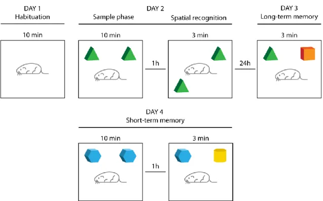
2.4.7 Novel Object Recognition test ...33
2.4.8 Morris Water Maze: Reference memory and Reversal tasks ...35
2.5 Measurement of Plasma Corticosterone Levels ... 35
2.6 Tissue Processing ... 35
2.7 Molecular Assays ... 36
2.7.1 Western Blot ...36
2.7.2 RNA isolation, cDNA synthesis and real time PCR analysis ...37
2.8 Morphological analysis: Golgi-Cox staining ... 38
2.9 Data analysis ... 39
III. RESULTS ... 41
3. RESULTS ... 43
3.1 Validation of the uCMS model of depression ... 43
3.2 Assessment of behavior dimensions affected by uCMS ... 44
3.2.1 Study of the role of ceftriaxone in the prevention of uCMS-induced behavior alterations ...44
3.2.2 Study of the impact of ceftriaxone in the reversion of uCMS-induced behavior alterations ...48
3.3 Cellular and molecular correlates of the ceftriaxone prevention of uCMS-related effects ... 53
3.3.1 Ceftriaxone effect in the expression of glutamate transporter GLT-1 ...53
3.3.3 Assessment of ceftriaxone impact on neuronal 3D morphology ...56
IV. DISCUSSION AND CONCLUSIONS ... 61
4. DISCUSSION AND CONCLUSIONS ... 63
V. FUTURE PRESPECTIVES ... 69
5. FUTURE PRESPECTIVES ... 71
VI. REFERENCES ... 73
ABBREVIATIONS LIST
% Percentage
μ Micrometer
AD Alzheimer’s disease
ADs Antidepressants
ALS Amyotrophic lateral sclerosis
ATP Adenosine triphosphate
BBB Blood brain barrier
BDNL Brain-derived neurotrophic factor
CA Cornus ammonis
cAMP Cyclic adenosine monophosphate
CEF Cetriaxone
CMS Chronic Mild Stress
cm Centimeters
CNS Central nervous system
CREB Clean renewable energy bonds
CT Control
CUS Chronic Unpredictable Stress
DAB 3,3'-Diaminobenzidine
DG Dentate gyrus
dlPFC Dorsolateral Prefrontal Cortex
EAAC1 Excitatory amino acid carrier 1
EAAT Excitatory amino acid transporter
EAE Experimental autoimmune encephalomyelitis
EPM Elevated Plus Maze
FLX Fluoxetine
FST Forced swimming test
GABA Gama-aminobutyric acid
GFAP Glial Fribilary Acidic Protein
GLAST Glumate-aspartate Transport
GLT-1 Glutamate transporter 1 GS Glutamine Synthetase Glu Glutamate HPA Hypothalamic-pituitary-adrene IMIP Imipramine IR Immunireactive LB Laemmi Buffer
MDD Major depressive disorder
min Minutes
MWM Morris Water Maze
NDMA Nmethy-D-aspartate
nm Nanometers
NOR Novel object recognition
NSC Neutral Steam Cell
OB Olfactory Bulb
OF Open field
PFA Paraformaldehyde
PFC Prefrontal Cortex
RMS Rotral Migratory Stream
rpm Rotations per minute
RT Room temperature
RT-PCR Real time polymerase chain reaction
SAL Saline
SDS Dicarboxylate Symporter Family
SDT Sweet Drive Test
SEM Standard error of the mean
SEZ Subependymal zone
SGZ Subgranular zone
SPT Sucrose preference test
SSRI Selective serotonin reuptake inhibitors
TBS Tris-buffered saline
1. INTRODUCTION
Major depressive disorder (MDD) is one of the world leading causes of morbidity, often associated to suicidal attempts. Despite of the importance of this disease in modern societies and the large investment of resources in its study, the processes underlying its pathophysiology remain poorly understood. The importance of glial cells, namely astrocytes in brain processes has been rising in the past years. Astrocytic actions may confer protection to vicinal neurons and may be used in the context of brain disorders as well. The use of astrocytic protective functions in the context of depression urges to be addressed.
1.1 DEPRESSION
1.1.1 State of the art
Depression is a highly prevalent mood disorder affecting more than 120 millions of people and is projected to be one of the major causes of worldwide burden by 2030 (Belmaker and Agam, 2008; WHO, 2008; Willner et al., 2013). Patients suffering from this disorder usually present loss of interest for experiencing pleasurable activities (anhedonia), changes in sleep and appetite, sadness, suicidal ideation and anergia. Furthermore, rather than low self-esteem, depressive patients present a deeply negative view of the world and the future, and display deficits of attention, interpretation and memory (Mathews and MacLeod, 2005). Evaluation reports on cognitive responses state a decreased control for processing negative information, which contributed to high levels of negative automatic thoughts and pathological rumination in these patients (Gotlib et al., 2008). Together with the observed inability to anticipate aversive events or rewards (Chase et al., 2010; McFarland and Klein, 2009; Pizzagalli et al., 2008), these facts provide a cognitive explanation for the core symptom of depression, namely anhedonia (Willner et al., 2013).
Depression and anxiety disorders are often comorbid with each other, since its symptoms are related. In fact, mood disorders such as depression and anxiety are frequently associated to patients suffering from other disorders such as chronic pain (Holley et al., 2013), inflammation (Slavich and Irwin, 2014), cardiovascular disorders (Van der Kooy et al., 2007), stroke
(Ramasubbu and Patten, 2003), Alzheimer’s disease (AD) (Green et al., 2003), epilepsy (Hesdorffer et al., 2000), diabetes (Mezuk et al., 2008) and cancer (Rooney et al., 2013). Furthermore, the emotional disruption presented in depression happens in parallel to cognitive impairments, namely in memory processes, which can be the cause of the onset and recurrence of depressive episodes. For all these reasons, depression is further characterized as a multidisciplinary disorder that affects three different behavioral dimensions: mood, anxiety and cognition (Bessa et al., 2009a). Even though there is no knowledge of a real cause for the precipitation of MDD, vulnerability or predisposition to become depressed may occur in several ways and throughout different life stages. It is accepted that early life experiences, particularly inadequate familial relations, increase the risk for precipitating depression (Slavich and Irwin, 2014; Willner et al., 2013). Also, it was previously identified some genetic predisposition to inherit this disorder in a range of 31 to 42% (Kendler et al., 2002, 2006; Sullivan et al., 2000). It is believed that there are “stress-provoking” genes passing on through generations and providing a stressful family environment (Slavich and Irwin, 2014). On the majority of the population only the interaction between multiple risk genes and environmental factors (e.g. stress-related factors or parental negligence) are sufficient to cause depression, specially at early childhood (Kendler et al., 2001; Widom et al., 2007). Personality characteristics intervening in social interaction and autonomy may also contribute to the vulnerability of oneself to express depressive symptoms. Actually, two types of depression named as “endogenous” or “reactive” are described to characterize autonomous or social-dependent people, respectively. Endogenous depression is associated with an interpersonal distance, feelings of failure, anhedonia, hopelessness, and blame, which minimize possible environmental precipitants (Willner et al., 2013). Interestingly, people suffering from a reactive depression tend to be socially dependent, relying on the satisfaction for approval of others. On the other hand, endogenous depressed people present an interpersonal sensitivity, guilt, anxiety and rumination, with temporary mood improvements but a constant need for attention within an exacerbated depressive state (Willner et al., 2013).
1.1.2 Pathophysiology and animal models
Although the neurobiological causes of this disorder are yet to be fully understood, depressive subjects present cell atrophy and loss in the brain (neurons and glial cells) which can be reverted by antidepressant (AD) treatment (Banasr et al., 2011; Bessa et al., 2009b; Martin et al., 2013).
Pathological changes in size of specific brain regions and alterations in neuronal morphology, neurochemical and signaling molecules; plus alterations in gene expression and epigenetic regulation are factors known to be affected in depression (Mateus-Pinheiro et al., 2011; Tsankova et al., 2007). Contributing to the etiopathological knowledge of depression are studies employing animal models of this disorder. The validity of an animal model for formulation of hypotheses and for the development of novel therapeutic strategies encompasses: use known etiological factors (etiological validity) to mimic the behavioral and neurological symptoms observed in human disease (face validity) and importantly, responsiveness to clinically effective treatments (predicted validity) (Berton et al., 2012; Bessa et al., 2009a; Patrício et al., 2013). There are several animal models of depression described in the literature: chronic unpredictable stress (CUS), chronic mild stress (CMS), social stress, early life stress, learned helplessness, fear conditioning and olfactory bulbectomy (Duman, 2010; Nestler et al., 2002). Nonetheless, these models do not mimic and recapitulate completely the complexity and heterogeneity of the human disease. The unpredictable Chronic Mild Stress (uCMS) protocol, based in the principles of the CMS and CUS protocols was proven to be a robust approach to study the human depression at our lab. In this model, it was observed that after the exposure to stress animals presented depressive-like symptoms such as anhedonia, anxiety and cognitive deficits (Bessa et al., 2009a; Mateus-Pinheiro et al., 2013a), showing therefore alterations in the three behavioral domains known to be affected in humans with depression (Figure 1.1). Additionally, a variety of behavioral tests were designed not only for validation of the depressive-like phenotype in animal models of depression, but also to validate ADs efficacies (Bessa et al., 2009b). Regarding the emerging knowledge given by the use of animal models and from post-mortem studies of depressive patients, the neurobiological causes of depression remains not fully understood.
Currently, several theories have been proposed to explain the causes of depression at a neuronal and molecular level. One of the classical theories of depression is the monoamine hypothesis, which is based on altered neurotransmitter pathways related to serotonin and norepinephrine (Hirschfeld, 2000). Giving the low availability of these two neurotransmitters in a context of depression, classical treatment with antidepressants (ADs) was developed to increase the serotonin and norepinephrine levels. Within the clinics, different classes of ADs are used, namely the serotonine-selective reuptake inhibitors (SSRIs; e.g.fluoxetine), norepinephrine-selective reuptake inhibitors, the tricyclic agents (e.g. imipramine) and other atypical (e.g. tianeptine and agomelatine). Despite its effectiveness for reverting impairments caused by
depressive episodes, around 50% of patients do not present total remission after ADs treatment (Nestler et al., 2002). Besides, the conventional ADs present undesired side effects such as sedation, increase of weight and sexual dysfunction, which may promote a low commitment to treatment, resulting in a break-up and further recurrence of depression (Keller et al., 2002; Lang and Borgwardt, 2013). Therefore, the urging need to prevent the depressive episodes by focusing on different possible physiological mechanisms and the treatment with alternative strategies beyond the monoamine hypothesis might help to obtain more effective therapies regarding this problem.
Other relevant hypotheses have been proposed to explain the etiology of depression: the hypothalamic-pituitary-adrenal (HPA) axis dysfunction hypothesis that is based on hyperactivity and/or disruption of the axis leading to increased levels of glucocorticoids (Pariante and Lightman, 2008); the neurogenic hypothesis that encompasses decreased neurogenesis in the
Figure 1.1 The effects of stress as a trigger factor of depression. In animal models of
depression stress exposure leads to neuroplastic alterations such as neuronal dendritic atrophy of hippocampal and pre-frontal cortex and impaired cytogenesis. These alterations correlate to behavioral deficits in mood, anxiety and cognitive dimensions. Antidepressant’s (AD’s) treatment restore the deleterious effects produced by unpredictable Chronic Mild Stress in emotional and cognitive profile, and reestablish neuronal dendritic morphology and cytogenesis in the hippocampal dentate gyrus (DG) which is associated to remission from depressive-like behavior and sustained long-term recovery (adapted from (Mateus-Pinheiro et al., 2013b).
hippocampus (Jacobs et al., 2000); the cytokines theory, that postulates an altered cytokine profile associated to depression (Miller et al., 2009; Schiepers et al., 2005); and the neurotrophin hypothesis, which is based on growth and trophic factors deficiencies, such as brain-derived neurotrophic factor (BDNF) reduction seen in the context of depression, that might contribute to neuroplastic and neurogenic alterations that enhance individuals vulnerability to depression (Duman, 2009; Hayley and Anisman, 2013). Many of these changes were also verified in animal models of depression, such as the uCMS model (Farooq et al., 2012; Gumuslu et al., 2014; Mateus-Pinheiro et al., 2013a).
Other urging theory is the glial hypothesis of depression. According to this hypothesis there are a series of molecular dysfunctions associated to glial neuroprotective functions, such as decreased flux through the glutamate/glutamine shuttle and consequently reduced glutamate reuptake at the synapse (Sanacora and Banasr, 2013). Additionally it was described that there is an impairment of the NMDA-receptor function, a disturbed neuronal metabolism for disruption of energy supply and reduced GABA synthesis (Choudary et al., 2005; Duman and Li, 2012). Glutamatergic dysfunctions in a context of mood disorders have already been described (Jun et al., 2014; Plitman et al., 2014). Moreover, blockage of glial glutamate uptake was able to induce behavioral alterations consistent with symptoms of mood disorders (Bechtholt-Gompf et al., 2010; John et al., 2012; Lee et al., 2007).
Although many theories have been put forward, none of these are mutually exclusive and may probably contribute together to the vast spectrum of depressive disorders.
1.2 NEUROPLASTICITY IN DEPRESSION
1.2.1 Remodeling of neuronal circuits
Since the first studies of Ramón y Cajal the brain is known to consist of neural cells (neurons and glia) and a fixed system of neuronal circuits (Ramón y Cajal, 1928). However, contrarily to what was then believed, in the recent years it is accepted that neuronal circuits and connections are dynamic and suffer modifications and reorganizations throughout life. This process of reorganization named neuroplasticity produces the generation of new cells and dendritic morphology changes in response to external and internal stimuli. Environmental alterations act as signals upon neuronal systems, brain nuclei, synapses and receptors promoting structural and
functional neuronal adaptation (Zilles, 1992). Basically, this process is responsible for development of new synapses and consequent retrograde of pre-existing ones due to continuous modifications such as axonal growth and collateral sprouting that will also change dendritic arborization and spines density, and therefore the number of post-synaptic sites (Carvalho et al., 2010; Serafini, 2012). Previous studies state that promoting the specific and multifaceted changes at the synapse may be the mossy fibers and hippocampal pyramidal neurons (Popov and Bocharova, 1992); also, chronic stress induced by corticosterone induced Cornus Ammonis (CA)3 pyramidal neurons dendritic shrinkage (Magariños et al., 1996; Sousa et al., 2000; Woolley et al., 1990) and reorganization of dendritic arbors in the medial prefrontal-cortex (Wellman, 2001).
Generally, neuroplasticity is a process of resiliency, meaning that encompasses the ability to adapt and react to environmental alterations that can also hold stressful life events. Several brain areas that are related to cognition or emotional responses can be involved in this process: pre-frontal cortex, hippocampus and amygdala. In fact, reduced hippocampal volume is commonly found in depressive patients and can be correlated to prolongation of the depressive episode (Lorenzetti et al., 2009; Sheline et al., 2003). This reduction is often associated to neural cell loss or compromised cytogenesis processes that involve somatodendritic, axonal and synaptic changes (Serafini, 2012). In animal models, this abnormal reduced hippocampal size was already related to: loss of dendritic spines, decreased number of synapses, loss of glia and impairment of neurogenesis (D’Sa and Duman, 2002; Duman, 2010; Pittenger and Duman, 2008). Loss in dendritic complexity and synaptic sites, which determine a reduction in connectivity, as well as loss in glial cells that have neuroprotective roles, and consequent reduction of neurotransmission are all putative changes contributing to neurogenesis impairments (Licznerski and Duman, 2013; Serafini, 2012). Furthermore, chronic stress is described to promote morphological alteration in the prefrontal cortex (PFC) of rats, namely dendritic retraction, loss of spines and decreased number and size of glial cells (D’Sa and Duman, 2002; Fuchs et al., 2004; Marsden, 2013). Post-mortem studies have shown reduced neuronal density and a relevant reduction of PFC thickness (Cotter et al., 2002; Rajkowska et al., 1999). Therefore, understanding neuroplasticity changes at a structural and functional level may help assessing the mechanisms accompanying such changes.
Many mechanisms may contribute to atrophy and loss of neurons, specifically failure of signaling cascades and target genes that control cell survival. Brain-derived neurotrophic factor
(BDNF) is critical for the survival and function of neurons during development and in the adult brain. BDNF has a role in processes of learning and memory long-term potentiation that may be associated with plasticity of the brain. In absence of this neurotrophic factor neurons undergo a process of programmed cell death or apoptosis (Duman et al., 2000), but the survival of the cell may also be due to the synaptic connections with other cells (neuroplasticity) (Goldberg and Barres, 2000). Signaling pathways as cAMP-CREB cascade when upregulated can inhibit cell death pathways and promote BDNF gene expression, leading to cell survival (Duman et al., 2000).
1.2.2 Neurogenesis as a process of neuroplasticity
Also contributing to neuroplasticity is the process of generating new neurons, neurogenesis. Often, in a context of depression adult neurogenesis is extensively studied and comparisons between pre-existing neurons and newly generated neurons are made. This process encompasses mitotic division of neural progenitors to produce new neurons in the adult brain, involving several steps such as, the commitment of the new cell to a neuronal phenotype, migration and maturation of the cells, and establishment of appropriate contacts that culminate with a full integration on pre-existent network (Patrício et al., 2013). It has already been described that adult neurogenesis happens in different brain regions, mainly in two restricted germinal zones: the subependymal zone (SEZ) of the lateral ventricles, and the subgranular zone (SGZ) of the hippocampal dentate gyrus (DG) (Zhao et al., 2008). In both regions, astrocytes act as primary precursors of the newly generated neurons (Seri et al., 2001). The neuronal progenitor cell population arising from astroglial cells composes the narrow layer of three nuclei wide, the SGZ, where neurogenesis occurs. This multipotent cell population is constituted by the neural stem cells (NSCs), which express nestin and glial fribilary acidic protein (GFAP; a marker also presented in mature astrocytes). NSCs can be divided in two cell subtypes according to their orientation in the SGZ: radial astrocytes/NSCs and horizontal astrocytes/NSCs. In adult mammalian brain, neurons born in the SGZ migrate into the granular cell layer of the DG and differentiate into glutamatergic granule cells (Zhao et al., 2008). Otherwise, neuroblasts born in the SEZ migrate along the rostral migratory stream (RMS) becoming mostly mature GABAergic granule and periglomerular interneurons in the olfactory bulb (OB). Despite of the controversy around this topic, some reports describe generation of new neurons in other brain regions such
as cortex, amygdala, hypothalamus, striatum and substancia nigra (Bédard et al., 2006; Ehninger and Kempermann, 2003; Gonçalves et al., 2008; Kodama et al., 2004; Kokoeva et al., 2005; Yoshimi et al., 2005). Furthermore, this process of hippocampal neurogenesis has already been correlated to emotional and memory processes (Burghardt et al., 2012; Denny et al., 2012; Malberg et al., 2000; Mateus-Pinheiro et al., 2013a).
1.3 GLIAL CELLS
Glial cells are the most abundant cells within the central nervous system (CNS). There are four types of glia cells in the brain: astrocytes, oligodendrocytes, microglia and NG2-positive cells. Oligodendrocytes are responsible for production of myelin along the axons, helping in the conduction of electrical signals, whereas microglia are smaller cells with phagocytic functions. NG2-positive cells are known as oligodendrocytes progenitor cells but can also give rise to astrocytes. Astrocytes are the most abundant glial cells in the brain and play important roles in metabolic support of neurons, in the control the blood flow and modulation of the blood brain barrier by supplying energy and nutrients to neurons and maintaining homeostasis.
1.3.1 Astrocytes
These cells are classically defined by their star-shape morphology and expression of glial fibrils and for their extended numerous processes surrounding neighboring neurons and blood vessels (Wang and Bordey, 2008b). As they establish connections to the capillaries of the blood brain barrier (BBB) they may influence the microenvironment in this region (Fuchs et al., 2004; Wang and Bordey, 2008b). Astrocytes also present neurotransmitters’ receptors that may lead to electrical and biochemical events inside the cell (Fuchs et al., 2004). Astrocytes express several membrane receptors, namely G-protein coupled receptors, ionotropic receptors and other receptors for growth factors, chemokines, steroids, and receptors involved in innate immunity such as Toll-like receptors (Abbracchio and Verderio, 2006; Franke and Illes, 2014; Heiman et al.; Wang and Bordey, 2008a). Moreover, these cells present a family of high affinity sodium-dependent glutamate transporters, which are responsible for the uptake of glutamate (Figure 1.3 a). Physiologically, there are three glutamate transporters in the rodent forebrain that provide glutamate clearance of the synaptic cleft during synaptic transmission: GLT-1 (a homolog of
EAAT2 in humans), glutamate-aspartate transporter (GLAST, homolog of EAAT1 in humans) that are expressed mainly in astrocytes (Rothstein et al., 1996; Wang and Bordey, 2008b); whereas, the excitatory amino acid carrier 1 (EAAC1, homolog of EAAT3) is expressed in neurons (Zink et al., 2010). Also part of these transporters family are the EAAT4 expressed in Purkinje cells (Huang et al., 2004) and EAAT5 which is widely expressed in peripheral tissues (Lee et al., 2013a). Interestingly, both variant 4 and 5 seem to act more like inhibitory glutamate receptors than glutamate transporters, mainly due to their high uncoupled anion conductance (Zhou and Danbolt, 2013). Nevertheless, together these transporters contribute to the balance of neurotransmitters at the synaptic cleft, preserving the responsiveness of glutamate receptors, and others such as Nmethyl-D-aspartate (NMDA) receptors and ATP receptors, and conferring neuroprotection against excitotoxicity (Lipski et al., 2007; Tzingounis and Wadiche, 2007).
GLT-1 and GLAST are highly efficient glutamate transporters and its inhibition with pharmacological blockers, antisense oligonucleotides or by transgenic knockout lead to increased levels of glutamate and consequent cell death (Izumi et al., 2002; Rothstein et al., 1996; Tanaka et al., 1997). Moreover, the use of pharmacological blockers cause Nmethyl-D-aspartate (NMDA) receptor-dependent cell death and enhances excitotoxicity induced by exogenous glutamate (Selkirk et al., 2005; Wroge et al., 2012), suggesting an interplay between this receptor and the astrocytic glutamate transporters.
1.3.2 Neuron-astrocyte interactions
The classical paradigm that brain information processing is exclusively the result of neuronal activity has been challenged by an emerging body of evidence (Araque et al., 2014; Kettenmann and Verkhratsky, 2008; Perea et al., 2009; Wang and Bordey, 2008a). Indeed, recent findings rather strongly support the concept of a “tripartite synapse” (Figure 1.2), in which a cross-talk between astrocytes and neurons complements and modulates the communication between pre- and post-synaptic structures (Araque et al., 2014). Upon an elevation of synaptically released neurotransmitters, astrocytes can increase the intracellular calcium ([Ca2+]i) resulting in the release of glutamate via regulated exocytosis (Rossi and Volterra, 2009). Data reports that this increase in [Ca2+]i is extremely important, in a functional view, for astrocyte-astrocyte and also astrocyte-neuron intercellular communication (Charles et al., 1991; Cornell-Bell et al., 1990; Sofroniew and Vinters, 2010). Indeed, one of main functions of astrocytes is the regulation of the
glutamate concentration in the synaptic cleft of glutamatergic neurons. Important for this astrocytic function is the glutamate-glutamine shuttle, which includes not only the uptake of extrasynaptic glutamate but also the production and release of glutamine from the astrocytes via glutamine synthetase (GS), which will be used by neuronal elements to refill glutamate supply (Chiang et al., 2007). In summary, the astrocytic features that favor this concept are: (i) the expression of functional neurotransmitter receptors (eg. for glutamate, ATP, GABA), which sense surrounding neuronal activity; (ii) the ability to process intracellular calcium signaling which may propagate to vicinal astrocytes; and (iii) the ability to release neuro- and vasoactive substances such as glutamate, d-serine, ATP, GABA, TNFα, prostaglandins, or peptides, which influence and regulate synaptic transmission, blood flow, the permeability of the blood brain barrier and metabolic support (Oliveira et al., 2011; Perea et al., 2009; Wang and
Figure 1.2 The tripartite synapse. As neurons, astrocytes express many receptors and transporters
that are activated upon neurotransmitters release from the presynaptic terminal. This activation increases calcium ions inside the astrocyte and parallel release of substances such as ATP. This gliotransmission will counter-act on neurons to either inhibit or enhance neuronal activity. Astrocytes also play an important role in modulation of presynaptic functions and postsynaptic responses to neurotransmitters. One of the main functions of astrocytes is to provide glutamate clearance from the synaptic cleft, creating a neuron-astrocyte interaction through a glutamate-glutamine shuttle. Glutamate, Glu; glutamine, Gln; glutamine synthetase, GS; red dots, neurotransmitter; green dots, gliotransmitter.
Bordey, 2008a). Alterations of cellular cross-talk conceptualized as the tripartite synapse (Perea et al., 2009) have been extensively demonstrated both in brain slice preparations (Henneberger et al., 2010; Martineau et al., 2013; Perea and Araque, 2007; Woo et al., 2012; Yang et al., 2003) and in vivo (Chen et al., 2013; Halassa et al., 2009; Han et al., 2012; Navarrete et al., 2013; Takata et al., 2011) in rodents and, more recently, in humans (Navarrete et al., 2013). These features provide astrocytes with the mechanisms to modulate neuronal function (Araque et al., 2014), thereby influencing network regulation and computation of behavior responses. Despite of the extensive demonstration of cellular and molecular cross-talk pointed out, little is known about the impact of neuron-astrocyte interactions on the production of network outputs.
Back in 2010, our team decided to study this phenomenon and dissect a putative role of astrocytes in network computation. Using an L-α-aminoadipate animal model in which astrocytes were affected specifically in the PFC, a severe impairment in cognitive tasks that depend on this brain region was induced in those animals (Lima et al., 2014). The PFC is intimately related to the computation of complex cognitive functions such as information integration, learning, memory processing and behavior flexibility (Clark et al., 2004; Goldman-Rakic, 1995), known to be affected in the model of depression used in this study. Additionally, cognitive function was studied in the dnSNARE mice model of impaired gliotransmission (impaired exocytosis specifically in astrocytes). It was gathered data indicating a need for gliotransmitter release from astrocytes for the PFC to produce a correct cognitive processing (data under review). This observation is in agreement with previous demonstrations using the same dnSNARE model, in which absence of gliotransmission was responsible for the exacerbation of cognitive deficits associated with sleep loss (Fellin et al., 2009; Halassa et al., 2009). On one hand, these observations, together with data obtained from different animal models of astrocytic dysfunction (Han et al., 2012; Pannasch et al., 2014), suggests that neuron-astrocyte communication is critical for correct production of cognitive outputs. On the other hand, the engraftment of rodent forebrain brain with human glial progenitors that differentiate to human-like astrocytes (described as being about 20x larger and having more complex morphology), enhanced the cognitive abilities of these animals (Han et al., 2013).
1.3.3 Roles of astrocytes in brain disorders
Moreover, an increasing significance has been attributed to glutamatergic system in the pathophysiology of several mood disorders such as schizophrenia, bipolar disorder and depression, and also neurodegenerative disorders as amyotrophic lateral sclerosis (ALS) and Alzheimer’s disease (AD) (Kruminis-Kaszkiel et al., 2014; Verkhratsky et al., 2010; Webster et al., 2005). In fact, it has been reported that a dysfunction or reduced number of glial cells in patients suffering from major depressive disorder (MDD) can result in an increase of levels of glutamate in blood and cerebral spinal fluid (Hashimoto, 2011), causing a toxic accumulation of glutamate. An overabundance of glutamate accompanied by the failure of astrocytes to remove it, may lead to neuronal excitotoxicity resulting in neuronal loss, as seen for motor neurons in ALS (Potokar et al., 2013). In the context of AD it is known to occur a reactive astrogliosis promoting the neurodegenerative processes observed. Among the histopathological features is the presence of senile plaques and neurofibrillary tangles, peripheral loss of cholinergic and cholinoceptive neurons (in PFC and hippocampus) and the presence of activated macrophages and reactive astrocytes (Markiewicz and Lukomska, 2006). The senile plaques are deposits of β-amyloid protein and in turn, neurofibrillary tangles are intraneuronal stuctures composed of tau protein. β-amyloid can be a potent neurotoxic agent that promotes activation of astrocytes and cellular mechanisms exacerbation leading to neuronal damage. Furthermore, it is known that inflammation contributes to neuropathology associated to AD. Indeed, upon activation, astrocytes and microglia can release inflammation-promoting mediators potentially neurotoxic, since these glial cells are the brain representatives of the immune system. Although it is considered as a beneficial role in defense and repair, an escalating pathological glial activation may contribute to secondary nerve-cell damage (Markiewicz and Lukomska, 2006). In response to the extracellular β-amyloid deposits, microglia cells can produce TNF-α and other cytokines, and consequently promote the secretion of reactive oxygen species, further enhancing neuronal damage. Microglia cells by releasing pro-inflamatory citokines and reactive oxygen species can activate surrounding astrocytes which will internalize the debris that are being released by dying neurons being important for plaque degradation. In addition, astrocytes activated by β-amyloid will also release pro-inflamatory cytokines, oxygen reactive species and other inflammation-involved molecules that will attract microglia and further contribute to neuronal damage caused by and exarcebation of released pro-inflamatory cytokines (Markiewicz and Lukomska, 2006).
Astrocytes are also involved in many chronic neurological disorders, such as epilepsy, since these cells undergo morphological and functional remodeling in the epileptic brain. Proliferation of reactive astrocytes (astrogliosis) is a common feature of temporal lobe epilepsy, which is one of the most prevalent forms of localization-related epilepsies in humans (Coulter and Eid, 2012). Besides observed neuronal loss, astrogliosis in this pathology is correlated with an increased number of seizures and slower clearance of extracellular glutamate in the hippocampal formation (Cavus et al., 2005). Reactive astrocytes are widely correlated to the epileptogenesis via their effects on glutamatergic regulation, role in buffering potassium and interstitial volume control (Benarroch, 2009; Wetherington et al., 2008). The formation of the epileptic foci in the hippocampus has been associated to reactive astrocytes (Ortinski et al., 2010) and abnormal neuronal excitability in adult model systems (Gómez-Gonzalo et al., 2010). Since there are clues to an astrocytic dysfunction and a role of these cells in post-natal synaptogenesis has been described; it has been hypothesized whether this abnormal astrocyte development could be the cause of predisposition to epileptogenesis of the developing brain adjacent to alterations in the excitatory-inhibitory balance (Molofsky et al., 2012).
1.3.4 Gliopathology and depression
Of the main cells in the CNS, astrocytes are highly implicated in glial pathology in MDD. In several studies of post-mortem brain tissue of depressive subjects was seen a decrease in packing density and number of Nissl-stained population glial cells (Rajkowska and Stockmeier, 2013). This glia pathology was observed in several brain regions including dorsolateral prefrontal cortex (dlPFC) (Cotter et al., 2002; Rajkowska et al., 1999), ornitofrontral cortex (Rajkowska et al., 1999), subgenual cortex (Ongür et al., 1998), anterior cingulate cortex (Cotter et al., 2001a; Gittins and Harrison, 2011) and amygdala (Bowley et al., 2002). However, in elderly patients of MDD the alterations in glial density in the orbitofrontal and anterior cingulate cortex were not observed (Khundakar et al., 2011a, 2011b). Specifically, dlPFC is involved in executive function and emotional regulation (Davidson et al., 2000; Milham et al., 2001); its neuropathological abnormalities in glial cells and in GABAergic interneurons and pyramidal neurons are believed to contribute to the pathophysiology of MDD (Oh et al., 2012; Rajkowska and Miguel-Hidalgo, 2007; Sanacora and Saricicek, 2007). Moreover, in the dlPFC of depressive patients was observed atrophy and reduction of neuronal and glial density (Rajkowska et al., 1999), decreased GFAP
expression (Miguel-Hidalgo et al., 2000; Si et al., 2004) and decreased density and size of calbindin-immunoreactive (IR) GABAergic neurons (Rajkowska et al., 2007). Oh et al. (2012) have recently shown that glutamate changes negatively correlates to GFAP expression levels and to calretinin-IR GABAergic neuronal density in the PFC of MDD patients; calretinin-IR GABAergic neuronal alterations were positively correlated to changes in glial cells and pyramidal neurons markers in the dlPFC. These findings provided some insight on mechanistic basis for neuronal and glial excitotoxic damage promoted by glutamatergic transmission, in the dlPFC of MDD patients (Oh et al., 2012).
Within the basis of cytokines hypothesis of depression an interpretation of astrocytic hypertrophy as a reflection of local inflammation was made (Maes et al., 2009). Detailed analysis of Golgi stained astrocytes revealed cell bodies and processes hypertrophy in the white matter of the anterior cingulate cortex of depressed subjects dying by suicide (Torres-Platas et al., 2011). Recently, it has been shown in post-mortem studies that the pathology in the white matter of prefrontal brain is promoted by decreased oligodendrocyte density, reduction in the expression of genes related to cell functions as well as molecular changes in intercellular cell adhesion molecule expression levels and a possible mechanism of ischemia (Tham et al., 2011).
1.3.5 Astrocytic mechanisms in depression
There is an increasing evidence of astrocytic dysfunction in the context of depression (Choudary et al., 2005; Cotter et al., 2001b; Gosselin et al., 2009; Oh et al., 2012; Sanacora and Banasr, 2013). Contrarily to neurodegenerative disorders in MDD there is no prominent neuronal pathology, no astrogliosis processes and the expression of GFAP and other astrocytic markers is decreased (Rajkowska and Stockmeier, 2013). This astrocyte dysfunction and the neuronal impairments have been thought as two related consequences of tripartite synapse disturbance. In addition, in several post-mortem studies of depressive-brain no reductions in neuronal density or in total number were found (Cobb et al., 2013; Van Otterloo et al., 2009; Stockmeier et al., 2004). Instead, it has been reported a decrease in cell bodies size or reductions in dendritic branching (Chana et al., 2003; Hercher et al., 2010; Stockmeier et al., 2004). Together these findings suggest that in MDD is present a neuronal atrophy rather than neuronal loss. Furthermore, measure of marked enolase neurons in the serum levels displayed no changes in patients with MDD (Schroeter et al., 2008, 2010). Contrastingly, in the same study
it was observed an increase of serum levels of the astrocytic marker S100β. Also, other studies showed that mRNA for glial markers, such as glutamate transporters and glutamine synthethase, was significantly reduced in the locus coeruleus of depressive patients; the expression of mRNA for neuronal markers tested were not significantly changed for the same brain region in these patients (Bernard et al., 2011). Interestingly, glial toxic ablation rather than neuronal ablation lead to depressive-like behaviors (Banasr and Duman, 2008; Lee et al., 2013b). Regarding these previous studies, it appears that there is a selective cellular pathology for glial cells in the context of depression (Rajkowska and Stockmeier, 2013). Notably, the promotion of each type of cellular pathology seen in depression appears to be age-related. In fact, the glial cellular pathology appears to be related to younger depressive subjects (less than 60 years of age); contrasting with neuronal pathology that appears to relate to older subjects (more than 60 years of age) (Khundakar and Thomas, 2009; Miguel-Hidalgo et al., 2000; Si et al., 2004). In the older subjects, the neuronal pathology of depression seems to be cause of prominent reductions in the density of pyramidal glutamatergic neurons in the orbitofrontalcortex (Khundakar and Thomas, 2009; Rajkowska et al., 2005). In addition, unaltered astrocytes density and GFAP levels were seen in the elderly patients of depression (Davis et al., 2002; Miguel-Hidalgo et al., 2000; Si et al., 2004). Contributing to this neuronal pathology may be the excitotoxicity promoted by an excess of glutamate at the synapse cleft of glutamatergic neurons due to reduced number of astrocytes and astrocytic glutamate transporters (Rajkowska and Miguel-Hidalgo, 2007; Rajkowska and Stockmeier, 2013).
These thrilling findings, suggest that astrocytic modulation of neuronal activity affects the network activity and consequent output production, both on healthy and pathological processes. Importantly, astrocytes regulate synaptic maturation, transmission and maintenance providing a correct development of synapses (Slezak and Pfrieger, 2003). However, the dynamic mechanisms underlying this cross-talk between neurons and glia and their implications on cellular plasticity in depression are still underexplored.
1.4 GLUTAMATE TRANSPORTER 1:GLT-1
Solute carrier family 1, member 2 (Slc1a2; also known as glial high affinity glutamate transporter, GLT-1) is a glial glutamate transporter predominantly expressed in the rat
hippocampus; is mainly present in astrocytes plasma membrane and together with GLAST play a role in glutamate clearance during synaptic transmission (Rothstein et al., 1996; Wang and Bordey, 2008b) (Figure 1.3 a).
Figure1.3 Impact of stress exposure and ceftriaxone effect on glutamatergic transmission.
Astrocytes uptake glutamate at the synaptic cleft of glutamatergic neurons through glutamate-glutamine shuttle, which includes not only the uptake of extrasynaptic glutamate (Glu) but also the production and release of glutamine (Gln) from the astrocytes via glutamine synthetase (GS) (a). After stress exposure, astrocytic transporters such as GLT-1 help the synapse to cope with the increase of glutamate release, diminishing excitotoxicity. The chronic exposure to stressors (uCMS) may overcome the capacities of the existing GLT-1 and cause excitotoxic effects at the synapse (b). Ceftriaxone (CEF) administration increases the expression of GLT-1 glutamate transporter in the astrocyte (c). Further elucidation of CEF beneficial potential to prevent or revert the installation of the deleterious effects of uCMS exposure (d) will be te main goal of this study.
1.4.1 Structure and function
GLT-1 gene is located at locus 3q31 and encodes three transcript variants (1, 2 and 3) that differ both in 3’ coding region and 5’ terminal exon. These transcript variants encode three distinct isoforms (-a, -b and -c). Transcript variants 2 and 3 encode isoforms with distinct –C and –N terminus, respectively, compared to isoform –a. Transcript variant 1 encodes the longest isoform, isoform –a, which results in a protein with 573aa. The other two variants result in smaller proteins: isoform –b protein has 562aa and isoform –c has 570aa. These are glycosylated proteins that have a palmitoylation in the Cys-38, which seems to be important for its function in glutamate uptake. Interestingly, only isoforms –a and –b present a conservative domain Sodium-dicarboxylate symporter family (SDS) and are known to be transmembranar
proteins (NCBI, 2014). It is also known that oligomerization is common feature of glutamate transporters and isoform –a and –b can interact and form hetero-oligomers in heterologous expression systems, in primary cultures from fetal rat and in the adult rat brain (González-González et al., 2009). However, this arrangement does not seem to interfere with the protein function and consecutively with glutamate transport, since its subunits present all the elements necessary for the translocation of glutamate (Grewer et al., 2005; Koch and Larsson, 2005). Furthermore, different expression was observed between post-natal and young-adult rats. With eight weeks of age, it was observed that GLT-1 isoform –a, represented 90% of total hippocampal GLT-1, but –b and –c represented only 1% each; however, at post-natal day 14, GLT-1 isoforms – b and –c were 1.7 and 2.5 times higher in relation to total GLT-1, respectively (Holmseth et al., 2009).
Although GLT-1 transporter was primarily detected in astrocytes, studies have also reported its presence also in neurons. The splice variant isoforms –a (GLT-1a) and –b (GLT-1b) are described to be widely expressed in astrocytes throughout different brain regions (Berger et al., 2005). Moreover, this splicing variation does not seem to alter transport characteristics of the isoforms. However, there has been descriptions that GLT-1a isoform is not exclusive for astrocytes and appears in CA3 neurons of hippocampus and in the olfactory nucleus of rat’s brain (Berger et al., 2005; Chen et al., 2004). Nonetheless, previous studies have observed a noticeable variation in labeling intensity of GLT-1a mRNA expression in astrocytes of pyramidal and molecular CA1 layers of the hippocampus and other brain areas, but homogeneous expression of GLT-1b isoform in astrocytes throughout several brain areas (Berger et al., 2005). Immunoreactives of
the neocortical brain extracts have shown higher GLT-1a distribution in the hippocampus, compared to –b and –c isorforms, and similar distribution of the three variants in the cerebral cortex (Holmseth et al., 2009). Additionally, assessment of intracellular localization of GLT-1a and GLT-1b expression on the astrocyte was verified that GLT-1a mRNA was expressed primarily in astrocyte processes, whereas GLT-1b mRNA was more restricted to the astrocyte cell body (Berger et al., 2005; Holmseth et al., 2009). Together these findings suggest that there are different mechanisms regarding the activity and functional regulation of the GLT-1 isoforms.
1.4.2 GLT-1 regulation in mood disorders
It is known that this transporter has a reduced expression in the pathophysiological context of depression (Lee et al., 2007; Rajkowska and Stockmeier, 2013; Sanacora and Banasr, 2013; Zink et al., 2010). The glial hypothesis of depression admits a glutamine/glutamate shuttle malfunction, with reduced reuptake of glutamate, causing excitotoxicity at the synaptic cleft (Figure 1.3 b). It is believed that since astrocytes were shown to be affected in the context of brain disorders in humans and animal models of chronic stress, loss of GFAP seems to lead to a disturbance in the transfer of GLT-1 from intercellular space to cell surface (Hughes et al., 2004) and that stress exposure possibly triggers a reduction of GLT-1 in the periaqueductal gray matter (Imbe et al., 2012). Compromising the function of GLT-1 will putatively lead to neuronal death, suggesting that an increase in glutamate uptake may correct deficits caused by GLT-1 malfunction in depressive subjects. In fact, learned helplessness animal model of depression showed suppressed expression of GLT-1 in the hippocampus and cerebral cortex (Zink et al., 2010).
For these reasons, it is hypothesized that by targeting glutamate transporter-activity we may increase neuroprotection and therefore diminish excitotoxicity (Figure1.3 c).
1.5 GLT-1 REGULATION BY B-LACTAM ANTIBIOTICS -CEFTRIAXONE
β-lactam antibiotics are widely used within the clinics and present a variable spectrum of antimicrobial activity (Asbel and Levison, 2000). β-lactams include: penicillins, cephalosporins, cephamycins and carbapenems. Since there are patients allergic to penincillin and the bacterial resistance may be a problem to validity and efficiency of these drugs, other antibiotics have
continuously been designed to treat aerobic gram-negative infections. Non-penicillin β-lactams like cephalosporins and cephamycins have a wider spectrum of antimicrobial activity, higher resistance to beta-lactamase enzymes and can be used against enterobacteriecae family. Ceftriaxone (CEF), a type of cephalosporin, can be use to treat: sepsis, meningitis, Lyme borreliosis, abdominal infections such as peritonitis and gastrointestinal infections, respiratory tract infections such as pneumonia, bone infections and genital infections. Moreover, this antibiotic has already been described to cross the blood brain barrier (BBB) (Barichello et al., 2014; Nau et al., 2010). Rothstein et al. (2005), previously demonstrated that this β-lactam antibiotic could be involved in neuroprotection mechanisms by inducing overexpression of the GLT-1 transporter preventing neuronal loss induced by malfunction of GLT-1 transport (Figure 1.3 c). Also in this study, an ability of this antibiotic to prevent motorneuron loss, as well as a rescue of the loss of muscular strength and extended survival in a mice model of amyotrophic lateral sclerosis (ALS) was shown (Rothstein et al., 2005). Recently, immunohistochemistry findings have shown an increase of GLT-1 protein expression in CA1, CA3 and DG regions of the hippocampus after treatment with CEF, specially at a dosage of 200 mg/Kg (Karaman et al., 2013). Following these reports, curiosity for the effects of CEF in neuroprotection has been emerging. These effects may be of importance in the context of various disorders involving neuronal degeneration, astrocytic dysfunction and inflammation mechanisms. In fact, it has already been shown to be a promising target to treat chronic pain; a daily intrathecal treatment of rats with CEF upregulates GLT-1 expression in lumbar spinal cord and attenuates opiod-induced pain and prevented associated astrocyte activation; it could also revert established neuropathic pain and prevent the progression of paralysis in a rat model of multiple sclerosis (experimental autoimmune encephalomyelitis; EAE) (Ramos et al., 2010). Moreover, in a mice model of spinal muscular atrophy CEF was able to ameliorate neuromuscular phenotype by protecting neuromuscular units and increased survival (Nizzardo et al., 2011). Again, this effect is due to several mechanisms including the overexpression of GLT-1 transcripts and protein levels. Similar effects have been reported in animal models of Parkinson’s disease, ischemia and in axotomy conditions (Chotibut et al., 2014; Inui et al., 2013; Soni et al., 2014; Yamada and Jinno, 2011). Remarkably, a study conducted in naïve mice has shown antidepressant-like effects of CEF in different behavioral domains (tail suspension test, forced swim test, and novelty-suppressed feeding test) (Mineur et al., 2007). These data suggests that enhancing neuroprotection by increasing the glutamate reuptake, may interfere with depressive-like behaviors.
Furthermore, studies carried out in primary human fetal astrocytes highlighted that the increased EAAT2 (GLT-1 homolog in humans) transcription levels by CEF is promoted by NF-κ B binding site at − 272 position (Lee et al., 2008). The authors further explain that CEF induces NF-κ B activation through degradation of Iκ Bα and induction of p65 nuclear translocation with further upregulation of its’ downstream target EAAT2. Increase of glutamate uptake by overexpression of EAAT2 across the plasma membrane of astrocytes results as an effect of CEF.
In the future, it would be interesting to dissect the beneficial potential of CEF administration on depression would be to assess its neuroprotective effect in preventing or reverse the depressive symptoms. This may result in a novel approach and therapeutic target to treat depression.
1.6 RESEARCH OBJECTIVES
The mail goal of this work was to study the use of astrocytic neuroprotective functions in the prevention and reversion of the negative effects of chronic stress in a rat model of depression (Figure 1.3 d). Taking in consideration the effects of CEF, this goal was sub-divided in two objectives:
Objective 1,
To explore if treatment with CEF can increase the protective effect of astrocytes and therefore prevent the deficits induced by exposure to unpredictable chronic mild stress (uCMS; model of depression in rats);
Objective 2,
To explore if the protective feature of astrocytes conferred by CEF administration can revert the established negative effects of uCMS exposure similarly to ADs treatment.
These objectives were accomplished by evaluating the behavioral, cellular and molecular alterations of rats exposed to uCMS pre- or post-treated with CEF (alone or combined with ADs), and ADs-treated rats, comparing with the respective controls (non-treated with CEF).
2. MATERIALS AND METHODS
In order to address the proposed objectives two set of animals were designed. One set of subjects was established to assess the effect of ceftriaxone (CEF) in the prevention of the effects of chronic stress. This set was named “Prevention”. The second set of subjects was established to evaluate the potential of CEF to revert the deleterious effects of chronic stress exposure. This set was named “Treatment”. Subjects from both sets were evaluated in order to measure behavioral, morphological and molecular alterations after exposure to chronic stress, treatment with CEF or antidepressants (ADs; alone or in combination), comparing with the respective controls (non-treated with CEF). Details on the subjects and treatments performed as given below.
2.1 ANIMALS
Male Wistar rats (Charles-River Laboratories), with 2 months of age and weighing 200-300g were group-housed (three per cage) under 12h light: 12h dark cycles, at 22°C, relative humidity of 55% and with food and water ad libitum.
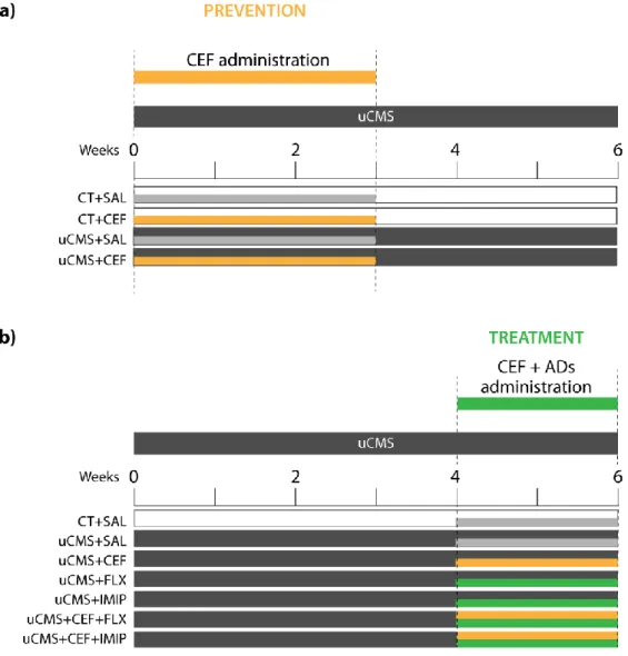
Ninety-four animals were randomly assigned to eleven experimental groups (n=7-10) as described in Figure 2.1. The Prevention set comprised 4 experimental groups: two control groups and two chronically stressed, treated either with saline or CEF (Figure 2.1, a). The Treatment set comprised 7 experimental groups: one control group and six chronically stressed groups treated with saline, CEF and/or ADs – fluoxetine or imipramine (Figure 2.1, b). Details on the treatments carried out are given below.
For the sake of simplicity, animals from the Prevention set, treated in the first 3 weeks of the uCMS protocol will be, from now on, referred to as “Prevention Groups” (CT+SAL, CT+CEF, uCMS+SAL, uCMS+CEF; Figure 2.1, a). Groups belonging to the Treatment set, treated in the last two weeks of the uCMS protocol will be referred to as “Treatment Groups” (CT+SAL, uCMS+SAL, uCMS+CEF, uCMS+FLX, uCMS+IMIP, uCMS+CEF+FLX, uCMS+CEF+IMIP; Figure 2.1, b).
All procedures were carried out in accordance with EU Directive 2010/63/EU and NIH guidelines on animal care and experimentation.
Figure 2.1 Schematic representation of the experimental groups. Ceftriaxone (CEF) was
administered in two different time points. a) administration in the first 3 weeks of the uCMS protocol (Prevention); saline (SAL) and ceftriaxone (CEF) was administrated either to control (CT) and stressed (uCMS) animals. b) administration in the last 2 weeks of uCMS protocol (Treatment); ceftriaxone (CEF) was administrated either to control (CT) and stressed (uCMS) animals; antidepressants (ADs) imipramine (IMIP) and fluoxetine (FLX), were administered alone or in combination with CEF.
2.2 UNPREDICTABLE CHRONIC MILD STRESS (UCMS) PROTOCOL
An unpredictable chronic mild stress (uCMS) protocol was applied for 6 weeks as previously described (Bessa et al., 2009b). Briefly, the uCMS protocol encompasses several mild stressors: confinement to a restricted space for 1h; overnight food deprivation followed by 1h of exposure to inaccessible food; overnight water deprivation followed by 1h of exposure to an empty bottle; overnight damp bedding; inverted light/dark cycles; exposure to stroboscopic lights during 4h
and noise exposure during 4. Animals are random- and uninterruptedly exposed to these stressors during 6 weeks. Control animals were handled gently every week throughout the 6 weeks protocol.
2.3 DRUG TREATMENT
Groups of stressed animals were administered with the different drugs and at distinct time points to address the different objectives proposed (Figure 2.1). In the Prevention set, ceftriaxone (CEF) was daily administered intraperitoneally (i.p.; 200 mg/Kg; Labesfal, Portugal) to non-stressed controls (CT) and non-stressed (uCMS) groups of animals during the first 3 weeks of uCMS exposure. In the Treatment set, CEF, as well as two antidepressants (ADs) from different classes, fluoxetine (FLX, selective serotonin reuptake inhibitor-SSRI; 10 mg/kg in ultra-pure water; Kemprotec, Middlesborough, UK) and imipramine (IMIP, Tricyclic antidepressant; 10 mg/kg in 0.9% saline solution; Sigma-Aldrich, St Louis, MO, USA) were daily administered, intraperitoneally, alone or in combination with CEF (200 mg/Kg; i.p.) to additional stressed groups of animals during the last 2 weeks of the uCMS protocol.
Non-stressed control (CT+SAL) and stress-exposed (uCMS+SAL) groups were administrated with saline (SAL), used as vehicle. The doses were chosen based on previous studies (Bessa et al., 2009a; Rothstein et al., 2005).
2.4 BEHAVIOR TESTS
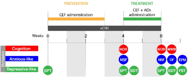
Behavioral tests were performed at week 4 and at the end of week 6 of the uCMS protocol as depicted in Figure 2.2.