Feedback control of chromosome separation by a
midzone Aurora B gradient
Olga Afonso,1* Irina Matos,1*† António J. Pereira,1 Paulo Aguiar,1,2 Michael A. Lampson,3 Helder Maiato1,4‡
1Chromosome Instability and Dynamics Laboratory, Instituto de Biologia Molecular e Celular, Universidade do Porto, Rua do Campo Alegre 823, 4150-180 Porto, Portugal. 2Center for Mathematics, Universidade do Porto, Rua do Campo Alegre 687, 4169-007 Porto, Portugal. 3Department of Biology, University of Pennsylvania, Philadelphia, PA 19104, USA. 4Cell Division Unit, Department of Experimental Biology, Faculdade de Medicina, Universidade do Porto, Alameda Prof. Hernâni Monteiro, 4200-319 Porto, Portugal.
*These authors contributed equally to this work. †Present address: Laboratory of Mammalian Cell
Biology and Development, Howard Hughes Medical Institute, The Rockefeller University, New York,
NY 10065, USA. ‡Corresponding author. E-mail: [email protected]
Originally published in Science 345(6194), 332–336, July 18, 2014.
DOI: 10.1126/science.1251121
Accurate chromosome segregation during mitosis requires the physical separation of sister chromatids before nuclear envelope reassembly (NER). However, how these two processes are coordinated remains unknown. Here, we identified a conserved feedback control mechanism that delays chromosome decondensation and NER in response to incomplete chromosome separation during anaphase. A midzone-associated Aurora B gradient was found to monitor chromosome position along the division axis and to prevent premature chromosome decondensation by retaining Condensin I. PP1/PP2A phosphatases counteracted this gradient and promoted chromosome decondensation and NER. Thus, an Aurora B gradient appears to mediate a surveillance mechanism that prevents chromosome decondensation and NER until effective separation of sister chromatids is achieved. This allows the correction and reintegration of lagging chromosomes in the main nuclei before completion of NER.
The formation of a nuclear envelope that compartmentalizes genomic DNA involves the recruitment of membranes around the decondensing chromatin and insertion of nuclear pore complexes (NPCs) at the anaphase-telophase transition of mitosis (1). However, it is unknown how cells coordinate nuclear envelope reassembly (NER) with the spatial separation of chromosomes during anaphase. Here, we found that the spindle elongation velocity and the respective duration of anaphase in Drosophila S2 cells were inversely correlated, which suggested that incomplete chromosome separation in spindles that elongate more slowly is compensated by increasing anaphase duration (fig. S1, A to C). Pharmacological or RNA
interference (RNAi)–based attenuation of spindle elongation velocity also correlated with an
in human cells (fig. S2, A to C and F to H, and fig. S3, A to C). NER was also delayed on lagging chromosomes and DNA bridges relative to the main nuclei (Fig. 2, A to C). In most cases (~50%), this promoted the correction and reintegration of lagging chromosomes into the main nuclei, and only a smaller fraction (~20%) formed micronuclei. Thus, the anaphase-telophase transition in metazoans cannot be explained by a “clock” model that is set at the onset of anaphase but must take into account the effective separation of sister chromatids before triggering NER. This spatial control of NER appears to be important for the fidelity of chromosome segregation.
The chromosomal passenger protein Aurora B relocates from centromeres to the spindle midzone in anaphase, which produces a phosphorylation gradient (2). Aurora B inactivation on chromatin is also required for chromosome decondensation and NER (3). We noticed that NER was inversely correlated with Aurora B activity on chromosomes and to the proximity of the
spindle midzone (Fig. 2, D and D′; fig. S3, D and D′; and fig. S4, A and A′). Measurement of Aurora
B activity in S2 cells using a chromatin-targeted Förster resonance energy transfer (FRET) sensor (2) revealed a lower Aurora B activity (FRET increase) as chromosomes separated during anaphase (fig. S4, B to G). Thus, chromosome position along the division axis appears to be monitored by a midzone-associated Aurora B activity gradient that spatially controls NER.
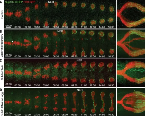
To test this idea, we performed laser microsurgery in metaphase S2 cells to generate acentric chromosome fragments (i.e., devoid of kinetochores and centromeric Aurora B) and found that NER was significantly delayed or inhibited on the lagging acentric fragments (9 out of 11 cells) (Fig. 3, A and B). Furthermore, Aurora B accumulation at the spindle midzone and the respective duration of anaphase were correlated (fig. S5, A and C). Prevention of Aurora B localization at the spindle midzone by depletion of the conserved kinesin-6 Subito (Mklp2) (4, 5) extended spindle elongation and anaphase duration (Fig. 3C and fig. S5, A, B, D, and E). Given that global Aurora B inhibition at anaphase onset with the Drosophila-specific Aurora B inhibitor Binucleine-2 (Bi-2) (6) did not perturb anaphase duration (fig. S6A and Fig. 4, A and B), we attributed the anaphase delay after Subito RNAi to Aurora B retention on chromatin (3, 4). NER occurred simultaneously on all chromosomes after Subito RNAi (7 out of 8 cells) or global Aurora B inhibition (12 out of 12 cells), independently of their position along the cell division axis (Fig. 3D and fig. S6A). Thus, Aurora B localization at the spindle midzone is required for spatial regulation of NER. Global Aurora B inhibition at anaphase onset compromised the separation of sister chromatids and led to the formation of polyploid cells with a single nucleus (Fig. 4, A to C, and fig. S7), which indicated that Aurora B activity is required to delay NER in response to incomplete chromosome separation. Similar findings were observed in human cells (fig. S2, D to H, and fig. S3, A to C).
PP1 and PP2A phosphatases are implicated in NER and counteract Aurora B activity on
chromosomes (7–9). Inhibition of PP1/PP2A with 300 nM okadaic acid (OA) at anaphase onset
prevented chromosome decondensation and NER and only slightly attenuated chromosome segregation velocity relative to controls (Fig. 4, A and C, and fig. S7). Specific inhibition of PP1-87B and PP2A-C by RNAi confirmed that both phosphatases were independently required for timely NER (Fig. 4, D and E, and fig. S8, A to F).
Aurora B regulates chromosome condensation by recruiting its substrate Condensin I
(Barren/Cap-H in Drosophila) to chromosomes (10–14), and its localization at the spindle
anaphase onset prevented Barren-GFP removal from chromatin, and chromosomes remained condensed (fig. S9, D and D′). Finally, RNAi-mediated depletion of Barren was reminiscent of Aurora B inhibition, with cells completing NER before effective chromosome separation (fig. S9, E to H). Histone H3 phosphorylation on S10 and S28 did not affect the anaphase-telophase transition in S2 cells (fig. S10, A to E). Condensin I recruitment to chromosomes during anaphase is thus spatially regulated by the counteracting activities of Aurora B and PP1/PP2A to control chromosome decondensation and NER.
To investigate whether Aurora B inhibition is sufficient to trigger chromosome decondensation and NER, we kept Cdk1 activity constitutively high by the transient expression of nondegradable
D90Cyclin B–GFP (fig. S11, A, B, and D), combined or not, with Aurora B inhibition by Bi-2 at
anaphase onset. Cells arrested in anaphase for several hours under both conditions (fig. S11, C and G). Thus, in addition to Aurora B, Cdk1 inhibition is required for chromosome
decondensation and NER, as shown previously (16–18). Expression of D90Cyclin B–GFP further
impaired spindle elongation and Aurora B relocalization from centromeres to the spindle
midzone during anaphase (fig. S11, E, F, H, and I), as reported in mammalian cells (18–20). In
contrast, Cdk1 inhibition at anaphase onset with RO-3306 accelerated NER in the main nuclei
relative to controls and Bi-2–treated cells, independently of chromosome separation, and also
decreased spindle elongation velocity (fig. S6, A and B; fig. S12, A to D; and fig. S13, A to D). Thus,
Cdk1 appears to work as a “clock” that temporally regulates the formation of a midzone Aurora
B–based “ruler,” which explains the observed minimal anaphase duration time (~5 min) (Fig. 1D and fig. S1C). Indeed, Cdk1 inhibition at anaphase onset led to a fast accumulation of Aurora B at the spindle midzone (fig. S12, E and F) and, contrary to Aurora B inhibition, NER never took place on lagging chromosomes (10 out of 10 cells) (fig. S6B). Simultaneous inhibition of Aurora B and PP1/PP2A or Cdk1 and PP1/PP2A (with or without Aurora B inhibition) at anaphase onset prevented timely chromosome decondensation (fig. S14). Thus, PP1/PP2A activities are required to dephosphorylate Aurora B and Cdk1 substrates during anaphase and for the spatiotemporal regulation of chromosome decondensation and NER.
sustain Aurora B activity to inhibit abscission during cytokinesis—the so-called NoCut
checkpoint that monitors clearance of chromatin from the spindle midzone (24–26). The critical
differences between these potential checkpoints are that the spatial regulation of NER involves a default mechanism mediated by a midzone Aurora B gradient that is active during a normal mitosis, whereas NoCut is activated only in the presence of chromatin at the cleavage furrow for successful completion of cytokinesis.
REFERENCES AND NOTES
1. S. Güttinger, E. Laurell, U. Kutay, Nat. Rev. Mol. Cell Biol. 10, 178–191 (2009).
2. B. G. Fuller et al., Nature 453, 1132–1136 (2008).
3. K. Ramadan et al., Nature 450, 1258–1262 (2007).
4. J. M. Cesario et al., J. Cell Sci. 119, 4770–4780 (2006).
5. U. Gruneberg, R. Neef, R. Honda, E. A. Nigg, F. A. Barr, J. Cell Biol. 166, 167–172 (2004).
6. Y. Smurnyy, A. V. Toms, G. R. Hickson, M. J. Eck, U. S. Eggert, ACS Chem. Biol. 5, 1015–1020
(2010).
7. P. Vagnarelli et al., Dev. Cell 21, 328–342 (2011).
8. M. H. Schmitz et al., Nat. Cell Biol. 12, 886–893 (2010).
9. J. Qian, M. Beullens, B. Lesage, M. Bollen, Curr. Biol. 23, 1136–1143 (2013).
10. N. Nakazawa, R. Mehrotra, M. Ebe, M. Yanagida, J. Cell Sci. 124, 1795–1807 (2011).
11. R. Giet, D. M. Glover, J. Cell Biol. 152, 669–682 (2001).
12. J. J. Lipp, T. Hirota, I. Poser, J. M. Peters, J. Cell Sci. 120, 1245–1255 (2007).
13. A. Takemoto et al., Nucleic Acids Res. 35, 2403–2412 (2007).
14. K. Tada, H. Susumu, T. Sakuno, Y. Watanabe, Nature 474, 477–483 (2011).
15. G. Neurohr et al., Science 332, 465–468 (2011).
16. S. Sigrist, H. Jacobs, R. Stratmann, C. F. Lehner, EMBO J. 14, 4827–4838 (1995).
17. D. H. Parry, P. H. O’Farrell, Curr. Biol. 11, 671–683 (2001).
18. S. P. Wheatley et al., J. Cell Biol. 138, 385–393 (1997).
19. M. Murata-Hori, M. Tatsuka, Y. L. Wang, Mol. Biol. Cell 13, 1099–1108 (2002).
20. S. Hümmer, T. U. Mayer, Curr. Biol. 19, 607–612 (2009).
21. L. H. Hartwell, T. A. Weinert, Science 246, 629–634 (1989).
22. A. Janssen, M. van der Burg, K. Szuhai, G. J. Kops, R. H. Medema, Science 333, 1895–1898
(2011).
23. K. Crasta et al., Nature 482, 53–58 (2012).
24. C. Norden et al., Cell 125, 85–98 (2006).
25. P. Steigemann et al., Cell 136, 473–484 (2009).
26. M. Mendoza et al., Nat. Cell Biol. 11, 477–483 (2009).
ACKNOWLEDGMENTS
Fig. 1. Incomplete chromosome separation delays the anaphase-telophase transition. (A) Control, Taxol-treated, and kinesin-like protein KLP10A RNAi S2 cells stably expressing Lamin
B–GFP and mCherry–α-tubulin (mCherry-TUB).Time shown as minutes: seconds. (B and C)
Fig. 4. PP1/PP2A phosphatases counteract Aurora B and are required to promote chromosome decondensation and NER. (A) Acute Aurora B (Bi-2) or PP1/PP2A inhibition (OA) at anaphase
onset in S2 cells stably expressing Lamin B–GFP and mCherry–α-tubulin. (B) Quantification of
anaphase duration until DNA decondensation or NER in control cells (n = 16 cells per condition)
and Bi-2–treated cells (n = 10 cells per condition). Time shown as minutes:seconds. (C)
Quantification of half-spindle elongation velocity. (D) RNAi against specific PP1 or PP2A
subunits (n = 8 cells per condition) in the Lamin B–GFP/mCherry–α-tubulin cell line

![Fig. 2. NER is delayed on lagging chromosomes and DNA bridges. (A and B) S2 cells stably expressing Nup107-mRFP and H2B-GFP showing a delay in NER on lagging chromosomes [white arrowheads in (A)] and (B) DNA bridges](https://thumb-eu.123doks.com/thumbv2/123dok_br/16776001.748319/7.892.134.683.194.654/delayed-lagging-chromosomes-bridges-expressing-showing-chromosomes-arrowheads.webp)