December 2012
Viviana Gomes Correia
Graduated in Molecular and Cell Biology
Genetic and Functional Analysis
of the Bacillus subtilis BSP1
gam Cluster
Dissertation to obtain the Master degree in Biotechnology
Supervisor: Isabel M. G. de Sá Nogueira,
Associated Professor, FCT-UNL
Jury:
President: Prof. Doutor Pedro Miguel Ribeiro Viana Baptista Examiner: Prof. DoutorAdriano José Alves Oliveira Henriques Member: Prof.ª DoutoraIsabel Maria Godinho de Sá Nogueira
III Genetic and Functional Analysis of the Bacillus subtilis BSP1 gam Cluster
Copyright© reserved to Viviana Gomes Correia, FCT/UNL
The Faculty of Science and Technology and the New University of Lisbon have the perpetual right, and without geographical limits, to archive and publish this dissertation through press copies in paper or digital form, or by other known form or any other that will be invented, and to divulgate it through scientific repositories and to admit its copy and distribution with educational or research objectives, non-commercial, as long as it is given credit to the author and editor.
V
A
CKNOWLEDGEMENTS
“Sê todo em cada coisa. Põe quanto és No mínimo que fazes.”
Ricardo Reis in Odes
First and foremost I offer my sincerest gratitude to my Professor and Supervisor, Isabel de Sá Nogueira, not only for her support and guidance throughout my thesis, but mainly for her contagious passion for science.
Thank you Isabel, Mário and Lia for the wise advices, precious teachings and of course, for the cheerful atmosphere at the 327 lab, even at late hours. One simply could not wish for a better environment to work in. I’ve learned so much from working with you guys! Also thanks Sónia, Diana, Liliana and Maria João.
I would also like to extend my thanks to every worker, researcher and professor at DCV, specially to Raquel, for the constant good mood and to Dr. Alan Phillips and Jorge Santos, for excellent previous scientific orientation.
Thanks Inês and Joana for the friendship; Pernes and Gralheira for the stupid jokes at lunch time; and all the Biotechnology Master’s colleagues of 2010/11 class. Thanks Sofia, Vasco, Tânia, Nadine, Filipa, Cátia, Rui and Marino, BCM’s future brightest scientists, for all the good moments.
I would also like to acknowledge all the teachers and friends that I´ve made along the way, especially to Professors Manuela, Mónica, Pedro and Ana Bela. Their commitment and enthusiasm motivated me to proceed with my studies and always give my best.
Most importantly, I wish to thank to the ones that made this possible, my family! My Mom, Anabela, for being my cornerstone, for made me believe in my value and for teaching me how to never give up. My Dad, João, for the financial support and the valuable lessons about hard working and dedication. Thanks Jonny, my not so little brother, for making me repeat 8th grade all over again while doing the thesis. Thanks Aliete and Gaspar for everything. Special thanks to my Grandparents, for being so proud of me. My work is dedicated to you.
Finally, I would like to thank Sérgio, for so many years of friendship, love and laughter. Thank you so much for always being there above all and no matter what.
A special thanks to Professor Adriano Henriques, Cláudia Serra (Microbial Development Lab - ITQB) and Dr. Ghislain Schyns (DSM) for kindly provide BSP1 strain and for sharing unpublished data.
I could not forget Bacillus subtilis, for its, not always easy, collaboration.
VII
A
BSTRACT
Mannans (linear mannan, glucomannan, galactomannan and galactoglucomannan) are the major constituents of the hemicellulose fraction in softwoods and show great importance as a renewable resource for fuel or feedstock applications. As complex polysaccharides, mannans can only be degraded through a synergistic action of different mannan-degrading enzymes, mannanases.
Microbial mannanases are mainly extracellular enzymes that can act in wide range of pH and temperature, contributing to pulp and paper, pharmaceutical, food and feed, oil and textile successful industrial applications. Knowing and controlling these microbial mannan-degrading enzymes are essential to take advantage of their great biotechnological potential.
The genome of the laboratory 168 strain of Bacillus subtilis carries genes gmuA-G dedicated to the degradation and utilization of glucomannan, including an extracellular -mannanase. Recently, the genome sequence of an undomesticated strain of B. subtilis, BSP1, was determined. In BSP1, the gmuA-G operon is maintained, interestingly, however, a second cluster of genes was found (gam cluster), which comprise a second putative extracellular β -mannanase, and most likely specify a system for the degradation and utilization of a different mannan polymer, galactoglucomannan.
The genetic organization and function of the gam cluster, and whether its presence in BSP1 strain results in new hemicellulolytic capabilities, compared to those of the laboratory strain, was address in this work.
In silico and in vivo mRNA analyses performed in this study revealed that the gam cluster, comprising nine genes, is organized and expressed in at least six different transcriptional units. Furthermore, cloning, expression, and production of Bbsp2923 in Escherichia coli was achieved and preliminary characterization shows that the enzyme is indeed a β-mannanase. Finally, the high hemicellulolytic capacity of the undomesticated B. subtilis BSP1, demonstrated in this work by qualitative analyses, suggests potential to be used in the food and feed industries.
K
EYWORDSIX
R
ESUMO
Os mananos (manano linear, glucomanano, galactomanano e galactoglucomanano) são as principais hemiceluloses presentes em gimnospérmicas (softwoods), constituindo uma importante fonte de material renovável. Como polissacáridos complexos, os mananos são apenas completamente degradados pela acção conjunta de várias enzimas hidrolíticas, denominadas mananases.
As mananases de origem microbiana são essencialmente enzimas extracelulares que podem actuar numa ampla gama de temperatura e pH, o que lhes confere excelentes qualidades para serem usadas nas indústrias da pasta e do papel, farmacêutica, alimentar e de rações, têxtil ou do petróleo. Conhecer e controlar estas enzimas é fundamental para tirar partido do seu potencial biotecnológico.
O genoma da estirpe de laboratório Bacillus subtilis 168 contém genes, gmuA-G, dedicados à degradação e utilização do glucomanano, os quais incluem uma β-mananase extracelular. Recentemente, foi determinada a sequência do genoma de uma estirpe “não domesticada” de B. subtilis, BSP1. Em BSP1, o operão gmuA-G é mantido mas, curiosamente, está presente um segundo aglomerado de genes (gam cluster) que inclui uma segunda possível β-mananase extracelular. O gam cluster deverá constituir um sistema para a degradação e utilização de um outro tipo de manano, o galactoglucomanano.
Neste projecto, foi estudada a organização genética e funcional do gam cluster, bem como se a sua presença no genoma de BSP1 lhe confere capacidades hidrolíticas adicionais, quando comparada com a estirpe laboratorial B. subtilis 168.
As análises realizadas in silico e in vitro ao mRNA, revelaram que o gam cluster inclui nove genes e está organizado em pelo menos seis unidades transcricionais. Além disso, Bbsp2923 foi clonada, expressa e produzida em Escherichia coli. A sua caracterização preliminar mostra que a enzima é de facto uma β-mananase. Finalmente, a elevada capacidade hemicelulolítica da estirpe “não domesticada” B. subtilis BSP1, demonstrada neste trabalho, sugere o seu potencial para aplicações nas indústrias alimentar e de rações-animais.
P
ALAVRAS-
CHAVEXI
C
ONTENTS
Acknowledgements ... V
Abstract ... VII
Resumo ... IX
Contents ... XI
Figures Index ... XIII
Tables Index ... XV
Abbreviations, Symbols and Notations ... XVII
1. General Introduction ... 1
1.1. Bacillus subtilis– An overview ... 3
1.2. Carbohydrate uptake ... 4
1.2.1. PTS system ... 5
1.2.2. ABC transporters ... 5
1.3. Hemicellulose degradation ... 6
1.3.1. Mannan structure ... 7
1.3.2. Mannan-degrading enzymes ... 9
1.3.3. Biotechnological application of Mannanases ... 11
1.4. Heteromannans degradation in B. subtilis ... 14
1.5. A novel mannan degrading system –gam cluster ... 16
1.6. Scope of the thesis ... 18
2. Materials and Methods ... 19
2.1. Substrates ... 21
2.2. Bacterial strains and growth conditions ... 21
2.3. DNA manipulation and sequencing ... 22
2.4. Plasmids construction ... 23
XII
2.6. Qualitative and comparative analysis of substrate hydrolysis ... 25
2.7. Hydrolytic activity assays ... 25
2.9. Protein analysis and Quantification ... 27
2.10. β-Mannanase activity assay ... 27
3. Results and Discussion ... 29
3.1. Genetic Analysis of the gam Cluster ... 31
3.1.1. Transcriptional organization of the gam cluster ... 31
3.1.2. Sequence Analysis of the gam cluster ... 35
3.2. Functional Characterization of the gam Cluster ... 43
3.2.1. Qualitative analysis of substrate hydrolysis ... 43
3.2.2. Hydrolytic activity assays ... 45
3.3. Cloning and Preliminary Characterization of the Bbsp2923 ... 46
3.3.1. Sequence analysis of Bbsp2923 ... 47
3.3.2. Expression of Bbsp2923 in E. coli ... 51
3.3.3. β-Mannanase activity assay ... 52
4. Concluding Remarks and Future Perspectives ... 55
5. References ... 61
6. Appendices ... 69
Appendix 6.1 ... 71
Appendix 6.2 ... 72
Appendix 6.3 ... 73
Appendix 6.4 ... 75
Appendix 6.5 ... 76
XIII
F
IGURES
I
NDEX
Figure 1.1– Typical mannan and heteromannans structure. ... 7
Figure 1.2 – Mannan-based saccharides and the mannanases responsible for their degradation. ... 9
Figure 1.3 – GmuA-G model for glucomannan utilization in B. subtilis ... 15
Figure 1.4–B. subtilis 168 chromosome region (A) replaced by gam cluster in B. subtilis BSP1 strain (B). ... 17
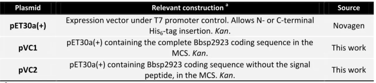
Figure 3.1 – RT-PCR results obtained with RNA extracted from cells grown in different conditions and using distinct pairs of primers ... 33
Figure 3.2– Transcriptional organization of the region Bbsp2929-2924 of gam cluster ... 34
Figure 3.3– Annotated sequence of the putative promoter regions of gam cluster ... 40
Figure 3.4 – Stem-loop structure of a putative transcription terminator found downstream of Bbsp2924 ... 42
Figure 3.5– Complete gam cluster scheme with identified promoter regions and hairpins ... 42
Figure 3.6– Hydrolysis test on Locust Bean Gum (LBG) and Birchwood Xylan ... 44
Figure 3.7 – Illustration of modular organization of Bbsp2923, adapted from BLASTp analysis. ... 47
Figure 3.8 – N-terminal sequence alignment of Bbsp2923 and other GH-26 members correlated with secondary structure prediction information ... 50
Figure 3.9 – Overproduction of recombinant Bbsp2923-His6 in E. coli BL21 pLysS (DE3) and E. coli Rosetta™ (DE3) pLysS strains harboring pVC2 ... 52
XV
T
ABLES
I
NDEX
Table 1.1– List of the major industrial enzymes produced by Bacillus spp. ... 4
Table 1.2– Galactose and mannose content of popular galactomannans ... 8
Table 1.3– Available information on commercial mannanases.. ... 11
Table 2.1– List of bacterial strains used in this work. ... 22
Table 2.2– List of oligonucleotides used in this project. ... 23
Table 2.3– List of used or constructed plasmids. ... 24
Table 2.4– List of oligonucleotides used for RT-PCR assays... 25
Table 3.1– Predicted signals for translation initiation in gam cluster sequence. ... 36
Table 3.2– Comparison of gam cluster products to other proteins in available databases using BLASTp tool. ... 37
Table 3.3– Identified cre sequences and location. ... 38
Table 3.4– Typical B. subtilis sigma-factor (σ) binding sequences identified in some gam genes promoters. ... 39
Table 3.5– Extracellular hydrolytic activity of B. subtilis BSP1 and 168 strains towards distinct substrates. ... 45
XVII
A
BBREVIATIONS
,
S
YMBOLS AND
N
OTATIONS
aa– Amino acid
ABC– ATP-binding cassette
ATP– Adenosine triphosphate
BLAST– Basic Local Alignment Search Tool
bp– Base pairs
BSA– Bovine Serum Albumin
CAZy– Carbohydrate-Active enZYmes Database
CBM – Carbohydrate Binding Domain
CCR– Carbon catabolite repression
Cm– Chloramphenicol
CRE – Catabolic responsive elements
DNA– Deoxyribonucleic acid
dNTPs– Deoxynucleotide Triphosphates
EDTA– Ethylenediamine tetra-acetic acid
gam– Galactoglucomannan utilization cluster
GH– Glycosyl Hydrolase
gmu– Glucomannan utilization operon
IPTG– Isopropyl-β-D-galactopyranoside
kb– Kilobase pairs
kDa– KiloDalton
Km– Kanamycin
LA– Luria-Bertani Agar, solid LB medium
LB– Luria-Bertani medium
LBG– Locust Bean Gum, source of galactomannan
XVIII
mRNA– Messenger ribonucleic acid
NBD– Nucleotide-binding domain
NCBI– National Center for Biotechnology Information
OD – Optical Density
ORF– Open Reading Frame
PC buffer– Citrate-Phosphate buffer
PCR– Polymerase chain reaction
PEP– Phosphoenolpyruvate
PMSF– Phenylmethylsulfonylfluoride
PTS– Phosphotransferase system
Rbs– Ribosome binding site
RNA– Ribonucleic acid
rpm– Revolutions per minute
RT-PCR– Reverse transcription polymerase chain reaction
SBP– Solute-binding protein
SDS-PAGE– Sodium dodecyl sulphate polyacrylamide gel electrophoresis
SF– Sugar Free medium
SFA– Sugar Free agar
TE– Tris-EDTA
TMD– Transmembranar domain
tRNA– Transfer ribonucleic acid
TSS – Translational start site
1
Chapter 1
3
1.
General Introduction
1.1.
Bacillus subtilis
–
An overview
Bacillus subtilis is a gram-positive, rod-shape, endospore-forming bacterium, ubiquitous found in soil, water sources or in association with plants and animal gastrointestinal tract (Priest, 1993; Casula & Cutting, 2002). B. subtilis, whose genome has been completely sequenced (Kunst et al., 1997), was the first non-pathogenic microorganism to be transformed (Anagnostopoulos & Spizizen, 1961) and since then has been used as the gram-positive paradigm for fundamental and applied research.
Bacillus strains, including B. subtilis, have developed a series of responses in order to cope with nutrient supply fluctuation, commonly felt in their natural environment. These responses can be observed in the laboratory towards the end of exponential growth phase or in stationary phase, and include onset of motility, production of hydrolytic enzymes, competence to take up exogenous DNA and sporulation (Harwood, 1992).
Sporulation is the ability to form endospores – highly differentiated dormant structures, resistant to desiccation, starvation or radiation, and used as a survival as well as new niche colonization techniques. Works of Casula & Cutting (2002) and Tam and colleagues (2006) proved that spores indeed germinate in the gastrointestinal animal tract, disproving the idea that vegetative cells only develop in the soil or in association with decaying vegetative material. Spores are widely used as probiotics in human diet improving gastrointestinal microflora health by preventing pathogenic bacteria onset (Casula & Cutting, 2002).
The metabolic diversity of Bacillus spp. led to the use of these organisms in a wide range of industrial microbial processes, also known as “white biotechnology”, which includes the production of carbohydrate-, lipid- and protein-degrading enzymes, antibiotics, fine biochemicals (vitamins) and insecticides (Harwood, 1992).
4
particular relevance for extracellular protein secretion (Ferrari et al., 1993). It’s estimated that 50-60% of the total enzymes market is produced or derived from Bacillus spp. (Schallmey et al., 2004; Westers et al., 2004).
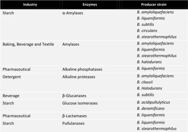
Table 1.1– List of the major industrial enzymes produced by Bacillus spp. Adapted from Harwood (1992) and Schallmey et al. (2004).
Industry Enzymes Producer strain
Starch α-Amylases B. amyloliquefaciens
B. liqueniformis B. subtilis B. circulans
B. stearothermophilus
Baking, Beverage and Textile Amylases B. amyloliquefaciens
B. liqueniformis B. stearothermophilus B. halodurans
Pharmaceutical Alkaline phosphatases B. liqueniformis Detergent Alkaline proteases B. amyloliquefaciens
B. clausii B. Halodurans
Beverage β-Glucanases B. subtilis
Starch Glucose isomerases B. acidipullulyticus
B. deramificans
Pharmaceutical β-Lactamases B. liqueniformis
Starch Pullulanases B. liqueniformis
B. stearothermophilus
1.2.
Carbohydrate uptake
5 Membrane transport systems found in all living organisms, including B. subtilis, can be classified into four major types: channel proteins, characterized by an energy-independent transport process; secondary active transporters, which use chemiosmotic energy to drive transport; primary active transporters such as ATP-binding cassette (ABC) transporters, that use chemical, electrical or solar energy as transportation force; and group translocating systems, including the phosphotransferase system (PTS) which transport and concomitantly phosphorylate the substrate. In B. subtilis, well characterized carbohydrates uptake systems belong mainly to PTS and ABC transporters families (Deutscher et al., 2002).
After entering the cell, all carbohydrates are converted into intermediates of one of the central carbohydrate-degrading pathways – glycolysis, pentose phosphate or Entner-Doudoroff pathways (Deutscher et al., 2002).
1.2.1.
PTS system
The phosphoenolpyruvate (PEP): carbohydrate phosphotransferase system or simply PTS, is a complex carbohydrate transportation system that exists in both positive and gram-negative bacteria. The PTS system is involved not only in the transport and phosphorylation of different carbon sources and in movement towards the carbohydrates (chemotaxis) but also in the regulation of other metabolic pathways (Postma et al., 1993).
This system catalyses the simultaneous transport and phosphorylation of the substrate through a phosphorylation cascade involving protein enzyme I (EI), HPr (histidine-containing protein) and PTS sugar-specific transporter proteins – IIA, IIB and IIC. PEP is used as energy source and phosphoryl group provider (Deutscher et al., 2002).
1.2.2.
ABC transporters
Existing from microorganisms to man, ABC transporters are membrane proteins that couple the translocation of several substrates across cellular membrane to the hydrolysis of ATP (Higgins, 1992).
6
(e.g. saccharides) or the excretion of toxic compounds, contributing to drug and antibiotic resistance (Davidson & Chen, 2004).
Depending on the direction of the transport, ABC transporters can be classified as exporters or importers. The latest type is only present in prokaryotes and requires a solute-binding protein (SBP, also known as periplasmic solute-binding protein) for substrate capture and delivery to the external face of the transporter. In gram-positive bacteria, like B. subtilis, the SBP is often a lipoprotein bound to the cytoplasmic membrane external side or fused to the ABC transporter, conferring high affinity, specificity and directionality to a certain substrate or solute family (Higgins, 2001).
Generally, the basic unit of an ABC transporter comprises four core domains: two transmembranar domains (TMDs) that provide a passageway for substrates and two cytoplasmic nucleotide-binding domains (NBDs) that bind and hydrolyse ATP. Usually, in ABC importers the TMDs and NBDs are encoded as separate polypeptides, although it is known that these domains can be fused to each other as multidomain polypeptides, in any combination (Higgins, 2001).
1.3.
Hemicellulose degradation
Hemicelluloses are polysaccharides of the plant cell wall with an important structural role in association with cellulose, lignin and pectin (McNeil et al., 1984).
Hemicellulose fraction corresponds to one third of total components available in plants and is the second most abundant heteropolymer in nature with great importance as a renewable resource for fuel or feedstock applications. This makes hemicellulases, the major industrially important enzymes right after proteases and cellulases (Dhawan & Kaur, 2007; Moreira & Filho, 2008; Chauhan et al., 2012).
7
1.3.1.
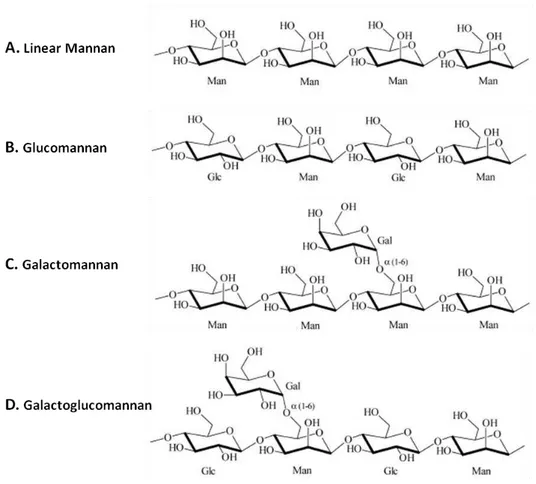
Mannan structure
Mannan is an important hemicellulose and can be classified in two main categories – homo- or linear mannans and heteromannans (glucomannans, galactomannans and galactoglucomannans) (Fig. 1.1). Each of these polysaccharides possesses a backbone of β -1,4-linked D-mannose residues (linear mannan) which might be combined with D-glucose (glucomannan) or/and D-galactose (galactomannan/galactoglucomannan). The ratio between different sugar residues alters mannan properties and is characteristic of each plant species.
Mannans can be found in gums and are used as thickening agents in the food (ice-creams and sauces) and pharmaceutical (hair gels, shampoos or tooth-pastes) industries (Chauhan et al., 2012).
Figure 1.1– Typical mannan and heteromannans structure. (A) Linear mannan with a backbone of β-1,4
linked mannose (Man) residues; (B) Glucomannan structure formed by a main chain of β-1,4 linked mannose and glucose (Glc) residues; (C) Galactomannan general structure, a main chain of β-1,4 linked
8
Linear mannans are homopolysaccharides with a backbone of β-1,4 linked D-mannose (β -D-mannopyranosyl) residues (Fig. 1.1-A). These mannans play a major structural role in woods and seed of several plants like ivory nuts (Phytelephas spp.), green coffee beans (Coffea spp.) or coconut kernel (copra). Mannans with linear chains are known to be highly insoluble in water (Moreira & Filho, 2008).
Glucomannans are formed by a chain of randomly arranged β-1,4 linked D-mannose and β -1,4 linked D-glucose (β-D-glucopyranosyl) residues (Fig. 1.1-B). Usually the observed ratio is of one glucose residue for each three mannans (1:3). Glucomannans backbone mannose or glucose units can be acetylated in the O-2 or O-3 position, providing branching points. These heteromannans can be obtained from several plants like ramie (Boehmeria nivea), lupins (Lupinus spp.) or Konjac (Amorphophallus konjac) (Moreira & Filho, 2008).
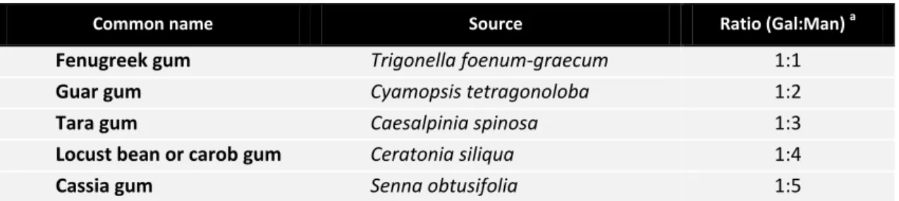
Table 1.2 – Galactose and mannose content of popular galactomannans. Ratio and polysaccharide source are referred. Adapted from Nishinari et al. (2007) and Moreira & Filho (2008).
Common name Source Ratio (Gal:Man) a
Fenugreek gum Trigonella foenum-graecum 1:1
Guar gum Cyamopsis tetragonoloba 1:2
Tara gum Caesalpinia spinosa 1:3
Locust bean or carob gum Ceratonia siliqua 1:4
Cassia gum Senna obtusifolia 1:5
a
Ratio between galactose (Gal) and mannan (Man) residues.
Galactomannans are formed by a backbone of β-1,4 linked D-mannose residues with side chains of single D-galactose (β-D-galactopyranosyl) groups attached though α-1,6 bonds along the main chain (Fig. 1.1-C). Similarly to what happen in other heteromannans, the ratio between different sugar residues characterizes mannan source, altering mannan properties. In table 1.2, galactose/mannose ratio of different popular galactomannans can be seen. The increasing amount of D-galactose side branches (highly hydrophilic) increases galactomannan polymer solubility in water (Moreira & Filho, 2008).
9 branches of α-1,6-linked D-galactose along the chain (Fig. 1.1-D). Some mannose units can be partially substituted by O-acetyl groups in the O-2 or O-3 position, on the average of one acetylated residue for each 3-4 residues. Like galactomannan, galactoglucomannan presents high solubility in water due to D-galactose side chains that prevents macromolecules from aggregate. Norway spruce (Picea abies) is an abundant softwood species that can contain up to 10-20% of acetylated galactoglucomannan (Moreira & Filho, 2008).
Due to the great variety of hemicelluloses available in nature, including mannans, microorganisms have developed over the time many enzymatic systems to overcome this problem and be able to take advantage of this carbon source abundance.
1.3.2.
Mannan-degrading enzymes
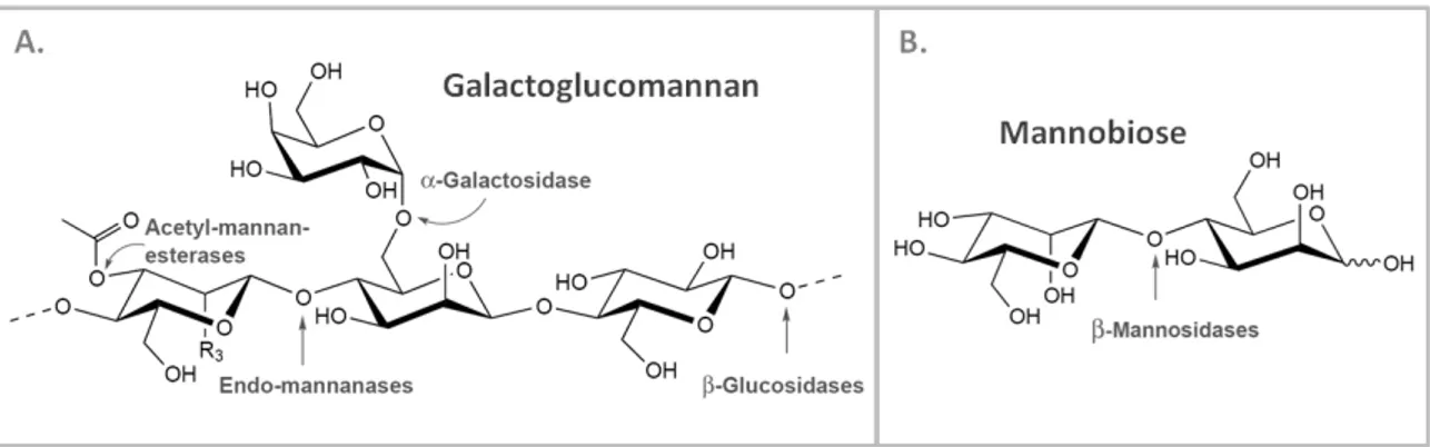
Complex polysaccharides such as mannans depend on a synergistic action of different mannan-degrading enzymes for complete degradation.
Mannan-degrading enzymes or mannanases involved in linear mannan degradation include β-mannanases, β-mannosidases and β-glucosidases. Heteromannans processing requires some additional enzymes like α-galactosidases and acetyl mannan esterases, for the side-chains substituents removal. Figure 1.2 illustrates the enzymatic action involved in the degradation of two representative mannan-based polymers, galactoglucomannan (Fig. 1.2 - A) and mannobiose (Fig. 1.2 - B).
10
β-Mannanases or endo-1,4-β-mannanases (EC 3.2.1.78, mannan endo-1,4-β -mannosidases) are hydrolases that catalyze the random cleavage of β-1,4 mannosidic linkages within the backbone of different homo- and heteromannans. β-Mannanase activity releases short β-1,4-manno-oligosaccharides, mainly mannobiose and mannotriose. Galactomannan and galactoglucomannan backbone substitution pattern possesses a great influence over β -mannanases activity (Dhawan & Kaur, 2007; Moreira & Filho, 2008). These are the most studied mannanases, mainly due to its abundance and biotechnological importance.
β-Mannosidases (EC 3.2.1.25, mannan exo-1,4-β-mannosidases) are exo-type enzymes that cleave β-1,4 mannosidic linkages releasing mannose units from the nonreducing ends of mannans and manno-oligosaccharides (Dhawan & Kaur, 2007; Moreira & Filho, 2008).
β-Glucosidases (EC 3.2.1.21, 1,4-β-D-glucoside glucohydrolases) constitute a group of exo-acting enzymes that hydrolyze β-1,4 bonds, releasing D-glucose residues from the nonreducing end of glucomannan or galactoglucomannan oligomers (Dhawan & Kaur, 2007; Moreira & Filho, 2008).
α-Galactosidases (EC 3.2.1.22, 1,6-α-D-galactoside galactohydrolases) act as debranching enzymes, cleaving α-1,6-side linked D-galactose units of galactomannans and galactoglucomannans backbones (Dhawan & Kaur, 2007; Moreira & Filho, 2008).
Acetyl mannan esterases or N-acetyl mannosamine esterases (EC 3.1.1.6) also act as debranching enzymes by releasing acetyl groups from galactoglucomannan polymers (Dhawan & Kaur, 2007; Moreira & Filho, 2008).
11
1.3.3.
Biotechnological application of Mannanases
Over the past years, mannanases have been isolated from many microbial sources like fungi, actinomycetes and bacteria.
Microbial mannanases are mainly extracellular enzymes that can act on a wide range of pH and temperature, which have contributed to many successful industrial applications (Dhawan & Kaur,2007).
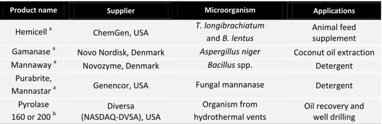
The induction of mannanases production by natural strains normally requires the use of expensive mannan-rich substrates, fact that compromises industrial use. So cloning and expression of mannanases in heterologous hosts for protein overproduction have become a very popular procedure. The production of genetic engineered mannanases with specific alterations and properties that suits commercial applications constitutes other market opportunity (Chauhan et al., 2012). Table 1.3 provides an updated list of commercial mannanases and their suppliers.
Table 1.3– Available information on commercial mannanases. Adapted from Dhawan & Kaur, 2007.
Product name Supplier Microorganism Applications
Hemicell a ChemGen, USA T. longibrachiatum and B. lentus
Animal feed supplement Gamanase a Novo Nordisk, Denmark Aspergillus niger Coconut oil extraction Mannaway a Novozyme, Denmark Bacillus spp. Detergent
Purabrite,
Mannastar a Genencor, USA Fungal mannanase Detergent Pyrolase
160 or 200 b
Diversa (NASDAQ-DVSA), USA
Organism from hydrothermal.vents
Oil recovery and well drilling
a
Endo-mannanase. b
Endo- and exo-mannanase.
12
Following paragraphs summarizes current industrial applications of mannanases.
Biobleaching in pulp and paper industry
The lignin extraction from wood is an essential step for pulp bleaching. Usually this was achieved through an alkaline pre-treatment that hydrolyze hemicelluloses covalently bound to lignin, thus facilitating lignin removal. However, this treatment releases extremely pollutant chlorinated compounds. Enzymatic treatment of pulps, using mannanases along with other hemicellulases as xylanases, constitutes an environmental friendly alternative for bleaching process. Mannanases specific for galactomannan (major hemicellulose in pulps) that remain active at high temperatures and pH are excellent for this application (Dhawan & Kaur, 2007; Chauhan et al., 2012).
Hydrolytic agent in detergent industry
Mannans have high tendency to adsorb to fibres and can be found in gums or thickening agents in many food or beauty products like ice-creams, sauces and hair gels. So, alkaline mannanases stable in detergents have found many applications in laundry segments as stain removal booster for fabrics, health care products and sanitization or hard surface cleaners (Chauhan et al., 2012). Mannaway, Purabrite and Mannastar are some commercially available products (Table 1.3).
Textile and cellulosic fiber processing
Mannanases in combination with other hemicellulases can be used in textile industry, by helping on fibres preparation and cleaning before transformation into yarn. Mannanases are also important in enzymatic scouring and desizing processes for cellulosic material preparation before dyeing (Dhawan & Kaur, 2007).
Hydrolysis of coffee extract
13
Food additives
Mammalian digestive enzymes aren’t able to degrade mannan based substrates. But when
mannans reach the large intestine, probiotic bacteria of the genera Bifidobacteria or Lactobacillus, which naturally produce mannanases, can readily metabolize mannans. The food enrichment with mannan degradation products, mannanoligosaccharides, was proved to have a probiotic effect, enhancing growth and proliferation of human beneficial intestinal microflora (Dhawan & Kaur, 2007).
Mannanases can also be used in food industry, helping fruit and vegetables maceration as well as fruit juices clarification (Chauhan et al., 2012).
Feed improvement
Feed ingredients are rich in mannans, xylans and arabinoxylans. Due to the lack of appropriate degrading enzymes in animals gut, these substrates are misused and nutritional value wasted. Mannanases can be incorporated in animal diets helping the digestion of these feeds, improving nutrient absorption and decreasing digested food viscosity. For this purpose, mannanases must be active over a wide pH range and resistant to proteases (e.g. tripsin, pepsin) in order to resist gastric conditions (Chauhan et al., 2012). Hemicell supplied by ChemGen is an example of industrial use (Table 1.3).
Pharmaceutical applications
Mannanases can be used for economical production of mannose from low-cost mannan rich substrates such as guar gum, copra meal or palm kernel cake. Mannose can be useful in urinary tract infections and intestinal disorders treatment. Besides, mannan can help reduce cholesterol and body fat without protein mass loss. Mannan is also used for conferring fast dissolving and structure properties to tablets (Chauhan et al., 2012).
Oil drilling and extraction
14
mannanases are required, due to the extreme temperatures in the wells (>80⁰C) (Moreira & Filho, 2008).
Mannanases can also be used in enzymatic oil extraction of coconut meat rich in mannans. The enzymatic process eliminates aflatoxin contamination, oxidative rancidity of the products and reduces refinement costs (Dhawan & Kaur, 2007).
Bioethanol production
An enzymatic cocktail, including mannanases, xylanases and cellulases, can be used to effectively hydrolyze lignocellulosic biomass to fermentable sugars for bioethanol production. This enzymatic treatment eliminates the usual expensive heat pre-treatment of biomass (Várnai et al., 2011).
Slime control agents
Alkaline mannanases combined with proteases can prevent slime formation in water purification or cooling systems and sewers or waste water treatment equipments. By degrading mannans rich substrates mannanases can control biofilms formation and bacterial adhesion (Chauhan et al., 2012).
1.4.
Heteromannans degradation in B. subtilis
The genes coding for polysaccharide-hydrolysing enzymes are generally organized in an operon or regulon, together with genes encoding proteins responsible for the uptake of the extracellular hydrolyzed products and the first intracellular steps in their metabolism (Deutscher et al., 2002).
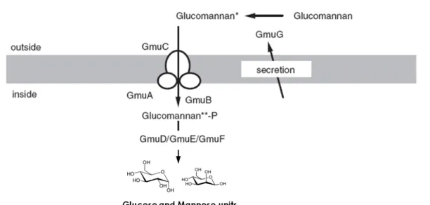
15 The operon gmuBACDREFG or simply gumA-G (formely ydhMNOPQRST) was found in the strain 168 and consists in eight genes encoding all the necessary enzymes for the uptake and degradation of glucomannan (Fig. 1.3) (Sadaie et al., 2008).
The gmuA-G operon codes for an external β-mannanase (GmuG), responsible for endo-cleavage of the β-1,4 linked glucomannan backbone; a transportation system (GmuA, GmuB and GmuC) homologous to the enzymes IIA, IIB and IIC of lactose-class phostransferase systems; a phospho-β-glucosidase (GmuD), a frutokinase (GmuE) and a mannose-6-phosphate isomerase (GmuF) for further oligo-mannans processing after cell internalization; and a DNA-binding transcriptional repressor (GmuR) containing a small molecule-DNA-binding UbiC transcription regulator-associated (UTRA) domain of the GntR family (named after the gluconate operon repressor) (Sadaie et al., 2008).
Experimental works suggests that the expression of the gmuA-G operon is induced by degraded glucomannan products, such as cellobiose and mannobiose, and repressed not only by the GmuR, but also by glucose in a CcpA-protein dependent manner (Sadaie et al., 2008).
Figure 1.3 – GmuA-G model for glucomannan utilization in B. subtilis. Adapted from Sadaie et al. (2008).
16
1.5.
A novel mannan degrading system
–
gam cluster
Recently, Barbosa and colleagues (2005) reported the discovery of an undomesticated gut-associated strain of B. subtilis, the BSP1 (#200). Like B. subtilis 168, BSP1 is a gram-positive, endospores-former bacterium, naturally competent and easily transformable by plasmid or genomic DNA.
BSP1 carries about 200 genes not found in B. subtilis 168 and some of these genes appear to be related with colony morphology, biofilm formation, mucosal adhesion or hemicellulose utilization. Most of these genes can be considered signatures for probiotic bacteria and evidence of B. subtilis cycling between soil and animal gastrointestinal tract (Schyns et al., 2009).
This strain also presents a potent anti-microbial activity and the ability to sporulate during growth, due to the increased activity of Spo0A, a key regulatory protein in B. subtilis. Both traits could be advantageous in the gut environment (Schyns et al., 2009).
In silico analysis of the BSP1 genome revealed that gmuA-G operon is maintained and all the encoded proteins present high similarity to the 168 strain operon. Moreover, a second cluster of genes was discovered in BSP1, which most likely specifies a putative system for the degradation and metabolization of another heteromannan, possibly galactoglucomannan due to putative function of the encoded enzymes (Fig. 1.4) (Schyns G., Serra C.R., Henriques A.O., et al. (unpublished data)).
18
Schyns G., Serra C.R., Henriques A.O. and co-workers (unpublished data) propose that Bbsp2923 is an endohydrolase responsible for randomly cleaving the β-1,4 bonds within galactoglucomannan backbone, releasing oligo-galactoglucomannans. These oligo-saccharides may then be imported by an ABC transporter, unlike the gmuA-G system which functions with a PTS importer, as previously referred. The putative ABC transporter consists of a binding protein (Bbsp2927), a channel of two transmembrane proteins (Bbsp2926 and Bbsp2928) and an ATPase (Bbsp2929).
After entry the cell, the oligo-mannans can be further processed by an α-Galactosidase (Bbsp2930), that acts on D-galactose α-1,6 linkages, a β-Glucosidase (Bbsp2925), that cleaves the β-1,4 bonds, removing D-glucose residues from oligo-saccharides non-reducing ends, and a N-acetyl-mannosamine kinase (Bbsp2931), possible esterase needed for the hydrolysis of D-mannose acetyl substitutions. As in gmuA-G operon, this system could be under the control of a CcpA-like protein (Bbsp2924).
1.6.
Scope of the thesis
Elucidation of microbial physiology and genetic organization is crucial to modulate and control metabolic pathways, leading interesting features discovery towards new biotechnological applications.
The gam cluster comprises a putative system for degradation of galactoglucomannan, a major component of softwood hemicellulose.
19
Chapter 2
21
2.
Materials and Methods
2.1.
Substrates
Konjac Glucomannan (60% mannose, 40% glucose; low viscosity), Carob Galactomannan (78% mannose, 22% galactose; low viscosity), Mannan (1,4-β-D-Mannan; 97% mannose, 3% galactose) and Lupin Galactan (88% galactose, 5% arabinose, 5% galacturonic acid, 1% rhamnose, 1% xylose) were purchased from Megazyme International Ireland, Ltd.
Locust Bean Gum (Galactomannan) from Ceratonia Síliqua seeds was acquired from Sigma-Aldrich Co. while Birchwood Xylan (89.3% xylose, 8.3% anhydrouronic acid, 1.4% glucose, 1% arabinose) from Fluka.
All substrates were prepared according to manufacturer’s instructions.
2.2.
Bacterial strains and growth conditions
The bacterial strains used in this project are listed in Table 2.1.
The strain E. coliDH5α™ (Gibco-BRL) was used for routine molecular cloning work, while E. coli BL21(DE3) pLysS (Studier et al., 1990) and E. coliRosetta™(DE3) pLysS (Novagen Inc.) were used as hosts for overproduction of recombinant Bbsp2923 enzyme. E. coli strains were grown in selective Luria-Bertani (LB) liquid broth (Miller, 1972) and on LA medium - LB solidified with 1.6% (w/v) Agar (Difco). Kanamycin (30 μg.mL-1), Chloramphenicol (25 μg.mL-1) and IPTG (1mM) were added as appropriate.
B. subtilis strains were grown on SF medium (gelatin peptone 7.5 g.L-1; tryptone 7.5 g.L-1; sodium chloride 5 g.L-1; pH7.4). Medium was inoculated with overnight grown culture from a freshly streaked colony, to an initial OD600nm of 0.05. When OD600nm reached 0.15-0.18 (early exponential growth phase – t0), B. subtilis BSP1 and 168 cultures were washed and
resuspended in the 20mL of SF medium + Konjac glucomannan or carob galactomannan 0.5% medium. Samples of cell culture were collected 2h (exponential growth phase - t2) and 4h (late
exponential growth phase - t4) after resuspension. Samples were centrifuged 6min at
22
Cultures were grown on an Aquatron® Waterbath Rotary Shaker at 37⁰C and 180rpm. Cellular growth was followed by OD600nm periodically measurements in an Ultraspec™ 2100 pro UV/Visible Spectrophotometer (GE Healthcare Life Sciences).
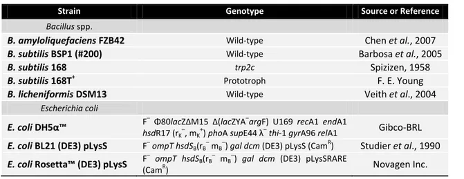
Table 2.1– List of bacterial strains used in this work.
Strain Genotype Source or Reference
Bacillus spp.
B. amyloliquefaciens FZB42 Wild-type Chen et al., 2007
B. subtilis BSP1 (#200) Wild-type Barbosa et al., 2005
B. subtilis 168 trp2c Spizizen, 1958
B. subtilis 168T+ Prototroph F. E. Young
B. licheniformis DSM13 Wild-type Veith et al., 2004
Escherichia coli
E. coli DH5α™ F
– Φ80
lacZΔM15 Δ(lacZYA–argF) U169 recA1 endA1
hsdR17 (rK
–
, mK +
) phoA supE44 λ–thi-1 gyrA96 relA1 Gibco-BRL
E. coli BL21 (DE3) pLysS F–ompT hsdSB(rB– mB–) gal dcm (DE3) pLysS (CamR) Studier et al., 1990 E. coli Rosetta™ (DE3) pLysS F
–
ompT hsdSB(rB
–
mB
–
) gal dcm (DE3) pLysSRARE
(CamR) Novagen Inc.
2.3.
DNA manipulation and sequencing
Regular DNA manipulations were carried out as described by Sambrook et al., 1989. PCR amplifications were performed with Phusion® High-Fidelity DNA Polymerase (Finnzymes) or NZYDNAChange Polymerase (NZYTech, Lda.). Designed oligonucleotides (Table 2.2) were purchased from Metabion International AG or StabVida Lda. DNA from agarose gels and PCR products were purified using GFX® PCR DNA and Gel Band Purification Kit (GE Healthcare).
All restriction enzymes were purchased from MBI Fermentas or New England Biolabs and used according to manufacturer’s instructions. The digested pET30a(+) (Novagen) vector DNA was dephosphorilated with an alkaline phosphatase (FastAP™, Fermentas). DNA ligations were performed using T4 DNA Ligase (MBI Fermentas).
Plasmid DNA was purified with the QIAGEN® Plasmid Midi Kit (Qiagen) or QIAprep® Spin Miniprep Kit (Qiagen). DNA samples were quantified using NanoDrop 2000c™ (Thermo Fisher Scientific Inc.). DNA sequencing reactions were performed by StabVida, Lda.
23 1xTAE, and stained with GreenSafe Premium (NZYTech, Lda.). NZYDNA Ladder III (NZYTech, Lda.) was the used molecular marker.
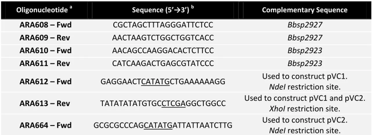
Table 2.2– List of oligonucleotides used in this project.
Oligonucleotide a Sequence (5’→3’)b Complementary Sequence
ARA608 – Fwd CGCTAGCTTTAGGGATTCTCC Bbsp2927
ARA609 – Rev AACTAAGTCTGGCTGGTCACC Bbsp2927
ARA610 – Fwd AACAGCCAAGGACACTCTTCC Bbsp2923
ARA611 – Rev CATCAAGACTGAGCGTATCCC Bbsp2923
ARA612 – Fwd GAGGAACTCATATGCTGAAAAAAGG Used to construct pVC1.
NdeI restriction site.
ARA613 – Rev TATATATATGTGCCTCGAGGCTGGCC Used to construct pVC1 and pVC2.
XhoI restriction site.
ARA664 – Fwd GCGCGCCCAGCATATGATTATTAATCTTG Used to construct pVC2.
NdeI restriction site.
a
(Fwd) forward primer and (Rev) reverse primer. b
Restriction sites in the primer sequence are underlined.
2.4.
Plasmids construction
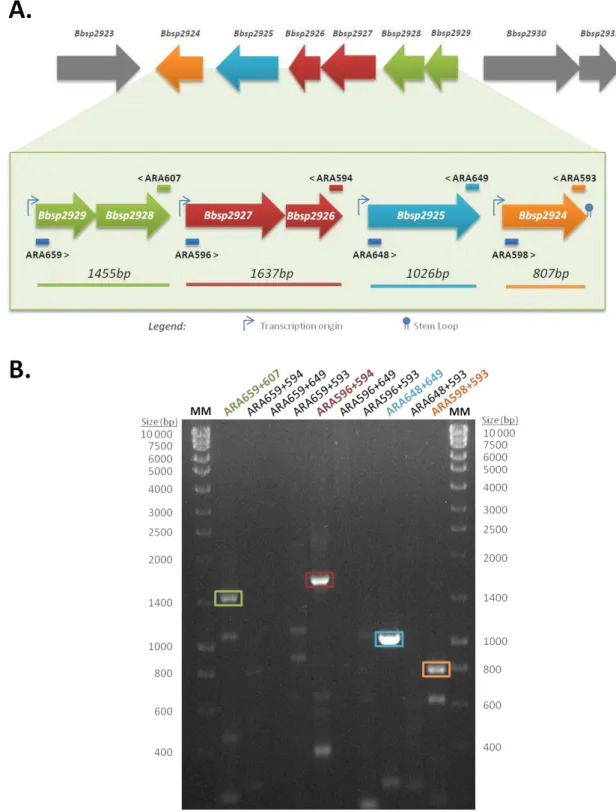
The coding sequences of Bbsp2923, with and without the signal peptide (see coding sequence in Appendix 6.3) were amplified by PCR using BSP1 chromosomal DNA as template. A nested PCR amplification step was used to increase obtained amplicon yield. The list of used primers is available in Table 2.2. These designed primers introduced unique restriction sites NdeI and XhoI, at the 5’ and 3’ end, respectively.
24
Table 2.3– List of used or constructed plasmids.
Plasmid Relevant construction a Source
pET30a(+) Expression vector under T7 promoter control. Allows N- or C-terminal
His6-tag insertion. Kan.
Novagen
pVC1 pET30a(+) containing the complete Bbsp2923 coding sequence in the
MCS. Kan. This work
pVC2 pET30a(+) containing Bbsp2923 coding sequence without the signal
peptide, in the MCS. Kan. This work
a
(kan) Kanamycin resistant. (MCS) Multiple cloning site.
2.5.
Total RNA extraction and Reverse transcription-PCR analysis
B. subtilis BSP1 was grown as described above. Cell samples of 1mL were harvest 2h (t2)
and 4h (t4) after growing in the absence or presence of different types of mannans - konjac
glucomannan or carob galactomannan. Total RNA was extracted using Absolutely RNA™ Miniprep kit (Stratagene – Agilent Technologies, Inc) according to manufacturer’s protocol for RNA isolation from Gram-positive bacteria. RNA samples were quantified using NanoDrop 2000c™ (Thermo Fisher Scientific Inc.).
For specific mRNA detection, a two-step reverse transcription polymerase chain reaction (RT-PCR) was used. In this procedure, first it is used a reverse transcriptase with a specific reverse primer, for generation of the cDNA first stand. The second step consists in a regular PCR using both specific oligonucleotides for the synthesis and amplification of the double stranded cDNA. In the first step, M-MuLV Reverse Transcriptase (MBI Fermentas), an RNA- and DNA-dependent DNA polymerase from Moloney Murine Leukemia Virus, was used according to the manufacturer’s instructions, with the exceptions: incubation at 37⁰C during 2h. PCR amplifications were performed with Phusion® High-Fidelity DNA Polymerase (Finnzymes). Used primers are listed in table 2.4.
25 Table 2.4– List of oligonucleotides used for RT-PCR assays.
Oligonucleotide a Sequence (5’→3’) Complementary Sequence
ARA593 – Rev GTTGCATAAGCATCATTGTCG Bbsp2924
ARA594 – Rev CCACGATACATATGTGAATAGC Bbsp2926
ARA596 – Fwd GGATCTGATACGTCAAGTGG Bbsp2927
ARA598 – Fwd AAAGCTTGGTGTAAGCAGCG Bbsp2924
ARA607 – Rev TTCCACCAGTAGTAGCGATACC Bbsp2928
ARA648 – Fwd GCCTACCGCACAAGCATTTC Bbsp2925
ARA649 – Rev CTTCATCTCACCTTGAGAGC Bbsp2925
ARA659 – Fwd GTCTCTTGTTCCCACTGTTGC Bbsp2929
a
(Fwd) forward primer and (Rev) reverse primer.
2.6.
Qualitative and comparative analysis of substrate hydrolysis
For qualitative and comparative analysis of the hemicellulose hydrolytic capabilities of the different Bacillus strains (Table 2.1), solid medium plates were prepared using 34 g.mL-1 of SFA (Sugar Free Agar) (LabM) and 10 g.mL-1 of the tested substrate, LBG (Galactomannan) or Birchwood Xylan. Strains were grown in liquid SF medium as described in section 2.2. After growing during 7 hours, cells were collected by centrifugation (6min, 6000rpm) and resuspended in new medium to a final OD600nm ≈ 0.8-0.9. Three blank paper discs, inoculated with 20μL of different strains growth, were placed on each plate. Duplicates of plates were prepared and incubated at room temperature during 4 days. Hydrolysis was recorded as the clear halo surrounding the paper discs. Pictures were taken after 48h and 96h, using ChemiDoc™ XRS Gel Documentation system and Quantity One® software (Bio-Rad).
2.7.
Hydrolytic activity assays
After hydrolysis of the tested polysaccharides, the reducing sugar content was determined by the Nelson-Somogyi method (Somogyi, 1952), using D-glucose as standard (Appendix 6.2).
B. subtilis strains were grown in liquid medium as described in section 2.2. Supernatants collected in t4 (4h after cell growth in the absence or presence of different types of mannans)
26
minutes at 37⁰C. Samples were prepared in triplicates. The hydrolytic activity was determined measuring the initial rates obtained from the linear portion of the progress curve and was expressed as the amount of glucose reducing-sugar equivalents (μg) produced per minute of reaction, per mL of supernatant and per OD600nm of cell culture.
2.8.
Small scale overproduction of recombinant Bbsp2923
Induction tests and small scale growth of E. coli BL21 (DE3) pLysS and E. coli Rosetta™ (DE3) pLysS cells harboring pVC1 and pVC2 were performed to assess the production and solubility of the recombinant proteins. Cells were grown at 37⁰C and 180rpm in 10mL of selective LB medium. Half of the culture was induced by IPTG 100mM, when culture OD600nm reached 0.6 . Both the induced and non-induced cultures where grown for further 3 hours, after which the cells were harvested by centrifugation at 13000rpmfor 5 minutes. Cells were resuspended in 100μL of French Press Buffer (Sodium phosphate buffer 20mM pH7.4, NaCl 500mM, Imidazole 100mM and Glycerol 10%(v/v)). For cells disruption, incubation with lysozyme (1mg.mL-1) for 10 minutes at 37⁰C, and three cycles of freezing in liquid nitrogen and thawing for 5 minutes at 37⁰C were performed, followed by incubation with 1μL of Benzonase Nuclease (Novagen®) and PMSF (10mg.mL-1), a serine protease inhibitor, at 37⁰C for 10 minutes. After centrifugation at 13000rpm and 4⁰C for 15 minutes, the soluble fraction (supernatant) was recovered and the insoluble fraction (pellet) was resuspended in 100μL of French Press Buffer.
27
2.9.
Protein analysis and Quantification
The analysis of the presence and molecular mass of the overproduced enzymes, in different tested fractions, were conducted by electrophoresis on 12.5% polyacrylamide gels containing sodium dodecyl sulphate (SDS-PAGE), stained with Coomassie Blue. Samples were prepared in 10x loading buffer solution (Tris-HCl 0.5M, pH6.8; SDS 20%; bromophenol blue 0.2%; β-mercaptonethanol 1M; glycerol 20%). NZYTech LMW Protein Marker (NZYTech, Lda.) was the protein marker used.
Supernatants concentration was determined using Bradford reagent (Bio-Rad Laboratories Inc.) with bovine serum albumin (BSA) as standard.
2.10.
β
-Mannanase activity assay
29
Chapter 3
31
3.
Results and Discussion
3.1.
Genetic Analysis of the gam Cluster
According to gene orientation, the gam cluster appears to be organized in at least three operons – Bbsp2923, Bbsp2924-Bbsp2929 and Bbsp2930-Bbsp2931 (Fig. 1.4). To address this question an evaluation of the genetic organization at the transcriptional level combined with in silico analysis of the gam cluster DNA sequence was performed.
3.1.1.
Transcriptional organization of the gam cluster
Based on the organization of the open reading frames of the gam cluster (Fig. 1.4), gene Bbsp2923 appears to be monocistronic and Bbsp2930-Bsp2931 may constitute a bicistronic transcriptional unit. The cluster of genes Bbsp2924-Bbsp2929 suggests they are organized as a single transcriptional unit comprising six cistrons. To elucidate the transcriptional organization of gam cluster Bbsp2924-Bbsp2929 genes, a two-step reverse transcription polymerase chain reaction (RT-PCR) approach was used.
32
difficult to disrupt (Sambrook & Russell, 2001). Total RNA was obtained using Absolutely RNA™ Miniprep kit, although other approach for total RNA extraction was attempted, using TRIzol® reagent (INVITROGEN™, Life Technologies Ltd) combined with RNeasy Mini kit clean up
(QIAGEN Inc.). Even with protocol alterations and longer periods of incubation with DNase, RNA kept on being DNA contaminated or degraded. Only Absolutely RNA™ Miniprep kit allowed the isolation of high-purity RNA. This kit procedure is based on the use of two different spin cups, a prefilter spin cup and a RNA binding cup, which, speeds up RNA extraction and diminishes exposure period to contaminants.
Total RNA was purified and tested from B. subtilis BSP1 grown in several conditions: cell samples harvested 2h (exponential growth phase - t2) and 4h (late exponential growth
phase - t4) in the absence or presence of different types of mannans - konjac glucomannan or
carob galactomannan (Fig. 3.1). The goal of the experiment was to determine the transcriptional organization of the cluster, but also analyse the response at the transcriptional level to the presence of different heteromannas and distinct growth stages. The obtained RT-PCR results are illustrated in Fig. 3.1.
The RT-PCR results illustrated in figure 3.1, show a mRNA amplification pattern that is identical for RNA extracted from cells grown in all tested conditions (data not shown). No differences were detected in the intensity of the amplicons bands obtained when cells were grown in the absence of sugars or in the presence of the heteromannans (glucomannan or galactomannan). Similarly, identical patterns and amplicon band intensity were observed with RNA extracted from cells during exponential growth and early stationary growth phase.
33 Figure 3.1– RT-PCR results obtained with RNA extracted from cells grown in different conditions and using distinct pairs of primers. (A) The position of each primer used in the experiments is indicated above (reverse primer) or below (forward primer) the genes. (B) Cells were grown in SF medium in the absence of sugars (SF Medium) in the presence of galactomannan (+Galactomannan), or in the presence of glucomannan (+Glucomannan) (top panel from left to right, respectively). Arrows indicated the time at different growth phases where cell were harvested and RNA extracted (t2) exponential growth phase
and (t4) late exponential-early stationary growth phase. The pairs of primers used in RT-PCR experiments
are indicated in the left column. (+) indicates positive amplification and (-) no detection of product/amplification.
A.
34
Figure 3.2– Transcriptional organization of the region Bbsp2929-2924 of gam cluster. (A) The transcripts detected by RT-PCR, and respective size of the amplicons are depicted below the cluster of genes. The position of each primer used in the experiments is indicated above (reverse primer) or below (forward primer) the genes. (B) Example of an obtained RT-PCR amplification pattern. In this example amplification products were obtained using RNA extracted from cells in exponential growth phase (t2)
grown in the presence of Konjac glucomannan. In each lane are indicated the pair of primers used and their sequence is available in Table 2.4. Amplification products were visualized under ultraviolet light in a 1% (w/v) electrophoresis agarose gel, buffered with 1xTAE and stained with GreenSafe Premium (NZYTech, Lda.). NZYDNA Ladder III (NZYTech, Lda.) was the used molecular marker (MM). Band size was
35
3.1.2.
Sequence Analysis of the gam cluster
As described in previous sections, Schyns G., Serra C.R., Henriques, A.O. and co-workers (unpublished data) reported that the gam cluster encodes all the necessary elements for the metabolization of the galactoglucomannan, including a β-Mannanase (Bbsp2923), an intracellular α-Galactosidase (Bbsp2930), a β-Glucosidase (Bbsp2925), an N-acetyl-mannosamine kinase (Bbsp2931), an ABC transportation system (Bbsp2926-29) and a CcpA-like regulatory protein (Bbsp2924).
An update of the predictions was performed in the databases, ORFs forecast was confirmed using DNA Strider version 1.4f6 (Douglas, 1995) and the correspondent gene products compared against available databases using BLASTp search (Basic local alignment search tool using a protein query) (Altschul et al., 1990). Ribosome binding sites (RBS) location and translation initiation were manually defined by complementarity with B. subtilis 16S rRNA sequence and canonical distance observed between RBS and start codon in bacteria [Snyder & Champess (1997); Hager & Robinowitz (1985)]. The putative location of RBS suggests start codons for Bbsp2923, Bbsp2926, Bbsp2927 and Bbsp2929, which are distinct from the ones annotated in the available sequence of the gam cluster (Schyns, G., Serra C.R., Henriques, A.O. et al., unpublished results). Table 3.1 summarizes the results.
Two distinct DNA regions of genes Bbsp2923 and Bbsp2927 were resequenced due to possible errors detected in the available nucleotide sequence. The primer pair ARA608-609 was design to amplify and sequence Bbsp2927 and primers ARA610-611 to gene Bbsp2923. In the DNA region of Bbsp2923 no mistakes were detected, however the DNA sequence of Bbsp2927 presented a wrongly annotated additional base (T) in a thymine rich zone leading to a frame shift. The corrected sequence is presented in appendix 6.1.
The update of the BLAST search for proteins that are homologous to those deduced from the nucleotide sequence, confirms that the gam cluster encodes a complete putative degradation system for galactoglucomannan, including degradation, uptake and catabolism.
36
Table 3.1– Predicted signals for translation initiation in gam cluster sequence.
Gene Orientation a RBS and start codon (5’-3’) b ΔG c (kcal/mol)
Bbsp2923 + AAGGAGGAACTGGTTTG -17.8
Bbsp2924 - AGGAGCGAGGATTTG -11.6
Bbsp2925 - AAGGAGTGCAAAGAAATG -12.8
Bbsp2926 - AAGGTGAAACGGATG -9.6
Bbsp2927 - AGGGAGTAATTCGATG -9.8
Bbsp2928 - GGAGGGAAAGTGGAGTG -14.4
Bbsp2929 - AAGGCGGGGTGGAAAATG -15.8
Bbsp2930 + AAGGAGTGGTGAATAGTAATG -12.8
Bbsp2931 + AAGGGGGATACCTATG -15.8
B. subtilis 16S rRNA consensus sequenced 3’ UUCCUCCA – N5-10 –Start codon a
Genomic orientation. (+) Same sense. (-) Anti-sense. b
Consensus elements in RBS are in boldface and translation start site is underlined. c
Free energy of binding for the interaction between the 3’ end of the B. subtilis 16S rRNA sequence and the putative RBS, calculated by the method of Tinoco et al. (1973).
d
B. subtilis 16S rRNA consensus sequence adapted from Snyder & Champess, (1997) and Hager & Robinowitz (1985). N denotes any nucleotide.
Bbsp2925 is 72% identical to a β-glucosidase from Clostridium sp. DL-VIII (Table 3.2). β -glucosidases are necessary for the hydrolysis of the β-1,4 linkage between glucose and D-mannose residues that constitute galactoglucomannan backbone (Shallom & Shoham, 2003).
Bbsp2930 is most probably an α-galactosidase 76% identical to one found in Geobacillus stearothermophilus (Table 3.2). α-galactosidases cleave the α-1,6 bond linking galactose with galactose, glucose or mannose residues (Shallom & Shoham, 2003).
37 Table 3.2– Comparison of gam cluster products to other proteins in available databases using BLASTp tool.
Gene Size
[aa (kDa)] Homolog protein
a Database entry Identity (%) Amino acid overlap
Bbsp2923 687 (75.5) Beta-Mannanase
[Bacillus sp. JAMB750] AB128831 57 486
Bbsp2924 339 (38.4)
LacI family transcriptional regulator
[Bacillus cellulosilyticus DSM 2522]
ADU29634.1 64 332
Bbsp2925 454 (52.1) 6-phosphobeta-glucosidase
[Clostridium sp. DL-VIII] EHI97801.1 72 449
Bbsp2926 205 (23.9)
unspecified sugar ABC transport ATP-binding protein [Mycoplasma canis UFG4]
EIE41424.1 31 109
Bbsp2927 422 (46.4)
sugar ABC transporter substrate-binding protein [Bacillus clausii KSM-K16]
BAD62861.1 45 418
Bbsp2928 293 (32.9) sugar ABC transporter permease
[Bacillus clausii KSM-K16] BAD62860.1 67 273 Bbsp2929 294 (33.4) sugar ABC transporter permease
[Bacillus clausii KSM-K16] BAD62859.1 73 295
Bbsp2930 746 (86.5) alpha-galactosidase
[Geobacillus stearothermophilus] AAF70204.1 76 742
Bbsp2931 322 (35.0) ROK family protein
[Geobacillus sp. C56-T3] ADI25265.1 57 301
a
Homolog protein found in database search and respective strain indicated between square brackets. Search performed with default settings.
38
Bbsp2924 most probably encodes a transcription factor belonging to the LacI family of transcriptional regulators (Weickert & Adhya, 1992).
The galactoglucomannan metabolization system encoded by gam cluster appears to be under the control of the regulatory mechanism of carbon catabolite repression (CCR) (Deutscher et al., 2002; Fujita, 2009). CCR is a common type of regulation for the use of alternative carbon sources, such as arabinose (Inácio et al., 2003). In silico analysis revealed the presence of several catabolic responsive elements (CREs) along the gam cluster sequence (Table 3.3). CcpA, a master regulator of CCR in gram-positive bacteria, functions as a repressor that in the presence of glucose binds to DNA operator sequences (CRE) usually located in the promoter region of a target gene, preventing or blocking transcription (Deutscher et al., 2002; Fujita, 2009). As indicated in Table 3.3, CREs were found in the promoter region of genes Bbsp2923, Bbsp2929 and Bbsp2930 (Fig. 3.3).
Table 3.3– Identified cre sequences and location.
Gene cre sequence a Distance to TSS (bp) b
Bbsp2923 TGAAGCGCTTTCA 42
Bbsp2929 TGTAAGCGTTTTCT 292
Bbsp2930 TGTAAGTGCTTTCT 54
B. subtilis consensus sequencec T G W N A N C G N T N W C A a
cre sequences were identified with DBTBS search tool using a p-value=5% (threshold). Conserved positions are in boldface type.
b
Distance in base pairs to the TSS (Translational start site) of the respective gene. c
B. subtilis cre consensus sequence (Deutscher et al, 2002). W= A or T, N= any base.
Transcription initiation within the gam cluster was analysed using the DBTBS search tool to identify putative promoter sequences, namely the -35 and -10 promoter regions that interact with sigma (σ)-factors, allowing the RNA polymerase to bind to DNA. DBTBS is a database of transcriptional regulation in Bacillus subtilis that contains intergenic conservation information (Sierro et al., 2008). The results are summarized in Table 3.4.