Brain aromatase mRNA expression in two populations of the peacock blenny Salaria
pavo with divergent mating systems
David Gonçalves
a,⁎
, João Saraiva
a, Magda Teles
a, Rita Teodósio
b, Adelino V.M. Canário
b, Rui F. Oliveira
a,ca
Unidade de Investigação em Eco-Etologia, CCMAR-CIMAR Laboratório Associado, Instituto Superior de Psicologia Aplicada, Rua Jardim do Tabaco 34, 1049-041 Lisboa, Portugal
bCentro de Ciências do Mar, Universidade do Algarve, Gambelas, 8005-139 Faro, Portugal c
Instituto Gulbenkian de Ciência, Rua da Quinta Grande, 6, P-2780-156 Oeiras, Portugal
a b s t r a c t
a r t i c l e i n f o
Article history: Received 25 May 2009 Revised 21 September 2009 Accepted 12 October 2009 Available online xxxx Keywords: Aromatase Reproductive behavior Salaria pavo Peacock blennyAlternative reproductive tactics Sex-role reversal
Aromatase, the key enzyme in the conversion of androgens to estrogens, regulates the availability of these hormones in tissues and controls many physiological and behavioral processes. Infish and other vertebrates, the regulation of aromatase expression in the brain has been implicated in the modulation of male sexual and aggressive behaviors. Here, the pattern of mRNA expression of the brain aromatase isoform (encoded by the CYP19A2 gene also referred as CYP19b) was quantified at the peak of spawning season in brain macroareas from males and females of the blenny Salaria pavo originated from two populations displaying male alternative reproductive tactics but differing in their mating systems. In Trieste (Adriatic) nesting males aggressively defend nests and take the initiative in courtship and perform sexual displays more often than females while in Ria Formosa (Southern Portugal) the pattern is reversed as a result of shortage of appropriate nesting sites. Nesting males from Ria Formosa had overall higher levels of brain aromatase mRNA expression than nesting males from Trieste, suggesting a higher brain estrogen synthesis in these males. Since in somefish species exogenous estradiol administration has been shown to decrease sexual and agonistic behaviors, the higher levels of brain aromatase in Ria Formosa nesting males may explain their reduced expression of sexual and aggressive displays when compared with nesting males from Trieste. Alternatively, the higher brain aromatase levels in nesting males from Ria Formosa could be a mechanism to decrease the putative androgen-induced activation of aggressive and sexual displays by reducing the local availability of androgens through their metabolization into estrogens. Although females and parasitic female-like males also differ in their displays between populations, the interpopulational pattern of brain aromatase mRNA expression was similar, suggesting that other neuroendocrine agents mediate the expression of female and female-like behaviors. In conclusion, brain aromatase availability seems like a probable mechanism to regulate the effects of steroids on the brain circuits underlying the expression of sexual and agonistic displays in S. pavo.
© 2009 Published by Elsevier Inc.
Introduction
The activation of male sexual displays in adult mammals and birds depends largely on brain aromatization of androgens into estrogens (reviewed byBall and Balthazart, 2004; Baum, 2003; seeOliveira, 2004 for a historical overview). In fishes, the role of brain aromatization in the regulation of sexual displays has been poorly investigated. This is surprising asfishes have the highest levels of brain aromatase across all vertebrate classes (Callard et al., 1990) and aromatase has been shown to occur in brain areas related to the control of reproduction (e.g.,Forlano et al., 2001; Menuet et al., 2003; Menuet et al., 2005) and to peak during the reproductive period in seasonal breeders (Forlano and Bass, 2005; Gelinas et al., 1998; Gonzalez and Piferrer, 2003). Studies conducted so far investigating
the role of aromatization in malefish reproductive behavior have produced conflicting results (Munakata and Kobayashi, 2009). For example, in a study on guppies, aromatase activity was pharmaco-logically inhibited with the drug fadrozole, administered in the water, and the frequency of two of three male sexual displays decreased (Hallgren et al., 2006). Also, in the goldfish testosterone (T) and estradiol (E2) injections in males stimulated approach behavior
toward females within minutes, while fadrozole blocked the stimu-latory effect of T (Lord et al., 2009). Finally, in the plainfin midshipman intramuscular injections of both 11KT and E2 to courting males
facilitated the duration of fictive vocalizations (similar to natural vocalizations produced in an agonistic and sexual context) measured in a neurophysiological brain preparation (Remage-Healey and Bass, 2004). These examples would suggest that, similarly to what has been found for birds and mammals, E2locally synthesized in the brain from
testosterone (T) facilitates some aspects of male sexual behavior in fishes. However, in five fish species for which exogenous E2 was Hormones and Behavior xxx (2009) xxx–xxx
⁎ Corresponding author.
E-mail address:[email protected](D. Gonçalves).
YHBEH-02921; No. of pages: 7; 4C:
0018-506X/$– see front matter © 2009 Published by Elsevier Inc. doi:10.1016/j.yhbeh.2009.10.007
Contents lists available atScienceDirect
Hormones and Behavior
j o u r n a l h o m e p a g e : w w w. e l s ev i e r. c o m / l o c a t e / y h b e h
administered to males sexual displays were reduced in four cases and remained unchanged in one case (see Table 3.1 in Oliveira and Gonçalves, 2008), and an inhibitory effect of estrogenic endocrine disruptors in male sexual behavior has been generally reported (e.g.,
Bayley et al., 1999; Bjerselius et al., 2001). Several issues limit the interpretation of these studies, in particular the fact that endocrine manipulations were systemic and thus it is possible that some of the behavioral effects were due to peripheral rather than central action of hormones. These findings show that more data from carefully controlled experiments are necessary in order to understand the role played by androgens and estrogens in the regulation of male sexual displays infishes.
Brain aromatase has also been implicated in intrasexual behavioral differences in species with male alternative reproductive tactics (ART). In the plainfin midshipman type I males reproduce by courting females into their nests and type II males try to approach the nests of type I males unnoticed and achieve parasitic fertilizations of eggs (Brantley and Bass, 1994). Type I males emit “hum” vocalizations in order to attract females, while both females and type II males do not emit these vocalizations (Bass and McKibben, 2003; Brantley and Bass, 1994). When compared with type I males, both females and type II males have high levels of aromatase activity in the hindbrain sonic motor nucleus that controls vocalizations (Schlinger et al., 1999). The high levels of aromatase activity in females and type II males were interpreted as a possible mechanism to avoid the testosterone-induced masculin-ization of the vocal motor nucleus through an increase in androgen to estrogen conversion (Schlinger et al., 1999).
Finally, aromatase has also been implicated in the regulation of aggression infish. Brain aromatase activity was found to be negatively correlated with the expression of aggression in a sex-changing goby (Black et al., 2005), and in general malefish exposed to estrogens or estrogen-like substances show reduced aggression (e.g.,Clotfelter and Rodriguez, 2006; Colman et al., 2009). These examples point towards a suppressive effect of estrogens onfish aggressive behavior.
Taken together the available data suggests that brain aromatase plays a significant role in the regulation of both sexual and aggressive displays infish.
In the peacock blenny Salaria pavo male ART and interpopulation variability in the mating system have been described. A population at the Adriatic, in Trieste, occurs in an area with a rocky bottom where nests are available in abundance. Males establish nests in rock crevices or holes and aggressively defend a territory around the nest. Males take the initiative in courtship and the frequency of male courtship displays is higher than the frequency of female courtship displays (Saraiva et al., unpublished data). In contrast, a population at the Ria Formosa (southern Portugal) occurs in a mudflat area where the only substrates available for nesting are artificial materials such as bricks and tiles used by clam culturists to delimit concessions. The scarcity of nest sites promotes a strong male–male competition for nests and only large competitive males are able to acquire a nest (Almada et al., 1994). After the breeding season starts males seldom leave their nests and do not defend any area around the nest, and it is common to observe males nesting in adjacent holes of the same brick (Almada et al., 1994). At the peak of the breeding season most nests arefilled with eggs and nest space may become a limiting factor for female reproduction (Almada et al., 1994). Females compete for the access to nests and the sex-roles are reversed with females taking the initiative and displaying courtship more often than males (Almada et al., 1995). Small males are unable to acquire nests and reproduce by mimicking the females' appearance and courtship displays in order to approach nesting males and parasitically fertilize eggs (Gonçalves et al., 2005; Gonçalves et al., 1996). Parasitic males switch into nesting males from their second breeding season onwards (Fagundes et al., unpublished data), thus undergoing major morphologic and behav-ioral modifications. Parasitic males have also been described for the
Trieste population but in a much lower frequency (Saraiva et al., unpublished data), suggesting that nest availability mediates the frequency of male ART. Interestingly, parasitic males seem to adjust the frequency of female-like displays to the frequency of female courtship behavior in the population. In the Ria Formosa, where females are most actively engaged in courting, female-like displays towards nesting males by parasitic males are very common. In Trieste, where males take the initiative in courtship and females assume a more passive role, parasitic males do not court males and for now it is unclear how they reproduce (Saraiva et al., unpublished data).
Sex steroids have been shown to influence sexual displays in S. pavo. Administration of both T and 11KT to parasitic males decreases the frequency of the female-like sexual displays and promotes the development of male secondary sexual characters (Gonçalves et al., 2007; Oliveira et al., 2001). This is consistent with the fact that circulating levels of both T and 11KT are higher in nesting males than in parasitic males (Gonçalves et al., 2008), and infish species with male ART higher levels of 11KT (but not T) have consistently been found in nesting males (Brantley et al., 1993; Oliveira, 2006). Brain aromatase activity is lower in parasitic than in nesting males (Gonçalves et al., 2008), and from the above it seems possible that this difference is related to the divergent sexual displays exhibited by the two male morphs.
Here, it was hypothesized that aromatase mediates the above described differences in the reproductive and aggressive behavior of females, nesting males and parasitic males across the Ria Formosa and the Trieste population. Unlike most mammals, for which only one aromatase coding gene has been described (Conley and Hinshelwood, 2001), two aromatase isoforms have been found in fish, one preferentially expressed in the ovary and encoded by the CYP19A1 (or CYP19a) gene and another preferentially expressed in the brain and encoded by the CYP19A2 (or CYP19b) gene (e.g.,Chang et al., 1997; Tchoudakova and Callard, 1998). In this study the CYP19A2 mRNA levels were compared in brain macroareas of females and parasitic, transitional and nesting males of S. pavo captured at Trieste and in the Ria Formosa.
Methods Fish collection
Fish were collected during the peak of the breeding season at Culatra island, southern Portugal, 36°59′N;7°51′W (11 females, 12 nesting males, 12 parasitic males and 10 transitional males) and Trieste, northern Adriatic Sea, 45°40′N;13°35′ E (9 females, 10 nesting males, 5 parasitic males and 5 transitional males). The criteria to discriminate the various morphotypes were as follows: nesting males had fully developed male secondary sexual characters and were defending a nest with eggs; females had swollen abdomens, an indicator of ripeness, and after dissection their ovaries were confirmed to have fully developed oocytes; parasitic males lacked male secondary sexual characters, sperm could be easily extruded from their vas deferens by gently pressing the abdomen and had enlarged testes in comparison with nesting males (confirmed during dissection), as described byGonçalves et al., 1996); transitional males were beginning to develop secondary sexual characters. These males have never been observed guarding eggs or courting nesting males with female-like displays and most likely they do not reproduce. Their testes are usually very small when compared with both parasitic and nesting males and this was confirmed during dissection (see
Gonçalves et al., 2008). In Ria Formosa, fish were collected during low-tide with a hand-net from bricks used as nests. Animals were immediately lightly anaesthetized with MS222 (Sigma-Aldrich, dilution 1:10,000) and euthanized by sectioning the spinal cord. Fish were kept in isolated plastic bags in a mixture of ice and water (approximately 0 °C) until dissection, which took place in afield
2 D. Gonçalves et al. / Hormones and Behavior xxx (2009) xxx–xxx
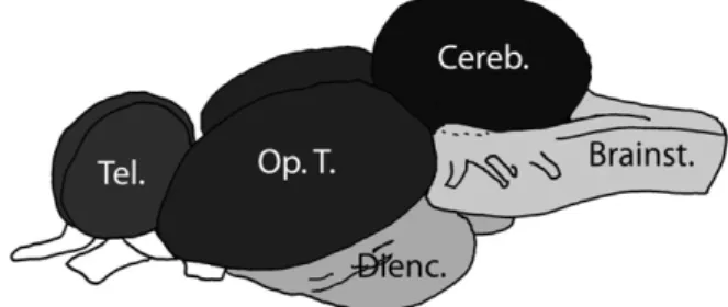
station within 1–2 h. In the field station, animals were measured, weighed and dissected. Brains were divided into five macroareas during dissection: telencephalon, optic tectum, cerebellum, dien-cephalon (excluding the pituitary) and brainstem (Fig. 1).
The brain macroareas werefirst briefly homogenized in a fixed volume (20–50 μl, depending on the macroarea) of chilled RNAse free 0.1 M, phosphate buffer pH 7.5. Half of this volume was transferred to a tube with 300μl of Tri reagent (Sigma, Spain) for RNA extraction and the other half to another tube for an enzymatic aromatase activity assay. Aromatase activity data has been published elsewhere (Gonçalves et al., 2008).
In Trieste fish were caught in food traps or fish nets while snorkeling and brains were preserved in RNAlater (Ambion, UK) at −20 °C until RNA extraction. Macroareas were transferred into 300 μl of chilled Tri reagent (Sigma, Spain) and homogenized as above.
During the experimental procedure the“ASAB guidelines for the use of animals in research” were followed.
CYP19A2 and 18S sequences
The CYP19A2 and 18S cDNAs were obtained by reverse transcription polymerase chain reaction (RT-PCR) of brain total RNA pooled from nesting males, females and parasitic males and extracted with TRI reagent (Sigma, Spain) following the manufac-turer's instructions. RNA was treated with DNAse I (Ambion, Portugal). For cDNA synthesis 5 μg of total RNA was reverse transcribed using oligo-dT primers and M-MLV reverse transcriptase (Promega, Spain). For CYP19A2, a 450 bp fragment was amplified using primers designed from the tilapia Oreochromis mossambicus ortholog sequence (forward: 5′-TCTTAGCAGGACTCGGTCCAAT- 3′; reverse: 5′-AGATGTCCAACACGATGGCTCT- 3′). The PCR mix contained 1× reaction buffer, 1.5 mM MgCl2, 200 μM of each
dNTP, 20 pmol of each primer, 1 U of Taq DNA polymerase (Promega) and 2 μl of first-strand cDNA. Cycling conditions were 2 min at 95 °C, 35 cycles of 1 min at 95 °C, 1 min at 59 °C and 1 min at 72 °C, followed by 5 min at 72 °C. RT-PCR products were run on a 2% agarose gel and the band corresponding to the predicted fragment size was cut from the gel, eluted (GFX PCR DNA and Gel Band Purification Kit, Amersham Biosciences, Piscataway) and cloned into pGem-T-easy vector (Promega). After sequencing in both directions (Macrogen, Inc., South Korea) a cDNA with highest similarity to other teleosts' CYP19A2 sequences was identified and received accession number FN356970. A 18S cDNA of 407 bp was obtained from total RNA using primers designed for conserved regions of other teleosts (forward: 5 ′-GTTCCGACCATAAACGATGC-3′; reverse: 5′-CTCAATCTCGTGTGGCTGAA-3′). The 18S sequence received accession number FN356969.
CYP19A2 relative mRNA expression
For quantitative real-time PCR (QPCR) specific primers were designed based on the above sequences (CYP19A2, forward: 5
′-TATGGCAGCATTACCAGGGT-3′, reverse: 5′-GCCGAATCTTGACGTGT-ACTG-3′, fragment size: 111 bp; 18S, forward: 5′-GCATGGCCGTTCT-TAGTTGGT-3′, reverse: 5′-TTAGCAAGCCGGAGTCTCGTT-3′, fragment size: 73 bp). The identity of the resulting PCR products was confirmed by DNA sequencing of PCR products.
Expression of CYP19A2 was normalized to the expression of 18S to account for variation in total RNA levels between samples and in reverse transcription reaction efficiencies. Total RNA from each brain macroareas was extracted, DNAse treated and reversed transcribed (1μg) into cDNA as described above except that random hexamers (Promega, Spain) were used during reverse transcription. QPCR reactions (25μl) were run in a Stratagene MX3000p thermocycler with Stratagene's Brilliant SYBR green QRT-PCR Master Mix (Strata-gene, Spain) and primers at 0.5μM. Thermocycling conditions were equal for both reactions and were as follows: 10 min at 95 °C, 40 cycles of 95 °C for 30 s, 59 °C for 30 s and 72 °C for 30 s. After PCR, a melting curve program from 55 to 95 °C with 0.5 °C change in 10-s intervals was applied and the presence of a single reaction product in each tube was confirmed. For the same animal, reactions for each macroarea and for both 18S and CYP19A2 were run in duplicate in a single PCR. Animals from the several morphotypes and from the two populations were randomly assigned to QPCR reactions. Controls without template were included for both primer sets and a set of samples were included in all reactions to determine interassay variation. Raw fluorescence data was submitted to PCR Miner (http://miner. ewindup.info/miner;Zhao and Fernald, 2005) to calculate reaction efficiencies and cycle thresholds from individual wells during the reaction. The average reaction efficiencies (E) were 1.8 for 18S and 1.9 for aromatase and the average intrassay and interassay coefficient of variation in cycle threshold (CT) were 1.7% and 5.3%, respectively. For each sample, the mean CT of 18S and aromatase was calculated and the relative initial template concentration (R0) of both genes determined from 1/1(1 + E)^CT (Zhao and Fernald, 2005). The relative aromatase mRNA expression was thus given by the ratio between the aromatase and 18S R0s. The 18S average R0s did not differ between macroareas, morphotypes or populations (data not presented), suggesting that this is an appropriate reference gene. Statistical analyses
A three-way ANOVA with factors morphotype (four levels), population (two levels) and brain macroarea (five levels) was applied to test differences in CYP19A2 relative mRNA expression. Data was Fig. 1. Division of the brain intofive macroareas for the quantification of CYP19b mRNA
expression. Tel.: telencephalon; Op.T.: optic tectum; Dienc.: diencephalon; Cereb.: cerebellum; Brainst.: brainstem.
Fig. 2. Average ± SE aromatase (CYP19A2) relative mRNA expression in the brain of females and nesting, transitional and parasitic males of S. pavo collected in the Ria Formosa and in Trieste. Different letters indicate significant differences (Pb 0.05) between morphotypes (average of both populations). Significant differences between populations for the same morphotype are marked with⁎.
3 D. Gonçalves et al. / Hormones and Behavior xxx (2009) xxx–xxx
squared-root transformed to comply with normality and homocedas-ticity assumptions. When the ANOVA results were significant, relevant differences were tested with contrast analysis.
Results
For the Ria Formosa samples, both aromatase activity and CYP19A2 mRNA expression were measured in each macroarea and it was possible to correlate these different measures. CYP19A2 mRNA expression levels in the brain macroareas were positively and significantly correlated with aromatase activity measured by Gon-çalves et al. (2008)—Pearson correlation coefficient: telencephalon, r = 0.71, n = 32, Pb 0.001; optic tectum, r = 0.54, n = 32, P = 0.001; diencephalon, r = 0.54, n = 36, P = 0.001; cerebellum, r = 0.57, n = 35, Pb0.001; brainstem, r= 0.72, n= 38, Pb 0.001.
Overall, CYP19A2 mRNA expression differed between the morpho-types (F3, 255= 21.6, Pb 0.001). Females and nesting males had higher
values than both transitional and parasitic males (Pb 0.001). The remaining comparisons were non-significant (PN 0.14). The overall pattern of CYP19A2 mRNA expression was similar between popula-tions (F1,255= 0.00, P = 0.984). However, there was a significant
location⁎ morphotype interaction (F3, 255= 4.5, P = 0.004,Fig. 2). In
Ria Formosa, males had overall higher aromatase mRNA expression values than in Trieste (P = 0.001). Females, transitional males and parasitic males did not differ between locations (PN0.17).
Aromatase mRNA expression differed between macroareas (F4, 255= 21.6, Pb 0.001, Fig. 3). The cerebellum had the lowest
expression (Pb 0.001 in the comparison with the other macroareas) followed by the brainstem (marginally different from the dienceph-alon, P = 0.078, and significantly different from the other macroareas, Pb 0.03). The optic tectum had significantly higher levels than the diencephalon (P = 0.025) and marginally than the telencephalon (P = 0.066). The diencephalon and telencephalon did not differ (P = 0.99). Differences between macroareas were similar between locations (location⁎ macroarea interaction: F4, 255= 1.6, P = 0.117,
Fig. 3).
The morphotype ⁎ macroarea interaction (F12, 255= 1.6,
P = 0.076) and the morphotype⁎ macroarea ⁎ location interaction (F12, 255= 1.1, P = 0.372) were not significant. However, because the
power to detect small interaction effects of this statistical approach is low with the current n (Murphy and Myors, 2004), differences in Fig. 3. Average ± SE aromatase (CYP19A2) relative mRNA expression measured in the five brain macroareas in Ria Formosa and in Trieste. Different letters indicate significant differences (Pb 0.05) between macroareas (average of all morphotypes).
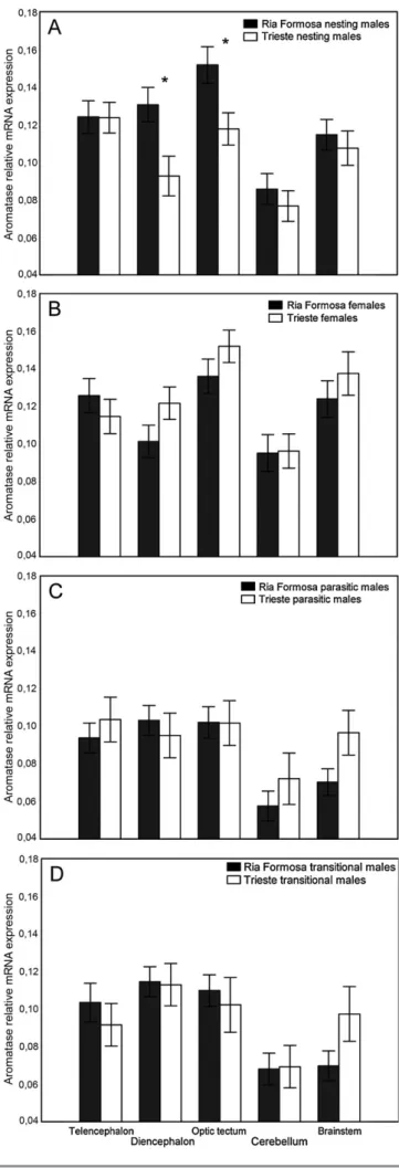
Fig. 4. Average ± SE aromatase (CYP19b) relative mRNA expression measured in the five brain macroareas of Ria Formosa and Trieste (A) nesting males, (B) females, (C) parasitic males and (D) transitional males. Significant differences between populations for the same macroarea are marked with⁎ (Pb 0.05).
4 D. Gonçalves et al. / Hormones and Behavior xxx (2009) xxx–xxx
aromatase expression between populations in the same macroareas of the same morphotypes were also tested with t-tests. Aromatase expression in the same macroarea did not differ in females, transitional males or sneakers from the two populations (PN 0.07). In males, differences were significant for the diencephalon (P = 0.043) and the optic tectum (P = 0.043) but not for the remaining macroareas (PN 0.35,Fig. 4).
Discussion
Correlation between aromatase activity and mRNA expression In this study the normalized CYP19A2 mRNA expression was positively and significantly correlated with aromatase activity measured in a previous study in the same brain samples (Gonçalves et al., 2008). In fish, significant positive correlations between aromatase activity and normalized mRNA levels of both the CYP19A1 (e.g.,Gen et al., 2001; Villeneuve et al., 2006) and CYP19A2 (Villeneuve et al., 2006) genes have been reported for ovarian tissue. However, the relationship between aromatase activity and CYP19A1 or CYP19A2 mRNA expression in the same brain samples had not been previously tested in fish. The positive correlation between brain CYP19A2 mRNA expression and aromatase activity found in this study, together with the fact that in the fish brain the CYP19A2 gene expresses much more than the CYP19A1 gene (e.g., Hinfray et al., 2006; Kishida and Callard, 2001; Villeneuve et al., 2006), suggests that CYP19A2 mRNA expression levels provide a good indication of the potential for aromatization in the S. pavo brain.
Differences between populations
The pattern of aromatase (CYP19A2 isoform) mRNA expression in the brain was similar between the two populations, with the exception of nesting males which presented higher levels in Ria Formosa. Aromatase has been implicated in the regulation of sexual behaviors in vertebrates (Ball and Balthazart, 2004; Baum, 2003) and thus it seems possible that the measured interpopulational difference in brain aromatase expression relates to the divergence in the nesting males' behavior. Previous studies have shown that the reproductive behavior of nesting males, but also of females and parasitic males, differs between populations. Field observations carried out during the species breeding season in Trieste and in the Ria Formosa revealed a 10-fold higher frequency of nesting male courtship displays in Trieste than at Ria Formosa and a 10-fold higher frequency of female courtship displays in the Ria Formosa than at Trieste (Saraiva et al., unpublished data). Sex-roles are thus reversed at Ria Formosa with females displaying most of courtship behavior while at Trieste males are the ones leading the role in courtship.
Infish, little is known about the central effects of sex steroids in male reproductive behavior. An experimental study testing the effects of aromatization in male guppy sexual displays found a significant decrease of these behaviors with aromatase blockage (Hallgren et al., 2006), and E2 promoted within minutes male approach behavior
towards females in the goldfish (Lord et al., 2009). On the contrary, in five studies where E2was administered to males, sexual displays were
reduced in four cases and remained unchanged in one (Oliveira and Gonçalves, 2008) and similarfindings have been described for fish exposed to other estrogen and estrogenic-like compounds (e.g.,
Colman et al., 2009). The interpretation of these results warrants some caution as in all these studies substances were administered peripherally and the observed behavioral effects could also have been the consequence of non-central effects. For example, E2
administra-tion to males has a suppressive effect on testicular development and plasma androgen levels (e.g.,Yamaguchi et al., 2006) and these could have accounted for the reported decrease in sexual displays of males
treated with E2. Additionally, estrogen manipulation in many of these
studies seems to have resulted in supraphysiological levels.
In the plainfin midshipman, a species that also presents male alternative reproductive phenotypes, injections of both 11KT and E2
facilitated the duration of fictive vocalizations in courting males measured in a neurophysiological brain preparation (Remage-Healey and Bass, 2004). However, the fact that courting males presented much lower levels of aromatase in brain regions implicated in the control of vocal courtship displays when compared with parasitic males and females (Schlinger et al., 1999) suggests that T conversion into E2is not a main mechanism facilitating male vocal courtship
behavior in this species. In S. pavo, differences in aromatase mRNA levels between males of the two populations were significant in two brain macroareas presumably containing important nuclei for the regulation of reproductive behavior, the diencephalon and the optic tectum (Fig. 4, see below). The higher brain aromatase levels in these areas in males from Ria Formosa will induce a higher local T to E2
synthesis, assuming a similar availability of T in these regions in males from both populations. However, although plasma T levels did not differ between males of both populations (Saraiva et al., in press), it was not possible to quantify the local availability of T in brain macroareas. With this limitation in mind, the fact that males from Ria Formosa exhibit a lower expression of sexual displays and have higher brain aromatase mRNA levels than males from Trieste points to a central inhibitory effect of E2in male sexual displays. Experiments
testing the effects of T and E2 in S. pavo male sexual displays are ongoing.
A second not exclusive hypothesis relates to the expression of aggressive behavior. In Trieste males aggressively defend an area around the nest from other competitors (Saraiva et al., unpublished data), while in the Ria Formosa males do not defend territories and very often have one or more males nesting in adjacent brick holes (Almada et al., 1994). Estrogens have been shown to reduce aggression in fish (e.g.,Bell, 2001; Munro and Pitcher, 1983) and increases in aggression were negatively correlated with brain aromatase activity in a sex-changing goby (Black et al., 2005). Importantly, E2administration to parasitic males of S. pavo from the
Ria Formosa population was also shown to inhibit aggression (Gonçalves et al., 2007). Thus, the higher brain aromatase levels in males from Ria Formosa probably increase local E2synthesis and this
may induce the necessary decrease in aggression due to the scarceness and aggregated nature of the available nests.
Although females and parasitic males exhibit notorious quantita-tive differences in sexual displays between these populations, the pattern of brain aromatase mRNA expression was similar. Females from Ria Formosa court more often nesting males than females from Trieste (Saraiva et al., unpublished data) and laboratory observations show that parasitic males follow the same pattern, courting very often nesting males in the Ria Formosa and not courting at all in Trieste (Saraiva et al., unpublished data). For now it is unclear how parasitic males reproduce in Trieste asfield observations in this area focusing on the parasitic males' behavior are lacking. The absence of interpopulational differences in brain aromatase levels is more unexpected for females than for parasitic males. Estradiol adminis-tration to parasitic males from Ria Formosa had no effect in the female-like displays (Gonçalves et al., 2007) and thus interpopula-tional differences are likely to be regulated by other neuroendocrine agents. Also, both female and parasitic female-like courtship beha-viors in S. pavo have been shown to depend on the action of the brain neuropeptide arginine vasotocin (AVT), thefish homologous of the mammalian arginine vasopressin (AVP). Females and parasitic males have higher preoptic levels of AVT mRNA than nesting males (Grober et al., 2002) and exogenous AVT administration to females and parasitic males promoted female courtship displays (Carneiro et al., 2003). This suggests that AVT generally promotes female and female-like displays, and interpopulational differences in female and parasitic 5 D. Gonçalves et al. / Hormones and Behavior xxx (2009) xxx–xxx
male sexual behaviour may be more related to variations in central AVT levels than to the direct action of steroids in the CNS.
Differences between morphotypes
Females and nesting males had higher brain aromatase mRNA levels than parasitic and transitional males. Transitional males had intermediate values between parasitic and nesting males, although the difference was only significant for nesting males. This is in accordance with what has been described in a previous study measuring brain aromatase activity in the same samples (Gonçalves et al., 2008). In that study, the increase in brain aromatase activity during the transition suggested a role for estrogen activation of male sexual displays, as demonstrated for other vertebrates. However, the data in this study argues against that hypothesis as males from Trieste, expressing more courtship behavior, had lower brain aromatase mRNA expression. One hypothesis is that male sexual displays are modulated by the direct action of androgens and not by estrogens. Androgens have been shown to promote male sexual displays infish (reviewed byBorg, 1994; Liley and Stacey, 1983). In S. pavo, levels of both 11KT and T are higher in nesting males (Gonçalves et al., 2008), and, in general, in species with male ART nesting males have higher levels of 11KT, a non-aromatizable androgen, but not of T when compared with parasitic males (Brantley et al., 1993; Oliveira, 2006). The administration of both T and 11KT to parasitic males inhibited the expression of felike displays, although it did not induce male-like sexual behaviors (Gonçalves et al., 2007; Oliveira et al., 2001). Those experiments only lasted for 8 days and it is possible that a longer time frame would be necessary for the putative masculinizing behavioral effects of androgens to be observed. Under that scenario, the higher aromatase levels in males of the Ria Formosa population, where sex-roles are reversed, could be a mechanism to locally reduce androgen levels and the androgen-induced activation of male sexual displays. A similar mechanism was proposed for the plainfin midshipman where the higher levels of aromatase in the brains of females and parasitic males in comparison with nesting males was suggested to prevent the masculinization by androgens of brain regions implicated in the production of mating calls (Schlinger et al., 1999). Experiments are ongoing to test the effects of androgens in S. pavo male sexual behavior.
Differences between brain macroareas
Aromatase levels were highest in telencephalon, diencephalon and optic tectum and lower in the cerebellum and brainstem. This is in agreement with the results reported forfish where aromatase levels have been reported to be higher in forebrain regions known to regulate reproductive behaviors, particularly the preoptic and hypothalamic periventricular nuclei, central telencephalon and optic tectum (e.g.,Forlano et al., 2001; Gelinas and Callard, 1993; Gelinas and Callard, 1997; Melo and Ramsdell, 2001). In theGonçalves et al. (2008)study, the pattern of aromatase activity across brain macro-areas differed between morphotypes, with females presenting higher levels in posterior brain regions when compared to males, a pattern also described for the medaka Oryzias latipes (Melo and Ramsdell, 2001). In this study the interaction between the macroarea and morphotype factor was marginally non-significant (P= 0.07), but females also presented somewhat higher values of aromatase mRNA expression in posterior brain regions, in particular in the cerebellum and brainstem, when compared with the other morphotypes (Fig. 4). In conclusion, the correlational data presented in this study are consistent with a major role for brain aromatase in the regulation of sexual displays phenotype in S. pavo. High aromatase mRNA expression levels were recorded in brain macroareas containing nuclei associated with the control of sexual displays. Regulation of aromatase levels may be used as a mechanism to control the
availability of both estrogens and androgens in local brain regions and, consequently, the effects of these steroids in the brain circuits underlying the expression of sexual and also aggressive behaviors. The described differences in the pattern of aromatase mRNA expression across the two populations and across morphotypes raises a number of hypotheses on the effects of estrogens and androgens in the regulation of behaviors in S. pavo that can be experimentally tested.
Acknowledgments
We thank the direction of the Ria Formosa Nature Park for providing essential logistic support. The study was funded by FCT (UI&D 331/2001 and PTDC/MAR/71351/2006).
References
Almada, V.C., Gonçalves, E.J., Santos, A.J., Baptista, C., 1994. Breeding ecology and nest aggregations in a population of Salaria pavo (Pisces, Blenniidae) in an area where nest sites are very scarce. J. Fish Biol. 45, 819–830.
Almada, V.C., Gonçalves, E.J., Oliveira, R.F., Santos, A.J., 1995. Courting females— ecological constraints affect sex-roles in a natural population of the blenniidfish Salaria pavo. Anim. Behav. 49, 1125–1127.
Ball, G.F., Balthazart, J., 2004. Hormonal regulation of brain circuits mediating male sexual behavior in birds. Physiol. Behav. 83, 329–346.
Bass, A.H., McKibben, J.R., 2003. Neural mechanisms and behaviors for acoustic communication in teleostfish. Prog. Neurobiol. 69, 1–26.
Baum, M.J., 2003. Activational and organizational effects of estradiol on male behavioral neuroendocrine function. Scand. J. Psychol. 44, 213–220.
Bayley, M., Nielsen, J.R., Baatrup, E., 1999. Guppy sexual behavior as an effect biomarker of estrogen mimics. Ecotoxicol. Environ. Saf. 43, 68–73.
Bell, A.M., 2001. Effects of an endocrine disrupter on courtship and aggressive behaviour of male three-spined stickleback, Gasterosteus aculeatus. Anim. Behav. 62, 775–780.
Bjerselius, R., Lundstedt-Enkel, K., Olsen, H., Mayer, I., Dimberg, K., 2001. Male goldfish reproductive behaviour and physiology are severely affected by exogenous exposure to 17beta-estradiol. Aquat. Toxicol. 53, 139–152.
Black, M.P., Balthazart, J., Baillien, M., Grober, M., 2005. Socially induced and rapid increases in aggression are inversely related to brain aromatase activity in a sex-changingfish, Lythrypnus dalli. Proc. R. Soc. London., B. Biol. Sci. 272, 2435–2440. Borg, B., 1994. Androgens in teleostfishes. Comp. Biochem. Physiol. 109C, 219–245. Brantley, R.K., Bass, A.H., 1994. Alternative male spawning tactics and acoustic signals in
the plainfin midshipman fish Porichthys notatus Girard (Teleostei, Batrachoididae). Ethology 96, 213–232.
Brantley, R.K., Wingfield, J.C., Bass, A.H., 1993. Sex steroid levels in Porichthys notatus, a fish with alternative reproductive tactics, and a review of the hormonal bases for male dimorphism among teleostfishes. Horm. Behav. 27, 332–347.
Callard, G.V., Schlinger, B.A., Pasmanik, M., 1990. Nonmammalian vertebrate models in studies of brain–steroid interactions. J. Exp. Biol. Suppl. 4, 6–16.
Carneiro, L., Oliveira, R., Canário, A.V.M., Grober, M., 2003. The effect of arginine vasotocin on courtship behaviour in a blenniidfish with alternative reproductive tactics. Fish. Physiol. Biochem. 28, 241–243.
Chang, X.T., Kobayashi, T., Kajiura, H., Nakamura, M., Nagahama, Y., 1997. Isolation and characterization of cDNA encoding the tilapia (Oreochromis niloticus) cytochrome P450 aromatase (P450arom): changes in P450arom mRNA, protein and enzyme activity in ovarian follicles during oogenesis. J. Mol. Endocrinol. 18, 57–66. Clotfelter, E.D., Rodriguez, A.C., 2006. Behavioral changes in fish exposed to
phytoestrogens. Environ. Pollut. 144, 833–839.
Colman, J.R., Baldwin, D., Johnson, L.L., Scholz, N.L., 2009. Effects of the synthetic estrogen, 17 alpha-ethinylestradiol, on aggression and courtship behavior in male zebrafish (Danio rerio). Aquat. Toxicol. 91, 346–354.
Conley, A., Hinshelwood, M., 2001. Mammalian aromatases. Reproduction 121, 685–695.
Forlano, P.M., Bass, A.H., 2005. Seasonal plasticity of brain aromatase mRNA expression in glia: divergence across sex and vocal phenotypes. J. Neurobiol. 65, 37–49. Forlano, P.M., Deitcher, D.L., Myers, D.A., Bass, A.H., 2001. Anatomical distribution and
cellular basis for high levels of aromatase activity in the brain of teleostfish: aromatase enzyme and mRNA expression identify glia as source. J. Neurosci. 21, 8943–8955.
Gelinas, D., Callard, G.V., 1993. Immunocytochemical and biochemical evidence for aromatase in neurons of the retina, optic tectum and retinotectal pathways in goldfish. J. Neuroendocrinol. 5, 635–641.
Gelinas, D., Callard, G.V., 1997. Immunolocalization of aromatase- and androgen receptor-positive neurons in the goldfish brain. Gen. Comp. Endocrinol. 106, 155–168.
Gelinas, D., Pitoc, G.A., Callard, G.V., 1998. Isolation of a goldfish brain cytochrome P450 aromatase cDNA: mRNA expression during the seasonal cycle and after steroid treatment. Mol. Cell. Endocrinol. 138, 81–93.
Gen, K., Okuzawaab, K., Kumakurab, N., Yamaguchic, S., Kagawabd, H., 2001. Correlation between messenger RNA expression of cytochrome p450 aromatase and its
6 D. Gonçalves et al. / Hormones and Behavior xxx (2009) xxx–xxx
enzyme activity during oocyte development in the red seabream (Pagrus major). Biol. Reprod. 65, 1186–1194.
Gonçalves, E.J., Almada, V.C., Oliveira, R.F., Santos, A.J., 1996. Female mimicry as a mating tactic in males of the blenniidfish Salaria pavo. J. Mar. Biol. Assoc. U.K. 76, 529–538.
Gonçalves, D., Matos, R., Fagundes, T., Oliveira, R., 2005. Bourgeois males of the peacock blenny, Salaria pavo, discriminate female mimics from females? Ethology 111, 559–572.
Gonçalves, D., Alpedrinha, J., Teles, M., Oliveira, R.F., 2007. Endocrine control of sexual behavior in sneaker males of the peacock blenny Salaria pavo: effects of castration, aromatase inhibition, testosterone and estradiol. Horm. Behav. 51, 534–541.
Gonçalves, D., Teles, M., Alpedrinha, J., Oliveira, R.F., 2008. Brain and gonadal aromatase activity and steroid hormone levels in female and polymorphic males of the peacock blenny Salaria pavo. Horm. Behav. 54, 717–725.
Gonzalez, A., Piferrer, F., 2003. Aromatase activity in the European sea bass (Dicentrarchus labrax L.) brain. Distribution and changes in relation to age, sex, and the annual reproductive cycle. Gen. Comp. Endocrinol. 132, 223–230. Grober, M.S., George, A.A., Watkins, K.K., Carneiro, L.A., Oliveira, R.F., 2002. Forebrain
AVT and courtship in afish with male alternative reproductive tactics. Brain Res. Bull. 57, 423–425.
Hallgren, S.L., Linderoth, M., Olsen, K.H., 2006. Inhibition of cytochrome p450 brain aromatase reduces two male specific sexual behaviours in the male Endler guppy (Poecilia reticulata). Gen. Comp. Endocrinol. 147, 323–328.
Hinfray, N., Palluel, O., Turies, C., Cousin, C., Porcher, J.M., Brion, F., 2006. Brain and gonadal aromatase as potential targets of endocrine disrupting chemicals in a model species, the zebrafish (Danio rerio). Environ. Toxicol. 21, 332–337. Kishida, M., Callard, G.V., 2001. Distinct cytochrome P450 aromatase isoforms in
zebrafish (Danio rerio) brain and ovary are differentially programmed and estrogen regulated during early development. Endocrinology 142, 740–750.
Liley, N.R., Stacey, N.E., 1983. Hormones, pheromones, and reproductive behavior in fish. In: Hoar, W.S., et al. (Ed.), Fish Physiology IX Part B. Academic Press, Inc., New York, pp. 1–63.
Lord, L.D., Bond, J., Thompson, R.R., 2009. Rapid steroid influences on visually guided sexual behavior in male goldfish. Horm. Behav.doi:10.1016/j.yhbeh.2009.09.002
Melo, A.C., Ramsdell, J.S., 2001. Sexual dimorphism of brain aromatase activity in medaka: induction of a female phenotype by estradiol. Environ. Health Perspect. 109, 257–264.
Menuet, A., Anglade, I., Le Guevel, R., Pellegrini, E., Pakdel, F., Kah, O., 2003. Distribution of aromatase mRNA and protein in the brain and pituitary of female rainbow trout: comparison with estrogen receptor alpha. J. Comp. Neurol. 462, 180–193.
Menuet, A., Pellegrini, E., Brion, F., Gueguen, M.M., Anglade, I., Pakdel, F., Kah, O., 2005. Expression and estrogen-dependent regulation of the zebrafish brain aromatase gene. J. Comp. Neurol. 485, 304–320.
Munakata, A., Kobayashi, M., 2009. Endocrine control of sexual behavior in teleostfish. Gen. Comp. Endocrinol.doi:10.1016/j.ygcen.2009.04.011
Munro, A.D., Pitcher, T.J., 1983. Hormones and agonistic behaviour in teleosts. In: Rankin, J.C., et al. (Ed.), Control processes infish physiology. Croom Helm, London, pp. 155–175.
Murphy, K.R., Myors, B., 2004. Statistical power analysis. Lawrence Erlbaum Associates, Publishers, London.
Oliveira, R.F., 2004. Social modulation of androgens in vertebrates: mechanisms and function. Adv. Study. Behav. 34, 165–239.
Oliveira, R.F., 2006. Neuroendocrine mechanisms of alternative reproductive tactics in fish. In: Sloman, K., et al. (Ed.), Behaviour and Physiology of Fish. Elsevier, New York, pp. 297–357.
Oliveira, R.F., Gonçalves, D., 2008. Hormones and social behaviour of teleostfish. In: Magnhagen, C., et al. (Ed.), Fish Behaviour. Science Publishers, Enfield, N.H., pp. 61–125. Oliveira, R.F., Carneiro, L.A., Gonçalves, D.M., Canário, A.V., Grober, M.S., 2001. 11-Ketotestosterone inhibits the alternative mating tactic in sneaker males of the peacock blenny, Salaria pavo. Brain Behav. Evol. 58, 28–37.
Remage-Healey, L., Bass, A.H., 2004. Rapid, hierarchical modulation of vocal patterning by steroid hormones. J. Neurosci. 24, 5892–5900.
Saraiva, J., Gonçalves, D., Oliveira, R. F., in press. Environmental modulation of androgen levels and secondary sex characters and in two populations of the peacock blenny Salaria pavo. Hormones and Behavior.
Schlinger, B.A., Creco, C., Bass, A.H., 1999. Aromatase activity in the hindbrain vocal control region of a teleost fish: divergence among males with alternative reproductive tactics. Proc. R. Soc. London., B. Biol. Sci. 266, 131–136.
Tchoudakova, A., Callard, G.V., 1998. Identification of multiple CYP19 genes encoding different cytochrome P450 aromatase isozymes in brain and ovary. Endocrinology 139, 2179–2189.
Villeneuve, D.L., Knoebl, I., Kahl, M.D., Jensen, K.M., Hammermeister, D.E., Greene, K.J., Blake, L.S., Ankley, G.T., 2006. Relationship between brain and ovary aromatase activity and isoform-specific aromatase mRNA expression in the fathead minnow (Pimephales promelas). Aquat. Toxicol. 76, 353–368.
Yamaguchi, S., Gen, K., Okuzawa, K., Matsuyama, M., Kagawa, H., 2006. Influence of estradiol-17 beta, testosterone, and 11-ketotestosterone on testicular develop-ment, serum steroid hormone, and gonadotropin secretion in male red sea bream Pagrus major. Fish. Sci. 72, 835–845.
Zhao, S., Fernald, R.D., 2005. Comprehensive algorithm for quantitative real-time polymerase chain reaction. J. Comput. Biol. 12, 1047–1064.
7 D. Gonçalves et al. / Hormones and Behavior xxx (2009) xxx–xxx