Hemodialysis aCess – a Creative
ATTiTuDE iS NECESSAry
Pedro Pinto Sousa1, Paulo Almeida2, Rui Almeida2, Pedro Sá Pinto2
1Centro Hospitalar de Vila Nova de Gaia/Espinho 2Centro Hospitalar do Porto *Contacto Autor: [email protected]
Abstract
Creating and maintaining a functional vascular access (VA) is a critical factor in the survival of a dialysis patient. It implies a creative attitude either to maintain its functionality or to build a new one wherever possible, being it auto-logous or synthetic.
We describe the VA history of a 59 years-old male patient, with extreme obesity, which started in 2012 with failed attempts of VA construction in both forearms until a functional brachiocephalic arteriovenous fistula (AVF) in the right upper limb was achieved. However, it required ligation due to severe venous hypertension secondary to central venous disease related to previous CVC use. As he had no good superficial conduit in the left arm we decided to harvest the arterialized right cephalic vein and implant it in the left arm, creating an autologous arteriovenous shunt between the brachial artery and axillary vein (AV). Despite initial patency, it failed irreversibly approximately one year after creation. As no more superficial veins were available in the upper limbs, a prosthetic access was the next step. We decided for a hybrid graft (HG) between the left brachial artery and the AV because of the patient’s biotype and scarred axilla that impeded a safe re-intervention on the AV. This graft was used between 2015 and 2017 with multiple interventions to maintain patency. In 2017 a significant diffuse prosthesis deterioration and reduced AVF flow were noticed with no pos-sible segmental reconstruction. We were then forced to proceed with subtotal graft substitution preserving the outflow stented segment of the HG, using an early cannulation graft to prevent CVC use. After this successful reconstruction, the patient started hemodialysis on the following day with no intercurrences registered.
INTRODUCTION
A detailed pre-operative history and physical exa-mination is essential in the evaluation of a patient with end stage kidney disease and indication for a vascular access construction. Complementary venous and arte-rial ultrasound mapping provides crucial information for planning the access construction and decreases primary failure rate.1-4
After construction, surveillance is important to attest the vascular access function and patency and iden-tify correctable lesions that may lead to access loss and it can be done during or outside the hemodialysis ses-sions.5,6 Duplex ultrasound should also be the first line imaging method in patients with suspected dysfunction.7,8
However, even when the monitoring is correctly done, under certain circumstances the access failures to mature, develops dysfunction or thromboses which make us search for alternatives. However, we might be faced with exhausted venous conduits for the creation of a new access, requiring our creativity.
CLINICAL CASE
The authors describe the vascular access history of a 59-year-old male patient, with morbid obesity. The patient weighted around 130 kilograms with particularly central obesity and end stage kidney disease with require-ment to enter a hemodialysis program.
His chronic kidney disease was secondary to diabe-tic nephropathy and his first vascular access was built in 2012. Planning the access creation was made with phy-sical examination and ultrasound evaluation with venous and arterial mapping, once he achieved stage four of chronic renal disease, approximately six months before planned vascular access requirement.
Despite what we consider to have been a good planning and surgical construction, we had to deal with primary failure of several autologous accesses constructed in both upper limbs, not only bilateral radio-cephalic fis-tulas but also a left brachio-cephalic fistula, until it was possible to achieve the maturation of a brachio - cepha-lic fistula in the right upper limb. During this period, the patient progressed to five stage of chronic renal disease
and was inevitable to use a central venous catheter (CVC) in both internal jugular veins and the right subclavian vein. The thrombophilia study conducted was negative.
In 2013, the patient developed severe right upper limb venous hypertension, secondary to central venous stenosis, requiring fistula ligation. It was a normal debit fistula with 740ml/min, measured with Doppler ultra-sound on the brachial artery, with turbulent flow and prolonged hemostasis time associated to venous hyper-tension. Angiography identified a long pre-occlusive cen-tral vein stenosis encompassing the right subclavian vein. Simple angioplasty was conducted at first but with reste-nosis before three months after the procedure. Due to the stenosis location and risk of eventual stent thrombosis, we performed a translocation of this cephalic vein and constructed a brachio-axillary vascular access in the left upper limb (Fig. 1). Meanwhile, it was necessary to use a new CVC in the femoral vein.
In 2014, one year after the cephalic transloca-tion, the quality of dialysis through this access started to decrease significantly precipitated by a cephalic arch vein stenosis and aneurysmal degeneration that culminated with access thrombosis, despite simple angioplasties and stenting of the cephalic arch. On physical and ultrasound evaluation we identified a fusiform, with long extension, aneurysm with organized thrombus, which made us abandon this cephalic vein for further investment.
After aneurysmatic cephalic vein excision, we crea-ted a new brachio – axillary access with an hybrid graft. An open surgical approach to the brachial artery was used to perform the proximal anastomosis. Distal anchoring of the Gore Hybrid Vascular Graft on the axillary vein was delivered through a 14French introducer using the wire and “peel–away” technique (Fig. 2).
Between June 2014 and May 2015, despite several interventions with simple angioplasties due to juxta-anas-tomotic area stenosis to maintain the primary assisted patency of this hybrid Graft, the access thrombosed. Correction was made with surgical thrombectomy and creation of a new proximal anastomosis. Since May until October, simple angioplasties due to neo-intimal prosthe-tic hyperplasia were still performed.
During this period, difficult cannulation access was identified by the nurses at the hemodialysis center so as deterioration of the graft in the context of multiple punc-tures, despite resort to ultrasound assisted cannulation. That was the motive why we decided to extract an impai-red graft segment and conduct an interposition graft with a ePTFE tubular graft (Fig.3).
Along the following period until 2017, the dialysis quality progressively decreased so as graft flow, reaching 350ml/min due to diffuse neointimal hyperplasia within the graft that caused hemodynamic effect with a decrease in lumen area superior to 70% . As a result, it was necessary to replace the graft. Since the distal segment anchored Figure 1 Right cephalic vein translocation to the upper left limb.
to the axillary vein was free of significant stenosis, it was possible to preserve that segment constructing an inter-position graft with an early cannulation ePTFE graft (Gore Acuseal®) (Fig. 4).
This latter procedure was uneventful. There was no necessity of CVC use and the patient started hemodialysis, through this vascular access on the next day.
He has now 16 months of follow up and required only one simple angioplasty, eleven months after the pro-cedure, due to distal juxta-anastomotic stenosis in order to preserve its primary assisted patency.
DISCUSSION / CONCLUSION
In general, central venous catheters are associated with inferior hemodialysis quality and reduced average life expectancy for this patients9. Therefore, all efforts should go towards building a functioning arteriovenous vascular access.
The appropriate vascular access for a patient depends on his individual available vessels and surgeon experience. Once this objective is reached, regular moni-toring of vascular access and a dedicated team to treat any complications that may arise is necessary to achieve the best functionality possible.
In complex situations like the one here exposed, the vascular surgeon experience is essential, as well the capacity to promptly diagnose access failure in order to explore the residual vascular patrimony of the patient and to select the most adequate solution, whether by open or endovascular surgery. There are some published studies concerning the functionality of vascular accesses in obese patients as described by Micah R Chan et al and Kats M et al,10,11 but the results are still debatable.
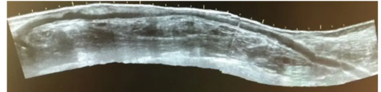
Considering the evolution of this particular vascular access so as the most recent published literature, once the functioning upper limb access was threatened by central venous stenosis, endovascular solutions should be conside-red. Still, there is some controversy about the best approach with some authors publishing good results, yet others presenting reduced primary patency on long-term follow up.12,12 In our daily practice, we tend to favor endovascular treatment of this central vein stenosis but, in this particu-lar case, once presented with early restenosis and conside-ring the lesion characteristics so as absence of immediately accessible venous patrimony in the left upper limb for an autologous access construction and the existence of an arterialized and matured right cephalic vein made us lead to that solution, which is someway also described in the literature. Great saphenous vein and femoral vein transloca-tion to the upper arm are commonly believed to have higher Figure 3 Ultrasound image after the segmental interposition with a simple ePTFE Graft.
complications rates, with poor maturation of the first one even considering good patency with the last one.14-18
The cephalic arch is prone to the development of hemodynamic significant stenosis being the most frequent location for stenosis of the upper arm dysfunctional vascu-lar accesses, corresponding to 30-55% of all stenosis sites.19 While endovascular solutions seem a reasonable option for the first pathology preventing an open surgical revision that might jeopardize the creation of a future basilic vein fistula, whether to choose between simple angioplasty, bare metal stent, graft stent or even drug eluting balloon is debatable with high recurrence rates.20-22 Vascular access aneurysms are frequently accompanied by pre or post-aneurysm ste-nosis. In our case, the thrombosed aneurysm was compli-cated by wall-adherent thrombus which implied ligation of the aneurysmal section and graft interposition. The option for the hybrid graft was made considering the biotype of the patient and despite having considered basilic vein employment, the low basilic vein diameter (<6mm, both arms), an axillary zone with a previous surgical approach and necessity of continuous dialysis sessions, preferably without novel use of CVC, made us elect the construction of a new access with an hybrid prosthesis.
The future of prosthetic grafts for AV access appears bright as new approaches and technology are being inves-tigated. Modifications in already existing grafts or develo-pment of new graft materials are being developed in an attempt to improve primary and secondary patency rates and reduce complications. Some grafts have also been upgraded with a silicone layer within the PTFE to allow early cannulation and to reduce needle hole bleeding.23
In this clinical case the use of a Hybrid Graft followed by simple graft and then early cannulation graft interpositions was described. Hybrid Grafts have hemody-namic advantages and also the possibility of anchoring its distal zone on what is expected to be a difficult surgical access region. Limitations come normally from the size of the graft that is 6 mm (according to the currently commer-cially available grafts) and of the outflow veins that should be larger than 4 mm. Other disadvantages are related to the published prosthesis infection rates that are around 7% and make this, together with the degradation at the puncture site, a long-term concernOn the other hand, early cannulation grafts allow an almost immediate cannulation and hemodialysis preventing unnecessary placement of a central venous catheter. The prosthesis is inserted into the vein, and the suture line is created on the outside surface of the prosthesis and is not located within the bloodstream at the graft-vein juncture with hemodynamic improvements. Nevertheless, prosthetic grafts still have significantly higher thrombotic threshold velocities than autologous grafts and require a higher flow to maintain patency.24-32
Acknowledgements: To the Vascular Surgery Department of Centro Hospitalar Universitário do Porto
Differential diagnosis should include malignancy and a special attention to this pathology in the transplant--recipient population should be present.
Despite mucormycosis being a rare infection, its prevalence is expected to raise together with increasing number and survival of the organ transplantation popula-tion as well as acquired immunodeficiencies. A high level of suspicion is recommended in the presence of the right clinical setting, as early diagnosis may be determinant for the prognosis.
ReFeReNCeS
1. Clinical practice guidelines for vascular access. Am J Kidney Dis 2006;48:S176e247.
2. Beathard GA. Physical examination: The forgotten tool. In: Gray RJ, Sands JJ, editors. Dialysis access: a multidisciplinary approach. Philadelphia, PA: Lippincott Williams & Wilkins; 2002. p. 111e8. 381;
3. Centers for Medicare & Medicaid Services. 2004 Annual Report: ESRD clinical performance measures project. Bal-timore, MD, Department of Health and Human Services, Centres for Medicare and Medicaid Services, Center for Beneficiary Choices, 2004. Am J Kidney Dis. 2005;46(Suppl 2):1. Available from: http://www.ajkd.org/article/S0272-6386(05)00922-4/pdf. Accessed Sep 12, 2017. 8.
4. Silva MB Jr, Hobson RW 2nd, Pappas PJ, et al: A strategy for increasing use of autogenous hemodialysis access proce-dures: Impact of preoperative noninvasive evaluation. J Vasc Surg 27:302-307, 1998
5. Trerotola SO, Scheel Jr PJ, Powe NR, Prescott C, Feeley N, He J, et al. Screening for dialysis access graft malfunction: compa-rison of physical examination with US. J Vasc Intervent Radiol JVIR 1996;7:15e20. 382 Schuman E, Ronfeld A, Barcl; 6. Robbin ML, Chamberlain NE, Lockhart ME, Gallichio MH,
Young CJ, Deierhoi MH, et al. Hemodialysis arteriovenous fis-tula maturity: US evaluation. Radiology 2002;225:59e64 7. Mihmanli I, Besirli K, Kurugoglu S, Atakir K, Haider S, Ogut
G, et al. Cephalic vein and hemodialysis fistula: surgeon’s observation versus color Doppler ultrasonographic findings. J Ultrasound Med 2001;20:217e22;
8. Beathard GA. The treatment of vascular access graft dysfunc-tion: a nephrologist’s view and experience. Adv Ren Replace Ther 1994;1:131e47. 380;
9. Vascular Access 2006Work Group. Clinical practice guidelines for vascular access. Am J Kidney Dis 2006;48(Suppl 1):S248-73 10. Chan MR, Young HN, Becker YT, Yevzlin AS; Obesity as a pre-dictor of vascular access outcomes: analysis of the USRDS DMMS Wave II study; Semin Dial. 2008 May-Jun;21(3):274-9. doi: 10.1111/j.1525-139X.2008.00434.x. Epub 2008 Apr 11. Kats M, Hawxby AM, Barker J, Allon M; Impact of obesity on
arteriovenous fistula outcomes in dialysis patients; Kidney Int. 2007 Jan;71(1):39-43. Epub 2006 Sep 27
12. Yang HT et al; A prospective randomized study of stent graft placement after balloon angioplasty versus balloon angioplasty alone for the treatment of hemodialysis patients with prosthetic graft outflow stenosis; J Vasc Surg 2018 Aug;68(2):546-553. doi: 10.1016/j.jvs.2017.12.062. Epub 2018 Apr 2
Central Venous Stenosis; Ann Vasc Surg 2018 Jan; 46:322-330. doi: 10.1016/j.avsg.2017.07.001. Epub 2017 Aug 12 14. Teruya TH, Schaeffer D, Abou-Zamzam AM, Bianchi C.
Arte-riovenous graft with outflow in the proximal axillary vein. Ann Vasc Surg 2009;23:95e8;
15. Rivers SP, Scher LA, Sheehan E, et al: Basilic vein transposi-tion: An underutilized autologous alternative to prosthetic dialysis angioaccess. J Vasc Surg 18:391-396, 1993
16. Scott JD, Cull DL, Kalbaugh CA: The mid-thigh loop arterio-venous graft: Patient selection, technique and results. Am Surg 72:825-828, 2006 33.
17. Brownie ER, Kensinger CD, Feurer ID, Moore DE, Shaffer D: A comparison of patency and interventions in thigh versus Hemodialysis Reliable Outflow grafts for chronic hemodialy-sis vascular access. J Vasc Surg 64:1392-1399, 2016 18. Takuya Hatakeyama et al; Introduction of arteriovenous
grafts with graft insertion anastomosis for hemodialysis access; J Vasc Surg 2017 Sep;66(3):952-957. doi: 10.1016/j. jvs.2017.03.441. Epub 2017 Jun 12
19. Rajan DK, Clark TWI, Patel NK, Stavropoulos SW, Simons ME. Prevalence and treatment of cephalic arch stenosis in dys-functional autogenous hemodialysis fistulas. J Vasc Intervent Radiol JVIR 2003;14:567e73 (12761309);
20. Patanè D, Giuffrida S, Morale W, L’Anfusa G, Pulliatti D, Bisceglie P, et al. Drug-eluting balloon for the treatment of failing hemodialytic radiocephalic arteriovenous fistulas: our experience in the treatment of juxta-anastomotic stenoses. J Vasc Access 2014;15:338e43;
21. Portugaller RH, Kalmar PI, Deutschmann H. The eternal tale of dialysis access vessels and restenosis: are drug-eluting balloons the solution? J Vasc Access 2014;15:439e47; 22. Liu YH, Hung YN, Hsieh HC, Ko PJ. Surgical thrombectomy
for thrombosed dialysis grafts: comparison of adjunctive treatments. World J Surg 2008;32:241-5.
23. Scher LA, Katzman HE: Alternative graft materials for hemo-dialysis access. Semin Vasc Surg 17:19-24, 2004 27; 24. Tordoir J, Canaud B, Haage P, Konner K, Basci A, Fouque
D, et al. EBPG on vascular access. Nephrol Dial Transplant 2007;22:ii88e117;
25. Hazinedaroglu SM, Tuzuner A, Ayli D, Demirer S, Duman N, Yerdel MA. Femoral vein transposition versus femoral loop grafts for hemodialysis: a prospective evaluation. Transplant Proc 2004;36:65e7;
26. Huber TS, Hirneise CM, Lee WA, Flynn TC, Seeger JM. Out-come after autogenous brachial-axillary translocated super-ficial femoropopliteal vein hemodialysis access. J Vasc Surg 2004;40:311e8;
27. Kitrou PM, Katsanos K, Spiliopoulos S, Karnabatidis D, Sia-blis D. Drug-eluting versus plain balloon angioplasty for the treatment of failing dialysis access: final results and coste-ffectiveness analysis from a prospective randomized control-led trial (NCT01174472). Eur J Radiol 2015;84:418e23; 28. Glickman M; Drug eluting grafts for hemodialysis access; J
Vasc Access. 2017 Mar 6;18(Suppl. 1):53-55. doi: 10.5301/ jva.5000671. Epub 2017 Mar 5
29. Berard X, Ottaviani N, Brizzi V, et al: Use of the Flixene vascu-lar access graft as an early cannulation solution. J Vasc Surg 62:128-134, 2015 28;
30. Glickman MH, Burgess J, Cull D, et al: Prospective multicen-ter study with a 1-year analysis of a new vascular graft used for early cannulation in patients undergoing hemodialysis. J Vasc Surg 62:434-441, 2015;
31. Glickman MH, Burgess J, Cull D, Roy-Chaudhury P, Schanzer H; Prospective multicenter study with a 1-year analysis of a new vascular graft used for early cannulation in patients undergoing hemodialysis; J Vasc Surg. 2015 Aug;62(2):434-41. doi: 10.1016/j.jvs.2015.03.020. Epub 2015 May 4 32. Al Shakarchi J, Houston G, Inston N; Early cannulation grafts
for haemodialysis: a systematic review; J Vasc Access. 2015 Nov-Dec;16(6):493-7. doi: 10.5301/jva.5000412. Epub 2015 Jun 12
33. Maytham GG, Sran HK, Chemla ES; The use of the early can-nulation prosthetic graft (Acuseal™) for angioaccess for hae-modialysis; J Vasc Access. 2015 Nov-Dec;16(6):467-71. doi: 10.5301/jva.5000390. Epub 2015 May 23
34. Windus DW, Jendrisak MD, Delmez JA. Prosthetic fistula sur-vival and complications in hemodialysis patients: effects of diabetes and age. Am J Kidney Dis. 1992;19(5):448-452