Chitosan scaffolds containing calcium phosphates and diclofenac for bone regeneration – Development, validation and optimization
106
0
0
Texto
(2) UNIVERSIDADE DE LISBOA FACULDADE DE FARMÁCIA. CHITOSAN SCAFFOLDS CONTAINING CALCIUM PHOSPHATE AND DICLOFENAC FOR BONE REGENERATION DEVELOPMENT, VALIDATION AND OPTIMIZATION. Lara Mariana Almeida Duarte das Neves. The dissertation was supervised by José Manuel Ventura, Ms and Lídia Diogo Gonçalves, PhD. Master in Biopharmaceutical Sciences. 2018.
(3) “O sucesso nasce do querer, da determinação e persistência em se chegar a um objetivo. Mesmo não atingindo o alvo, quem busca e vence obstáculos, no mínimo fará coisas admiráveis” José de Alencar. iii.
(4) ABSTRACT. In the last decades a growing need for bone substitutes has been detected. This is mainly related to the increasing aging of the population which leads to the increase of muscle and bones disorder. Bone loss may result from congenital malformations, tumour resections, trauma, shortterm and acute onset pathologies, and chronic diseases or even from aging, which may promote physical incapacity and prolonged pain. The application of implants in order to solve these losses is not always positive and some inconveniences may occur, such as the infection development, immune rejections or local tissues death. At the moment, the market offers numerous solutions with different biomaterials with singular properties combinations that mimic the original bone tissue constitution. However, cases where bone regeneration is associated with local inflammatory response control after surgery are rare. In order to overcome this limitation, a threedimensional porous matrix (scaffold) incorporating diclofenac will be developed with the goal of release anti-inflammatory drug in a controlled and localized way as it degrades. This drug promotes the inflammation reduction and it is widely used in the treatment of diseases of bones and joints. This porous matrix of chitosan, calcium phosphate and sodium diclofenac was subjected to the lyophilization process, and it was later characterized in more detail in terms of porosity and structures surfaces by scanning electron microscopy (SEM), of chemical composition and crystallographic structure of calcium phosphate granules by X-ray diffraction (XRD), of particle size by laser beam diffraction, of release profile measured in phosphate buffered saline (PBS) and determined by UV-Vis spectroscopy and absorption capacity by the captive bubble method. Since no viable microorganisms should exist in medical devices, the scaffolds were subjected to a sterilization process by gamma radiation, guaranteeing the inactivation of microorganisms and providing biological safety. The scaffolds ability to release drug was studied for 41 days by the diclofenac release assay, with a faster exchange within the first 7 hours. Through the destruction of the samples it was possible to determine that 20% of diclofenac was still remaining in the non-irradiated samples and 40% in the irradiated ones. In this way, it was verified that the radiation influences the release profile. The swelling assay has demonstrated that the absorption capacities of the different structures exhibit similar behaviours, although the irradiated samples appear to have lower values. iv.
(5) The captive bubble assay has led to the conclusion that all the produced samples are hydrophilic, thus promoting better biological interactions between the cells and the material. Calcium phosphate granules are the major component of the scaffold and, through XRD analysis, it is a biphasic mixture composition that was found to be around 75% Hap and 25% β-TCP, allowing a more efficient osseointegration. As visualized by the SEM images, the small porosity inside the granules along with the scaffolds porosity will allow a total material vascularization. The SEM images allowed to observe the irregular and rough surface of calcium phosphate granules and the structure of the sterilized and unsterilized scaffolds, allowing to conclude that both have characteristics favorable to cell adhesion. Cytotoxicity assays, evaluated by the direct-contact method, showed moderate cytotoxic effect only in 25kGy scaffolds with L929 fibroblast cells. According to the results obtained in the several tests that were carried out, it was concluded that the developed scaffold that showed the best performance during the study were the samples sterilized. These scaffolds may be a potential medical device for localized delivery of diclofenac.. Keywords: scaffold, calcium phosphates, chitosan, diclofenac, controlled release.. v.
(6) RESUMO. Nos últimos tempos têm-se vindo a verificar uma crescente necessidade de utilização de substitutos ósseos. Isto deve-se à incapacidade de regeneração óssea acompanhar o aumento do defeito ósseo. A perda óssea pode advir de malformações congénitas, ressecção de tumores, traumatismos, patologias de curta duração, doenças crónicas, patologias de início agudo, ou mesmo do envelhecimento, podendo promover incapacidade física e dor prolongada. O recurso à aplicação de enxertos ósseos ou implantes para resolução destas perdas nem sempre é positivo, surgindo alguns inconvenientes como o desenvolvimento de infeções, rejeições imunológicas e morte dos tecidos locais. De momento, o mercado disponibiliza inúmeras soluções, com diversas combinações de biomateriais com diferentes propriedades que mimetizam a constituição original do tecido ósseo. No entanto, são raros os que permitem a regeneração óssea associada ao combate da resposta inflamatória local após cirurgia. Com a finalidade de colmatar esta limitação, desenvolveu-se neste projeto uma matriz tridimensional porosa (scaffold), em cuja estrutura se associou um anti-inflamatório não esteróide, que promove a redução da inflamação. A inflamação é caracterizada como uma complexa cascata de eventos fisiológicos, necessária para o processo de cicatrização. Inicia-se uma hora após a agressão, com a afluência inicial de neutrófilos e posteriormente de linfócitos e macrófagos ao local lesionado. O pico da resposta inflamatória aguda é atingido após 24 horas e é concluído ao fim de sete dias. A implantação cirúrgica de um biomaterial desencadeia uma resposta à agressão e durante essa fase, as propriedades físicas e químicas do biomaterial vão delimitar a duração do processo inflamatório. Desta forma, propôs-se criar um substituto ósseo que permitisse libertar um anti-inflamatório de forma controlada e localizada, de modo a eliminar as tomas diárias de comprimidos por parte dos utentes. A libertação localizada, faz com que a presença de fármaco no organismo seja menor e mais eficaz, evitando potenciais problemas renais e do trato gastrointestinal. O scaffold desenvolvido, teve de respeitar os seguintes requisitos para poder futuramente ser considerado e utilizado em aplicações médicas: capacidade de libertar de forma controlada e localizada o anti-inflamatório, ser biocompatível, ter uma degradação controlada, apresentar resistência mecânica suficiente para suportar tensões existentes do ambiente onde é inserido, demonstrar poros com tamanho e morfologia apropriada para o transporte de nutrientes, ter propriedades químicas. vi.
(7) apropriadas para adesão celular, ser de fácil esterilização e de processar em diferentes geometrias tridimensionais. A composição da matriz porosa em estudo, conta com a mistura de alguns componentes, como: •. Quitosano, polímero natural desacetilado do polissacarídeo quitina extraído do exoesqueleto de crustáceos, apresenta excelentes propriedades a nível de biodegradabilidade, toxicidade,. biocompatibilidade,. imunogenicidade. e. tem. atividade a. antibacteriana,. capacidade. de. baixa. melhorar. a. neovascularização. É um biopolimero de manipulação relativamente fácil, apresentando solubilidade em soluções aquosas ácidas. •. Fosfatos de cálcio (hidroxiapatite e β-TCP), estruturalmente são semelhantes aos componentes inorgânicos do tecido ósseo, capazes de atuar de forma apropriada, numa aplicação específica, promovendo assim a ligação química com o osso e sua regeneração.. •. Diclofenac sódico, anti-inflamatório não esteroide com propriedades antiinflamatória, analgésica, antipirética e antirreumática. Provoca a inibição da biossíntese de prostaglandinas. Apresenta propriedades higroscópicas e a sua solubilidade varia com o pH, podendo ser mais solúvel no meio básico e pouco solúvel em meio ácido.. A mistura dos fosfatos de cálcio e de quitosano, já é comercializada pela empresa Ceramed S.A, sob a forma de pasta injetável com aspeto pastoso e amarelado, denominada comercialmente por k-IBS® . No início do processo de fabrico das amostras, estudou-se a melhor forma de incorporar o diclofenac na pasta k-IBS®. Após o sucesso da mistura, seguiu-se o processo de liofilização, colocando a pasta em moldes específicos. Este é um processo estabilizante através do qual a mistura é inicialmente congelada, passando para a fase de secagem primária, onde a quantidade de solvente é reduzida por sublimação seguida da dessorção. A liofilização faz com que ocorra transformação da mistura pastosa para um sólido com porosidade associada, capaz de ser cortado com as dimensões pretendidas. A ausência de microrganismos viáveis é uma obrigatoriedade para os dispositivos médicos. Todavia, as condições em que foram produzidas as amostras deste trabalho, contam com microrganismos provenientes das matérias-primas, do processo produtivo mais concretamente do operador, dos equipamentos e do ambiente. O processo de esterilização é o método de eliminação de contaminação microbiológica, que permite transformar as amostras produzidas em material estéril. Assim sendo e de acordo com a norma Internacional ISO 11137, esterilizaram-se as amostras liofilizadas com doses de radiação gama a 15kGy e a 25kGy. vii.
(8) O perfil de libertação do diclofenac foi medido em solução salina tamponada de fosfato (PBS) e determinada por espectroscopia UV-Vis. A capacidade que os scaffolds têm de libertar fármaco foi estudada durante 41 dias. Observou-se uma libertação mais rápida nas primeiras 7 horas, como expectável. Ao fim dos 41 dias, verificou-se que para os scaffolds não irradiados faltava libertar 20% do fármaco e para os scaffolds irradiados, tanto a 15 como 25kGy, faltava libertar 40% de diclofenac. Desta forma, comprovou-se que a radiação influencia o perfil de libertação. Este facto, pode advir da reticulação provocada pela radiação gama na cadeia do quitosano. De certa forma, os resultados dos ensaios de swelling, vieram corroborar os ensaios de libertação, exibindo uma capacidade de absorção inferior para as amostras irradiadas. Uma vez que o diclofenac parece ser libertado quando há desagregação de matéria, a expansão do scaffold promovida pela absorção de líquido, leva a um aumento de volume que promove a libertação de fármaco. O ensaio da bolha cativa permitiu chegar à conclusão, de que todas as amostras produzidas eram hidrofílicas. A nível biológico este parâmetro é essencial. As interações biológicas (adesão e proliferação celular) ocorrem de forma positiva, quanto mais hidrofílico for o material. Os grânulos de fosfato de cálcio são a maior componente do scaffold e através da análise de DRX, verificou-se que a sua composição de mistura bifásica ronda os 75%HAp e 25%β-TCP com alto grau de pureza e ausência de outras fases cristalográficas. Desta forma, e devido à semelhança da composição com a fase mineral do osso humano, a osteointegração ocorrerá de forma rápida. O fosfato tricálcico ao dissolver-se mais rapidamente que a hidroxiapatite acelera o processo de reabsorção e formação de novo osso. E como visualizado pelas imagens SEM, a pequena porosidade existente no interior dos grânulos juntamente com a porosidade do scaffold irá permitir uma vascularização total do material. As imagens de SEM juntamente com a análise FTIR, não permitiram observar a presença do diclofenac nos scaffolds irradiados e não irradiados. A quantidade de fármaco adicionado à matriz é inferior a 1%, podendo apenas ser identificada nos ensaios de libertação. As imagens SEM permitiram observar a superfície dos scaffolds esterilizados e não esterilizados e os grânulos de fosfatos de cálcio. A estrutura dos grânulos esféricos apresenta uma superfície irregular e rugosa que quando ampliada, pode-se visualizar uma estrutura com pouca compactação e desarranjo das partículas de grandes dimensões. Estas características são favoráveis à adesão celular.. viii.
(9) Comprovou-se através da análise granulométrica, que os tamanhos dos grânulos utilizados no processo variam entre os 135µm e 355µm. Os resultados obtidos revelaram a existência de homogeneidade de tamanhos. Os ensaios de citotoxicidade, avaliados através do método de contacto direto, revelaram ser citotóxicos para as células MG63 e L929 fibroblastos, quando o poço se encontrava coberto por 1/10 da amostra. Realizou-se novo ensaio apenas com as células L929, onde a amostra ocupava 1/20 do poço. Os scaffolds com diclofenac esterilizados a 25kGy deram moderadamente citotóxicos, com viabilidade entre os 60-65% As restantes amostras, com e sem fármaco esterilizadas a 15kGy e não esterilizadas, e as amostras sem diclofenac irradiadas a 25kGy, não tinham evidencias de efeitos citotóxicos, apresentando uma viabilidade celular entre 70%-82%, que estão acima do limite citotóxico definido pela ISO standard. De acordo com os resultados obtidos nos vários ensaios realizados, concluiu-se que os scaffolds desenvolvidos que melhores performances demonstraram ao longo do estudo, foram as amostras esterilizadas, tanto a 15kGy como 25kGy. Estes scaffolds podem ser um potencial dispositivo médico para entrega localizada de diclofenac.. Palavras-chave: scaffold, fosfatos de cálcio, quitosano, diclofenac, libertação controlada.. ix.
(10) ACKNOWLEDGEMENTS To all those who contributed to the realization of this master’s thesis directly or indirectly, my sincere thanks. First of all, I am deeply grateful to my supervisor José Ventura, who has been tireless throughout this project and has always demonstrated an extraordinary availability. I would like to thank you for all the knowledge you have transmitted to me, all the help and guidance you have given me, which has been contributed to my personal and professional growth. To Doctor Lídia Gonçalves I would like to thank for his availability, help and wisdom when it was needed. To Ceramed – Cerâmicos para aplicações médicas S.A. for providing the raw material for the development of this project and for the availability it demonstrated in relation to the accomplishment of analyzes. My gratitude goes also to all my co-workers and friends, Thierry, Sara, Carlos, Ana, Andreia L., Alexandre, Telma, Vanessa, Pedro, Joel and Mário, for all the help, guidance and good disposition. Thanks you also to Dr.Eduardo Pires, CEO of Ceramed S.A, for the help and financial support. To Doctor Lígia Figueiredo, Doctor Luis Pinto and Doctor Andreia Pimenta, for their help, knowledge and friendship. A special thanks goes to Doctor Ana Paula Serro (IST), who opened the doors of his laboratory to help me with several techniques, all support and transmitted knowledge. To Eng. Ana Topete (IST) and Eng.Diana Silva (IST), for help with the equipment and all the knowledge transmitted. Eng.Isabel Nogueira (IST), for her help in SEM analysis, Prof. Amélia Almeida (IST), who helped perform the specimens to be analyzed in SEM. To Doctor Nuno Faria and Ricardo Pereira (IST) for all assistance with the centrifuge and the lyophilization processes. To Doctor Helena Gil and Doctor Jorge Guiomar (U.Coimbra) for providing the diclofenac and help. Last, but definitely not least, a huge thank you to my family for understanding my absences and for all the unconditional support during this journey, especially to my parents, brothers and Ana Almeida. To my friend Miguel for all patience, strength, support and help. You have always motivated me to achieve more. Thank you very much to all who have accompanied me.. x.
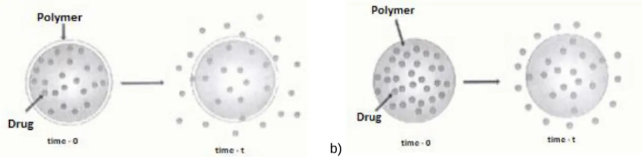
(11) CONTENTS ABSTRACT ................................................................................................................. iv RESUMO ..................................................................................................................... vi ACKNOWLEDGEMENTS ............................................................................................. x CONTENTS ................................................................................................................. xi LIST OF FIGURES ..................................................................................................... xiii LIST OF TABLES .......................................................................................................xvi ABREVIATIONS ........................................................................................................ xvii INTRODUCTION .......................................................................................................... 1 MOTIVATION AND GOALS .......................................................................................... 3 1.. THEORETICAL FUNDAMENTS ............................................................................ 4. 1.1.. Tissue Engineering ............................................................................................ 4. 1.2.. Contributing Factors to the Clinical Success of Bone Substitutes Scaffolds ....... 4. 1.3.. Bone Tissue ....................................................................................................... 5. 1.4.. Mechanism of Bone Growth .............................................................................. 7. 1.5.. Regeneration Process ....................................................................................... 8. 1.6.. Bone Substitutes ................................................................................................ 9. 1.6.1.. Biomaterials .................................................................................................. 11. 1.6.1.1.. Synthetics Biphasic Bones Substitutes ...................................................... 12. 1.6.1.2.. Synthetic Biphasic Bones Substitutes with Chitosan Incorporation ............ 13. 1.6.1.2.1. Chitosan .................................................................................................... 14 1.7.. Nonsteroidal Anti-Inflammatory Drugs .............................................................. 16. 1.8.. Mechanisms of Action of NSAIDs ..................................................................... 16. 1.8.1. 1.9. 1.10.. Diclofenac Sodium – Structure and main properties ..................................... 18 Incorporation of an Active Substance ............................................................... 20 Controlled Drug Delivery ............................................................................... 20. 1.10.1. Polymers in Drug Delivery Systems .............................................................. 21 1.10.2. Mathematical Description of the Drug Release Kinetics from the Scaffolds .. 24 1.11.. Sterilization ................................................................................................... 29 xi.
(12) 1.11.1. Gamma Radiation ......................................................................................... 29 1.11.2. Gamma sterilization influence in drug release............................................... 30 2.. PROCESS, MATERIALS AND METHODS .......................................................... 31. 2.1.. Freeze Drying Process ..................................................................................... 31. 2.2.. Scaffolds Preparations ..................................................................................... 32. 2.2.1.. Optimization Process .................................................................................... 34. 2.2.2.. Sterilization Process ..................................................................................... 36. 2.3.. Diclofenac Controlled Release Experiments ..................................................... 36. 2.4.. Chemical and Structural Scaffold Characterization ........................................... 38. 2.4.1.. Microscopy ................................................................................................... 39. 2.4.2.. X-ray Diffraction Method ............................................................................... 42. 2.4.3.. Particle Size Analyser ................................................................................... 43. 2.4.4.. Wettability – Contact Angle (Captive bubble) ................................................ 44. 2.4.5.. Swelling ........................................................................................................ 47. 2.4.6.. FTIR - Fourier-transform Infrared Spectroscopy ........................................... 48. 2.5.. Biological Analysis ........................................................................................... 50. 2.5.1. 3.. In Vitro cytotoxicity test ................................................................................. 50. RESULTS AND DISCUSSION............................................................................. 54. 3.1.. Diclofenac Release System on CaPs and CS Scaffolds ................................... 54. 3.2.. Scaffolds and Components Characterization Analysis ...................................... 61. 3.2.1.. XRD Analysis................................................................................................ 61. 3.2.2.. Particle Size Distributions ............................................................................. 62. 3.2.3.. Wettability ..................................................................................................... 63. 3.2.4.. Swelling ........................................................................................................ 64. 3.2.5.. Scanning Electron Microscopy / Energy Dispersive X-Ray Spectroscopy ..... 66. 3.2.6.. FTIR ............................................................................................................. 67. 3.2.7.. In Vitro cytotoxicity test ................................................................................. 69. 4.. CONCLUSION AND FUTURE WORK ................................................................. 73. 5.. BIBLIOGRAPHY .................................................................................................. 77 xii.
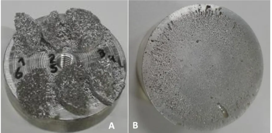
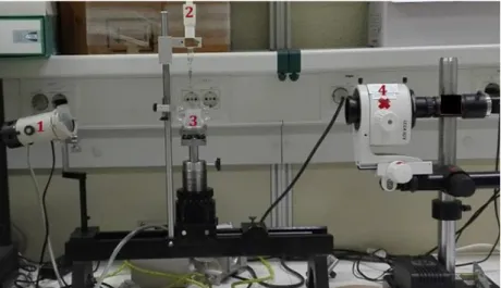
(13) LIST OF FIGURES Figure 1 - Example of scaffold application developed in this project. ............................ 3 Figure 2 - Composition of bone tissue ......................................................................... 6 Figure 3 – Structure cortical and cancellous bone. ....................................................... 7 Figure 4 - Bone matrix and cells that constitute it . ....................................................... 8 Figure 5 - Chemical structure of chitin. ....................................................................... 14 Figure 6 - Chemical structure of chitosan. .................................................................. 15 Figure 7 – Mechanism of action of non-steroidal anti-inflammatory drugs. ................. 18 Figure 8 - Chemical structure of DF. .......................................................................... 19 Figure 9 - Schematic representation of chemically controlled release. a) biodegradable system; b) pendant-chain system................................................................................ 22 Figure 10 - Schematic representation of osmotically controlled release systems and swelling system. a) Type A contains an osmotic core with drugs; b) Type B contains a drug reservoir surrounded by osmotic core. ................................................................ 23 Figure 11 - Schematic representation: a) reservoir diffusion-controlled release systems, b) monolithic diffusion-controlled release systems ...................................................... 23 Figure 12 - Water's phase diagram ............................................................................ 32 Figure 13- Schematic illustration of scaffolds production (adapted Asadian-Ardakani et al.). ............................................................................................................................. 34 Figure 14 - Example of irradiated scaffolds. These were inserted in Tyvek bags with yellow gamma indicator and sent to Aragogamma. After the radiation, the indicator gets red, as a sign that the radiation occurred. ................................................................... 36 Figure 15 - Preparation of the irradiated and non-irradiated samples with and without diclofenac, to the release assay and subsequent spectrophotometer analysis. ........... 37 Figure 16 - overnight shaking of the tubes that contains scaffolds emerged in PBS. .. 38 Figure 17 –A) scaffold emerged in PBS before the shaking. b) Scaffold destroyed after the centrifugation. ....................................................................................................... 38 Figure 18 - epoxy formers with granules of calcium phosphates, on the left and with scaffolds on the right. .................................................................................................. 39 Figure 19 - Scanning Electron Microscope from HITACHI, model S-2400 by IST. ...... 41 Figure 20 – A) scaffolds half’s without DF (1, 2 and 3) and with DF (4, 5 and 6) on the SEM support; B) Epoxy former of the CaPs granules overlayed with gold-palladium. . 41 Figure 21 - Sessile drop (left image); Captive bubble (right image). ........................... 44. xiii.
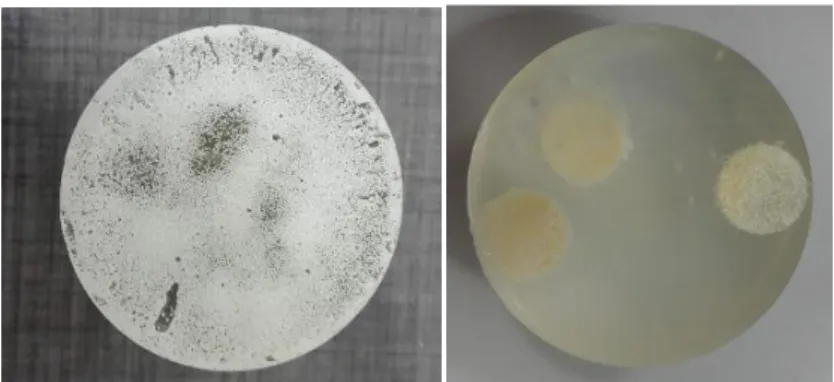
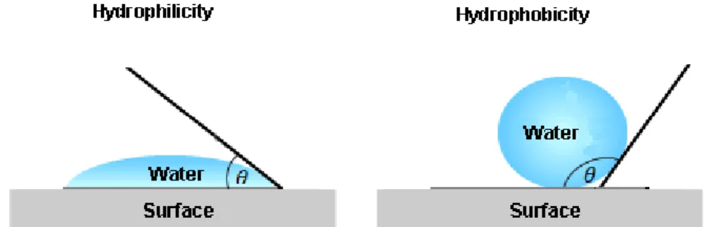
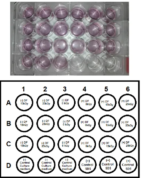
(14) Figure 22 - The water contact angle (θ) on a hydrophilic surface and a hydrophobic surface. ....................................................................................................................... 45 Figure 23 - Hydrophobic and hydrophilic water contact angle .................................... 45 Figure 24 - Micrometer syringe with a needle inverted in the edge and the liquid cell. 46 Figure 25 – Goniometer: 1) Light source 2) Micrometer syringe 3) Liquid cell 4) Video camera........................................................................................................................ 46 Figure 26 - Scaffold dried used in swelling assay. ...................................................... 47 Figure 27 - IRAffinity-1 Shimadzu equipment. ............................................................ 49 Figure 28 – Example of KBr tablet with scaffold powder blend. .................................. 49 Figure 29 – 24-well plate real and layout for the MTT cytotoxicity test, accounting for the 6 different samples of scaffolds and respective positive and negative controls. .......... 52 Figure 30 - SEM image of the lyophilized scaffold, obtained from the dissolution of DF in 1/3 of water and lactic acid in 2/3 of water. Resolution of 20.0kV and magnification of 60x.............................................................................................................................. 54 Figure 31 – A – CaPs granule exterior; B,C CaPs granule interior with different amplifications; D – scaffold’s porosity. ........................................................................ 55 Figure 32 - DF release profiles in terms of released mass accumulated by sample mass. Error bars: standard deviation (n=5). The smaller graph is an enlargement of the larger graph of the first 7h of release. ................................................................................... 57 Figure 33 – Fraction of released DF in relation to that introduced in scaffold. Error bars: standard deviation (n=5). The smaller graph is an enlargement of the larger graph for the first 7h of release. The represented lines are only intended to facilitate the profiles identification. ............................................................................................................... 58 Figure 34 - X-ray diffractogram representative of biphasic ceramic granules used in the manufacturing process of scaffolds. ............................................................................ 61 Figure 35 - Distribution of the particle size of the biphasic granules used in the production of scaffolds in function of the sample volume through laser diffraction. ....................... 62 Figure 36 – Epoxy mold with spherical CaPs granules used in scaffolds.................... 63 Figure 37 - Water contact angle of samples with and without diclofenac and nonsterilized and sterilized. ............................................................................................... 63 Figure 38 - An air bubble sitting beneath a surface of specimens: a) non-sterilized without DF, b) sterilized at 15kGy without DF, c) sterilized at 25kGy without DF, d) nonsterilized with DF, e) sterilized at 15kGy with DF, f) sterilized at 25kGy with DF. ........ 64 Figure 39 - Swelling profile as a function of time. ....................................................... 65 Figure 40 - SEM images from the scaffold surface. A) non-irradiated (bar indicates 400µm); B) irradiated at 15kGy (bar indicates 400µm); C) irradiated at 25kGy (bar indicates 400µm); D) 25kGy irradiated scaffold amplification (bar indicates 200µm); .. 66 xiv.
(15) Figure 41 - Distribution chart of the chemical elements present in the analysed scaffold. ................................................................................................................................... 67 Figure 42 – Comparation between raw material with scaffold, with DF and not irradiated. ................................................................................................................................... 68 Figure 43 - Comparation between scaffolds with DF, irradiated and not irradiated. .... 69 Figure 44 - A) Scaffold covering 1/10 of the well; B) Scaffold covering 1/20 of the well. ................................................................................................................................... 70 Figure 45 - Graphic representation of cell viability (%) values obtained for the different scaffolds tested with the different line cells. ................................................................ 71 Figure 46 - Graphic representation of cell viability. ..................................................... 72 Figure 47 – Comparative graphic of cell viability between different amounts of material introduced into the wells. ............................................................................................ 72 Figure 48 - FTIR spectra corresponding to DF. ............................................................. i Figure 49 - FTIR spectra corresponding to CS. ............................................................ ii Figure 50 - FTIR spectra corresponding to CaPs. ........................................................ iii. xv.
(16) LIST OF TABLES. Table 1 - Commonly prescribed drugs with diclofenac, dose and the recommended dosage ........................................................................................................................ 19 Table 2 - Interpretation of diffusional release mechanisms from polymeric films, taking into account the exponent of the Korsmeyer-Peppas model. ...................................... 28 Table 3 - Mixing optimization processes. .................................................................... 35 Table 4 - Determination coefficient (R2) obtained through the application of the mathematical models to the DF release profiles of the different prepared samples. .... 60 Table 5 - Values of the parameter 𝑛 obtained from the Korsmeyer-Peppas model. .... 60 Table 6 - Cell viability percentage of the samples tested with 1/10 area of cell monolayer. ................................................................................................................................... 70 Table 7 - Cell viability percentage of the samples tested with 1/20 area of cell monolayer. ................................................................................................................................... 72. xvi.
(17) ABREVIATIONS CaP. Calcium Phosphate. CIFs. Crystallographic information files. COX. Cyclooxygenases. CS. Chitosan. DD. Deacetylation Degree. DF. Diclofenac Sodium. DMSO. Dimethyl sulfoxide. FTIR. Fourier-transform Infrared Spectroscopy. HAp. Hydroxyapatite. IL. Interleukin. IR. Infrared spectroscopy. ISO. International Standards Organization. MAUD. Materials Analysis Using Diffraction. MSCs. Mesenchymal Stem Cells. MTT. 3-(4,5-dimethylthiazol-2-yl)-2,5-diphenyl-tetrazolium bromid. NSAID. Nonsteroidal Anti-Inflammatory Drug. PBS. Phosphate Buffered Saline. PDS. Polydioxanone. PEG. Polyethylene glycol. PGA. Polyglycolic acid. PLA. Polylactic Acid. SAL. Security assurance level. SEM/EDS. Scanning Electron Microscopy/ Energy Dispersive X-Ray Spectroscopy. TCP. Tricalcium Phosphate. TNF-α. Tumor necrosis factor – alfa. UV-VIS. Ultraviolet-Visible. XRD. X-ray powder diffraction. β-TCP. β-tricalcium phosphate. xvii.
(18) INTRODUCTION Healthy bones are extremely important in overall health and in human life quality. They allow the mineral storage, essential for the proper body functioning, mobility, support and protection1. Due to congenital anomalies, traumas, diseases or even aging, the bone deteriorates, promoting pain and physical incapacity, thus compromising daily activities, sometimes even leading to the need of high medical costs treatments 2–4. Since the mid-twentieth century, there has been a significant increase in the use of orthopedic and maxillofacial implants for bone replacement5,6. Generally, these implants need to be associated with drug shots that can reduce the inflammatory process, counteract rejection and promote a more efficient regeneration. However, many of these bone fixation and repair devices rely on the use of metals, such as stainless steel, titanium and its alloys7. These metal implants are not biodegradable and they frequently require more surgeries to remove them8. Due to a major boost in science, there are already several biodegradable bone substitutes that support the surrounding cells and tissues, in order to promote tissue regeneration and repair, providing mechanical support and simultaneously the implant's degradation by the body9. In order to mimic the bone structure, several composites of different ceramic types and biodegradable polymers have been developed. Recently, chitosan has played an important role in bone regeneration, and more attention has been paid to chitosan composites and their applications in bone tissue, due to their ability to reduce adverse reactions to a foreign body, both being biocompatible, biodegradable and because of their ability to be shaped into various geometries and shapes. These scaffolds degrade at a rate similar to bond tissue regeneration, are osteocunductors and possess an interconnected pore structure that allow bone in-growth. Chitosan (CS) and calcium phosphates CaPs namely Hydroxyapatite (HAp) and β-tricalcium phosphate (β-TCP) composites scaffolds have revealed great potential in bone regeneration since CaPs are the main inorganic component in natural bone10. Bioceramics such as HAp and β-TCP allow the occurrence of osteogenesis and strongly bind to bone tissue10. HAp has been amply used as coating in orthopedical implants, due to its osteoconductivity, despite of presenting limitations by the fact that this is a fragile material2. Chitosan, a natural polymer, as mentioned above, has come to be studied as a scaffold material since it is nontoxic, it can be shaped into complex structures, promotes adhesion and cellular migration, improves wound cicatrization and is biodegradable at a rate that depends only of controllable material characteristics such as deacetylation degree, molecular weight and crystallinity11,12.. 1.
(19) The inflammatory process is associated to the occurrence of lesions and traumas and it can be divided in multiple steps: the acute phase, which is characterized by a localized vasodilatation and an increase off capillary permeability; a sub-acute phase, comprised of leukocytes and phagocytic cells infiltration; and a proliferative chronical phase, at which tissue degeneration and fibrosis occurs. Nonsteroidal anti-inflammatory drugs (NSAID) are generally used to reduce pain and inflammation resulting from several types of lesions. Thereby, the incorporation of NSAIDs in the scaffolds allows the reduction of the inflammatory process1, avoid adverse gastrointestinal effects and offer the possibility to administer the drug in the absence of inactivation over the effect of first transition at a hepatic level, as that release happens in the oral area13. This work aims to gather a wide range of knowledge about the development and characterization of chitosan and calcium phosphates composite scaffolds based on the commercially available injectable bone substitute k-IBS® produced at Ceramed- Cerâmicos para Aplicações Médicas S.A. in which NSAID has incorporated. Despite the diversity of commercially available products aimed at bone regeneration, they can have high costs. Therefore, one of the great challenges is to develop a biodegradable scaffold from low cost raw materials, with easy access and with the proper characteristics to accomplish the envisioned function. In accordance with market demands, the pretension of this work is to obtain a composite scaffold product easy to manipulate, that can be cut to fit bone defects independent of their shape and that stimulates bone growth and tissue formation. .. 1. (If the inflammatory process reaches the chronic phase, excessive phagocytic activity of the neutrophil is observed and the bone regeneration process will be impaired). 2.
(20) MOTIVATION AND GOALS This project's main objective is to create a scaffold composed of CaPs and CS combined with an anti-inflammatory drug, sodium diclofenac, which can be used for bone regeneration and targetted treatment of the initial inflammation. It is intended to study the composite (calcium phosphates/chitosan) influence and the irradiation process influence on drug release, mechanical strength, body fluid absorption capacity, stability and degradation level. More specifically, it is intended to: •. Improve scaffolds’ properties through changes in k-IBS® composition;. •. Prepare chitosan-calcium phosphates composite scaffolds with incorporated diclofenac;. •. Understand the influence of diclofenac in the obtained scaffolds properties;. •. Evaluate the kinetic release behavior for the application of diclofenac drug delivery;. •. Optimize process parameters in order to obtain non-cytotoxic scaffolds and promote bone formation;. •. Study the effects of different gamma radiations on the scaffolds properties.. Figure 1 - Example of scaffold application developed in this project. Adapted 14. 3.
(21) 1. THEORETICAL FUNDAMENTS 1.1.. Tissue Engineering. Like Rami Mhanna and Anwarul Hansan15 said “the tissue engineering is an interdisciplinary field that utilizes cells, biomaterials, biochemical and physical signals, as well as their combinations to generate tissue-like structures”. In order to promote cell growth and proliferation, tissue engineering utilizes biomaterial manipulation, from both artificial and natural biomaterials that supply the structural basis for the controlled tissue growth. Biomaterial is a material in contact with biological fluids or tissues and its objective is to constitute partially or as an whole living structure. 16,17. . It can. also be utilized as a drug transport through several formulation types (foams, hydrogels, fibers and microparticles)18. Tissue regeneration matrices, also designated as scaffolds, are three-dimensional structures that work as a support and have physical, chemical and mechanical properties appropriated for new tissues formation both in vivo or in vitro. These structures goals are to mimic the extracellular matrix, promote cellular migration and adhesion, provide biochemical factors that will allow nutrient diffusion, and to tridimensionaly orientate new tissue formation17,19. For this reason, the scaffold architecture and morphology are important factors in the development of these structures due to their impact in tissue regeneration (e.g. porosity level and pore size and distribution)20. Other factors must be taking into account when developing a scaffold for a specific application, namely the damaged tissue location and the size of the defect.. 1.2.. Contributing Factors to the Clinical Success of Bone Substitutes Scaffolds. To achieve clinical success, scaffolds must have a good osteointegration, adhesion, proliferation, and a good cellular differentiation and migration. Generally, the goal is to potentiate the mechanical, physical and chemical properties from a variety of materials that complement each other21. Usually it is required that a scaffold demonstrate the following properties: •. Mimic the native extracelular matrix;. 4.
(22) •. Biocompatibility – the material and it's degradation products should not promote an immune system response or not to possess an unacceptable level of toxic substances22;. •. Adequate degradation rate – the matrix should be bioabsorved or degraded in a very well determined time period. This period must be equivalent to the time needed for the previously occupied tissue space to be substituted by new tissue;. •. Chemical properties that allow adhesion, proliferation and cellular differentiation;. •. Controlled degradation – different tissues have different regeneration rates. Thus, the scaffold's regeneration rate must be adjusted to the injured tissue;. •. Mechanical properties – the material should have mechanical resistance similar to the resistance of the implantation local tissue;. •. Pore interconnectivity and scaffold porosity – the pore size and structure influence the nutrients transport. The pore interconnectivity is desired, since it elevates the diffusion rates from the scaffold's exterior to its interior, potentiating vascularization, residues elimination and oxygen transport. The microporosity facilitates the regulatory agents and drugs inclusion into the scaffold, aiding the tissue regeneration;. •. Sterilization – before its implementation, the scaffold must be submitted to one of the following techniques, in order to eliminate microbiological contamination: high temperatures, gamma radiation or ethylene oxide vapor;. •. Facility and versatility – the scaffold must be easy to process at different geometries and manufacture level22,23.. 1.3.. Bone Tissue. Bone is a tissue that present several important properties in our organism, working as body support, internal organs protector and allowing the storage and exchange of ions with the extracellular liquid24,25. Bone is a living, vascularized and dynamic tissue, notable for its hardness and regenerative capacity. It remains active throughout the life of the organism, being responsible for several functionalities and the capacity of multiple response to stimuli26–28. It is the biggest reservoir of calcium and phosphorus in the human body. Calcium is particularly important in the regulation of homeostasis process, also in the process of intracellular signalization on the cytoplasmic membranes and in the control of the proteins extracellular function, namely the ones related with the coagulation cascade1,29.. 5.
(23) As indicated in figure 2 and according to the definitions of several authors24,26,28,30–32, bone tissue is a highly specialized type of connective tissue composed of a mineral phase, formed by calcium phosphate crystals, in the form of hydroxyapatite [Ca10(PO4)6(OH)2], which confers rigidity and maintains some degree of elasticity, and by an organic matrix formed essentially (95%) of type I collagen fibers, figure 2.. Figure 2 - Composition of bone tissue 24.. The bone matrix is primarily collagen which is responsible for the tensile strength. The mineral component of bone is calcium phosphate, which imparts compressive strength to the bone tissue. There are two types of bone tissue, as illustrated in figure 3: cortical (compact), and cancellous (trabecular)4. •. Cortical bone is dense and the collagen matrix is organized in the form of concentric lamellae, usually around the central vascular canal constituted by the Havers system;. •. Trabecular bone has a porous matrix, organized in trabeculae (100 to 150μm thick) in a three-dimensional network, which follows the lines of mechanical forces, giving bone a good resistance to loads transmitted by the articular surfaces. The trabeculae are composed of bony lamellae that delimit the intercommunicating cavities by bone marrow26,32.. Compact bone has a Young’s modulus of elasticity ranging from 17–20GPa and compressive strength in the range of 131–224MPa, while Young’s modulus and compressive strength for trabecular bones are 50–100MPa and 5–10MPa respectively4.. 6.
(24) Figure 3 – Structure cortical and cancellous bone33.. 1.4.. Mechanism of Bone Growth. Bone originates mainly in pluripotent mesenchymal cells during embryonic development. These cells, by definition, have the ability to autoregenerate and repopulate all appropriate cell lines, being able to differentiate into osteoblastic, myoblastic, adipogenesis, chondrogenic, neurogenic endothelials26,27. Formation of bone tissue occurs during embryonic development, but also during the growth phase, in the remodelling process, in the treatment of fractures and after the implantation of osteoinductive medical devices27. Figure 4 shows specialized cells found in bone tissue, responsible for all bone homeostasis: osteoblasts, osteoclasts and osteocytes. •. Osteoblasts are highly differentiated, non-migratory cells responsible for the synthesis (type I collagen, glycoproteins, cytokines and growth factors) and deposition of the organic component of the extracellular matrix, called osteoid, and its subsequent mineralization (inorganic component). After completing their biological activity, and with terminal differentiation, they suffer from one of three destinations: apoptosis, differentiation in coating cell, or differentiation in osteocytes5.. •. Osteocytes are mature osteoblasts that are trapped in the bone matrix and are responsible for bone maintenance and homeostasis. These are found within the bone matrix, located between the lamellae, which communicate between them through canaliculi to effect small molecules and ions exchanges.. •. Osteoclasts are derived from hematopoietic stem cells that differentiate along the monocyte/macrophage lineage. Cells responsible for bone resorption through bone acidification promoting its dissolution and demineralization by the enzymatic degradation of the bone matrix5. 7.
(25) Figure 4 - Bone matrix and cells that constitute it. 1.5.. 34.. Regeneration Process. When developing implants that will replace defective bone or even lack thereof, one must take into account factors that affect the growth of bone tissue, and it is therefore important to know the regeneration process The regeneration of the bone tissue is a dynamic and complex process in which the repaired tissue is restored to its original function and anatomic structure. It does not occur the formation of conjunctive fibrous tissue, by opposition to what happens in soft tissues. In the bone regeneration process, from the lesion to the final reshuffle, there are an enormous number of cellular phenotypes involved. They are responsible for the coordination of the proliferation, migration, cellular differentiation functions and the synthesis of the extracellular bone matrix. The development of those functions in a determinate and correct temporal sequence, by several cellule lines presents, or mobilized to the lesion local and in reply a specific stimuli, allows the success of this process 34,35. However, there are situations where there is a total or partial loss of bone tissue by various pathological and traumatic processes, which then need prolonged and expensive treatments. In order to accelerate the postoperative, with the improvement of the patient's health status, the development of new therapies that promote and accelerate the process of bone regeneration has been increasing. The regeneration process occurs without affecting the shape and density of the bone, through a sequence of events including osteoclastic activation, bone resorption, osteoblast activation and the formation of new bone27. However, the patterns of remodeling have changed dramatically depending on age and disease. Generally, the regeneration process begins with an inflammatory response phase, immediately after the trauma, with the formation of a hematoma.. 8.
(26) The inflammatory response is necessary for the healing process, causing the hematoma to coagulate between the ends of the lesion and within the medulla, forming a model for the formation of the bone callus36. The inflammatory response lasts for approximately 2/3 weeks, however, proinflammatory molecules continue to perform important functions at the end of regeneration27. The initial proinflammatory response involves the secretion of tumor necrosis factor-α (TNFα), interleukins (IL), macrophages, inflammatory cells and mesenchymal cells. These factors recruit inflammatory cells and stimulate angiogenesis27,36. TNF-α, stimulates the function of osteoclasts and promotes the recruitment of mesenchymal stem cells (MSCs), inducing apoptosis of hypertrophic chondrocytes during endocondral bone formation37. After the cartilaginous matrix, which eventually mineralizes, the bone transition occurs, with the start of reabsorbing mineralized cartilage. The formation of the primary bone after 12 weeks is followed by remodeling, in which the initial bone callus is modified by formation and secondary bone resorption to restore the anatomic structure that supports mechanical loads27,36,38,39. The expression of IL, mainly IL-1 and IL-6, increases in association with remodeling during secondary bone formation, whereas TNF-α expression increases in association with reabsorption of mineralized cartilage at the end of the endochondral repair phase of the fracture36. When implantation of a biomaterial occurs, the sequence of biological processes includes: is tissue regeneration in the presence of cells capable of forming new tissue (osteogenesis), which can adhere and proliferate throughout the material (osteoconduction) and the phenotypic differentiation of osteoblasts (osteoinduction)5. In order for bone regeneration to occur, when a biomaterial is implanted, it is strictly necessary that there is porosity on the material. The minimum diameter for cell growth to occur, and to form blood capillaries that infiltrate the biomaterial leading nutrients to the internal regions, should be 100-500μm. Smaller pores will only allow the passage of fibrous tissue, which will help the mechanical fixation of the matrix40.. 1.6.. Bone Substitutes. Synthetic osseous grafts can be divided into metals, polymers, ceramics and composites41,42. Polymers are used in several situations, as bone fixation elements, suture materials, prostheses, dental material, tridimensional porous structures to bone filling, membranes and release pharmaceutical products systems43. Ceramics like calcium phosphates (HAp and βTCP), calcium sulfate, coral derivates, alumina (aluminum oxide) and zirconium are inorganic 9.
(27) nonmetallic solids42. Ceramic materials can form biological interactions with bone, but they have weak biomechanical properties. Some of these materials, by having a chemical composition too similar to the osseous tissue and a good adherence to it, had created big expectations in the usage as substitutes of stiff tissues. However, the limitations of its mechanical properties reduces its range of applications. Tissues substitution in non-loads subject areas (osseous defects) and in the coating of metallic implants (for leverage its capacity of quickly accession to bone tissue) are the principal indications of these materials42,44,45. In the orthopedic biomaterials market CaPs have a big importance21. Bone substitutes are being increasingly used in medicine, especially in traumatology, spine surgery and revision prosthetic surgery. Applications include the fill of cavities or defects, the treatment of osseous cysts and tumors, union delays, articulations immobilization, column arthrodesis, fractures fixation, maxillofacial and periodontal surgeries, etc.46. More than two million bone grafting procedures are performed every year, which represents an estimated market of five billion euros that is increasing 10% every year47,48 In accordance with literature, Gutierres et al. (2006)21 and Fernandez-Yague et al. (2014)49, and with the final goal to mimic the properties of bone tissue and accelerate the bone regeneration, several synthetic bone substitutes present different combinations of materials in order to tailor the final mechanical and chemical characteristics. The most used are: •. Calcium phosphates;. •. Calcium sulphates;. •. Bioglass.. Nevertheless, due to its excellent biocompatibility and bioactivity, the CaPs are the ones that more stands out. HAp is one of the more studied CaPs, due to its chemical similarity with the mineral component of the bone tissue. HAp is known for its osteoconductive properties, but its clinical usage presents limitations due to its slowly biodegradation50. In the osseous reshuffle process, implants reabsorption rate should, ideally, be equal to the new bone formation rate, in a way that total implant substitution is enabled and in order not to affect bone mechanical, physical and chemical properties. Several mechanisms are involved in implants reabsorption as the dissolution caused by in situ pH modification, the physical disintegration in minor particles and biological factors as phagocytosis. The cellular component mainly performed by the osteoclasts, in these mechanisms, presents a central role in the implant’s biodegradation. The osteoclasts degrade the implant by dissolution and its by product is CaP crystals, throughout a in situ pH decrease next to its membrane and by phagocytosis, originating CaP crystals degradation process in its cytoplasmic vacuoles. However, there are 10.
(28) other cells that are also responsible for the ceramic degradation by CaP crystals phagocytosis such as macrophages and giant cells. The implants surface characteristics have a huge role in the determination of which type of cellules will adhere to the implant. The rough surfaces are the ones that have evidenced a bigger osteoclastic accession if compared to smooth surfaces 51. The most common combination of CaPs is HAp and β-TCP. This mixture allows a chemical stability (HAp role) and a quick reabsorption immediately after the implementation (TCP role). The TCP frequently used has a Ca/P ratio of 1.5,which allows the quickly reabsortion of the biomaterial without biocompatibility nor osteoconductive problems. However, it is known that Ca/P ratios of 1.5 are too much soluble and the degradation rate on the organism is too high, making this option inappropriate for some medical applications42.. 1.6.1. Biomaterials Biomaterials development has proven itself fundamental in the improvement of our life quality. A biomaterial, according to Gutierres et al.21, is a substance or combination of substances, pharmacologically inert, natural or synthetic in nature, to be implanted or incorporated, for any period of time, in a system that treats, augments or replaces any tissue, organ or function in the human body. The selection criteria for a biomaterial to produce a matrix, for a given purpose, is one of the most important steps. The choice should be made taking into account similar characteristics, physical or chemical, that the material presents in comparison to the tissue to be replaced. For this analysis, there is a specific set of properties that become fundamental for the use of biomaterials in living tissue21,52,53: biocompatibility; structure and morphology; porosity; chemical composition; mechanical resistance; surface topography; superficial energy; corrosion resistance; absence of toxicity and degradation53. The most important characteristic is biocompatibility, since we can only consider a biomaterial adequate and biofunctional if it does not cause local or systemic damages (toxic, cancerous or radioactive) in the surrounding tissues, having the capacity to induce in the host a proper response to a specific application. Biocompatibility of a material can only be assessed when all the mechanisms are understood, being those chemical, biochemical, physiological, physical or other that involve the interaction between the implant and the tissues of the human body. Depending on the application for the biomaterial, biodegradability might also be an important property, for instance in the support of tissue growth and drug release systems, in which the implanted matrices are not removed after the terminus of their function but rather degraded by the organism52,54.. 11.
(29) Biomaterials are classified in accordance to the reactions they cause in the biological tissues. They can be bioinert, biotolerable, bioactive and biodegradable. As for the interaction with the tissues, biomaterials can be classified according to their chemical nature: synthetic (PLA, PGA, PDS…) or natural (hyaluronic acid, silk, chitin, collagen, gelatin, chitosan, silk, polyhydroxyalkanoates,…)55. A bioinert material presents a minimal interfacial response that does not result in binding or rejection by the host tissue43. Bioactive materials potentiate the interaction in the implant site through the occurrence of osteo-integration, that arises without the presence of fibrous containers, in the replacement of bone tissue53. The bone tissues connect with the implant depending on the chemical similarity between the materials and the mineral part of the bone, allowing the osteoconductivity to happen over the surface of the material with bone cells. The main materials for this class are HAp and TCP. HAp is the most chosen ceramic due it good biocompatibility and compression resistance. Although It has been widely used in bone repair, it is a material with high elasticity and density when compared to polymers. TCP is considered a ceramic with fast resorbance and exists in allotropic form α e β. Its actuation mechanism results of a high concentration of calcium and phosphorous at the surface. Its chemical formula is Ca3(PO4)2 with molar ratio Ca/P=1.5, containing 39% calcium and 20% phosphorus. The high concentration of calcium and phosphorous. improves. its. osteo-integration,. initiating. biomineralization,. stimulating. osteoclasts and influencing the phenotypical differentiation of osteogenic cells.. 1.6.1.1.. Synthetics Biphasic Bones Substitutes. The biphasic osseous substitutes, based on HAp and TCP, have the capacity to form chemical connections directly with the bone, allowing, that way, a strong bonding. Interface formations between implant and bone is the result of cellular interaction with the material surface and the carbonated hydroxyapatite formation, identical to the bone mineral phase, by dissolution processes and the CaP crystals precipitation42,50. The CaP crystals dissolution has the capacity to proportionate an increase of the calcium and phosphorus concentration in loco. As a consequence, the local breakthrough near to the implant takes to the occurrence of apatite crystals precipitation and the formation of carbonated hydroxyapatite, through incorporation of ions mostly carbonates obtained by corporal fluids. The osseous apatite crystal uses the material surface as local of nucleation, support and growing. Being chemically identical, the crystals are in perfect continuity with the implant crystalline net. This region of coalescence works as a matrix to the cellular adherence and the 12.
(30) osseous growing. During the extracellular matrix production and the collagen fibers mineralization occurs a crystals incorporation of carbonated hydroxyapatite recently formed. The bioactivity of calcium phosphate biphasic osseous substitutes is important to reduce the micro-displacements between the bone and the implant, avoiding the formation of the fibrous capsule and accelerating the patient recovery process during post-surgery period. However, if the bone grafts objective is its quick substitution by new osseous tissue which requires an osteoconductive material to allow cellular colonization in all implant area. This property depends on the macroporosity (pores with dimensions superiors to 100μm) and microporosity (pores with dimensions lower to 10μm) of the bone substitute, allowing, this way, the nutrients and fluids diffusion thought the matrix 42,45.. 1.6.1.2.. Synthetic Biphasic Bones Substitutes with Chitosan Incorporation. In the biphasic osseous substitutes, their interconnected porosity and chemical similarity with the carbonated hydroxyapatite mimics the inorganic bone phase. To mimic the organic phase, polymer incorporation must be performed. To accomplish this goal, several polymers, either of natural or synthetic origin, were tested by some authors47,56. Combinations of ceramics and polymers (i.e. composites) have been developed to combine within a single bone substitute the advantageous properties of both materials. The advantages are the strength provided by the ceramic phase, and the toughness and plasticity provided by the polymer phase57. Due to their extracellular matrix-like properties, necessary for cell survival and function, and low toxicity, natural polymers are attractive materials in the development of ceramic-polymer composites57. Actually, in the market we can find a polymer of animal origin that possesses interesting properties - chitosan. This multifaceted biopolymer is obtained through the alkaline deacetylation of chitin. Chitin is the second organic substance more abundant in the nature, after cellulose, present in all vegetables and also in some microorganisms, being chitin, very abundant in the invertebrates exoskeletons and in the cellular walls of some fungi and algae. 58. . Chitin,. illustrated in figure 5, is a polysaccharide very stable and linear, constituted by monomers of β-1,4-N-acetyl-glucosamine and β-1,4-glucosamine distributed in the polymer chain. When the number of N-acetyl-glucosamine is superior to 50%, the biopolymer is called chitin. In opposition when the number of N-acetyl-glucosamine is inferior to 50%, biopolymer is called chitosan. 58. . The deacetylation degree (DD) is a parameter that defines the quantity of. deacetylated units in the polymer chain and should be between 70% and 95% because of biological properties54. 13.
(31) Figure 5 - Chemical structure of chitin.. 1.6.1.2.1. Chitosan Chitosan (figure 6) is a semi-crystalline co-polymer, that results from the partial deacetylation of chitin, the second most abundant natural polymer12. CS is studied in different investigation lines in the biomedical area, as for example, in the osseous regeneration, wounds healing and controlled pharmaceutical products release. It is a biodegradable, hemostatic, biocompatible, antimicrobial, osteoconductive, osteoinductive polymer, with low toxicity and immunogenicity and also with antioxidant capacity 49,55. Due to its chemical and physic characteristics, namely its solubility in acid aqueous solutions, the chitosan is a biopolymer of relatively easy manipulation. Several of its properties are associated to its positive charge character and the connection that establishes with cellular components of negative charge. CS properties are influenced by its viscosity, crystallinity, deacetylation degree and molecular weight12,54. The degree of deacetylation influences properties, like biodegradability and immunological activity since it acts on the solubility, hydrophilicity, viscosity and cross-linking ability of the polymer59. The molecular mass is a parameter that determines the solubility of CS. Chitosan solution viscosity increase with the concentration of the polymer and the degree of deacetylation and decreases with temperature. The molecular mass also influences biological properties such as: cicatrization, osteogenesis, and biodegradation of the polymer by lysozyme60. Due to the protonation of (-NH2) groups of CS, the solubility is affected. The greater the degree of deacetylation, the greater the number of free amine groups in the chain and consequently the number of amine groups protonated in solution. Ruel-Gariépy et al.60, point out that chitosan with a low degree of deacetylation induces an inflammatory response, as it degrades more rapidly causing accumulation of aminosaccharides, while that of a high degree of deacetylation causes minimal response in adjacent 14.
(32) tissues due to low degradation rate. However, chitosan’s with high degree of deacetylation have a positive effect in the adhesion and cellular proliferation. In other words, the bigger the deacetylation degree, the bigger will be the quantity of free amines groups (-NH2) which can form cations (-NH3+), making the surface positive. CS has chemical properties that helps to fix the proteins and enzymes61. Also, allows them to be incorporated in its matrix different therapeutically agents, growing factors or cells 62. There are numerous studies and applications using CS as hemostatic material. The CS hemostatic mechanism is ruled by two different phenomena: - The CS and the platelets adhere one to each other through the mediation proteins and, after that, the complex formed by chitosan/platelets, accelerates the fibrin monomers polymerization and forms clots; - The CS induces the erythrocytes aggregation and stimulate the vasoconstriction. Consequently, local thrombosis is formed and the wound is sealed. It is believed that the ion interaction between the positively changed polymer chain of chitosan and the cellular membranes, negatively charged, of the erythrocytes is responsible for the clot formation. This mechanism can act independently of the events cascade that occurs in the normal coagulation, which results in the fibrin formation. This way, chitosan can form a stable blood clot in the fibrins absence 63. The hemostatic agents, available in the market, depend essentially of the fibrins formation. An agent that works in an independent way or inside the events cascade of the normal blood clot process, can be very utile in the cases where the fibrins formation is inhibited pharmaceutically (by heparin or anticoagulant therapy) or, in some cases, due to diseases that affects the normal coagulation process 63,64.. Figure 6 - Chemical structure of chitosan.. 15.
(33) 1.7.. Nonsteroidal Anti-Inflammatory Drugs. Nonsteroidal Anti-inflammatory Drugs (NSAIDs) belongs to the pharmaceutical group of antiinflammatory drugs, and they are amongst the most commonly prescribed drugs in medicine for the resolution of inflammatory pathologies65. There are a group of drugs with antipyretic, anti-inflammatory and analgesic action that aims to suppress pain and inflammation, but not cure. These drugs act as inhibitors of cyclooxygenases (COX) which are key enzymes responsible for the conversion of compounds involved in inflammation. They are usually used for long periods of time in the treatment of diseases such as rheumatoid arthritis, osteoarthritis, post-operative and postoperative pain66. The analgesic effect is related to the inhibition of the production of prostaglandins when a tissue aggression occurs. The anti-inflammatory effect is associated with the inhibitory effect of cyclooxygenase and consequently on the synthesis of prostaglandins. The antipyretic effect occurs from the inhibition of prostaglandins of the synthesis at hypothalamus level, mainly prostaglandin, which regulates body temperature. Sill, not all NSAIDs demonstrate the same analgesic, anti-inflammatory, and antipyretic activity67. The dose of NSAID that is required to provide the anti-inflammatory effect is greater than the dose required to promote the antipyretic and analgesic effect. Pharmacological actions and adverse effects are closely related to the mechanism by which the drugs act, that is, the inactivation of cyclooxygenases, leading the production of prostaglandins decrease, which are responsible for several pathophysiological processes in the organism67,68.. 1.8.. Mechanisms of Action of NSAIDs. Most NSAIDs inhibit the two COX isoforms, however there are anti-inflammatory drugs that selectively inhibit each of them. Its selectivity can be expressed on the basis of the COX Ic50, i.e., the concentration required to inactivate 50% COX activity69,70. COX-1 is a constructive isoenzyme found in most tissues (blood vessels, platelets, stomach, intestines, and kidneys) in which its inhibition by NSAIDs is associated to adverse renal and gastric effects. COX-2 is induced during the inflammatory process and when inhibited it is related to anti-inflammatory actions68. In general, NSAIDs vary considerably in their selectivity for COX-1 or COX-2. NSAIDs can be divided into 3 distinct groups according to their enzymatic selectivity: 16.
(34) •. Inhibitors without selectivity (without cyclooxygenases preference, similarly inhibit the two isoforms);. •. Selective inhibitors (prefer one of the isoforms);. •. Specific inhibitors (exclusively inhibit COX-2)70.. According to the Ic50 COX-2 / Ic50 COX-1 ratio, diclofenac has a value of 0.7, indicating that it is a non-selective NSAID69,70. This explains the existence of more adverse effects than in NSAIDs selective for COX-2. NSAIDs entering in the bloodstream bind to plasma proteins, demonstrating a variable plasma half-life. The diclofenac’s half-life is short, since it is lower than 6 hours. Figure 7 shows the pharmacological action of NSAIDs, in which the process starts after cell membrane damage, where the enzyme phospholipase A2 is activated by proinflammatory cytokines, such as interleukin-1 (IL-1). This promotes the degradation of phospholipids, from which results the formation of arachidonic acid. The acid to be metabolized, forms leukotrienes due to the action of the enzyme lipoxygenase, and through the action of the enzyme COX originates prostaglandins, prostacyclins and thromboxanes68. COX, by oxygenation, converts arachidonic acid into prostaglandin G2 and prostaglandin H2. These are subsequently transformed by isomerases into prostacyclin, thromboxane A2, and prostaglandins D2, F2α and E2. COX 1 and COX 2 enzymes, although very similar at the level of protein structure, have distinct functions once they are encoded by different genes. COX 1 undergoes NSAID action, reduces gastric protection, platelet aggregation, vascular homeostasis, and maintenance of renal blood flow. In contrast, when COX 2 is expressed by cells involved in the inflammatory process and when it undergoes NSAIDs, it reduces the production of prostaglandin thus controlling inflammation, fever and pain sensitivity68,71.. 17.
(35) Figure 7 – Mechanism of action of non-steroidal anti-inflammatory drugs Adapted 69.. 1.8.1. Diclofenac Sodium – Structure and main properties At the structural level, most NSAIDs present at the structural level an acidic group and a carboxyl group, which may be required for efficient drug binding to their site of action. The aromatic planar group and lateral chain, belonging to the same structure, allow to verify the good disposition of the drug by the hydrophilic and hydrophobic tissues of the organism66. Diclofenac (2(2,6-dichlorophenyl)amino) benzeneacetic acid, as shown in figure 8, is a sodium salt of an amino phenyl acetic acid that is quickly absorbed by the organism after oral administration, and it reaches peak plasma levels in 1 to 2 hours. DF has a short plasma halflife and it circulates in a bound-to-free equilibrium with plasma proteins. This acetic acid is hydroxylated by the liver and undergoes enterohepatic recirculation. It can cause some side effects, such as gastritis, peptic ulcer and bleeding. However, diclofenac sodium appears to be an effective anti-inflammatory drug in the treatment of rheumatic arthritis with a good safety profile72. 18.
(36) This sodium salt presents a solubility that varies with the pH, being more soluble in alkaline medium and little soluble in acid medium73.. Figure 8 - Chemical structure of DF.. Table 1 shows some of the drugs marketed, as well as the dose and the dosage, where the active principle common to all is DF.. Table 1 - Commonly prescribed drugs with diclofenac, dose and the recommended dosage74–79.. Active Substance. Medical Product’s Name. Indication. Voltaren ® Dorcalor ® Diclofenac. Flameril ® Fenil-V ® Olfen ®. Pain and oedema post-surgical; Inflammation. Dosage Form Tablet Suppositories. Posology 50-150mg/day (2 to 3 doses). Inject. 75 mg/ 3ml. Gel. 10-23mg/g. Eye drops. 1 mg/ml. Most medications used to treat inflammation require several doses per day to maintain the concentration of the active ingredient at the recommended therapeutic level80. However, this thesis intends to develop a controlled release system of anti-inflammatory for direct and convenient application in the target site (in loco), allowing not only to eliminate the number of daily doses, but also maintain the therapeutic level appropriate thus avoiding hepatic metabolism. This system would increase the effectiveness of the drug and the patient's quality of life.. 19.
Imagem


+7

Documentos relacionados