UNIVERSIDADE DE LISBOA
FACULDADE DE CIÊNCIAS
DEPARTAMENTO DE QUÍMICA E BIOQUÍMICA
Characterization of Orphan CFTR Mutations
Sofia Santos Ramalho
Mestrado em Bioquímica
Especialização em Bioquímica Médica
Dissertação orientada por:
Acknowledgments/Agradecimentos
Ao concluir este trabalho não posso deixar de agradecer a todos os que, de alguma forma, me acompanharam e contribuíram para a sua realização.
Em primeiro lugar gostaria de agradecer ao Professor Carlos Farinha, não só por me terem dado a oportunidade de desenvolver o meu trabalho no seu laboratório e grupo de investigação, como também pela orientação, acompanhamento e confiança depositada. Não posso deixar de agradecer também à Professora Margarida Amaral, por toda a ajuda, interesse e confiança depositada.
Quero agradecer também a todos os meus colegas de laboratório, pois todos me ajudaram de alguma forma. Primeiramente queria agradecer à Susana, que não só foi incansável a supervisionar de perto todo o meu trabalho neste projeto, mas também pela boa disposição e doidice, pelos lanches, pelos conselhos e até pelos momentos de “patinho feio”; à Sara por me ter introduzido no laboratório e por me ensinar desde o inicio e com muita paciência como se trabalha num laboratório; à Filipa (Pipa) por me aturar em todos os meus dramas, pelo apoio constante, pelas noitadas no laboratório a escrever esta tese, pelo excelente gosto musical, por tudo; à Margarida (Pequenita) pela motivação e confiança que por vezes me faltava (mesmo quando faço coisas “básicas” ahah), e pelos múltiplos vídeos de cães partilhados!; à Madalena, pelas sessões de Game of Thrones, pela simpatia e pela paciência quando mostrava fotos/videos de bebés; and I also really need to thank Nikhil, for all he taught me about physiology, for making me like physiology and mainly for all the time “lost” because of my experiments. Obrigada igualmente ao Luís pela simpatia, pela paciência a resolver os meus problemas informáticos e pelos “obos” moles bindos de Abeiro!; à Iris Silva, pela energia e boa disposição contagiantes; à Sofia lab manager que apesar de não nos largar da mão está lá para dar confiança; ao Daniel, companheiro de clonagens até me abandonar para ir para os EUA; I also want to thank Lisa, for all your help, friendship and for my little Benji, I will visit you someday e à Iris Lameiro que entrou nisto comigo e ao Miquéias, pelos momentos de loucura.
Agradeço às minhas “Petit Gâteau”, nomeadamente à Diana, Beatriz, Inês, Clara e Íris, porque apesar de não estarmos muitas vezes juntas, mas quando estamos nada muda. Adoro os nossos jantares com muita gordice à mistura. Agradeço especialmente à Clara, porque atura todos os meus dramas mesmo estando a aproximadamente 3000 km de distância.
Obrigada aos “Pinguins”, nomeadamente ao António, Catarina, João, Dinis e Maria Inês. Por me obrigarem a ir de férias mesmo quando tinha uma tese para escrever, pelos fins-de-semana e momentos de descontração e diversão, sem eles não teria conseguido escrever esta tese.
Obrigada às minhas amigas de sempre e para sempre, Rita e Alexandra, por todo o apoio incondicional, pela amizade e pela companhia nem que seja virtual.
Obrigada ao Daniel, pela amizade e apoio, sei que mesmo passando o tempo a gozar comigo, estarás lá sempre que precisar.
A toda a minha família, um obrigada por estarem sempre lá para mim, por nunca me deixarem desistir, por acreditarem mim sempre. Obrigada ao meu pai pelo o apoio constante e confiança infinita em mim. Obrigada à minha mãe pelo carinho infinito, pela preocupação e apoio constantes. Obrigada ao meu irmão António, que me apoia ao máximo e que não me acha louca por fazer o que faço. Obrigada a todos os meus tios, primos e avós, somos a família mais divertida!
Por fim não posso deixar de agradecer àqueles que apesar de não falarem, têm a capacidade de me por um sorriso na cara sempre: aos meus bebés, Benji e Quinn, que me dão força incondicional.
Summary
Cystic Fibrosis (CF) is the most common lethal autosomal recessive disorder in the Caucasian population, affecting 1 in 2,500-6,000 newborns. CF is caused by mutations in the CF Transmembrane Conductance Regulator (CFTR) gene, being the most common a deletion of a phenylalanine residue at position 508 (F508del) that disrupts its traffic and function, due to protein misfolding. CFTR functions as a cyclic AMP-regulated chloride (Cl-) and bicarbonate (HCO3-) channel at the apical membrane of a variety of epithelial cells, also controlling several other ion channels and transporters, such as the epithelial sodium (Na+) channel (ENaC). CFTR influences the ion and water content of the airway
surface liquid (ASL). Its dysregulation, leads to a reduced ASL volume and consequently thick and dehydrated airway mucus. This thick mucus then impairs the mucociliary clearance (MCC) and causes the accumulation of bacteria and other pathogens, leading to persistent infections, inflammation and eventually to airway fibrosis and lung destruction, which is the primary cause of mortality in CF patients.
CFTR is synthesized at the endoplasmic reticulum (ER) where it is co-translationally core-glycosylated, generating an immature form (called band B). The protein is then processed during its trafficking through the Golgi apparatus to produce its fully-glycosylated mature form (band C) that functions as a channel at the cell surface.
About 2,000 alterations have been described to date in the CFTR gene, most presumed to be CF-causing. Ultimately, all of them result in defective cAMP-regulated Cl- secretion by epithelial cells
but due to different reasons. This great diversity led to the grouping of CFTR alterations into several classes according to the basic functional defect caused by each mutation, so as to be targeted by the same therapeutic strategy.
Among the ~2,000 CFTR mutations, many of them are very rare variants - termed “orphan” mutations due to their low frequency, >1,000 existing in less than 5 patients worldwide. For some of these mutations, the prediction of disease outcome is difficult, since the functional defect has not been characterized. In addition, as they affect very few patients, it is difficult not only to establish the diagnosis and prognosis, but also to assess the potential of new mutation-based therapies. Nevertheless, it is likely that some of these mutations will respond to already approved CFTR modulator drugs. Thus, there is an unmet need to functionally characterize these orphan mutations to establish validated drugs for mutation-based therapies.
The main goal of the present work was to better characterize a set of 11 CFTR orphan mutations which occur in Portuguese CF patients (P205S, L206W, R334W, R347P, I507del, R553P, R560S, L997F, H1079P, M1101K and D1152H), at the cellular level by novel cellular models and to test the efficacy of existing CFTR corrective drugs on these mutations. To accomplish this goal, the following tasks were proposed: (i) to generate novel cell lines stably expressing each of the CFTR mutants in study; (ii) to assess the molecular and functional effect of each CFTR mutant, using different approaches (Western blot, immunofluorescence and Ussing chamber); (iii) to test the efficacy of already approved drugs (for more common CFTR mutations) on these CFTR mutants expressed in the novel cell lines.
The first task was accomplished for the mutations P205S, R334W, R560S and H1079P. CF bronchial epithelial (CFBE) cell models stably expressing the above mutants were generated. The remaining mutations were studied using HEK293T cells transiently transfected with each CFTR mutant. All the above mutations were studied in terms of processing (no appearance of fully glycosylated form, i.e. band C in WB assays) and for both R560S and P205S, also in terms of function (Ussing chamber assays). For the mutations that showed processing defects, the corrector VX-809, part of the FDA/ EMA-approved lumacaftor/invacaftor drug, was tested.
Overall the results showed that:
1. Mutations P205S, L206W, I507del, R553P, R560S, H1079P and M1101K cause processing defects (abrogating or drastically reducing the production of band C).
2. Among these seven mutations affecting processing, P205S-, L206W-, R553P- and M1101K-CFTR are rescued by the corrector VX-809.
3. Mutations R334W, R347P, L997F and D1152H did not cause a defect in processing – thus are likely to cause CF by impairing CFTR function.
The continuation of the work developed during this project will be important to further characterize the cellular effect of each mutation and to assess whether they can be rescued by the already approved drugs in order to contribute to bringing these novel therapeutic approaches to CF patients carrying such rare mutations.
Resumo
A fibrose quística (FQ) é a doença autossómica recessiva letal mais comum na população caucasiana, afetando 1 em 2500-6000 recém-nascidos. Esta doença é causada por mutações no gene
Cystic Fibrosis Transmembrane Conductance Regulator (CFTR). A proteína CFTR funciona como
um canal de cloreto (Cl-) e bicarbonato (HCO3-) regulado por cAMP e é expressa na membrana apical de várias células epiteliais. Para além disso, a CFTR também regula outros canais de iões e transportadores, entre os quais o canal de sódio (Na+) epitelial (ENaC). Desta forma, a CFTR
influencia o conteúdo em água e iões do líquido que reveste as vias respiratórias (airway surface
liquid - ASL). A sua desregulação leva a uma redução do volume do ASL e consequentemente a um
muco muito espesso e desidratado que impede a limpeza mucociliar e leva à acumulação de bactérias e agentes patogénicos. Isto leva a infeções persistentes, inflamação e, eventualmente, à fibrose e destruição do tecido das vias respiratórias, sendo esta a principal causa de morte.
A proteína CFTR pertence à superfamília dos transportadores ABC (do inglês ATP binding
cassette), sendo constituída por 5 domínios distintos: dois domínios transmembranares (MSDS1 e
MSD2), formados por 6 segmentos transmembranares cada, e que em conjunto constituem o poro do canal; dois domínios de ligação ao ATP (NBD1 e NBD2) e um domínio regulador (R), que contém múltiplos locais de fosforilação – sendo responsáveis pela abertura e fecho do canal
A proteína CFTR é sintetizada no retículo endoplasmático onde é glicosilada co-traducionalmente, dando origem a uma forma imatura (designada band B). Esta proteína sofre depois processamento, durante o seu tráfego pelo complexo de Golgi, originando uma proteína madura (banda C) que vai para a membrana celular onde funciona como canal de cloreto.
Até à data, já foram descritas mais de 2000 mutações no gene CFTR, a maior parte das quais causadoras de fibrose quística. A deleção do resíduo de fenilalanina na posição 508 (F508del) é a mutação mais comum e causa um defeito no trafego e na função devido ao misfolding da proteína. Eventualmente, todas as mutações resultam num transporte deficiente de água e iões no epitélio. No entanto, de acordo com o defeito provocado por cada mutação, essa alteração no transporte pode ser mais ou menos grave (ausência total ou transporte residual de cloreto no epitélio, respetivamente). Assim, as mutações foram agrupadas em sete classes de acordo com o defeito causado. Nas mutações de classe I não há produção de proteína, sendo normalmente mutações nonsense, nas quais a existência de um codão stop prematuro leva à degradação do mRNA por nonsense-mediated decay (NMD); as mutações de classe II levam ao folding e processamento incorreto da CFTR, ficando a proteína retida no reticulo endoplasmático (RE) e posteriormente degradada pelo sistema ubiquitina-proteassoma; as mutações de classe III impedem o funcionamento do canal, sendo maioritariamente mutações localizadas nos NBDs; as mutações de classe IV levam a uma baixa condutância, localizando-se maioritariamente nos MSD1 e MSD2; as mutações de classe V resultam em diminuição dos níveis de CFTR, normalmente devido a defeitos no splicing; nas mutações de classe VI, a proteína é pouco estável na membrana celular; e por fim, as mutações de classe VII levam a que não haja produção de mRNA CFTR, sendo muito difícil a sua correção usando fármacos (ex: grandes deleções).
As consequências funcionais causadas por uma determinada mutação podem gerar defeitos celulares que resultam na inclusão de uma dada mutação em mais que uma classe - é o caso da mutação F508del que possui defeitos que tanto a classificam como uma mutação de classe II (retenção intracelular) como de classe III (abertura reduzida do canal).
De entre as cerca de 2.000 mutações no gene CFTR, muitas são variantes pouco comuns, sendo designadas de mutações “órfãs”. Para estas mutações, é difícil prever os efeitos da mutação pois a mutação ainda não foi caracterizada em termos funcionais e porque na maioria dos casos existem em heterozigotia com outras mutações. Para além disto, como estas mutações afetam poucos pacientes, é
difícil não só fazer o diagnóstico, mas também testar os fármacos já existentes (e aprovados) para mutações mais comuns. Assim, há uma necessidade de fazer uma caracterização funcional destas mutações órfãs a fim de se poder fazer a validação de compostos que levem à correção especifica de cada mutação.
Para o estudo da fisiopatologia da fibrose quística têm sido desenvolvidos vários modelos. De forma a se poder estudar a CFTR no seu ambiente natural, ou seja, na membrana das células, é importante usar células com um fenótipo epitelial e que tenham capacidade de polarizar e estabelecer
tight junctions para se poder medir o transporte iónico. Estas células são normalmente imortalizadas e
são manipuladas de forma a que expressem a proteína CFTR wt ou mutante. Quando é requerido um modelo que represente melhor o epitélio das vias respiratórias, são usadas células primárias do epitélio nasal ou dos brônquios - estas podem ser obtidas através de biópsias/escovados ou pólipos nasais ou isolados a partir de materiais de doentes após transplante pulmonar. Outro tipo de sistema são os organoides intestinais, que são produzidos a partir de células estaminais primárias das criptas do reto e podem ser cultivados em matrigel por longos períodos de tempo sem modificações genéticas. Este sistema tem a vantagem de poder ser usado para ensaios funcionais nos quais a ativação da CFTR pela forskolin leva à secreção de sais e fluido para o lúmen no organoide, levando ao seu inchamento
(forskolin-induced swelling - FIS).
Assim, embora seja fisiologicamente relevante o uso de materiais de doentes para estudar os mecanismos de doença e testar possíveis estratégias terapêuticas, estes necessitam de ser complementados com modelos celulares, pois a maioria das mutações aparecem em heterozigotia, e é difícil de perceber o efeito de cada uma individualmente. Assim as linhas celulares produzidas neste trabalho são indispensáveis para o estudo de mutações raras.
Este trabalho teve como principal objetivo a caracterização de mutações órfãs no gene CFTR - nomeadamente as mutações P205S, L206W, R334W, R347P, I507del, R553P, R560S, L997F, H1079P, M1101K e D1152H. Estas foram estudadas a nível funcional e molecular, usando novos modelos celulares. Para além disto, testámos se os fármacos já existentes para mutações comuns são eficazes nestas mutações. Para isto foram levadas a cabo as seguintes tarefas:
(i) Criação de novas linhas celulares (baseadas na linha CFBE) que sobreexpressam de forma estável cada uma das mutações em estudo;
(ii) Estudo do efeito de cada mutação a nível funcional e molecular usando diferentes técnicas (Western blot, imunofluorescência, câmara de Ussing);
(iii) Avaliação do efeito de drogas (já aprovadas para mutações comuns) nestas novas linhas celulares.
A primeira tarefa foi concluída para as mutações P205S, R334W, R560S e H1079P. Células CFBE (Cystic fibrosis bronchial epithelial) foram usadas como modelo celular, tendo sido estavelmente transduzidas com cada mutante (com recurso a vetores lentivirais). As restantes mutações foram estudadas usando células HEK293T transfetadas transientemente. Todas as mutações foram estudadas em termos de processamento (presença ou ausência da forma matura da proteína - banda C - por WB) da proteína CFTR e as mutações P205S e R560S foram também estudadas do ponto de vista funcional (determinação de transporte transepitelial em câmara de Ussing). Para as mutações em que se observou um defeito no processamento, foi testado o corrector VX-809 - componente do fármaco combinado lumacaftor/ivacaftor aprovado pela FDA e EMA.
Os resultados neste trabalho mostram que:
• As mutações P205S, L206W, I507del, R553P, R560S, H1079P e M1101K levam a um defeito no processamento (banda C ausente ou bastante reduzida).
• O defeito de processamento das mutações L206W, R553P e M1101K é corrigido pelo corretor VX-809.
• O defeito de processamento causado pelas mutações I507del e H1079P não é corrigido pelo VX-809.
• O defeito de processamento causado pela mutação R560S não é corrigido por nenhum dos compostos testados (VX-809, VX-661, cisteamina – isolada ou combinada com o epigalocatequinagalato) nem pela incubação a baixa temperatura.
• As mutações R334W, R347P, L997F e D1152H não levam a um defeito no processamento, gerando possivelmente uma deficiência a nível funcional.
A continuação do trabalho desenvolvido neste projeto será importante para a melhor compreensão do efeito de cada mutação e para a identificação de compostos já disponíveis (aprovados ou em ensaio clínico) levam à sua correção. Assim este trabalho contribuirá para a descoberta de novas abordagens terapêuticas para pacientes que possuem mutações raras como as que aqui foram estudadas.
Index
Index of figures ... IX Index of tables ... X Abbreviations ... XI 1. Introduction ... 1 1.1. Cystic Fibrosis ... 11.2. CFTR gene and protein ... 3
1.2.1. CFTR gene ... 3
1.2.2. CFTR – Protein structure and function ... 3
1.2.3. CFTR mutation classes ... 4
1.2.4. Orphan mutations ... 6
1.3. Models to study CF ... 6
2. Objectives of the present work ... 8
3. Materials and Methods ... 9
3.1. Generation of cell lines overexpressing wild-type and mutant CFTR ... 9
3.1.1. Plasmids and cDNAs ... 9
3.1.2. Mutagenesis ... 9
3.1.3. Bacteria Transformation ... 10
3.1.4. Cloning ... 11
3.1.5. Extraction and Purification of plasmid DNA ... 12
3.1.6. Plasmid cDNA Sequencing ... 12
3.2. Cell culture ... 13
3.2.1. Cell lines and culture conditions ... 13
3.2.2. Transient transfections ... 13
3.2.3. Production of Lentiviral Particles ... 13
3.2.4. Lentiviral infection – Production of stable cell lines ... 14
3.2.5. Treatment with CFTR modulators ... 14
3.2.6. Polarized cultures ... 14
3.3. Protein Analysis ... 15
3.3.1. Immunofluorescence ... 15
3.3.2. Image acquisition, processing and analysis ... 15
3.3.3. Western Blot ... 15
3.4. Functional Analysis ... 16
3.4.1. Micro-Ussing chamber ... 16
3.4.2. Forskolin-induced swelling (FIS) assay ... 17
3.5. Statistical analysis ... 17
4. Results ... 18
4.1. Characterization of CFTR orphan mutations ... 18
4.1.1. P205S mutation ... 20
4.1.2. L206W mutation – assessment of protein processing ... 22
4.1.3. R334W mutation – assessment of protein processing ... 24
4.1.4. R347P mutation – assessment of protein processing ... 25
4.1.5. I507del mutation – assessment of protein processing ... 26
4.1.7. R560S mutation ... 28
4.1.8. L997F mutation – assessment of protein processing ... 32
4.1.9. H1079P mutation – assessment of protein processing ... 33
4.1.10. M1101K mutation – assessment of protein processing ... 34
4.1.11. D1152H mutation – assessment of protein processing ... 35
5. Discussion ... 37
6. Future Perspectives ... 41
Index of figures
Figure 1.1. Pathogenetic cascade that causes cystic fibrosis lung disease. ... 1
Figure 1.2. CFTR protein structure. ... 4
Figure 1.3. Classes of CFTR mutations. ... 4
Figure 1.4. Drugs already on clinical trials and drugs available to treat CF patients. ... 6
Figure 4.1. Schematic representation of the location of each CFTR mutation in study on the CFTR protein. ... 18
Figure 4.2. Cloning of R560S-CFTR into pLVX-puro after mutagenesis. ... 19
Figure 4.3. Assessment of the P205S CFTR mutation protein processing and the effect of VX-809 using WB assays. ... 20
Figure 4.4. Original Ussing Chamber (open-circuit) recordings showing transepithelial voltage measurements (Vte) obtained from CFBE stably expressing wt- or P205S-CFTR. ... 22
Figure 4.5. Effect of VX-809 on processing of L206W-CFTR and F508del- (control) CFTR protein assessed by WB. ... 23
Figure 4.6. Western Blot analysis of R334W-CFTR protein expression. ... 24
Figure 4.7. Western Blot analysis of R347P-CFTR protein expression. ... 25
Figure 4.8. Effect of VX-809 on processing of I507del-CFTR and F508del- (control) CFTR protein assessed by WB. ... 26
Figure 4.9. Effect of VX-809 on processing of R553P-CFTR and F508del- (control) CFTR protein assessed by WB. ... 27
Figure 4.10. Effect of correctors on processing of R560S-CFTR and F508del- (control) CFTR protein assessed by WB. ... 29
Figure 4.11. Immunostaining images of CFTR in CFBE cells stably expressing wt-, R560S- and F508del-CFTR. ... 30
Figure 4.12. Original Ussing Chamber (open-circuit) recordings showing transepithelial voltage measurements (Vte) obtained from CFBE stably expressing wt- or R560S-CFTR. ... 31
Figure 4.13. FIS assay of organoids with the R560S mutation. ... 32
Figure 4.14. Western Blot analysis of L997F-CFTR protein expression. ... 33
Figure 4.15. Effect of VX-809 on processing of H1079P-CFTR and F508del- (control) CFTR protein assessed by WB. ... 34
Figure 4.16. Effect of VX-809 on processing of M1101K-CFTR and F508del- (control) CFTR protein assessed by WB. ... 35
Figure 4.17. Western Blot analysis of D1152H-CFTR protein expression. ... 36
Index of tables
Table 3.1. List of primers used for site-directed mutagenesis. ... 10
Table 3.2. PCR program for the mutagenesis reaction. ... 10
Table 3.4. PCR program for cDNA amplification wt and mutant CFTR. ... 11
Table 3.5. Primers used for the amplification of each CFTR mutant and colony PCR reaction. ... 11
Table 3.6. List of primers used to confirm the insertion of mutations. ... 12
Table 3.7. List of primers used for sequencing the whole CFTR. ... 12
Table 3.8. Primary and secondary antibodies used in immunofluorescence assays. ... 15
Table 3.9. Primary and Secondary antibodies used in western-blot assays. ... 16
Table 4.1. List of CFTR orphan mutations in study (ordered by aminoacid number). ... 18
Abbreviations
ABC ATP binding cassette
ALI Air-liquid interface
APS Ammonium persulfate
ASL Airway surface liquid
ATP Adenosine triphosphate
AUC Area under the curve
BSA Bovine serum albumin
cAMP Cyclic adenosine monophosphate
cDNA Complementary DNA
CF Cystic Fibrosis
CFBE41o- / CFBE Cystic fibrosis bronchial epithelial (cell line)
CFTR Cystic Fibrosis Transmembrane Conductance
Regulator
C-terminal Carboxyl-terminal
DMSO Dimethyl sulfoxide
DNA Deoxyribonucleic acid
DTT Dithiothreitol
EGCG Epigallocatechin gallate
ENaC Epithelial sodium channel
EMEM Eagle’s minimum essential medium
ER Endoplasmatic reticulum
ERQC Endoplasmatic reticulum quality control
FBS Fetal bovine serum
FIS Forskolin induced swelling
FRT Fisher rat thyroid
Fsk Forskolin
hBE Human bronchial epithelial cells
HEK 293 T Human embryonic kidney (cell line)
hNEC Human nasal epithelial cells
IBMX 3-isobutil-1-metilxantina
ICL Intracellular loop
LB Lysogeny broth MI Meconum ileus MSDs Membrane-spanning domains NBD Nucleotide-binding domains NMD Nonsense-mediated decay N-terminal Amino-terminal
PAGE Polyacrilamine gel electrophoresis
PBS Phosphate buffered saline
PCR Polymerase chain reaction
PFA Paraformaldehyde
PI Pancreatic insufficiency
PM Plasma membrane
PTC Premature termination codon
PVDF Polivinylidene difluoride
RD Regulatory domain
RNA Ribonucleic acid
RT Room temperature
SDS Sodium dodecyl sulfate
TEER Transepithelial electrical resistance
TEMED Tetramethylethylenediamine
Vte Transepithelial voltage
WB Western Blot
1. Introduction
1.1. Cystic Fibrosis
Cystic fibrosis (CF) is the most common life-shortening autosomal recessive disease in the Caucasian population [1] affecting one in 2,500-6,000 new-borns and having a carrier frequency of 1 in 25 to 40 individuals, depending on the geographic region [2].
The first description was provided by Dorothy Anderson in 1938, that called the disease “cystic fibrosis of the pancreas” reflecting the destruction of the pancreas that is one of hallmarks of CF [3]. Although a defect in chloride (Cl-) transport was identified as a characteristic of CF, it was only in 1989 that the gene responsible for CF was identified and named CF Transmembrane Conductance Regulator (CFTR) [4]. This gene encodes a cAMP-dependent Cl- and bicarbonate (HCO
3-) channel
expressed at the apical plasma membrane (PM) of epithelial cells [4]. Mutations in the gene were found in all CF patients analysed [5], the most common of which is F508del, a three-nucleotide deletion causing the deletion of phenylalanine residue at position 508 leading to defective processing and trafficking of CFTR protein [6].
The basic defect in CF is associated with decreased Cl- ion conductance across the apical membrane of epithelial cells [4]. Although CFTR functions mainly as a chloride channel, it is a major epithelial ion regulator, that includes inhibition of sodium (Na+) transport through the epithelial Na+
channel (ENaC) [7]. Lack of CFTR leads to a general dysregulation, that will cause dehydration of the airway surface liquid layer (ASL), resulting in enhanced mucus viscosity and in impaired mucociliary clearance (MCC) [8]. As MCC is an important defence mechanism against pathogens, its reduction in CF patients leads to chronic lung infections normally by Pseudomonas aeruginosa associated with inflammation and scarring resulting in progressive loss of lung function. This process is the so-called CF pathogenesis cascade [2] (Figure 1.1).
Figure 1.1. Pathogenetic cascade that causes cystic fibrosis lung disease. Cystic fibrosis is caused by mutations in the
Other CF symptoms include pancreatic dysfunction, elevated sweat Cl- and male infertility, but the obstructive lung disease remains the leading cause of morbidity and is responsible for 80% of mortality [10], [11].The symptoms vary across CF patients and may change over the patient’s life [10], [12], [13]. Exocrine pancreatic insufficiency (PI) is present in about 85% of CF patients. Pancreatic disease is believed to result from a reduced volume of pancreatic secretion with low concentrations of HCO3-. Without sufficient fluid and HCO3- digestive proenzymes are retained in
pancreatic ducts being prematurely activated and leading to tissue destruction and fibrosis [14]. Moreover, the CF phenotype may include less common symptoms or complications such as meconium ileus (MI), distal intestinal obstruction syndrome, pancreatitis, liver disease or diabetes, among others [13]. Concerning the reproductive system, 98% of men are infertile and women are also frequently affected [14].
Since CF patients have elevated Cl-concentrations in the sweat, the sweat test remains the
most readily available and clinically useful way of making the diagnosis of CF [10]. A sweat chloride concentration above 60mmol/L on repeated analysis corresponds to a CF diagnosis, but about 5% of these cases are false negative. Genotyping the most common CFTR mutations is then used to confirm the diagnosis [14] – however, the diagnosis will be missed if the patient is affected by a mutation that is not screened [10].
Typically, the CF symptomatic treatments include mucolytics to dissolve the thick mucus, antibiotics to treat or prevent infections and anti-inflammatory agents to reduce chronic inflammation [9]. Because of all the progress in the study of this disease the median predicted survival age in 2015 for the USA was 41.6 years, having increased 8.2 years since 2013 [15].
Despite of these major therapeutic advances, the burden of CF care is still very high and life expectancy and quality of life of most CF patients are still limited [11]. The on-going study of the CFTR gene and protein has provided new opportunities to develop novel therapeutic approaches to the treatment of CF. These may include CFTR gene replacement, suppression of nonsense mutations, restoration of folding and function of mutant CFTR channels [16]. In vivo gene therapy trials in patients with CF have been done with viral vectors and cationic lipids, however long-term effects were limited, so it is presently not a treatment option [14]. Alternatively, the approaches with small molecules that aim to rescue intracellular production, trafficking or activation of CFTR channel appear to be the most attractive in a short- to medium-term period [11]. The available CFTR modulators are correctors and potentiators which are able to rescue F508del-CFTR protein to the PM and can restore gating of the mutant channel, respectively [16]. Nonetheless, there are still mutations that do not respond to these compounds, therefore, therapeutic approaches that include read-through of premature termination codons (PTCs), nucleic acid approaches or take advantage of other ion channels and transporters to compensate the CFTR absence are necessary for the treatment of CF patients [17].
1.2. CFTR gene and protein
1.2.1. CFTR gene
The CFTR gene was identified by positional cloning techniques and located on the long arm of chromosome 7 (7q31). It consists of 27 exons and encodes a 1,480 amino-acid protein. Analysis of 3.8 kb of genomic sequence upstream of exon 1 of this gene revealed a high GC content (65%), no TATA box and multiple transcriptional start sites [18].
More than 2,000 alterations have so far been described, including missense (39.6%), frameshift (15.6%), splicing (11.4%), nonsense (8.3%), large in-frame deletions or insertions (2.6%, 2.0%, respectively), promoter (0.7%) mutations and non-pathological variants (15%)[9], [19]. All these mutations have been reported in the CFTR1 database [19], with the clinical features available for the most common variants in the CFTR2 database [20].
1.2.2. CFTR – Protein structure and function
CFTR is a symmetrical, polytopic protein that belongs to the superfamily of the ATP-binding cassette (ABC) transporters, which bind ATP and use the energy to drive the transport of a wide variety of substrates across extra- and intracellular membranes [21]. CFTR, in particular, has adapted the ABC structural motif to form a tightly regulated anion channel at the apical surface of many epithelia [22].
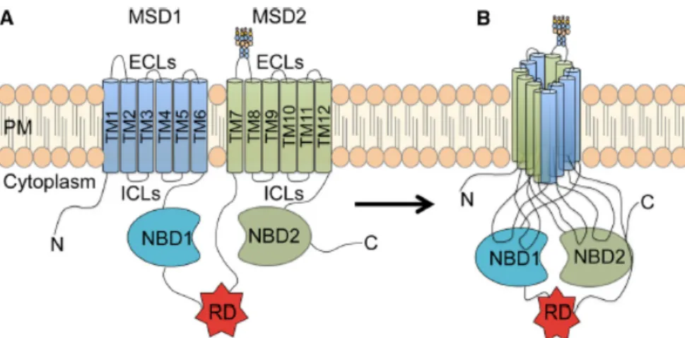
CFTR is composed by five domains (Figure 1.2): two membrane-spanning domains (MSDs) each containing six alpha-helical segments, that form the channel pore; two nucleotide-binding domains (NBD1/2) that interact with ATP to control channel gating; and a central unique regulatory domain (RD), absent in other ABC transporters and containing multiple consensus phosphorylation sites [23], [24]. Both the amino (N) and carboxyl (C) terminal tails of CFTR are in the cytoplasm and mediate the interactions of CFTR with a variety of binding proteins [21]. When ATP binds to CFTR, NBDs heterodimerize and hydrolysis of one of the ATPs disrupts the NBD1-NBD2 interaction and closes the channel gate, leading to the termination of the anion flow.
Like other proteins from the secretory pathway, CFTR assembly begins with the synthesis and folding in the endoplasmic reticulum (ER), where it is core-glycosylated. Once checked for correct folding by the ER quality control (ERQC), this immature form of CFTR migrates to the Golgi complex, where it undergoes processing to achieve its mature form [23].
Figure 1.2. CFTR protein structure. It is composed of five domains: two membrane-spanning domains (MSD1 and
MSD2), each one composed of six transmembrane segments (TM1-6 and 7-12), two cytosolic nucleotide binding domains (NBD1 and NBD1), and a regulatory domain (RD)[24].
1.2.3. CFTR mutation classes
Mutations in CFTR result in abnormal epithelial ion and water transport, and according to the defect in CFTR they may be “severe” (leading to total absence of Cl- transport through CFTR) or
“mild” (residual Cl- transport through CFTR)[25], [26]. CFTR mutations have been classified according to their functional defect into seven classes (Figure 1.3).
Figure 1.3. Classes of CFTR mutations. Mutations are grouped into seven functional classes [9]. Examples of the most
common mutations in each class are shown.
Class I mutations impair protein production, being often nonsense mutations that generate premature stop codons leading to nonsense-mediated decay (NMD); Class II mutations affect CFTR processing causing the protein to misfold, leading to retention in the ER and early degradation through the ubiquitin-proteasome pathway; Class III mutations impair gating of the CFTR channel and are mainly mutations located in the NBDs; Class IV mutations cause decreased ion conductance, and localize mostly to the MSD’s; Class V mutations reduce CFTR protein levels often by affecting splicing; Class VI mutations decrease the retention and stability of CFTR at the cell surface; and Class
VII mutations lead to a total absence of CFTR mRNA production and have been termed "unrescuable" mutations because they cannot be pharmacologically rescued (e.g. large gene deletions) [9], [27].
To add further complexity to this classification system, certain mutations may lead to more than one class of functional defect, as for example F508del results in class II and III defects [2], [28]. It has been proposed to group CFTR variants into theratypes according to their effect on the CFTR protein and in response to correctors and potentiators [12]. This way, unclassified variants can be provisionally assigned to theratypes on the basis of their effect on CFTR quantity and function in studies performed in cell lines.
Mutation specific therapies
Major efforts have identified strategies that rescue different defects of mutant CFTR. One of the major developments on this regard was the identification of its temperature sensitivity. Incubation at a lower temperature than the physiological 37oC promotes the appearance of a fully-glycosylated mature
form of CFTR [27] and was the first evidence that the protein can be rescued.
Mutation-specific therapies have become an important area of drug discovery for CF based on the classification of the mutations into classes [29]. For class I mutations, aminoglycoside antibiotics have been reported to supress premature termination codons by read-through leading to expression of full-length CFTR [27], [29]; For class II mutations, chemical, molecular or pharmacological chaperones, normally called correctors, were reported to stabilize protein structure and promote folding leading to the expression of the mutants at the PM. For class III and IV mutations, in which the CFTR is located at the PM, the modulation of these variants aims at activating the dysfunctional channel (either its gating or its conductance, respectively) with potentiators [9], [27], [29]. For class V mutations, splicing factors that promote normal exon inclusion or factors that promote abnormal exon skipping can increase levels of properly spliced transcripts. Single-stranded antisense RNA-based oligonucleotides can act as guide sequences to repair the targeted abnormal mRNA [9]. For class VI mutations compounds that enhance CFTR retention/ anchoring at cell surface will benefit these mutants [2]. Finally, for patients with unrescuable mutations, class VII mutations, the most straightforward approach is to target alternative non-CFTR anion channels to restore the ionic homeostasis of the epithelia, such as the calcium (Ca2+)-activated Cl- channels (CaCCs) i.e., anoctamins 1 or 6 or some of the members of the SLC26 family of transporters [2], [9].
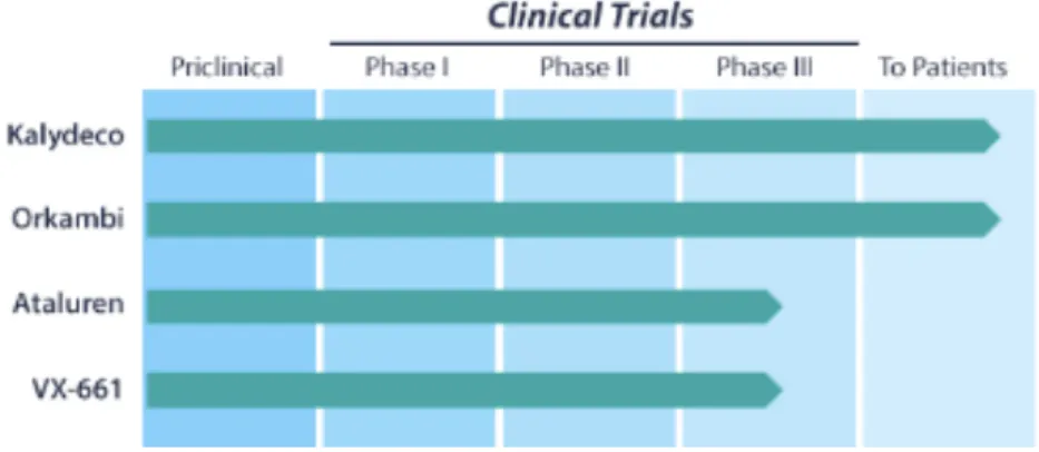
Three chemical compounds identified by high-throughput (HT) screening have led to new perspectives in CF treatment, particularly for class II correctors: lumacaftor (VX-809) and tezacaftor (VX-661) and for III mutations, potentiator ivacaftor (VX-770). Lumacaftor and tezacaftor are correctors that improve the conformational stability of CFTR bearing F508del, resulting in increased processing and trafficking to the cell surface. Ivacaftor is a potentiator that activates CFTR conductance when the protein is already at the PM [30]. Two of these drugs reached the market - Kalydeco (VX-770) and Orkambi (VX-770 + VX-809), being thus available for CF patients (Figure
1.4) [31].
Despite that 40-45% of patients (most of them F508del-homozygous) can already benefit from these approved therapies, there are still <50% of the patients for whom finding an effective treatment is still an unmet need.
Figure 1.4. Drugs already on clinical trials and drugs available to treat CF patients.
1.2.4. Orphan mutations
Among the ~2,000 CFTR mutations, many of them are very rare variants, these are the so-called orphan mutations. For these mutations, the prediction of disease outcome is difficult, since the functional defect has not been defined. Many of these mutations may result in partial (residual) CFTR function and milder "atypical" forms of CF [25]. As each of these mutations affect very few patients worldwide, it is difficult not only to establish the diagnosis, but also assess the potential of the new mutation-based therapies. Nevertheless, some of these patients bearing these rare mutations are likely to respond to existing CFTR modulators. Thus, there is a need to do a molecular and functional characterization of these orphan mutations in order to establish validated compounds for mutation-based therapies [9], [25], [32].
1.3. Models to study CF
To study the many aspects of CF pathology, different model systems have been developed. To allow the study of CFTR in its natural environment – i.e. the membrane of a differentiated epithelial cell - many immortalized cell lines have been used [33]. Cell lines with an epithelial phenotype - with respect to polarization, tight junctions (TJs) and ion transport are desirable to study CF – have been engineered to express normal or mutant CFTR and can be used to the study of rare CFTR mutations or HT approaches to search for novel rescuing strategies (modulators or others). These cell lines have over-expression of CFTR and are normally quiescent, hence they do not present the complete phenotype of the parent tissue, decreasing its physiological relevance [34].
When more physiological models of airway epithelium are required, primary cultures of human nasal epithelial (HNE) or bronchial epithelial (HBE) cells are used. These cells can be obtained from nasal brushings, nasal polyps and lung explants or biopsies. When grown on porous supports at an air-liquid interface (ALI), they recapitulate many features of the native epithelium, hence, this model has been extensively used to study CFTR function and to test CFTR modulators. Nevertheless, good patient material is limited, it is time consuming and the reproducible differentiation can be difficult to achieve. Moreover, ALI cells are difficult to transfect with plasmids or infect with viral vectors [34], [35].
A variant procedure using primary intestinal organoids have been described. These are produced out of primary adult stem cells from the rectum crypts and can be cultured in matrigel for long times in
vitro without genetic modifications. CFTR activation at the apical membrane by forskolin (Fsk) results
does not happen in organoids from patients with CF [9]. The magnitude of swelling can be correlated with CFTR activity, for this reason they can be used to study the effect of CFTR modulators, leading to the next step, testing the clinical benefit in vivo.
Although physiological relevant, the use of patient-derived materials needs to be complemented by cellular models – most mutations exist in compound heterozygosity and it is hard to ascertain if an observed effect comes from one or other allele. Thus, the production and use of novel cell lines is a requirement in the study of rare mutations.
2. Objectives of the present work
The main goal of the present work is to characterize CFTR orphan mutations at the cellular and functional level using novel cellular models and also, whenever available, to compare these results with those in patient-derived materials. We focused on very rare mutations many of which however occur in Portuguese CF patients, but which have not yet been characterized or likely misclassified. In addition, we also propose to assess the efficacy of existing CFTR corrective drugs on such CFTR mutations.
In order to achieve this goal, we propose the following specific objectives:
1) To generate stable cell lines expressing each mutant by:
a. generating CFTR constructs carrying orphan mutations (P205S, L206W, R334W, R347P, I507del, R553P, R560S, L997F, H1079P, M1101K and D1152H) by site-directed mutagenesis using as a backbone CFTR cDNA cloned in the mammalian expression vector pcDNA5/FRT;
b. sub-cloning each CFTR mutant cDNA into the lentiviral vector pLVX-Puro;
c. using lentiviral transduction of CFBE cells to generate novel stable cell lines expressing each of the above CFTR mutants;
2) To assess processing, expression and localization of CFTR mutants, by Western blot (WB) and immunofluorescence (IF);
3) To assess function of CFTR mutants by assessing CFTR-mediated transepithelial Cl- transport
of polarized monolayers in Ussing chamber;
4)
To test the efficacy of approved corrective drugs, by testing CFTR response in our newly created cellular models (as well as in patient-derived intestinal organoids).Through the functional characterization of CFTR orphan mutations using our novel in vitro cellular systems and as well as organoids, primary cells and native tissues (whenever available and also studied in our lab), we will be able to confirm the diagnosis of many patients with a suspicion of CF and to get information on their prognosis. Moreover, by testing the response of these rare mutations to approved drugs in such systems, we can predict the in vivo response of patients carrying these orphan mutations to such drugs.
3. Materials and Methods
3.1. Generation of cell lines overexpressing wild-type and mutant CFTR
3.1.1. Plasmids and cDNAs
cDNA of human wild-type CFTR (wt-CFTR) was available in our lab cloned into pNUT and pcDNA™5/FRT vectors [36]. Each mutation under study was first introduced by site-directed mutagenesis into pcDNA™5/FRT carrying the full-length wt-CFTR cDNA (kindly given by Garry R. Cutting, M.D.) using KOD HOT start DNA polymerase (Novagen, Darmstadt, Germany). pNUT-CFTR-wt and pcDNA™5/FRT-CFTR-mutants (CFTR-Mut) are 8.1kb and 9.6kb vectors, respectively, both containing an ampicillin resistance gene, which was used for selection of transformed bacteria. Inserts from pNUT-CFTR-wt and pcDNA™5/FRT-CFTR-mutants where then subcloned into pLVX-Puro (Clontech, USA) vector. pLVX-pLVX-Puro is a 8.1kb lentiviral vector with ampicilin and puromycin resistence genes, used to produce lentiviral particles to transduce cells and create stably transfected cell lines.
For cDNA and exon nomenclature, mutations are numbered based on the legacy name (Cystic Fibrosis Mutation Database [19]).
3.1.2. Mutagenesis
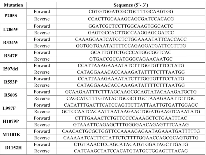
Each CFTR mutation in study was inserted into wt-CFTR by site-directed mutagenesis. The mutagenesis reactions were performed using the KOD Hot Start Kit (Novagen, USA) with the primers described in Table 3.1. In this PCR reaction, the primers are not completely complementary to the template sequence, having a mismatch at the selected mutation location. The PCR program can be found in Table 3.2. The PCR reaction results in a mixture of the template plasmid and the new plasmid containing the mutation. After confirmation of the amplification (by electrophoresis using a 1% agarose gel), the PCR products were incubated 1h at 37 ºC with DpnI (Invitrogen, USA), that hydrolyses methylated DNA. The template DNA, heavily methylated, is degraded, leaving the mutated DNA intact.
Competent bacteria were then transformed with the mutated DNA and grown in LB agar plates with 100 µg/mL ampicillin (selection antibiotic). The plasmid DNA of some colonies were extracted and purified and the mutation insertion was confirmed by DNA sequencing.
Table 3.1. List of primers used for site-directed mutagenesis. Mutation Sequence (5’- 3’) P205S Forward CGTGTGGATCGCTGCTTTGCAAGTGG Reverse CCACTTGCAAAGCAGCGATCCACACG L206W Forward GGATCGCTCCTTGGCAAGTGGCACTC Reverse GAGTGCCACTTGCCAAGGAGCGATCC R334W Forward CAAAGGAATCATCCTCTGGAAAATATTCACCACC Reverse GGTGGTGAATATTTTCCAGAGGATGATTCCTTTG R347P Forward GCATTGTTCTGCCCATGGCGGTCAC Reverse GTGACCGCCATGGGCAGAACAATGC
I507del Forward CCATTAAAGAAAATATCTTTGGTGTTTCCTATG
Reverse CATAGGAAACACCAAAGATATTTTCTTTAATGG R553P Forward CCATTAAAGAAAATATCTTTGGTGTTTCCTATG Reverse CATAGGAAACACCAAAGATATTTTCTTTAATGG R560S Forward GCAAGAATTTCTTTAGCAAGCGCAGTATACAAAGATGCTG Reverse CAGCATCTTTGTATACTGCGCTTGCTAAAGAAATTCTTGC L997F Forward CATATTTGACTTCATCCAGTTCTTATTAATTGTGATTGGAGC Reverse GCTCCAATCACAATTAATAAGAACTGGATGAAGTCAAATATG H1079P Forward CTTTGAAACTCTGTTCCCCAAAGCTCTGAATTTAC Reverse GTAAATTCAGAGCTTTGGGGAACAGAGTTTCAAAG M1101K Forward CAACACTGCGCTGGTTCCAAAAGAGAATAGAAATGATTTTTG Reverse CAAAAATCATTTCTATTCTCTTTTGGAACCAGCGCAGTGTTG D1152H Forward CTGTAAACTCCAGCATACATGTGGATAGCTTGATG Reverse CATCAAGCTATCCACATGTATGCTGGAGTTTACAG
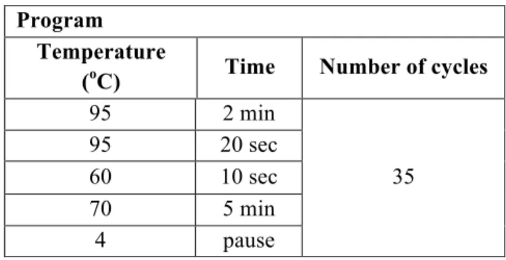
Table 3.2. PCR program for the mutagenesis reaction.
Program
Temperature (oC) Time Number of cycles
95 2 min 23 95 20 sec 48 10 sec 70 4 min 4 pause 3.1.3. Bacteria Transformation
100ng of DNA were added to a 200 µL aliquot of competent cells (the XL1-Blue bacterial strain (Stratagene, USA) was used to prepare competent cells). Bacteria were incubated 30min on ice followed by heat-shock 1.5 min at 42°C, incubated for 2 min on ice and then incubated in LB medium for 1h at 37°C at 220 rpm. After centrifugation, the supernatant was discarded and the pellet was ressuspended. This bacterial suspension was then plated into LB-agar plates (Sigma-Aldrich, USA) supplemented with 100 mg/ml ampicillin (selection antibiotic) and left growing overnight at 37°C at 220 rpm. The following day the plates were stored at 4⁰C.
cultures were grown overnight at 37⁰C at 220rpm. The following day the plasmid DNA was extracted and purified.
3.1.4. Cloning
wt and mutant CFTR cDNAs were subcloned into pLVX-puro using the In-Fusion® HD
Cloning Kit (Clontech, USA, 631187). pLVX with the insert was then used to produce lentiviral particles to transduce human cells and create stably transfected cell lines expressing either wt or mutant CFTR.
cDNAs from the original vectors were PCR amplified (Table 3.3) with primers designed to amplify CFTR and to create 15bp extensions at both the C- and N- terminal (using Primer Design tool for In-Fusion® HD Cloning Kit, Clontech, USA) (Table 3.4).
pLVX-Puro was linearized using the restriction enzyme XhoI (Thermo Scientific, USA) (3h at 37 ºC) to create sticky ends which are complementary to the primer extensions. Both linearized vector and wt or mutant inserts with extensions were spin-column purified using the NZYGelpure kit (NZYTech, Portugal). Then, the In-Fusion cloning reaction (In Fusion HD Cloning Kit, Clontech, USA) was performed and used to transform competent bacterial cells. The insertion was confirmed through colony PCR using CFTR specific primers (Table 3.4). The colony PCR reaction is a regular PCR reaction using DNA directly from bacterial colonies without extraction and purification. Cloning of the cDNA into the pLVX vector was also confirmed by restriction analysis with the enzyme EcoRV (Promega, USA) – detection of 2 bands with a different size from the empty vector when analysed by agarose gel electrophoresis was indicative of successful cloning. The full constructs were then sequenced (outsorced to StabVida).
Table 3.3. PCR program for cDNA amplification wt and mutant CFTR.
Program Temperature
(oC) Time Number of cycles
95 2 min 35 95 20 sec 60 10 sec 70 5 min 4 pause
Table 3.4. Primers used for the amplification of each CFTR mutant and colony PCR reaction.
Primers for cDNA ampification
Name Sequence (5’- 3’)
Forward GGACTCAGATCTCGAATGACATCACAGCAGGTCA
Reverse GAAGCTTGAGCTCGACTAAGAGGCTGTGTCTGG
Primers for colony PCR
Name Sequence (5’- 3’)
Ex2F (forward) AAGGATACAGACAGCGCC Ex5R (reverse) GGAGACTAACAAGTTGTCC
3.1.5. Extraction and Purification of plasmid DNA
Plasmid DNA purification was performed using the NZYMiniprep kit (NZYTech, Portugal) based on the alkaline lysis of bacterial cells followed by adsorption of plasmid DNA into a silica gel-based spin column. Other impurities such as proteins, salts, oligos were washed away. In the end, the DNA is eluted in water.
Plasmid DNA concentration was assessed using a Nanodrop ND1000 Spectrophotometer (Thermo Scientific, USA) (absorbance at 260nm).
3.1.6. Plasmid cDNA Sequencing
Plasmid cDNA sequencing was performed by StabVida (Costa Caparica, Portugal) using the Sanger sequencing method. The primers used were specific for CFTR and are shown in Table 3.5 and
Table 3.6. Each CFTR mutant sequence was then compared with a CFTR-wt reference sequence
(ENST00000003084.10) using the open-source software Geneious.
Table 3.5. List of primers used to confirm the insertion of mutations. Primers
Mutation Name Sequence (5’- 3’)
P205S Forward Ex4F CTTCCTATGACCCGGATAA L206W R334W B2R GGAAGGCAGCCTATGTGAGA R347P I507del B3R=R3R AATGTAACAGCCTTCTGGGAG R553P Ex10F GGCACCATTAAAGAAAATATCATCTT R560S L997F CF Ex15F TGCAGTGATTATCACCAGC H1079P I1R CTCACAGCAACTCAAACAAC M1101K D1152H Int17A GCATATTTCCTCCAAACC
Table 3.6. List of primers used for sequencing the whole CFTR.
Name Sequence (5’- 3’)
AC1L Reverse GAAACCAAGTCCACAGAAGGC
CMV Forward CGCAAATGGGCGGTAGGCGTG Ex5F CTCCTTTCCAACAACCTGAAC B3R AATGTAACAGCCTTCTGGGAG C2R AGCAGTATACAAAGATGCTG D1R GACAACAGCATCCACACGAA E1R AGATTCTCCAAAGATATAGC Ex18F AACTCCAGCATAGATGTGG Ex22F AGCAGTTGATGTGCTTGGC
3.2. Cell culture
3.2.1. Cell lines and culture conditions
The novel stable cell lines produced in this work (CFBE pLVX-P205S, CFBE pLVX-R334W, CFBE pLVX-R560S, CFBE pLVX-H1079P) were generated as described in 3.1. by lentiviral transduction of the Cystic Fibrosis Brochial Epithelial (CFBE41o-) cell line. CFBE41o-, futher referred to as CFBE were developed from brochial epithelial cells from a F508del-CFTR homozygous CF patient [37].
Before producing stable cell lines and to test the novel constructs, transient transfections were performed using Human Embryonic Kidney (HEK 293T) cells [38], [39]. All cell lines were maintained at 37ºC in a humidified atmosphere of 5% (v/v) CO2.
3.2.2. Transient transfections
Transient transfections using Lipofectamine 2000
Lipofection uses a cationic lipid to form an aggregate with the DNA, which is negatively charged, with these aggregates or liposomes being then internalized by the cells. HEK 293T cells were transfected using Lipofectamine 2000 (Invitrogen, USA).
The procedure consists in preparing two mixtures - one containing lipofectamine 2000 diluted in opti-MEM Reduced Serum Medium (Gibco, USA), and another containing the DNA also diluted in opti-MEM. The two dilutions were then mixed and incubated for 5min at room temperature. The DNA-lipofectamine complex was then added to the cells, which were seeded the day before (70-80% confluent). 24h transfection, the medium was changed to remove the complexes. 48h post-transfection, protein or RNA were extracted for further analysis.
Calcium Phosphate transient transfections
A precipitate containing calcium phosphate and DNA is formed slowly by mixing a HEPES-buffered saline solution with a solution containing calcium chloride and DNA. The precipitate adheres to the cell surface and the day after transfection should be visible black dots on the medium. This protocol was performed for the production of lentiviral particles used to transduce parental CFBE cells (see below).
3.2.3. Production of Lentiviral Particles
Lentiviral particles with pLVX-puro-CFTR-wt or pLVX-puro-CFTR-mutant cDNAs were produced in packaging cells - human embryonic kidney (HEK) 293T cells. 5x10⁵ cells were seeded
per well in a 6-well plate containing Eagle’s Minimum Essential Medium (EMEM, Lonza-BioWhittaker, Switzerland) plus 10% of Fetal Bovine Serum (FBS) (Gibco Life Technologies) and incubated for 24h at 37ºC, 5% CO2. The next day, the cells in each well were transfected with 5 µg of
pLVX-puro-CFTR, 4 µg of packaging plasmid pCMV-dR8.74psPAX2 and 0.4 µg of envelop plasmid VSV-G/pMD2.G. The transfection was performed using a calcium-phosphate transfection protocol. Cells were then incubated for 24h at 37ºC, 5% CO2, after which the medium was changed to remove
the lentiviral particles was collected and the packaging cells were discarded. The lentiviral particles were immediately used or stored at -80ºC for further use.
3.2.4. Lentiviral infection – Production of stable cell lines
CFBE 41o- cells were seeded at a density of 4.0x10⁵ cells per well on a 6-well plate
containing EMEM plus 10% FBS and were incubated for 24 h at 37oC, 5% CO2. Cells were infected
with 1mL of medium containing pLVX-puro-lentiviral particles, 1mL of EMEM containing 10% FBS and the infection enhancer Polybrene (8 µg/mL) (Hexadimethrine bromide, Sigma-Aldrich, H9268-5G). Plates were centrifuged at 220rpm for 1h at 25ºC and then incubated for 24h at 37oC, 5% CO
2.
The medium was changed to EMEM plus 10% FBS with 2 µg/mL of puromycin (Sigma-Aldrich, USA), which is half of the concentration needed to kill all the non-infected cells. The cells were incubated for 24h at 37oC, 5% CO2 and the medium was then changed to EMEM plus 10% FBS
supplemented with 5 µg/mL of puromycin. The cells were kept in culture at 37ºC, 5% CO2.
3.2.5. Treatment with CFTR modulators VX-809
VX-809 or lumacaftor (Selleckchem, USA) is a CFTR corrector [40]. CFBE cells seeded in a 24-well plate were incubated with VX-809 3µM diluted in EMEM plus 0.1% v/v FBS. 24h post-treatment the protein was extracted for further assays.
VX-661
VX-661 or tezacaftor (Selleckchem, USA) is a CFTR corrector [41].CFBE cells seeded in a 24-well plate were incubated with VX-661 3µM diluted in EMEM plus 0.1% of FBS and after 24h the protein was extracted.
Cysteamine and Epigallocatechin gallate
Cysteamine (Sigma- Aldrich, USA), the reduced form of cysteamine, is an FDA-approved drug [42]. Epigallocatechin gallate (EGCG) (Sigma- Aldrich, USA) is a green tea flavonoid [43]. CFBE cells seeded in a 24-well plate (day before) were incubated with cysteamine 150µM alone or combined with EGCG 50µM.
3.2.6. Polarized cultures
To perform functional analysis in Ussing chambers, polarized cultures of CFBE cells were prepared. The cells were seeded on collagen IV (Sigma-Aldrich, USA) coated 12mm Snapwell filter (Corning, USA) inserts with 0.4 µm pore polyester membrane and 1.12 cm2 surface area. Cells were
seeded at a density of 3x10⁵ cells per filter on the apical side (top side) and the filters were maintained
in liquid-liquid interface. The medium was changed every other day and the transepithelial electrical resistance (TEER) was measured using a volt-ohmmeter (Milicell-ERS, Millipore, MERS00001). Cells took 2-7 days to polarize. When the TEER value was above 600 Ω/cm2, the experiments were performed.
3.3. Protein Analysis
3.3.1. Immunofluorescence
CFBE cells grown on glass coverslips in 24-well plates were washed 3 times with PBS supplemented with calcium chloride (CaCl2) and magnesium chloride (MgCl2) (PBS++), and fixed for
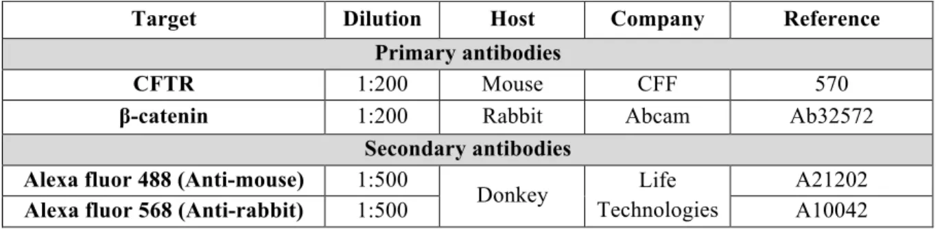
10 min with paraformaldehyde (PFA) 4% (v/v) (Merck Millipore, USA). Then, the cells were washed 3 times with PBS++, permeabilized for 10 min with Triton X-100 0.1% (v/v) (Amersham Biosciences, UK) and washed again with PBS++. After this, cells were incubated at room temperature with primary antibodies (see table 7 below) diluted in PBS with Bovine serum albumin (BSA) (Sigma-Aldrich, USA) 1% (w/v) for 1h. The cells were then washed 3 times with PBS++ and incubated at room
temperature in the dark with secondary antibody (Table 3.7) diluted in PBS with BSA 1% (w/v) for 1h. The cells were again washed 3 times with PBS++ and incubated 10 min in the dark with Hoechst 33342 Fluorescent Stain (Life Technologies, USA). The cells were washed one last time with PBS++ and the cover slips were mounted in glass slides with mounting solution (0,5% n-propyl-gallate; 1 mL PBS 10X; 9 mL glycerol for microscopy) and sealed. Immunofluorescence staining was observed and acquired in a Leica DMI 6000B fluorescence microscope.
Table 3.7. Primary and secondary antibodies used in immunofluorescence assays.
Target Dilution Host Company Reference
Primary antibodies
CFTR 1:200 Mouse CFF 570
β-catenin 1:200 Rabbit Abcam Ab32572
Secondary antibodies
Alexa fluor 488 (Anti-mouse) 1:500
Donkey Life
Technologies
A21202
Alexa fluor 568 (Anti-rabbit) 1:500 A10042
3.3.2. Image acquisition, processing and analysis
Images were acquired with a Leica DMI6000B system equipped with a metal halide light source (EL6000) and a DFC365 FX CCD camera (Leica). Fluorescence was assessed in three different channels: Alexa Fluor 488 (excitation 490-510, emission 520-550) and Alexa Fluor 568 (excitation 515-560, emission >590) and Hoechst (excitation 340– 380, emission 450–490).
3.3.3. Western Blot Cell lysis and protein extraction
Lysates from cells grown in 24-well plates were prepared by washing cells twice with cold PBS and adding the appropriate volume of sample buffer (31.25 mM Tris HCl (Sigma, USA) pH 6.8; sodium dodecyl sulfate (SDS) 1.5% (v/v) (Gibco, USA); glycerol 5% (v/v) (Sigma, USA); bromophenol blue 0.02% (w/v) (Sigma-Aldrich, USA); dithiothreitol (DTT) 50 mM (Sigma, USA)). Benzonase (Sigma-Aldrich, E1014) 25 U/mL was added to shear the DNA, in the presence of MgCl2
Western Blot
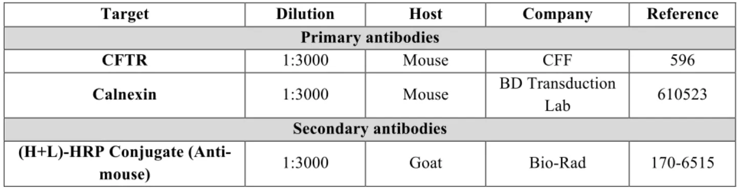
Samples were separated by SDS-PAGE using a 7% (w/v) separating gel (TrisHCl 375 mM pH 8.8; acrylamide 7% (v/v) (Bio-Rad, USA); glycerol 0.1% (v/v); SDS 0.1% (v/v); ammonium persulfate (APS) 0.075% (v/v) (Bio-Rad, USA); tetramethylethylenediamine (TEMED) 0.06% (v/v) (Sigma- Aldrich, USA)) and a 4% stacking gel (Tris HCl 125 mM pH 6.8; acrylamide:bisacrylamide 4% (v/v); glycerol 0.1% (v/v); SDS 0.1% (v/v); APS 0.075% (v/v); TEMED 0.08% (v/v)) gels. Samples run on Tris-Glycine-SDS buffer (Bio-Rad, USA) for 90 to 210min at 65 to 120V. The next step was the transfer onto polivinylidene difluoride (PVDF) membranes (Merck Millipore, USA) using Tris-Glycine buffer (Bio-Rad, USA), at 400mV for 1h30min. The membranes were blocked for 1h with 5% (w/v) skimmed milk (Nestlé, Molico, Switzerland) in PBS supplemented with Tween 20 0.1% (v/v) (PBS-T) and then incubated overnight at 4ºC with primary antibodies (Table 3.8) diluted in 5% milk in PBS-T. After washing 3 times with PBS-T the membranes were incubated with horseradish peroxidase (HRP)-conjugated secondary antibody 1h at room temperature.
Chemiluminescent detection was performed using Chemidoc XRS plus analyser (BioRad, USA) using a 1:1 mixture of peroxide:luminol/enhancer solution (BioRad, USA). Quantification of the intensity of the bands was performed using the ImageLab software (Bio-Rad, USA).
Table 3.8. Primary and Secondary antibodies used in western-blot assays.
Target Dilution Host Company Reference
Primary antibodies
CFTR 1:3000 Mouse CFF 596
Calnexin 1:3000 Mouse BD Transduction
Lab 610523
Secondary antibodies (H+L)-HRP Conjugate
(Anti-mouse) 1:3000 Goat Bio-Rad 170-6515
3.4. Functional Analysis
3.4.1. Ussing chamber
CFBE cells overexpressing each CFTR variant were seeded onto previously collagen IV-coated snapwell filters at a density of 3x10⁵ cells per filter. Transepithelial electrical resistance (TEER) was routinely measured using a volt-ohmmeter (see above).
For assessment of transepithelial transport, the basolateral surface of CFBE cells was continuously perfused with Ringer solution (NaCl 145 mM, KH2PO4 0.4 mM, K2HPO4 1.6 mM,
D-glucose 5 mM, MgCl2 1 mM, Ca-gluconate 1.3 mM) and the apical surface with a low Cl- Ringer
solution (NaCl 38 mM, KH2PO4 0.4 mM, K2HPO4 1.6 mM, D-glucose 5 mM, MgCl2 1 mM,
Ca-gluconate 1.3 mM).
Following a 20 min equilibration period, baseline values were recorded. Values were then recorded following the sequential addition of forskolin (2 µM) and IBMX (100 µM) - CFTR agonists - at the apical side, the CFTR potentiators VX-770or genistein (50 µM), and finally the CFTR inhibitor
Values for the transepithelial voltage (Vte) were referred to the serosal surface of the
epithelium. Transepithelial resistance (Rte) was determined by applying intermittent (1s) current pulses
(0.5 µA). The equivalent short-circuit current (Isc) was calculated according to Ohm’s law (Isc=Vte/Rte),
after appropriate correction for fluid resistance.
3.4.2. Forskolin-induced swelling (FIS) assay
The FIS assay was carried out as previously described [44]. Isolation of crypts and organoids preparation was performed by Nikhil T.A. according to [44], [45]. Rectal CF organoids (Passage 3-25), from a 7-9-day-old culture were seeded in a pre-warm 96-well plate (Thermo Fisher Scientific, USA) with 5µl 50% matrigel containing approximately 20-60 organoids immersed in 50µl complete medium. The organoids were treated with the corrector VX-809 (3 µM) which was prepared in complete media. Potentiators or stimulators were prepared in DMEM-F12. One day after, the organoids were incubated with calcein green (3µM) (Invitrogen, USA) for 30 min. After calcein green staining, forskolin was added with and without potentiators (VX-770) at different concentrations. Live cell imaging was performed on a Leica DMI 6000B Fluorescence microscope (5x objective) for 120 min at 37 oC.
Quantification of forskolin-induced swelling
Forskolin-induced swelling was quantified using Cell profiler software. The area under the curve (AUC; t=60; baseline=100%) was calculated using GraphPad Prism version 5.01. A paired t-test was used to calculate statistical differences.
3.5. Statistical analysis
Data are presented as a mean and standard error of the mean (SEM). When necessary, Statistical Analysis Student’s t-test for unpaired samples was performed, with p<0.05 considered as the level of statistical significance.
4. Results
4.1. Characterization of CFTR orphan mutations
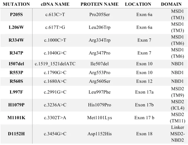
In the present study we studied the following CFTR mutations (Table 4.1):
Table 4.1. List of CFTR orphan mutations in study (ordered by amino acid number).
MUTATION cDNA NAME PROTEIN NAME LOCATION DOMAIN
P205S c.613C>T Pro205Ser Exon 6a MSD1 (TM3) L206W c.617T>G Leu206Trp Exon 6a MSD1 (TM3) R334W c.1000C>T Arg334Trp Exon 7 MSD1 (TM6) R347P c.1040G>C Arg347Pro Exon 7 MSD1 (TM6)
I507del c.1519_1521delATC Ile507del Exon 10 NBD1
R553P c.1790G>C Arg553Pro Exon 10 NBD1
R560S c.1680A>C Arg560Ser Exon 12 NBD1
L997F c.2991G>C Leu997Phe Exon 17a MSD2
(TM9)
H1079P c.3236A>C His1079Pro Exon 17b MSD2
(ICL4)
M1101K c.3302T>A Met1101Lys Exon 17 b MSD2
(TM11)
D1152H c.3454G>C Asp1152His Exon 18
Linker MSD2-NBD2
Figure 4.1. Schematic representation of the location of each CFTR mutation in study on the CFTR protein. 1 2 3 4 5 6 NBD1 7 8 9 10 11 12 R NBD2 TMD1 TMD2 P205S L206W R334W R347P I507del R553P R560S L997F H1079P M1101K D1152H Out In
In order to characterize these orphan mutations (Table 4.1) at cellular and functional levels and to test their responsiveness to already approved corrective drugs, it was necessary to develop novel cellular models overexpressing each mutant CFTR.
CFTR constructs bearing each CFTR mutation were generated by site-directed mutagenesis using the wt-CFTR cDNA cloned into pcDNA5 mammalian expression vector as a backbone and sub-cloned into the lentiviral vector pLVX-Puro (Figure 4.2). In the end, the sequence of all constructs was confirmed by sequencing (see Methods). In order to test the newly created constructs and before the production of stable cell lines, HEK 293T cells were transiently transfected with each of the constructs. HEK293T cells were also used to produce lentiviral particles for each construct. These lentiviral particles were then used to transduce CFBE cells (a human bronchial epithelial cell line, thus a physiologically relevant cell model) and thus obtain stable cell lines expressing each of the CFTR mutants. CFBE cells were originally isolated from the bronchial epithelium of CF patients and immortalized, they do not express endogenous CFTR [46] and thus after transduction will express only the specific CFTR variant.
Figure 4.2. Cloning of R560S-CFTR into pLVX-puro after mutagenesis. (A) By site-directed mutagenesis the mutation
R560S-CFTR was inserted into the pcDNA5-CFTR-wt vector. (B) Results of the sequencing used to confirm the insertion of the R560S mutation. (C) Results of the sequencing used to confirm the cloning of the R560S-CFTR sequence into the pLVX-Puro vector. The same approach was applied to develop the other cell lines described in this study.
Expression and processing CFTR mutants was assessed by WB. Two of the mutations (P205S and R560S) analysed were further analysed in terms of function by Ussing chamber and immunofluorescence to assess protein intracellular localization (only R560S).
The results obtained for each mutation are described below, using the same order with which they are listed in the table above.
M (bp) 10000 7500 6000 5000 ◄ 9956 bp CFTR-wt CFTR-R560S
![Figure 1.1. Pathogenetic cascade that causes cystic fibrosis lung disease. Cystic fibrosis is caused by mutations in the CFTR gene, which leads to a series of events and ultimately leads to loss of lung function [9]](https://thumb-eu.123doks.com/thumbv2/123dok_br/18490703.901054/14.892.166.725.762.1098/figure-pathogenetic-cascade-fibrosis-fibrosis-mutations-ultimately-function.webp)