UNIVERSIDADE DE LISBOA
FACULDADE DE CIÊNCIAS
DEPARTAMENTO DE BIOLOGIA ANIMAL
Characterization of a new malaria vaccine
candidate against Plasmodium vivax using
genetically modified rodent Plasmodium
parasites
Miguel Filipe Duarte
Dissertação de Mestrado
MESTRADO EM BIOLOGIA HUMANA E AMBIENTE
UNIVERSIDADE DE LISBOA
FACULDADE DE CIÊNCIAS
DEPARTAMENTO DE BIOLOGIA ANIMAL
Characterization of a new malaria vaccine
candidate against Plasmodium vivax using
genetically modified rodent Plasmodium
parasites
Miguel Filipe Duarte
Dissertação para a obtenção do Grau de Mestre orientado por: Doutor António Mendes (Instituto de Medicina Molecular, Faculdade de Medicina da Universidade
de Lisboa) e Professora Doutora Deodália Dias (Faculdade de Ciências da Universidade de Lisboa)
MESTRADO EM BIOLOGIA HUMANA E AMBIENTE
The citation format used on this thesis is based on the citation style used by Nature, one of the most prominent scientific journals.
Acknowledgements/Agradecimentos
Em primeiro lugar, quero-te agradecer a ti Miguel pelo voto de confiança e pela oportunidade que me deste de realizar a minha dissertação de mestrado no teu grupo de investigação. Ao longo deste ano foste sem dúvida uma fonte de inspiração, pela forma como conduziste o grupo, pelo teu espírito crítico e rigor científico.
A ti António, o maior e mais sincero obrigado pela tua orientação, pela paciência, pelo apoio, disponibilidade, por tudo o que me conseguiste transmitir. Sem ti, este projecto não teria sido possível. Quando as coisas não correram tão bem, apelaste sempre à minha perseverança. Obrigado por tudo o que me ensinaste, pois mesmo quando não tinhas a resposta, conseguiste sempre indicar-me o caminho certo para a encontrar. Não poderia pedir melhor orientação. :)
Quero também agradecer à orientadora interna desta dissertação de mestrado, Deodália Dias, pelo voto de confiança, disponibilidade e pelo apoio ao longo destes dois anos de mestrado.
Quero agradecer a todos os membros do grupo por me acolherem e por me fazerem sentir em casa: A ti Marta, obrigado pelo todo o apoio e amizade ao longo deste ano, és das pessoas mais impecáveis que tive o prazer de conhecer até hoje, apesar dos mil e um nomes que inventaste para me chamar (:p). Já não imagino aquele laboratório sem ti por lá a correr de um lado para o outro. À Patricia, desde que eu cheguei ao laboratório que, para além dos bons conselhos, nunca me faltou com um sorriso. À Inês, que apesar de já não fazeres parte do grupo, me ensinaste imensa coisa; mas mais importante ainda, obrigado por teres criado o “meu” parasita, sem o qual o meu projecto não existiria. À Filipa, “a mulher furacão” do nosso insetário, por toda a ajuda, boa disposição e animação constante. À Joana, a mestre das “genetiquices” por toda a ajuda com os PCRs e por seres uma fixe. My thanks to you Marija for being so helpful and supportive. I wish I had the time to learn more Serbian words, other than “Kako ste?” And “Hvala” :). À Margarida (a grande rival de Lasertag!) pela boa disposição e simpatia. À Cláudia pela ajuda com o Western Blotting. Ao Mário, que apesar de já te teres ido embora (boa vida), me ajudaste imenso enquanto cá estiveste. Carolina, o nosso membro mais recente, a ti desejo-te boa sorte para este teu ano que se segue. Te garanto que estás bem entregue!
Quero agradecer ao pessoal do Mota Lab pela ajuda e por serem tão prestáveis, em especial a ti Ana, pela ajuda durante os meus tempos de insetário. Um obrigado aos nossos laboratórios vizinhos; UPAR pelas gargalhadas e UPAMOL pela ajuda, em especial a ti Jorge, pelas duvidas que me foste esclarecendo. Obrigado também à Ana e ao António da unidade de Bioimaging do IMM por tudo o que me ensinaram, pela ajuda e pelos bons conselhos.
Um grande obrigado a todos os meus amigos, que me acompanharam ao longo das etapas mais importantes da minha vida, e que nos bons e maus momentos me fazem sentir abençoado por vos ter como amigos. Um obrigado especial ao João Vitor e ao Tiago que ao fim destes anos todos já são quase como irmãos. Ao Gonçalo,
não só pela amizade mas também pelos conselhos e ajuda prestada ao longo do mestrado.
A ti Carolina, pela paciência, pelo apoio, por estares sempre presente nos bons e nos maus momentos.
Para terminar, o meu maior obrigado é sem dúvida à minha família pelo apoio incondicional. Em especial à minha mãe, por todo o teu apoio nas minhas decisões, por acreditares que eu podia ir sempre mais além e pelo esforço que tens feito para eu conseguir perseguir os meus sonhos. Sem ti, certamente não seria o que sou hoje. Obrigado.
Um obrigado também a todos os que não foram aqui mencionados, mas que directa ou indirectamente me ajudaram a alcançar este objectivo.
Abstract
Malaria is an infectious disease transmitted by Anopheles mosquitoes and caused by protozoan parasites of the genus Plasmodium. Despite countless efforts, there is still no effective vaccine against any of the human infective Plasmodium parasites of which P. falciparum and P. vivax are the most clinically significant.
A new whole organism-vaccine has been proposed which is based on the use of genetically modified rodent parasites (P. berghei) as platforms for the delivery of immunogenic antigens of human infective Plasmodium species. The efficacy and safety of P. berghei parasites as a platform to deliver immunogenic antigens is warranted by their ability to infect human hepatocytes without being able to develop inside human erythrocytes. A genetically modified P. berghei expressing P. falciparum circumsporozoite (CS) protein, under the control of the P. berghei UIS4 promoter, (Pb(PfCS@UIS4)), was already developed and characterized as a potential vaccine candidate. Immunization of mice with Pb(PfCS@UIS4) parasites has successfully elicited an immune response capable to recognize and bind to P. falciparum sporozoites, and inhibit infection by this parasite.
P. vivax malaria is the most widespread of the human-infective Plasmodium species and leads to a great socioeconomic burden worldwide. In light of this fact, and given the promising results obtained for the vaccine candidate Pb(PfCS@UIS4), a new genetically modified P. berghei expressing the P. vivax CS protein, Pb(PvCS@UIS4) was created, as a vaccine candidate against P.vivax malaria. This parasite was generated using the GIMO (Gene Insertion Marker Out) transfection method.
The work presented in this thesis aims to characterize Pb(PvCS@UIS4) parasites in terms of infectivity and development across the sporogonic and pre-erythrocytic stages of the parasite life cycle, both in vitro and in vivo. Sporogonic parasite development and infectivity in the mosquito was assessed using Anopheles stephensi mosquitoes by counting oocysts in the midgut and sporozoites in the salivary glands at 10 and 21 days post infectious blood meal, respectively. No significant differences were observed between the development of Pb(PvCS@UIS4) and PbGIMO (wild-type P. berghei - transfection motherline) parasites. Pre-erythrocytic development was evaluated in vitro on Huh7 and HepG2 human hepatoma cell lines (48h p.i) and in vivo experiments on C57BL/6J mice (44 hpi), revealing that Pb(PvCS@UIS4) sporozoites can infect and develop within hepatocytes to a similar extent to PbGIMO sporozoites. Blood stage development experiments also revealed that Pb(PvCS@UIS4) parasites infectivity and development during this stage is comparable to PbGIMO. These results indicate that the insertion of the PvCS gene in P. berghei 230p neutral locus does not appear to have an impact on the parasite‟s ability to infect and develop throughout its life cycle. Additionally, CS expression was also assessed on Pb(PvCS@UIS4) sporozoites and exoerythrocytic forms (intrahepatic forms of the parasite), showing that both the endogenous PbCS and the exogenous PvCS are indeed being co-expressed on both stages.
This characterization represents one of the first steps on the development of this new vaccine candidate against P.vivax malaria and the results here presented
provide valuable insight in order to proceed to future studies regarding the immunogenicity and efficacy of Pb(PvCS@UIS4) as a vaccine candidate.
Keywords: Malaria, Plasmodium vivax, Plasmodium berhgei, whole-organism vaccine,
Resumo
A malária é uma doença infecciosa causada por um parasita protozoário do género Plasmodium que causa a morte entre 650.000 a 1.200.000 pessoas todos os anos, das quais aproximadamente 85% são crianças com menos de 5 anos de idade.1 Existem 5 espécies de Plasmodium capazes de causar malária em humanos, sendo P. falciparum e P. vivax as espécies responsáveis pela grande maioria dos casos.1 Estes parasitas são transmitidos sob a forma de esporozoíto através da picada de mosquitos fêmea do género Anopheles.1 Após a picada, os esporozoítos invadem a corrente sanguínea do hospedeiro e migram até ao fígado onde, após atravessarem vários hepatócitos, acabam por invadir e se desenvolver dentro de um.10,11 No interior do hepatócito, o parasita replica-se, dando a origem a milhares de merozoítos.12 Estes são libertados para a corrente sanguínea no interior de uma estrutura denominada merosoma, terminando assim a fase pré-eritrocitária da infeção.10 Uma vez na corrente sanguínea, o merosoma rompe-se e liberta os merozoítos que por sua vez vão infetar os eritrócitos, dando origem à fase sanguínea e sintomática da doença.10 Dentro dos eritrócitos os parasitas vão novamente replicar-se, dando origem a novos parasitas capazes de perpetuar o ciclo de infeção na corrente sanguínea mas dando igualmente origem a formas sexuais (gametócitos) aptas para serem ingeridas aquando da picada por novos mosquitos e de se desenvolverem dentro dos mesmos, dando origem a um novo ciclo de infeção.13 Apesar da malária causada por P. falciparum ter uma sintomatologia mais severa, a malária causada por P. vivax tem uma maior distribuição geográfica e está geralmente associada a longos períodos de morbilidade devido à capacidade que o parasita possui de gerar formas adormecidas (hipnozoítos), que podem levar a uma reincidência dos sintomas da doença.5,6
Atualmente, as medidas de combate à malária passam pelo uso de inseticidas para diminuir as populações de mosquitos transmissores, uso de redes mosquiteiras e a prescrição de fármacos profiláticos contra a malária. No entanto, a eficácia tanto dos inseticidas como dos fármacos tem vindo a diminuir ao longo do tempo devido ao aparecimento de “perfis” de resistência quer nas populações de mosquitos contra os inseticidas, quer nas populações de Plasmodium contra os fármacos existentes.1,15,17
Dadas as limitações das medidas existentes, tornou-se consensual entre a comunidade científica de que a criação de uma vacina é uma componente essencial no combate à malária, uma vez que esta permitiria não apenas prevenir a sintomatologia da doença mas também a sua transmissão. Contudo, até ao momento não existe uma vacina licenciada contra a malária.17
A RTS,S, a vacina que se encontra actualmente no estadio mais avançado de desenvolvimento, é uma vacina de subunidade, que consiste na administração de um fragmento da proteína CS do parasita Plasmodium falciparum fundido com uma matriz transportadora proveniente da superfície do vírus da hepatite B, mostrou oferecer um nível de proteção bastante modesto em humanos.27 Em particular, as respostas imunitárias observadas após vacinação com RTS,S foram maioritariamente mediadas por células T CD4+ e anticorpos contra a proteína CS, sem que os níveis de células T
CD8+ detectáveis fossem muito significativos, as quais estão demonstradas como sendo as principais células efectoras na proteção imunitária contra a malária.22,30,31
Como alternativa, outros tipos de vacina contra a malária estão a ser desenvolvidos, como por exemplo, as vacinas de organismo inteiro. As vacinas de organismo inteiro consistem na administração de parasitas inteiros, previamente atenuados, com o objetivo de espoletar uma resposta imunitária capaz de inibir a infeção causada por organismos não atenuados. Vários estudos realizados com esporozoítos atenuados (P. falciparum), têm demonstrado que estas estratégias de vacinação podem garantir um alto nível de proteção em humanos.33-35,45 Além disso, as respostas espoletadas com esta estratégia de vacinação são maioritariamente mediadas por células T CD8+.32 Foi também observado que o nível de proteção é tanto maior quanto maior for a progressão do desenvolvimento do parasita no fígado.32
No entanto, estas vacinas baseiam-se na assunção que todos os parasitas administrados estão totalmente atenuados, e como tal, não progridem para a fase sanguínea da infeção. Dito isto, é fácil perceber que este princípio representa um risco para os indivíduos vacinados, uma vez que basta que um parasita escape ao processo de atenuação, para que haja progressão da infeção para a fase sintomática da doença.
Como resposta a esta limitação inerente às vacinas que se baseiam no uso de parasitas atenuados, a equipa do Prudêncio Lab no IMM está a desenvolver uma nova estratégia de vacinação que se baseia no uso de parasitas de roedores (P. berghei) geneticamente modificados, como plataforma de apresentação de antigénios pertencentes às espécies de Plasmodium que causam malária em humanos, de forma a promover imunidade contra os mesmos quer por mecanismos de proteção cruzada entre espécies quer por mecanismos de proteção especifica contra os antigénios dos parasitas causadores de malária em humanos.
Com o intuito validar esta nova estratégia, foram realizadas experiências no sentido de verificar que P. berghei consegue invadir e desenvolver-se em hepatócitos humanos sem progredir para a fase sanguínea, e que parasitas P. berghei geneticamente modificados para expressar a proteína CS de P. falciparum, são capazes de espoletar respostas imunológicas capazes de reconhecer e inibir P. falciparum. Foi então desenvolvido e caracterizado um novo candidato a vacina, que consiste em usar P. berghei geneticamente modificado de forma a que este expresse a proteína CS de P. falciparum sob o controlo do promotor UIS4 de P. berghei, o parasita Pb(PfCS@UIS4).
Os resultados deste estudo demonstraram que P. berghei consegue de facto infetar hepatócitos humanos sem causar doença, o que faz deste parasita uma potencial plataforma de administração de antigénios imunogénicos para humanos. Por outro lado, a imunização de ratinhos com Pb(PfCS@UIS4) mostrou conduzir a respostas imunitárias capazes de reconhecer e ligar-se a esporozoítos de P. falciparum, inibindo a infeção por este parasita.
Dados os resultados satisfatórios obtidos com a caracterização do parasita Pb(PfCS@UIS4), foi produzido um novo candidato a vacina contra P. vivax, o parasita Pb(PvCS@UIS4), que de um modo semelhante ao candidato anterior, consiste em
usar P. berghei geneticamente modificado de forma a expressar a proteína CS de P.vivax sob o controle do promotor UIS4 de P. berghei.
Deste modo, esta tese de mestrado teve como objetivo principal caracterizar o desenvolvimento deste novo parasita durante o seu ciclo de vida dentro do mosquito assim como caracterizar a sua infetividade e desenvolvimento em culturas celulares in vitro, e in vivo com ratinhos C57BL/6J. Adicionalmente, a sequência do gene PvCS usada na criação deste parasita foi analisada em detalhe e comparada com sequências referencia para o mesmo gene disponíveis na base de dados PlasmoDB de modo a identificar diferenças significativas entre a sequência encontrada no isolado de P. vivax utilizado e as diferentes populações existentes mundialmente. Ao compararmos a sequencia da PvCS usada na criação do Pb(PvCS@UIS4) com as sequencias existentes na base de dados, concluímos que as regiões N-terminal e C-terminal da proteína se mantêm conservadas, enquanto que na zona “central de repetição”, existe alguma variabilidade. De acordo com estudos anteriores, concluímos que a variabilidade encontrada no nosso isolado não é superior à que normalmente se verifica para este gene e que as alterações especificas encontradas são muito provavelmente características comuns das populações de P. vivax existentes na região geográfica de onde foi obtido o isolado utilizado neste estudo (Tailândia).47,51
De forma a caracterizar o desenvolvimento dos parasitas Pb(PvCS@UIS4) dentro do mosquito, foram contados quer os oocistos presentes no estômago do mosquito ao dia 10 após infeção quer esporozoítos presentes nas glândulas salivares entre o dia 20 e 22 após infeção. Estes números foram então comparados com os números obtidos com parasitas não-transfetados, pertencentes à linha mãe (PbGIMO) usada na transfeção dos parasitas Pb(PvCS@UIS4). Os resultados mostraram que não existem diferenças significativas entre os parasitas Pb(PvCS@UIS4) e PbGIMO, o que nos leva a concluir que a inserção da PvCS não aparenta ter qualquer impacto nesta fase de desenvolvimento do parasita.
A fim de caracterizar a capacidade deste parasita se desenvolver e invadir células in vitro, foram usadas 2 linhas celulares de hepatomas humanos; Huh7 e HepG2. Estas linhas celulares foram infetadas com esporozoítos recolhidos das glândulas salivares de mosquitos infetados a partir do dia 20 após infeção. Passadas 48h da infeção, as células foram fixadas e marcadas com anticorpos de marcação nuclear e anticorpos específicos contra o parasita (anti-HSP70). Desta forma, através de microscopia de fluorescência, foi quantificado o nível de infeção e comparado entre parasitas Pb(PvCS@UIS4) e PbGIMO. Não foram observadas diferenças significativas entre os níveis de infeção observados com ambos os parasitas, o que demonstra que o desenvolvimento do parasita na fase hepática não é afetado pela presença da proteína CS exógena. Resultados idênticos foram também obtidos in vivo, em experiencias em que ratinhos C57BL/6J foram infetados por injeção i.v. de esporozoítos. Nestas experiencias, decorridas 44h após a infeção, os ratinhos foram sacrificados e os seus fígados foram recolhidos de forma a ser possível analisar os níveis de infeção por qRT-PCR e microscopia de imunofluorescência.
Concomitantemente com as experiencias anteriores, a expressão da PbCS e da PvCS foi também observada com recurso a técnicas de microscopia de imunofluorescência. Como era esperado, nos parasitas Pb(PvCS@UIS4), a PbCS e a
PvCS estão a ser co-expressas em esporozoítos e nas formas exoeritrocitárias presentes em hepatócitos infetados. Esta observação é extremamente relevante para a estratégia de vacinação, uma vez que é esperado que parte da imunidade gerada seja especificamente contra a PvCS.
Para terminar a caracterização dos parasitas Pb(PvCS@UIS4), foi também realizada uma experiencia no sentido de caracterizar a capacidade destes parasitas infetarem e se desenvolverem durante a fase eritrocitária da doença ao longo de 9 dias após infeção. Os resultados demonstraram que não existem diferenças significativas entre a capacidade de infectar e se replicar no interior dos eritrócitos por parte dos parasitas Pb(PvCS@UIS4) quando comparados com parasitas PbGIMO.
Concluindo, os parasitas Pb(PvCS@UIS4) demonstraram não apresentar nenhuma diferença significativa em termos de capacidade infetiva e de desenvolvimento ao longo do seu ciclo de vida quando comparados com a linha materna usada para transgénese. No contexto de desenvolvimento de uma vacina é importante que o parasita mantenha a sua funcionalidade de forma a conseguir apresentar o maior número possível de antigénios e desta forma, tornar a vacina mais eficaz. Adicionalmente, a PvCS está a ser expressa tanto em esporozoítos como durante a fase hepática, o que mais uma vez, no contexto desta estratégia de vacinação, é extremamente importante dado que a maior parte da imunidade contra Plasmodium é gerada durante a fase hepática. Os resultados aqui obtidos estão em concordância com os resultados previamente obtidos com o candidato a vacina contra P. falciparum, o Pb(PfCS@UIS4). Os dados aqui apresentados representam um primeiro passo na caracterização deste candidato a vacina e, como tal, serão necessárias mais experiencias, nomeadamente do ponto de vista imunológico, para averiguar a sua eficácia.
Palavras-chave: Malaria, Plasmodium vivax, Plasmodium berhgei, vacinas de
1
Abbreviations
18S Plasmodium 18S ribosomal RNA NK Natural Killer cells 5-FC 5-fluorocytosine PABA p-aminobenzoic acid BSA Bovine serum albumin Pb(PfCS@UIS4)
Plasmodium berghei expressing Plasmodium falciparum circumsporozoite protein under
the control of the PbUIS4promoter
cDNA Complementary
Deoxyribonucleic acid Pb(PvCS@UIS4)
Plasmodium berghei expressing Plasmodium vivax circumsporozoite protein under
the control of the PbUIS4promoter CS Circumsporozoite protein PbCS Plasmodium berghei
circumsporozoite protein DMEM Dulbecco Modified Eagle‟s
Medium – cell culture medium PbGIMO
Plasmodium berghei (Gene Insertion Marker Out) motherline DNA Deoxyribonucleic acid PBS Phosphate buffered saline
solution
EEF Exoerythrocytic forms PCR/qRT-PCR
Polymerase chain reaction/quantitative real time
polymerase chain reaction ER Endoplasmatic reticulum PfCS Plasmodium falciparum
circumsporozoite protein GAP Genetically Attenuated Parasites p.i Post-infection GMEP Global Malaria Eradication
Program PvCS
Plasmodium vivax circumsporozoite protein HC-04 Human hepatocyte cell line PVM Parasitophorous vacuole
membrane hdhfr Selectable markar human
dehydrofolate reductase RAS Radiation attenuated sporozoites HEPES 4-(2-hydroxyethyl)
-1-piperazineethanesulfonic acid RBC Red blood cell HepG2 Human hepatoma cell line RNA Ribonucleic acid
HPRT Hypoxanthine Guanine
Phosphoribosyl Transferase RPMI
Roswell Park Memorial Institute medium – cell culture medium HSPG Heparin sulphate proteoglycans RPM Rotations per minute
Huh7 Human hepatoma cell line SEM Standard error of the mean IFN-y Interferon gamma STAT Signal transducers associated
with transcription iNOS Nitric oxide synthase TSR Thrombospondin repeat region
i.p. Intraperitoneal WT Wild type
i.v. Intravenous yfcu Yeast fcu selectable marker MHC Major Histocompatibility Complex HSP70 Heat shock protein 70
2
Índex
Abstract ... 6 Resumo ... 8 Abbreviations ... 1 Índex ... 2 1. Introduction ... 4 1.1 Socioeconomic burden ... 41.2 Etiology and distribution ... 4
1.3 Symptoms and diagnosis ... 6
1.4 Plasmodium life cycle ... 6
1.5 Control measures ... 8
1.6 Vaccines against Malaria ... 8
1.6.1 Historical background ... 9
1.6.2 Immunity in Malaria ... 9
1.6.3 Malaria pre-erythrocytic vaccines ... 10
2. Aims ... 18
3. Materials and Methods ... 19
3.1 Mice (C56BL/6J AND BALB/C) ... 19
3.2 Parasite lines ... 19
3.3 Rearing of Anopheles stephensi mosquitoes ... 19
3.4 Maintenance of Plasmodium berghei infections ... 20
3.5 In vitro culture of hepato-cellular carcinoma celllines ... 21
3.6 In vitro infection ... 22
3.7 In vivo experiments ... 22
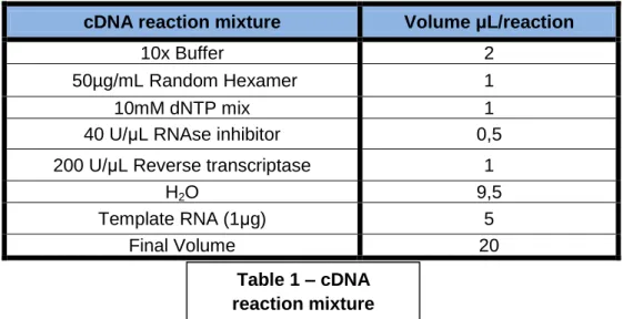
3.8 RNA extraction and cDNA synthesis ... 22
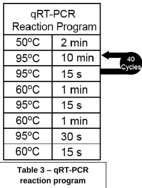
3.9 Real Time quantitave PCR ... 23
3.10 Genomic DNA extraction from blood stage parasites ... 24
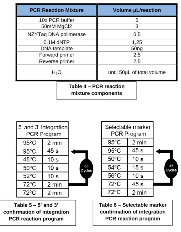
3.11 Confirmation PCR for genomic integration ... 24
3.12 Antibody production and acquisition ... 25
3.13 Imunofluorescence microscopy ... 26
3.14 Statistical analyses ... 27
4. Results ... 28
4.1 Characterization of the PvCS sequence ... 28
4.2 PCR confirmation of the genomic integration for the PvCS@UIS4 gene cassette ... 29
3
4.3 Mosquito stage development of Pb(PvCS@UIS4) parasites ... 31
4.3.1 Oocyst Development of Pb(PvCS@UIS4) parasites ... 31
4.3.2 Sporozoite development and salivary gland infectivity of Pb(PvCS@UIS4) parasites ... 31
4.3.3 PbCS and PvCS expression in Pb(PvCS@UIS4) parasites in mosquito stages ... 32
4.4 In vitro characterization of Pb(PvCS@UIS4) parasites ... 34
4.4.1 In vitro infection assays in Huh7 hepatoma cells ... 34
4.4.2 Expression assays in Huh7 hepatoma cells ... 35
4.4.3 In vitro infection assays in HepG2 hepatoma cells ... 35
4.4.4 Expression assays in HepG2 hepatoma cells ... 37
4.5 In vivo characterization of Pb(PvCS@UIS4) parasites... 39
4.5.1 Infection load quantified by qRT-PCR ... 39
4.5.2 Infectivity and development analysis by immunofluorescence microscopy .. 40
4.5.3 In vivo expression assays ... 41
4.6 Characterization of blood-stage development of Pb(PvCS@UIS4) parasites .... 41
5. Discussion ... 44
6. References ... 49
7. Appendix section ... 54
Appendix-1. Sequences of primers used. ... 54
Appendix -2. Consensus sequences from plasmids (With PvCS@UIS4 cassette) obtained from transformed bacteria before transfection. ... 55
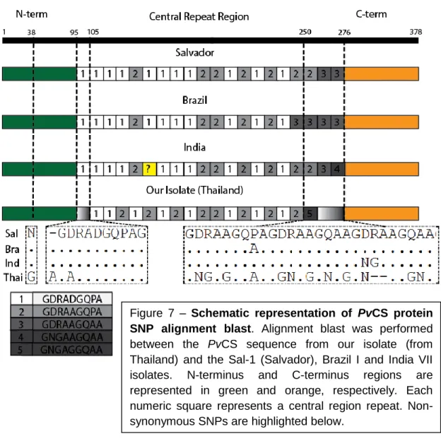
Appendix 3. Alignment Blast between our PvCS sequence and PvCS reference sequences ... 56
Supplementary figure 2: Alignment Blast between our PvCS sequence and PvCS reference sequences present on PlasmoDB; Pv_Sal1_chr08 (Salvador), BrazilI (Brazil) and IndiaVII (India). ... 57
4
1. Introduction
1.1 Socioeconomic burden
Malaria represents one of the most serious public health problems worldwide; and although malaria incidence has been decreasing over the past 14 years, every year there are more than 216 million clinical episodes resulting in 650.000 to 1.200.000 deaths, 85% of which occur in children under 5 years old. In 2012 alone, an estimated 3.4 billion people (47% of the world‟s total population) were at risk of malaria, of which 1.2 billion, mostly in the African and South-East Asia regions, were at high risk (>1 case per 1000 population) of contracting this disease.1 Malaria was once present worldwide. However, between 1940 and 1969, with the establishment of the Global Malaria Eradication Programme (GMEP) by WHO, it was possible to eliminate malaria from USA and Canada, Europe, and Russia.2 Nevertheless, the excessive use of insecticides and anti-malarial drugs, such as chloroquine, which were the core tools of the GMEP, led to a resurgence of malaria prevalence in some tropical countries on the following years.3 Additionally, the selective pressure issued by those two factors (insecticides and anti-malarials) led to the appearance of insecticide and drug resistant mosquito and Plasmodium populations, respectively.3 Nowadays, malaria is still present in 104 countries, in which it is considered endemic.1
1.2 Etiology and distribution
Malaria is an infectious disease caused by protozoan parasites of the genus Plasmodium, which are transmitted by female Anopheles mosquitoes. There are numerous species of Plasmodium parasites which can infect a plethora of vertebrate animal species, although only five Plasmodium species (P. falciparum, P. vivax, P. ovale, P. malariae and P. knowlesi) are known to cause disease in humans.1 P. falciparum and P. vivax raise the greatest concerns in terms of public health as the remaining three species are reported to have much lower incidence rates.1 P. falciparum malaria is indeed the most deadly form of human malaria but it predominates mostly in Africa (Fig.1.A); as for P. vivax malaria, although not as severe presents a wider distribution (Fig.1.B) ranging from South American to South-eastern Asian regions.1 Despite the fact that most malaria-related deaths are due to P. falciparum infections, there is no denying the impact of P. vivax infections, mostly because P. vivax, unlike P. falciparum, is able to evolve into dormant forms (hypnozoites) that can result in disease relapses.4 Relapses associated to P. vivax malaria add to the morbidity burden and make this species of Plasmodium even harder to eliminate.5 The low transmission of P. vivax in African countries is due to the predominantly Duffy negative human populations. The Duffy receptor is a chemokine receptor present on the erythrocyte surface of Duffy positive individuals and it is known to be essential for P. vivax parasites to recognize and invade erythrocytes.6 Therefore, populations with a Duffy negative trait are resistant to P. vivax infections.6
5 Figure 1 – Malaria transmission map – World map showing the areas with P. falciparum (top map) and P. vivax malaria transmission. Regions coloured in pink represent areas with unstable malaria transmission (annual case incidence reported is lower than 0,01%). Regions colored in red represent areas with stable malaria transmission (annual case incidence reported is higher than 0,01%).68,69
6
1.3 Symptoms and diagnosis
The first malaria symptoms usually appear about 7-9 days post infection (depending on the Plasmodium species). In endemic areas, malaria is the most common cause of fever, although uncomplicated malaria includes other flu-like symptoms, such as headache, fatigue, muscle ache, anemia, nausea, and after a few days, a palpable spleen.7 Severe malaria may have other manifestations, most commonly in children or immunocompromised adult individuals, such as acute pulmonary oedema, kidney injury, jaundice, generalized seizures and ultimately coma (cerebral malaria).8
The most common method for malaria diagnosis is by the microscopic observation of Giemsa-stained blood films. However, depending on resource availability, there are other methods such as antigen detection kits or by polymerase chain reaction (PCR).9
1.4 Plasmodium life cycle
Female Anopheles mosquitoes regularly bite mammalian hosts to take a blood meal and gather the required proteins to generate eggs. During a bite, the mosquito injects saliva in order to generate an anesthetic, vasodilator and anti-coagulant effect.10 When a mosquito is infected, a Plasmodium form called sporozoite migrates to the mosquito‟s salivary glands and, upon a blood meal, will be injected into the host‟s skin (Fig.2.1).11 Some of the injected parasites will use gliding motility to move to the nearest blood vessel and enter the bloodstream. Upon reaching the bloodstream, sporozoites remain in circulation until they eventually reach the liver sinusoids, where they traverse endothelial, Kupffer cells and a few hepatocytes until productively infecting a final hepatocyte (Fig.2.2). When hepatocyte infection occurs, sporozoites will form a parasitophorous vacuole and, within it, transform into spherical hepatic stages called exoerythrocytic forms – EEFs.12 P. vivax and P. ovale parasites can develop into dormant hepatic forms (hypnozoites) that can be reactivated later and proceed with the normal course of infection (Fig.2.3). Parasites multiply inside the cell, ultimately forming a schizont, which is made up of thousands of merozoites (Fig.2.4).10 After the process of maturation, merozoites are released inside merosomes, which eventually burst and release the merozoites to the bloodstream where they will infect erythrocytes (Fig.2.5). Inside the erythrocytes, making use of the infected cell‟s resources, more merozoites will develop. After reaching erythrocyte capacity, the cell will burst and release all the merozoites into the bloodstream, leading to the symptoms of malaria (Fig.2.6).10 In some cases, infected erythrocytes will lead to the formation of gametocytes, the sexual forms of the parasite (Fig.2.7).13The parasite‟s life cycle is eventually perpetuated when a new mosquito bites an infected individual and collects Plasmodium gametocytes in its blood meal. Upon reaching the mosquito midgut, Plasmodium male gametocytes are exposed to mosquito-specific factors and environmental factors forming eight motile male gametes (exflagellation) that undergo a process of fusion with female gametes leading to a new form, the zygote.13 The zygote will mature into a motile ookinete (24h post-blood meal) that will penetrate the midgut epithelium, moving from the lumen to the basal side, beginning the transformation into
7 an oocyst (Fig.2.8). Oocyst development takes between 7 and 14 days (depending on mosquito-parasite species combination). About 11-18 days after mosquito‟s infection, thousands of sporozoites have been produced inside the oocysts (through budding process), leading to the oocyst burst and sporozoite release into mosquito‟s hemolymph.13 When in the hemolymph, sporozoites will migrate and infect the salivary glands along with the formation of a parasitophorous vacuole.10 Sporozoites will then be ready to start a new cycle of infection upon the mosquito‟s next blood meal (Fig.2.9).
Figure 2 – Plasmodiumspp life cycle – 1 – During the bite of an infected mosquito, spozoroites are injected into the host‟s skin. 2 – The sporozoites migrate to the liver where they traverse several hepatocytes until effectively infecting one. 3 (Only in P. vivax and P. ovale infections) – The parasites inside the hepatocyte develop into a dormant form, the hipnozoite. 4 – Merozoites replicate inside the hepatocyte, forming a schizont. 5- After finishing the development inside the hepatocyte, the merozoites grouped inside the merosome, will be released into the blood stream, where the merosome membrane will burst releasing the infectious merozoites. 6 – The merozoites will infect erythrocytes where they will replicate, leading to the formation of sexual (gametocytes) and asexual forms. Asexual forms will then perpetuate the cycle of infection inside the host by infecting more erythrocytes. 7 – Gametocytes in circulation are taken by uninfected mosquitoes that bite an infected host. 8 – Inside the mosquito, a zygote is formed and matured into motile ookinetes which will invade the midgut epithelium and develop into oocysts. As oocysts mature, sporozoites are formed within. When sporozoite‟s development is complete the oocyst will burst and the sporozoites will migrate to the mosquito salivary glands. 9 – The mosquito will infect a new host upon its next blood meal.
8
1.5 Control measures
Nowadays the most relevant malaria control measures are the use of indoor residual spraying to control Anophelian populations, the distribution of insecticide-treated bednets and the prescription of prophylactic/therapeutic drugs.1 However, the efficacy of these measures is dramatically decreasing.14 Specifically, the intensive process of positive selection created by the uncontrolled use of insecticides led to the appearance of Anophelian populations which are more likely to have multiple mechanisms of resistance against common insecticides15, and bednets are insufficient since some mosquitoes have peak-biting hours before bedtime.16Likewise, many of the existing drugs are becoming ineffective as resistance is developed by Plasmodium populations.17 The World Health Organization (WHO) currently recommends artemisinin-based combination therapies for uncomplicated P. falciparum malaria. Chloroquine, the malaria wonder drug during the 1950s and 1960s, is still a valid option for some countries (especially against P. vivax), although with a lower efficacy due to chloroquine-resistant Plasmodium profiles.1
1.6 Vaccines against Malaria
Given all the limitations associated with vector control measures and anti-malarial drugs, it became consensual within the malaria scientific community that a vaccine will always be an essential component of any malaria eradication/control strategy, as it would prevent malaria symptoms from occurring effectively, reducing morbidity and mortality on malaria endemic regions.17 For the past 75 years, efforts have been made to create a vaccine that would grant immunity against Plasmodium parasites, although no licensed vaccine has been developed so far. The main reasons for such a delayed progress toward an effective malaria vaccine are the complexity of the parasite, its genetic diversity, the incomplete and temporary nature of naturally acquired immunity and also the fact that parasite material must be obtained from infected hosts or mosquitoes, as only P. falciparum can be grown in continuous culture.18 To put all its complexity into context, the P. falciparum genome has more than 23 million bases of DNA organized into 14 chromosomes and about 5.000 genes. This is a significantly more complex genome than that of many of the pathogens against which vaccines have already been developed.18 Despite all the hardship on malaria research, and particularly on malaria vaccine development, in the past couple of decades, with the increased funding and the development of new molecular biology techniques, there has been an increase of viable vaccine candidates. Malaria vaccines can fall into three groups based on their target in the parasite‟s life cycle; blood-stage vaccines, transmission-blocking vaccines and pre-erythrocytic vaccines.19
9
1.6.1 Historical background
The first attempts to generate a malaria vaccine took place during the early 1930s. These studies used avian animal species as a model and used inactivated, killed, whole parasites or parasite extracts as vaccines, often accompanied by immune-boosting adjuvant systems, though with no success.18 In 1945, Jules Freund reported that he had partially protected ducks against intravenous challenge with the avian malaria P. lophurae by immunizing them with formalin-inactivated malaria-infected blood cells and an adjuvant system consisting of a lanolin-like substance, paraffin oil, and killed tubercle bacilli.18 Shortly afterwards, a similar experiment was done with Rhesus monkeys and P. knowlesi, also successfully. These initial studies showed that whole-organism vaccines appeared to be a good and viable approach and that it is important to use good adjuvants to elicit an immune protection against the parasite.18 It was only in 1967 that the first successful pre-erythrocytic targeting vaccine was reported; Ruth Nussenweig‟s group showed that radiation-attenuated P. berghei sporozoites injected intravenously in mice could indeed elicit protective immunity.20 These findings already suggested that the capacity of the parasite to infect is important to elicit protective immunity, as past attempts showed that immunizations with dead sporozoites grant no protection against subsequent infections.21
1.6.2 Immunity in Malaria
Despite all the years of human and animal research in malaria, the basis of protective immunity against malaria is still poorly understood. However, some recent studies have been aiming to establish which specific immune responses have an essential role in protective immunity. For a long time, it was believed that the mechanism responsible for the protection induced by the radiation-attenuated sporozoites was mainly antibody-meadiated.22 Nowadays, not excluding the importance of the humoral response, it is commonly accepted that CD8+ T cell specific for the parasite-derived peptide/class I MHC molecule complexes on the infected hepatocyte surface may be the primary immune effectors in protective immunity.22 CD4+ T cells also seem to play a role in recognizing parasite-derived peptide/class II MHC molecule complexes.22,23 The mechanism through which CD8+ T cells were initially believed to eliminate infected hepatocytes was by direct cytolysis, but more recent data revealed that infected hepatocyte elimination is mediated by the release of interferon-gamma(IFN-γ) by CD8+ T cells (Fig.3).24
Individuals living in malaria-endemic regions are constantly exposed to Plasmodium infections leading to some naturally acquired immunity; however this immunity is slowly developed and only seems to attenuate the severity of the disease symptoms. 25 A possible explanation for this could be that the dose of sporozoites inoculated is too low to induce and boost immune responses or that suppression of anti-sporozoite immunity may occur due to concurrent skin infecting sporozoite stages and concurrent blood stage infections.25
In addition to acquired immunity, there are also some genetic factors that can offer some degree of protection against malaria, although through completely different mechanisms. Sickle cell trait and other haemoglobinopathies are examples of genetic
10 traits that protect against malaria26; or in the case of P. vivax malaria, as previously mentioned, individuals that lack erythrocyte Duffy receptors are also protected against blood-stage infection.
1.6.3 Malaria pre-erythrocytic vaccines
Pre-erythrocytic vaccines can belong to one of 2 groups: subunit vaccines and whole-organism vaccines.
Figure 3 - Proposed mechanism of protective immunity directed against the Plasmodium
infected hepatocyte.[Image adapted from [22]] - Within the infected hepatocyte, cytoplasmic
malaria proteins are transformed into short peptides through a proteolytic process by proteosomes. These peptides are imported into the endoplasmatic reticulum (ER) via transporters associated with antigen processing, TAP1 and TAP2. At the ER, peptides are associated with MHC class I molecules and pass through the Golgi apparatus to the cell surface. When on the surface, the peptide/MHC complex is recognized by antigen-specific CD8+ T cells, the primary immune effectors in malaria protective immunity. Upon activation, CD8+ T cells will produce IFN- γ which may be upregulated by a positive feedback loop involving dendritic cells, macrophages, NK cells or CD4+ T cells. IFN- γ will then activate nitric oxide synthase (iNOS), via signal transducers associated with transcription (STAT), inducing the Larginine dependent nitric oxide pathway to eliminate the hepatocyte/intrahepatic schizonts.22
11
Subunit vaccines
Subunit vaccines consist of delivering a specific parasite antigen (without introducing any organism) to the vaccinee in order to elicit immune protection against the parasite. This type of vaccine‟s major handicap is their lack of immunogenicity, and therefore, they often require the use of immunostimulating adjuvants or other similar strategies to elicit strong immune response against that specific antigen. RTS,S, the most well-known and advanced vaccine candidate against P. falciparum malaria to date is an example of this; consisting of a central repeat (NANP) of the circumsporozoite (CS) protein of the parasite (R), fused to a region know to contain T cell epitopes (T), fused in turn to the hepatitis B surface antigen as a carrier matrix (S), self-assembled with unfused S antigen („S)(Fig.4).27 This subunit particle is administered in combination with a potent adjuvant (AS01B) that contains the immunostimulants, toll-like receptor 4 agonist, and QS21.28 RTS,S has advanced to Phase III testing and the results showed a 55% reduction on clinical malaria‟s acquisition and a 35% reduction on progression to severe malaria episodes.29 A study performed on volunteers, immunized with RTS,S, showed that 6 months post-immunization, none of the vaccinees was protected. The vaccine induced both antibody and CD4+ T cell responses, but CD8+ T cell responses were not detectable.30,31 Despite the modest results from RTS,S Phase III trials, there are ongoing studies to find a stronger antigen-adjuvant combination.
Figure 4 – Schematic representation of the RTS,S particle. [Image adapted from [27]]
12
Whole-organism vaccines
Whole-organism vaccines consist of administering an attenuated organism in order to elicit immune protection against the same organism or a very close related organism. In this case, the parasitic form used on whole organism vaccine strategies is the sporozoite.
However, as previously mentioned, it has been shown that dead Plasmodium sporozoites fail to induce protection; which can be explained mostly because the parasite needs to invade the liver cells in order to elicit differential protection and CD8+ T cell responsiveness, in addition to this, it was also shown that the later the liver-stage arrest of the parasites, the more efficacious is the protection elicited.32
Radiation-attenuated sporozoites
The whole idea of using radiation-attenuated sporozoites (RAS) as a vaccination strategy originated on the previously mentioned work of Ruth Nussensweig‟s group, showing that immunization of mice with X-irradiated P. berghei sporozoites successfully elicits protective immunity.20 In this first approach, sporozoites were subjected to a radiation dose between 8.000 and 10.000 rads and 75.000 sporozoites were injected per mouse. Control and immunized mice were then challenged with 1.000 sporozoites per mouse and, strikingly, only 37% of the immunized mice developed patent parasitemia as compared with 90% of the controls.20 Given these encouraging results, in the following years, immunization studies with human volunteers, showed that sterile immunity against P. falciparum and P. vivax was possible with RAS administered by infected mosquito bites, although in order to be effective it would require hundreds of mosquito bites to deliver enough sporozoites to elicit protection, therefore it was for many years considered impractical as a vaccination strategy.33 More recent studies aimed to compare the efficacy of two administration routes for aseptic purified RAS in human volunteers; subcutaneously and intradermally.34 The immunization protocol, varied in terms on immunization dose (7.500, 30.000 or 135.000 sporozoites per immunization) and number of immunizations (4 and 6). When challenged, 3 weeks after last immunization, not only all the immune response levels were low but merely 2 of the 80 immunized volunteers developed immune protection; 1 immunized 4 times intradermally with 30.000 sporozoites and 1 immunized 4 times subcutaneously with 30.000 sporozoites.34 Later, another study was performed using the same formulation of aseptic purified RAS in humans injected intravenously.35 A similar experimental setup to the previous study was performed (same immunization doses administered in 4 or 5 immunizations). The results showed that the 6/9 volunteers who received 135.000 sporozoites on the 4-dose group and all of the 6 volunteers who received 135.000 sporozoites on the 5-dose group were protected and did not develop parasitemia while groups that received lower doses were not significantly protected.35 This led to the conclusion that not only the administration route seems to be of most importance for eliciting protection, but also the number of sporozoites used as well as the number of doses received by each volunteer.34,35 Another concern is that this vaccine candidate uses whole P. falciparum sporozoites as the immunogen, leading to possible breakthrough infections to the vaccinees. Based on the human trials, it is believed that sporozoite exposure to 15.000-20.000 rads is
13 optimal, as no breakthrough infections were observed and still elicited immune protection, although, this approach still represents a risk, as it would require only one sporozoite to escape attenuation in order to cause disease.33,36
Genetically-attenuated sporozoites (GAP)
With the advancements in the genetic engineering field, the increased knowledge of the Plasmodium genome and the possibility of its manipulation, it was demonstrated that sporozoites lacking certain stage-specific genes become impaired to complete their development inside hepatocytes and confer protective immunity. 37 Studies with rodent malaria parasites, P. berghei and P. yoelii, showed that these parasites could be attenuated by deleting pre-erythrocytic stage-specific genes, such as UIS3 and UIS4, and confer sterile protection against wild type parasites.38 These genes are involved in the formation of the parasitophorous vacuole membrane during the hepatocyte invasion; therefore, their deletion completely arrests parasite development during the pre-erythrocytic stage.37 However, unlike in the case of uis3- parasites, the use of uis4- parasites led to occasional breakthrough infections, raising concerns about the complete attenuation of these parasites.39,40 Other examples include the p52-/p36- P. berghei mutants which also showed an early arrest on liver stage development, while inducing protective immunity against wild type parasites.41 However, further studies showed that these parasites are not effectively attenuated and that breakthrough infections may occur depending on the mice strain used.42 The only GAPs clinical trial performed so far used p52-/p36- attenuated P. falciparum sporozoites as a vaccine candidate.43 The immunization protocol consisted on the administration of sporozoites by the bite of infected mosquitoes (5 bites on the first immunization and 200 on the second). After the second immunization, 1 out 6 volunteers developed parasitemia after being immunized with 263 bites of infected mosquitoes. This event triggered a stopping rule for the clinical trial that led to its abortion.43 A PCR was performed and confirmed that the parasites present on the volunteer‟s blood were indeed p52-/p35- parasites, showing that even with a successful deletion of these genes, rare events may occur whereas the attenuated parasites can still progress to blood stage and cause disease.43 Considering all the available GAS vaccine candidates, effective attenuation is still a limitation and therefore none has successfully undergone phase I trials yet.
Live-parasite immunization under antimalarial therapy
This type of vaccine is based on the administration of a drug-susceptible, non-attenuated parasite along with the drug itself, to elicit immunization without development of disease. This approach has proven successful in mice with administration of azithromycin with concomitant transmission of P. berghei, eliciting protection against further challenges.44 The same kind of study was performed on human volunteers with administration of P. falciparum (NF54 chloroquine-sensitive strain) and chloroquine, achieving similar long-term immune protection. Subjects were exposed to 12-15 infected mosquito bites once a month for 3 months while taking chloroquine.45 Individuals were then challenged 2 months after last immunization and none of them developed parasitemia, another challenge was performed 28 months after the first challenge and only 2 out of 6 individuals developed (delayed)
14 parasitemia.45 Although this vaccination strategy has shown to be efficacious, it is difficult to be implemented in the field, given the long immunization protocol and the fact that it relies on the administration of live, infectious, non-attenuated P. falciparum sporozoites.45 Nonetheless, it encourages further studies regarding the development of more whole-organism malaria vaccines.
Rodent Plasmodium parasites as a vaccination strategy
Given all the limitations presented by the existing whole-organism, pre-erythrocytic vaccine candidates, either in terms of safety (use of P. falciparum as immunogen), or efficacy, the Prudêncio Lab of the Instituto de Medicina Molecular (IMM) has been working on the development of a new whole-organism vaccine using fully infectious, genetically modified, P. berghei parasites as a platform for the delivery of the immunogenic antigens of human-infective Plasmodium species.
This new vaccination strategy allows the administration of non-attenuated P. berghei sporozoites expressing a protein that is known to be immunodominant in human-infective Plasmodium parasites. When comparing the RAS vaccine strategy with live-parasite immunization under antimalarial therapy in terms of immunization dose required to elicit protection, we can easily understand that in some cases attenuated parasites may constitute a suboptimal immunogen as they require a much higher immunization dose in order to be effective.43 Therefore, when using non-attenuated P. berghei sporozoites expressing P. falciparum or P. vivax immunodominant proteins we expect to unfold a higher antigenic potential than RAS without causing a blood stage infection.
To validate this new strategy as a potential vaccine candidate against malaria, the initial project consisted of(I) evaluating if rodent P. berghei can infect and develop in human hepatocytes, therefore following a natural route of infection needed for a pre-erythrocytic vaccine; (II) showing that P. berghei parasites are unable to cause human pathology, thereby ensuring a high safety level. (III) Determining whether a genetically modified P. berghei expressing a highly immunogenic human infective Plasmodium antigen is able to elicit an immune response able to recognize and inhibit a subsequent infection with a similar human parasites.
In order to assess if P. berghei could or not infect human hepatocytes, several in vitro, in vivo and ex vivo experiments were performed. In vitro infection assessment was performed using 2 hepatoma cell lines (HepG2 and Huh7) and one immortalized hepatocyte cell line (HC-04). Ex vivo infection was performed using primary hepatocyte/fibroblast co-cultures. Lastly, In vivo infection was assessed in liver-humanized mice with wild-type (WT) P. berghei sporozoites. Results showed that P. berghei is able to infect human hepatocytes and therefore is a valid option to deliver immunizing antigens to human liver cells, where the immunization process is initiated.
P. berghei is the most commonly used laboratory model of malaria and is not known to cause disease in humans. Nonetheless, in order to demonstrate that P. berghei cannot complete its development and multiply inside red blood cells, blood-humanized mice (engrafted with human erythrocytes) were infected with this parasite. Results showed that P. berghei has a very low ability to infect human erythrocytes and,
15 most relevantly, the few merozoites that are able to infect these cells, are unable to multiply and cause disease.
In order to establish the proof-of-principle of the proposed immunization strategy, a highly immunogenic antigen expressed by Plasmodium parasites during sporogonic and intrahepatic stages of infection was selected; the circumsporozoite (CS) protein.
The CS protein is a major surface multifunctional protein, expressed on sporozoites and early liver stage forms.46 It is believed to be required for the hepatocyte invasion process through interaction with heparin sulphate proteoglycans (HSPGs) but also appears to be extremely important in the invasion of mosquito‟s salivary glands by sporozoites and their exit from mature oocysts.46 The structure of the CS protein is highly conserved among Plasmodium species infecting rodents, primates, and humans.47 It consists of a central repeat region that varies greatly in repeat sequence and repeat number throughout Plasmodium species, flanked by 2 highly conserved domains; Region I at the N-terminus and a thrombospondin repeat (TSR) motif at the C-terminus, region II.48 A model has been suggested where CS protein seems to have 2 conformational states at the sporozoite‟s surface; an adhesive state where the TSR motif is exposed, which is displayed during development inside oocysts and hepatocyte invasion, and a non-adhesive state where the TSR motif is masked by the N-terminus, displayed by sporozoites during salivary gland invasion and migration to the liver.46 Previous studies have shown that the CS protein is the immunodominant protein of the humoral response to Plasmodium infections.49 CS protein‟s non-conserved repeat region varies in terms of repeat sequences and number of repeats for different Plasmodium species but there is also some variation within the same species.50–52 P. vivax CS protein for instance, exists in 3 variations of the predominant central repeat sequences; VK210 (GDRA(A/D)GQPA), VK247 (ANGA(G/D)(N/D)QPG) and P. vivax-like (APGANQ(E/G)GGAA). 52
The first approach to the proposed P. berghei-based vaccination strategy employed an already existing transgenic P. berghei line, which had its endogenous CS protein replaced by P. falciparum‟s CS protein (Pb(PfCS)).48
Results revealed that the parasite had relatively low mosquito salivary gland, in vitro, ex vivo and in vivo infectivity. A justification for this observation is that the endogenous CS protein might be necessary for infection of both mosquito salivary glands and host hepatocytes.48,53 In light of this, a new transgenic parasite was generated, a P. berghei line expressing P. falciparum CS protein under the control of the P. berghei UIS4 promoter (Pb(PfCS@UIS4)). This latter approach differs from the previous Pb(PfCS) as the PfCS@UIS4 genomic cassette was inserted on an inert genomic locus (230p) rather than replacing PbCS with PfCS, allowing both CS proteins (Pb and Pf) to be expressed. UIS4 is known to be expressed by sporozoite and liver stages; therefore it was expected to lead to the expression of PfCS during those same stages. Also, the results showed that salivary gland and hepatocyte infectivity are similar for wild-type (WT) and Pb(PfCS@UIS4) parasites, overcoming the initial limitations of the Pb(PfCS) parasite. Post-immunization results revealed that sera from Pb(PfCS) and Pb(Pf@UIS4) immunized mice; contains high titers of PfCS specific antibodies, are able to elicit a cellular immune response; and those antibodies are also able to
16 recognize and bind with high avidity to P. falciparum sporozoites. Additionally there is some evidence that there is some cross-species protection between P. berghei and P. falciparum conserved epitopes, at the cellular immunity level.54
The results available so far indicate that this approach can potentially be more versatile, efficient and safer than other malaria vaccination strategies.
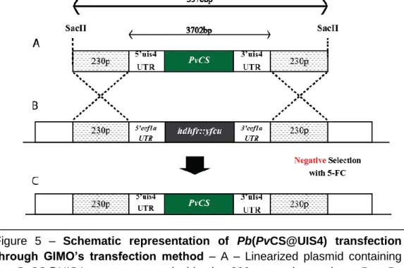
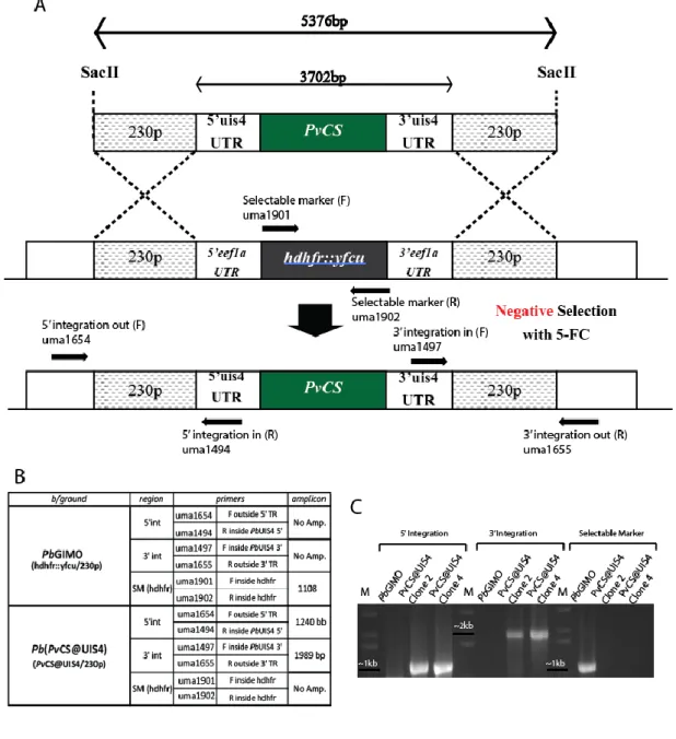
Recently, Inês Albuquerque from IMM‟s Prudêncio Lab, using a blood sample obtained from a P. vivax-infected patient in the field (Thailand), was able to isolate the gene encoding the P. vivax CS protein and insert it on a plasmid containing the 230p targeting region and the promoting regions of the UIS4. In order to generate a new P. berghei line expressing the P. vivax CS protein, Pb(PvCS@UIS4), a transfection was performed according to the GIMO (Gene Insertion Marker Out) method 55, similarly to what was done in order to generate the Pb(PfCS@UIS4) transgenic parasite (Fig.5).
The GIMO transfection method consists of using a genetically modified P. berghei or P. yoelii as recipient motherline for transfection. These transgenic parasites have a positive/negative selection marker (hdhfr::yfcu) stably integrated into the silent 230p locus. Using a linearized plasmid with a gene of interest fused to the 230p targeting region (Fig.5.A), is possible to transfect the recipient motherline by double cross-over homologous recombination leading to simultaneous replacement of the
Figure 5 – Schematic representation of Pb(PvCS@UIS4) transfection
through GIMO’s transfection method – A – Linearized plasmid containing
the PvCS@UIS4 gene cassette inside the 230p targeting regions. B – By double cross-over homologous recombination, PbGIMO parasites‟ positive/negative selectable marker (hdhfr::yfcu) is replaced by the PvCS@UIS4 cassette. C – Transfected parasites are negatively selected using the 5-FC drug, which will eliminate all parasites that still have the selectable marker on their genome, leading to populations where all parasites are Pb(PvCS@UIS4).
17 selection marker with the gene of interest (Fig.5.B). After this, the parasites are negatively selected with 5-fluorocytosine (5-FC). The 5-FC drug will eliminate any parasite that still has the yfcu selectable marker, resulting on the generation of parasites with the integrated gene of interest and no drug resistance gene (Fig.5.C).55
Based on the results obtained with Pb(PfCS@UIS4) parasites, this new vaccination strategy represents a good alternative to the ones that have and are being developed so far. However the newly generated transgenic parasite, P. berghei expressing P. vivax CS under the control of the PbUIS4 promoter (Pb(PvCS@UIS4)), requires developmental characterization in order to proceed to more advanced immunological studies. After the parasite‟s transfection, 2 clones of Pb(PvCS@UIS4) were selected for characterization; Pb(PvCS@UIS4) clone 2 and Pb(PvCS@UIS4) clone 4.
18
2. Aims
As mentioned before, in order to proceed with further, immunological studies, the vaccine candidate Pb(PvCS@UIS4), must be fully characterized in terms of infectivity and development. . In the specific context of this vaccine candidate, it is also important to characterize the expression of the inserted CS gene, to ensure that it is being expressed correctly throughout various development stages. Therefore, in order to characterize the new vaccine candidate Pb(PvCS@UIS4), the following aims were established for this thesis:
- To compare the PvCS gene sequence from the P. vivax isolate used in this study with sequences of the same gene found in isolates from different geographic regions across the world. This will ensure that the CS protein being inserted into our vaccine candidate is not significantly different from the CS protein found in most P. vivax populations worldwide. To do this we shall perform a sequence alignment between our PvCS sequence against reference sequences found on a Plasmodium genome database (PlasmoDB).
- To characterize Pb(PvCS@UIS4) parasite development and infectivity during sporogonic development. This task is to be performed by registering oocyst numbers in the mosquito midgut at 10 days post infectious blood meal and sporozoites in the mosquito salivary glands at 21 days post infectious blood meal.
- To assess Pb(PvCS@UIS4) parasite‟s ability to infect and develop hepatocytes in vitro. In order to do this, two different hepatoma cell lines, Huh7 and HepG2, will be infected with freshly isolated sporozoites. Infection and development will be assessed by immunofluorescence microscopy at 48h post infection.
- To assess the ability of Pb(PvCS@UIS4) parasites to infect and develop in vivo. C57BL/6J mice will be infected through i.v. injection of sporozoites. Infected livers will be analyzed by qRT-PCR and immunofluorescence microscopy.
- To observe CS protein expression on sporozoites and exo-erythrocytic parasite forms, in vitro and in vivo. This will be done by immunofluorescence microscopy, using specific antibodies against PbCS and PvCS.
- Blood-stage development of Pb(PvCS@UIS4) will be analyzed throughout time to assess the parasite‟s ability to exit the liver, infect and replicate inside erythrocytes.
Throughout the whole study, Pb(PvCS@UIS4) results will be compared to PbGIMO parasites as the latter represent the non-transfected, wild-type parasites.
19
3. Materials and Methods
3.1 Mice (C56BL/6J AND BALB/C)
In vivo experiments were performed on C56BL/6J and BALB/C mice, ordered from Charles River Laboratories international. Ages ranged between 4-6 weeks and were housed, upon arrival, in specific pathogen-free conditions, at the animal house facility of Instituto de Medicina Molecular (IMM). All the experimental work involving animals was performed according to the EU regulations and was approved by the Animal Care and Ethical Committees of IMM.
3.2 Parasite lines
-Plasmodium berghei GIMO (Gene Insetion Marker Out)
In order to produce our transgenic parasites, PbGIMO was used as transfection motherline, and therefore used as control on characterization experiments. PbGIMO has a positive/negative selection marker cassette (hdhfr::yfcu) on the 230p locus.
-Plasmodium berghei expressing the Plasmodium vivax CS protein under the control of the PbUIS4 promoter (Pb(PvCS@UIS4))
This transgenic parasite was produced through the GIMO transfection method (by Inês Albuquerque). It expresses P. vivax‟s CS protein under the control of the P. berghei UIS4 promoter. Confirmation of integration will be shown in the results section of this thesis. After confirmation, 2 clones were selected to proceed with characterization; Pb(PvCS@UIS4) Clone 2 and Pb(PvCS@UIS4) Clone 4.
3.3 Rearing of Anopheles stephensi mosquitoes
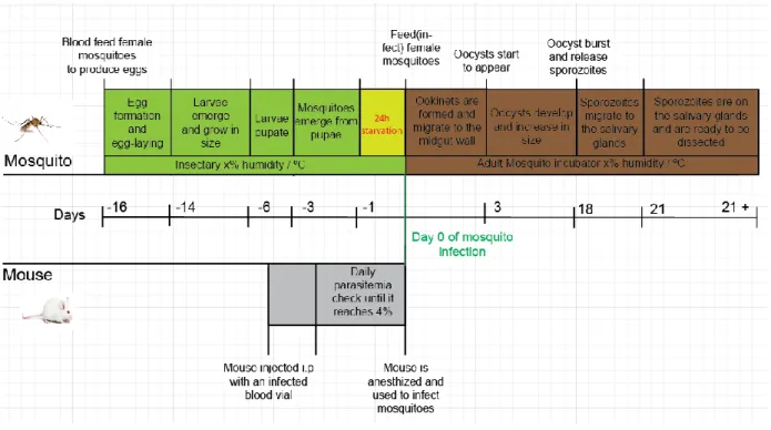
Anopheles stephensi mosquitoes were reared in IMM‟s insectary at 27ºC and 80% humidity. Larvae developed in trays with water and standard fish food while adult mosquitoes were maintained on a 10% sucrose solution with 0,05% of p-aminobenzoic acid (PABA), on a 12h light/dark cycle, according to standard rearing conditions.56 The rearing schedule is described below (Figure 6);
20
3.4 Maintenance of Plasmodium berghei infections
Sporozoite collection
Sporozoites were freshly extracted from the salivary glands of female Anopheles stephensi mosquitoes by hand-dissection and collected in RPMI medium. To obtain free sporozoites the salivary gland suspension was mechanically homogenized and filtered through a 70 μm strainer. Sporozoites were counted in a Neubauer-chamber. Figure 6 – Mosquito rearing and infection Schedule – Day 0 corresponds to the day that mosquitoes were fed with an infectious blood meal. (Schedule was repeated on a weekly basis) Adult female mosquitoes were fed with rodent blood in order to produce eggs (16 days before the infectious blood meal). Through the 2 days after feeding, female mosquitoes laid their eggs. Eggs hatched and larvae emerged (-16 to -14). Larvae grew in size through the following 7-8 days (-14 to -6). At day -6, larvae began to pupate. Also, on the same day, mice were injected i.p. with an vial of frozen parasite stabilates. At day -3, mosquitoes started to emerge from pupae. Mouse parasitemias were checked daily until 4% parasitemia. Male gametocyte exflagellation was accessed as it is a strong indicator of transmission capacity of infected mice. At the day previous to blood infection, adult mosquitoes were moved from the insectary to a mosquito incubator and left on starvation for 24h. The following day (day 0), infected mice were anesthetized and used to feed starved mosquitoes. Within the mosquito, ookinets are formed and develop into oocysts on the first 3 days post infectious blood meal. From day 3 to 18, oocysts grow in size. At day 18, oocysts start to burst and release sporozoites, which migrate to the salivary glands through the following 3 days. At day 21, sporozoites are already on the salivary glands and ready for dissection.

![Figure 3 - Proposed mechanism of protective immunity directed against the Plasmodium infected hepatocyte.[Image adapted from [22]] - Within the infected hepatocyte, cytoplasmic malaria proteins are transformed into short peptides through a prote](https://thumb-eu.123doks.com/thumbv2/123dok_br/18488748.900875/21.892.134.769.211.644/proposed-mechanism-protective-plasmodium-hepatocyte-hepatocyte-cytoplasmic-transformed.webp)
![Figure 4 – Schematic representation of the RTS,S particle. [Image adapted from [27]]](https://thumb-eu.123doks.com/thumbv2/123dok_br/18488748.900875/22.892.149.751.588.1076/figure-schematic-representation-rts-s-particle-image-adapted.webp)