1. Pediatric Rheumatology Unit, Universidade Federal de São Paulo 2. Pediatric Rheumatology Unit, Faculdade de Medicina da Universidade de São Paulo (USP)
3. Division of Rheumatology, Faculdade de Medicina da Universidade de São Paulo (USP)
4. Pediatric Rheumatology Division, São Paulo State University (UNESP) – Faculdade de Medicina de Botucatu
5. Pediatric Rheumatology Unit, Hospital Infantil Darcy Vargas, São Paulo – Brazil
ment such as intravenous immunoglobulin in two pa-tients, hydroxychloroquine and azathioprine in two and intravenous cyclophosphamide in one patient. Sepsis was observed in three cSLE patients. Two pa-tients required intensive care and death was observed in one patient.
Conclusion: Our study identified SJS and overlap
SJS--TEN as rare manifestations of active cSLE associated with severe multisystemic disease, with potentially lethal outcome.
Keywords: Stevens-Johnson syndrome; Toxic
epider-mal necrolysis; Childhood-onset systemic lupus ery-thematosus; Systemic lupus eryery-thematosus; Childhood
INTRODUCTION
Systemic lupus erythematosus (SLE) is a rare multisys-tem autoimmune disease more common in adults (aSLE) with 10% to 20% of cases beginning in children and adolescents1. Childhoodonset SLE (cSLE) is cha
-racterized by the involvement of various organs and systems, such as mucocutaneous, which has been repor ted in 50 to 85% at the time of diagnosis or du -ring the course of the disease2,3.
Stevens-Johnson syndrome (SJS) and toxic epider-mal necrolysis (TEN) are severe cutaneous adverse re-actions with high morbidity and mortality, usually in-duced by medications or infections4,5. They result in
acute onset of target lesions followed by detachment of the epidermis and epithelia resulting in extensive areas of denuded skin, necrosis and mucosal erosions, ge -nerally with the presence of Nikolski's sign4-7. Both di
-seases are part of a single spectrum of severe epider-molytic reactions, differing mainly by the extent of skin detachment and type of cutaneous lesions.
Stevens-Johnson syndrome and toxic epidermal
necrolysis in childhood-onset systemic lupus
erythematosus patients: a multicenter study
Sakamoto AP1, Silva CA2,3, Saad-Magalhães C4, Alencar AN1, Pereira RMR3, Kozu K2, Barbosa CMPL5, Terreri MT1
ACTA REUMATOL PORT. 2017;42:250-255
ABSTRACT
Objective: To assess Stevens-Johnson syndrome (SJS)
and toxic epidermal necrolysis (TEN) in a large popu-lation of childhood-onset systemic lupus erythemato-sus (cSLE) patients.
Methods: Multicenter study including 852 cSLE
pa-tients followed in Pediatric Rheumatology centers in São Paulo, Brazil. SJS was defined as epidermal de-tachment below 10% of body surface area (BSA), over-lap SJS-TEN 10-30% and TEN greater than 30% of BSA.
Results: SJS and TEN were observed in 5/852 (0.6%)
cSLE female patients, three patients were classified as SJS and two patients were classified as overlap SJS-TEN; TEN was not observed. The mean duration of SJS and overlap SJSTEN was 15 days (range 722) and antibio -tics induced four cases. Regarding extra-cutaneous manifestations, hepatomegaly was observed in two cSLE patients, nephritis in two and neuropsychiatric involvement and conjunctivitis were observed respecti -vely in one patient. Hematological involvement in-cluded lymphopenia in four, leucopenia in three and thrombocytopenia in two patients. The mean SLEDAI--2K score was 14.8 (range 6-30). Laboratory analysis showed low C3, C4 and/or CH50 in two patients and the presence of anti-dsDNA autoantibody in two patients. One patient had lupus anticoagulant and another one had anticardiolipin IgG. All patients were treated with steroids and four needed additional
treat-The initial lesions are characterized by atypical tar-gets and/or purpuric macules commonly in upper tor-so, proximal limbs, and face, spreading to the trunk and distal limbs. Palms and soles are often involved. Mucosal membranes of the eyes, mouth, nose and geni talia can initially appear, leading to an erosive and hemorrhagic mucositis7,8.
Other clinical features such as fever, malaise and upper respiratory tract symptoms and ocular inflam-mation can precede the eruption by several days. Necrosis can occur in the epithelia of the respiratory tract, causing bronchial and tracheal obstruction and ventilatory compromise; in gastrointestinal tract, lead-ing to exuberant diarrhea; and in kidneys, causlead-ing hy-poperfusion, acute tubular necrosis and acute kidney injury. Mild elevation of liver enzymes is usual, al-though significant hepatitis or hepatic impairment are rare 4,7.
Of note, SJS and TEN can occur simultaneously with SLE6,9,10and particularly in cSLE patients11-14.
There are, however, no reports characterizing their prevalence and describing SJS and TEN in a large popu lation of cSLE patients.
meThODS
STUDy DeSIgN AND pATIeNTS
A retrospective multicenter cohort study was per-formed including 1,017 consecutive cSLE patients fol-lowed in ten Pediatric Rheumatology tertiary referral centers in São Paulo state, Brazil. The charts were re-vised from 2012-2014. One hundred and sixty five patients were excluded due to: incomplete medical charts (n=96), undifferentiated connective tissue di -sorder with 3 or fewer American College of Rheuma-tology (ACR) criteria for SLE15(n=43), isolated
cuta-neous lupus erythematosus (n=11), neonatal lupus erythematosus (n=8), drug-induced lupus (n=5) and other autoimmune diseases (n=2)16. All the remaining
852 cSLE patients fulfilled the ACR criteria for SLE15,
with disease onset before 18 years of age17and current
age up to 25 years old. In all the participant centers the Committee for Research Ethics approved the study.
An investigator meeting was held in the beginning of the study in the city of São Paulo to define the pro-tocol, including definitions of clinical, laboratory and treatment parameters, disease activity, SJS and TEN characteristics and outcomes (intensive care unit stay and death). All investigators used the same specific
database. All patient’s medical charts were reviewed according to this standardized protocol.
SJS was defined as epidermal detachment below 10% of body surface area (BSA) with purpuric macules or flat typical targets, overlap SJS-TEN with detach-ment of 10-30% of BSA with purpuric macules or flat typical targets and TEN with detachment of greater than 30% of BSA with or without purpuric macules or flat typical targets, also known as spots4,7,8.
All cSLE patients with the suspicion of SJS, overlap SJSTEN or TEN were evaluated by a local dermatolo -gist.
DemOgRAphIC DATA, ClINICAl evAlUATION, DISeASe ACTIvITy AND DRUg TheRApy
Demographic data analysis included: gender, age at SJS/TEN onset, duration of cSLE before SJS/TEN in months and duration of SJS/TEN in days. Descriptors and definitions of SLE clinical manifestations were based on SLE Disease Activity Index 2000 (SLEDAI--2K) score of disease activity18, and were evaluated at
the manifestation of SJS/TEN. Other SJS/TEN characte -ristics included the identification of the triggering agent, presence of blisters/vesicles and mucosal in-volvement, percentage of body surface area with epi-dermal detachment and extra-cutaneous involvement. Other cumulative SLE clinical manifestations inclu ded hepatomegaly [based on physical exam with liver edge ≥ 2 cm below the right costal margin or imaging (ul-trasound or computer tomography when available)] and splenomegaly [based on physical exam with pal-pable spleen or imaging (ultrasound or computer to-mography when available)]. Neuropsychiatric lupus included 19 syndromes according to ACR classifica-tion criteria19.
Complement levels (CH50, C3 and C4) were as-sessed by immunodiffusion, turbidimetric im-munoassay or immunonephelometry. Anti-double--stranded DNA (anti-dsDNA) was assessed by indirect immunofluorescence or Enzyme Linked Immuno Sor-bent Assay (ELISA); anticardiolipin (aCL) IgG and IgM autoantibodies by ELISA. All the autoantibodies pro-file were carried out at each center. The cutoff values from the kit manufacturer were used to define abnor-mal. Lupus anticoagulant was detected according to the guidelines of the International Society on Throm-bosis and Hemostasis20.
Drug treatment data (prednisone, intravenous methylprednisolone, chloroquine diphosphate, hy-droxychloroquine sulfate, azathioprine, cyclosporine,
hematological involvement, such as lymphopenia <1,500/mm3in four, leucopenia <4,000/mm3in three,
and thrombocytopenia<100,000/mm3in two cSLE
pa-tients.
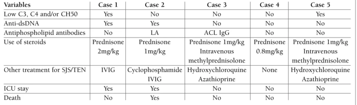
The mean SLEDAI-2K score was 14.8 (range 6-30). The laboratory tests analysis showed low C3, C4 and/or CH50 in two patients and the presence of anti-dsDNA antibodies in two patients. One patient had lupus anticoagulant positive and another one had anticardio -lipin IgG.
Therapy in cSLE patients with SJS and TEN com-prised of steroid use either in intravenous methyl-prednisolone or prednisone. Four patients needed additional treatment, such as intravenous immuno -globulin in two patients, combined hydroxychloro-quine and azathioprine in two and intravenous cy-clophosphamide in one patient. Regarding outcome, two patients required intensive care unit and death was observed in one patient.
DISCUSSION
Our multicenter study characterized SJS and overlap micofenolate, intravenous cyclophosphamide,
intra-venous immunoglobulin) were also recorded.
ReSUlTS
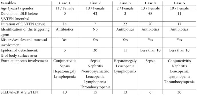
SJS and TEN were observed in 5/852 (0.6%) cSLE pa-tients, all girls, with age ranging between 2 to 18 years old. Three patients were classified as SJS and two pa-tients were classified as overlap SJS-TEN; TEN was not observed. In one patient these manifestations occurred at SLE diagnosis and in four patients during the fol-low-up. The mean duration of SJS and overlap SJS-TEN was 15 days (range 7-22) and four cases were induced by antibiotics. Demographical and clinical characteris-tics as well as disease activity (SLEDAI-2K) of the five cSLE patients were shown in Table I. Presence of au-toantibodies, treatment and outcome were shown in Table II.
All patients had blisters, vesicles and mucosal in-volvement. Regarding extra-cutaneous manifestations, sepsis was observed in three patients, hepatomegaly in two, nephritis in two, conjunctivitis in two and neu-ropsychiatric involvement in one. Four patients had
TABle I. DemOgRAphICAl, ClINICAl ChARACTeRISTICS AND DISeASe ACTIvITy Of The fIve SJS/TeN cSle pATIeNTS
Variables Case 1 Case 2 Case 3 Case 4 Case 5
Age (years) / gender 11 / Female 18 / Female 2 / Female 13 / Female 10 / Female
Duration of cSLE before 0 43 2 48 11
SJS/TEN (months)
Duration of SJS/TEN (days) 14 7 22 20 17
Identification of the triggering Antibiotics No Antibiotics Antibiotics Antibiotics agent
Blisters/vesicles and mucosal Yes Yes Yes Yes Yes
involvement
Epidermal detachment, 5 20 11 Less than 10 Less than 10
% of body surface area
Extra-cutaneous involvement Conjunctivitis Sepsis Hepatomegaly Sepsis Conjunctivitis
Sepsis Nephritis Leucopenia Nephritis
Hepatomegaly Neuropsychiatric Lymphopenia Leucopenia
Lymphopenia Leucopenia Lymphopenia
Lymphopenia Thrombocytopenia
Thrombocytopenia
SLEDAI-2K at SJS/TEN 10 15 13 6 30
*SJS: Stevens-Johnson syndrome, TEN: toxic epidermal necrolysis, cSLE: childhood-onset systemic lupus erythematosus, SLEDAI-2K: Systemic Lupus Erythematosus Disease Activity Index 2000.
cal criteria: the pattern of individual skin lesions, their distribution and the maximum extent of epidermal de-tachment7-9. Importantly, due to the large detachment
of the skin, TEN patients have higher mortality rates4,7.
The alternative diagnosis to SJS and TEN such as ery-thema multiforme and drug induced lesions, pemphi-gus, pemphigoid, acute graft versus host disease and other immunobullous diseases as well as infectious disea ses like Mycoplasma, Herpes virus and HIV must be ruled out7.
In some cases, the diagnosis of lupus-induced TEN and classical TEN is intriguing. In both cases the clas-sical clinical and histopathological features will be pre-sent, with epidermolytic reactions and skin detachment due to inflammatory dermatoses with keratinocyte apoptosis and the same molecular mediators in-volved13. Lupus-induced TEN tend to have a subacute
presentation of weeks with the absence of systemic in-volvement and no history of drug ingestion, while clas-sical TEN has an acute evolution within 3 to 4 days or sometimes within hours, with a close drug-related causality and negative immunoflorescence4,21. In
con-trast to SJS-TEN, cutaneous lupus demonstrates the prototypic interface dermatitis with deposition of munoglobulins (IgG, IgM and/or IgA, C3) in direct im-munofluorescence13,22,23.
In both manifestations, renal involvement is com-mon. It is known that in cSLE renal disease has been reported in almost two thirds of the patients and more frequent than the adult population24, 25. Due to systemic
drug-induced hypersensitivity, SJS and TEN can also SJS-TEN as rare manifestations in a large cSLE
lation. The advantage of including a large cSLE popu-lation selected in tertiary referral centers allowed a bet-ter evaluation of these rare and potentially lethal manifestations. The use of a standardized combined database, with proper SJS, overlap SJS-TEN and TEN definitions minimized possible bias. In addition, in two large prospective European studies characterizing SJS and TEN, association with SLE and other autoimmune rheumatologic diseases was found in approximately 0.6 to 5% of all patients with these severe manifestations8,9.
We found five (0.6%) cSLE patients with SJS and over-lap SJS-TEN in our cohort population, and one of them was previously reported (patient 1)11. TEN seemed to
be rarer since we did not find any patient in our cohort study.
However, the main limitation of this study was the retrospective design and possible missing data. In addi -tion, skin biopsies were not a routine procedure in all participant Pediatric Rheumatology centers. Only one patient had SJS features confirmed by autopsy (case 2). However, all other patients had a close drug causality and were evaluated by a local experient dermatologist. These skin vesiculobullous diseases are severe and life-threatening cutaneous adverse reactions mainly caused by drugs, infections or sometimes, unidentified causes7-9, 21. Antibiotics are the most frequent cause
followed by antiinflammatories and analgesics, as obser -ved herein4. Other organs are described to be involved
as we found in most cases.
Classification of SJS and TEN is based on three clini
-TABle II. SeROlOgICAl AND lABORATORIAl ChARACTeRISTICS, TReATmeNT AND OUTCOme Of The fIve SJS/TeN cSle pATIeNTS
Variables Case 1 Case 2 Case 3 Case 4 Case 5
Low C3, C4 and/or CH50 Yes No No No Yes
Anti-dsDNA Yes Yes No No No
Antiphospholipid antibodies No LA ACL IgG No No
Use of steroids Prednisone Prednisone Prednisone 1mg/kg Prednisone Prednisone 1mg/kg 2mg/kg 1mg/kg Intravenous 0.8mg/kg Intravenous
methylprednisolone methylprednisolone Other treatment for SJS/TEN IVIG Cyclophosphamide Hydroxychloroquine None Hydroxychloroquine
IVIG Azathioprine Azathioprine
ICU stay Yes Yes No No No
Death No Yes No No No
*SJS: Stevens-Johnson syndrome, TEN: toxic epidermal necrolysis, cSLE: childhood-onset systemic lupus erythematosus, Anti-dsDNA: double-stranded DNA, LA: Lupus anticoagulant, ACL IgG: Anticardiolipin IgG, IVIG: Intravenous immunoglobulin, ICU: Intensive Care Unit
ciana Tudech Pedro Paulo); Hospital Municipal Infantil Menino Je-sus (Simone Lotufo, Tânia Caroline Monteiro de Castro) and Pon-tifical Catholic University of Sorocaba (Valéria C. Ramos).
fUNDINgS
This study was supported by grants from Conselho Nacional de De-senvolvimento Científico e Tecnológico (CNPq 303422/2015-7 to Clovis Artur Silva, 301805/2013-0 to Rosa Maria Rodrigues Pereira, 305068/2014-8 to Eloisa Bonfá, 301479/2015 to Claudia Saad-Ma-galhães and 303752/2015-7 to Maria Teresa Terreri), Federico Foun-dation (to Clovis Artur Silva, Rosa Maria Rodrigues Pereira and Eloisa Bonfá) and by Núcleo de Apoio à Pesquisa “Saúde da Criança e do Adolescente” of USP (NAP-CriAd) to Clovis Artur Silva.
CORReSpONDeNCe TO
Maria Teresa Terreri Rua Ipê 112- apto 111
São Paulo – SP, Brazil, 04022-005 E-mail: teterreri@terra.com.br
RefeReNCeS
1. Tarr T, Dérfálvi B, Györi N, et al. Similarities and differences be-tween pediatric and adult patients with systemic lupus erythe-matosus. Lupus 2014; 0: 1-8.
2. Levy DM, Kamphuis S. Systemic lupus erythematosus in chil-dren and adolescents. Pediatr Clin N Am 2012; 59: 345-364. 3. Mina R, Brunner HI. Pediatric lupus – are there differences in
presentation, genetics, response to therapy, and damage accru-al compared with adult lupus? Rheum Dis Clin N Am 2010; 36: 53-80.
4. Letko E, Papaliodis DN, Papaliodis GN, Daoud YJ, Ahmed AR, Foster CS. Stevens-Johnson syndrome and toxic epidermal necrolysis: a review of the literature. Ann Allergy Asthma Im-munol 2005; 94: 419-436.
5. Harr T, French LE. Severe cutaneous adverse reactions: acute generalized exanthematous pustulosis, toxic epidermal necroly -sis and Stevens-Johnson syndrome. Med Clin North Am 2010; 94: 727-742.
6. Cetin GY, Sayar H, Ozkam F, Kurtulus S, Kesiei F, Sayarhoglu M. A case of toxic epidermal necrolysis-like skin lesions with sys-temic lupus erythematosus and review of the literature. Lupus 2013; 22: 839-846.
7. Creamer D, Walsh SA, Dziewulski p, et al. U.K. guidelines for the management of Stevens-Johnson syndrome/toxic epidermal necrolysis in adults 2016. British Jourmal of Dermatology 2016; 174: 1194-1227.
8. Auquier-Dunant A, Mockenhaupt M, Naldi L, Correia O, Schröder W, Roujeau JC; SCAR Study Group, Severe Cutaneous Adverse Reactions. Correlations between clinical patterns and causes of erythema multiforme major, Stevens-Johnson syn-drome, and toxic epidermal necrolysis. Arch Dermatol 2002; 138 (8): 1019-1024.
9. Ziemer M, Kardaun SH, Liss Y, Mockenhaupt M. Stevens-John-son syndrome and toxic epidermal necrolysis in patients with lupus erythematosus: a descriptive study of 17 cases from a na-tional registry and review of the literature. Br J Dermatol 2012; 166 (3): 575-600.
10. Konda S, Fernandez AP, Berman B, Elgart G, Milikowski C, Alonso-Llamazares J. Toxic epidermal necrolysis in the setting of systemic lupus erythematosus. Int J Dermatol 2011; 50 (10): 1270-1275.
lead to drug-induced acute interstitial nephritis in al-most 20% of the cases26. In our cohort, 2 patients had
lupus nephritis prior to cutaneous involvement (cases 2 and 5). One of them had worsening of the renal disea se with hypertension and acute renal failure at SJS manifestation (case 2).
We also observed that all patients presented a SLEDAI-2K score higher or equal to 6. We hypothe-sized that SJS-TEN occurs in the exacerbation of lu-pus, concomitantly with the disease presentation or during flares. This finding should be further analyzed in order to establish a possible association and has not been des cribed yet in the literature.
There is no optimal treatment for this condition but glucocorticoids are frequently beneficial and intra-venous immunoglobulin (IVIG) can be administered as an additional treatment7,11,12. The exclusion of any
trig-gering agent is required and antibiotics should be promptly discontinued, as occurred in four of our cSLE patients.
In conclusion, we have presented five patients with cSLE who presented SJS and overlap SJS-TEN at the time of cSLE diagnosis or during the course of the disea se. Most patients had a clear drug causality main-ly related to antibiotics and required an intensive care unit treatment, showing how rare and lethal these mani festations can occur in our population. Further efforts may be needed for the search of risk factors and treatment in the pediatric lupus patients.
ACkNOwleDgemeNTS
Our gratitude to Ulysses Doria-Filho for the statistical analysis. The authors thank the following Pediatric Rheumatology Divisions and colleagues for including their patients: Pediatric Rheumatology Unit, FMUSP (Adriana Almeida de Jesus, Adriana Maluf Elias Sal-lum, Cristina Miuki Abe Jacob, Gabriela Blay, Gabriela Nunes Leal, Gabriella Erlacher Lube de Almeida, Heloisa Helena de Souza Mar-ques, João Domingos Montoni da Silva, Joaquim Carlos Rodrigues, Juliana Caíres de Oliveira Achili Ferreira, Laila Pinto Coelho, Lu-ciana dos Santos Henriques, Maria Helena Vaisbich, Nadia Emi Aikawa, Lucia Maria Arruda Campos, Victor Marques, Werther Brunow de Carvalho); Pediatric Rheumatology Unit, UNIFESP (Anandreia Simões Lopes, Claudio Arnaldo Len, Daniela Petry Pi-otto, Giampaolo Faquin, Gleice Clemente Souza Russo, Luis Eduardo Coelho Andrade, Maria Odete Esteves Hilário, Melissa Ma -riti Fraga, Octavio Augusto Bedin Peracchi, Vanesssa Bugni Miotto e Silva); Division of Rheumatology, FMUSP (Juliane A. Paupitz, Glauce Leão Lima); UNESP (Priscila R. Aoki, Juliana de Oliveira Sato, Silvana Paula Cardin, Taciana Albuquerque Pedrosa Fernandes); Irman-dade da Santa Casa de Misericórdia de São Paulo (Andressa Guari-ento, Maria Carolina dos Santos, Natali Weniger Spelling Gormen-zano); State University of Campinas (Maraísa Centeville, Renata Barbosa); Ribeirão Preto Medical School – University of São Paulo (Gecilmara Salviatto Pileggi, Paola Pontes Pinheiro, Virginia Paes Leme Ferriani); Hospital Infantil Darcy Vargas (Jonatas Libório,
Lu-19. American College of Rheumatology Ad Hoc committee on neu-ropsychiatric Lupus Syndromes. The American College of Rheumatology nomenclature and case definitions for neu-ropsychiatric lupus syndromes. Arthritis Rheum 1999; 42: 599--608.
20. Brandt JT, Triplett DA, Alving B, et al. Criteria for the diagnosis of lupus anticoagulants: an update. On behalf of the Subcom-mittee on Lupus Anticoagulant/Antiphospholipid Antibody of the Scientific and Standardisation Committee of the ISTH. Thromb Haemost 1995; 74: 1185-1190.
21. Mandelcorn R, Shear NH. Lupus-associated toxic epidermal necrolysis: A novel manifestation of lupus? J Am Acad Derma-tol 2003; 48: 525-529.
22. Walling HW, Sontheimer RD. Cutaneous lupus erythematosus. AM J Clin Dermatol 2009; 10: 365-381.
23. Camisa C, Sharma HM. Vesiculobullous systemic lupus ery-thematosus: report of cases and review of literature. J Am Acad Dermatol 1983; 9: 924-1033.
24. Klein-Gitelman M, Reiff A, Silverman ED. Systemic lupus ery-thematosus in childhood. Rheum Dis Clin North Am 2002; 28: 561-577.
25. Benseler SM, Silverman ED. Systemic lupus erythematosus. Pe-diatr Clin N Am 2005; 52: 443-467.
26. Hung CC, Liu WC, Kuo MC, Lee CH, Hwang SJ, Chen HC. Acute renal failure and its risk factors in Stevens-Johnson syn-drome and toxic epidermal necrolysis. Am J Nephrol 2009; 29 (6): 633-638.
11. Cavalcante EG, Guissa VR, Jesus AA, Campos LM, Sallum AM, Aikawa NE, et al. Stevens-Johnson syndrome in a juvenile sys-temic lupus erythematosus patient. Lupus 2011; 20: 1439--1441.
12. Baker MG, Cresce ND, Ameri M, Martin AA, Patterson JW, Kim-pel DL. Systemic lupus erythematosus presenting as Stevens--Johnson syndrome/toxic epidermal necrolysis. J Clin Rheuma-tol 2014; 20: 167-171.
13. Lee HY, Tey HL, Pang SM, Thirumoorthy T. Systemic lupus ery-thematosus presenting as Stevens-Johnson syndrome and to xic epidermal necrolysis: a report of 3 cases. Lupus 2011; 20: 647--652.
14. Samimi SS, Siegfried E. Stevens-Johnson syndrome developing in a girl with systemic lupus erythematosus on high-dose cor-ticosteroid therapy. Pediatr Dermatol 2002; 19 (1): 52-55. 15. Hochberg MC. Updating the American College of Rheumatolo
-gy revised criteria for the classification of systemic lupus ery-thematosus. Arthritis Rheum 1997; 40: 1725.
16. Gomes RC, Silva MF, Kozu K, Bonfá E, Pereira RM, Terreri MT, et al. Features of 847 Childhood-Onset Systemic Lupus Erythematosus Patients in Three Age Groups at Diagnosis: A Brazi -lian Multicenter Study. Arthritis Care Res (Hoboken) 2016; 68 (11): 1736-1741.
17. Silva CA, Avcin T, Brunner HI. Taxonomy for systemic lupus erythematosus with onset before adulthood. Arthritis Care Res (Hoboken) 2012; 64: 1787-1793.
18. Gladman DD, Ibañez D, Urowitz MB. Systemic lupus erythe-matosus disease activity index 2000. J Rheumatol 2002; 29: 288-291.