1. Head of the Brachial Plexus Surgery and Microsurgery Unit; Senior Consultant at the Plastic and Reconstructive Surgery Department, São José Hospital, Lisbon, Portugal
2. Junior Consultant at the Plastic and Reconstructive Surgery Department, São José Hospital, Lisbon, Portugal
3. Instructor at the Anatomy Department, Medical Sciences Faculty, New University of Lisbon
Brachial plexus tumors in a tertiary referral center:
a case series and literature review
ACTA REUMATOL PORT. 2015;40:372-377
AbstrAct
Introduction: Brachial plexus (BP) tumors are very rare tumors, with less than 800 cases been described in the literature worldwide since 1970. These tumors often present as local or radicular pain, with scant or no neu-rological deficits. These symptoms are shared by many other more common rheumatologic diseases, thus ma -king their diagnosis difficult in most cases. Additional-ly, these tumors often present as lumps and are there-fore biopsied, which carries a significant risk of iatro-genic nerve injury.
Material and Methods: In this paper the authors des -cribe their experience in the management of 5 patients with BP tumors, followed-up for at least 2 years. There were 4 males and 1 female. The median follow-up time was 41 ± 21 months. The average age at diagnosis was 40,0 ± 19,9 years. The most common complaints at presentation were pain and sensibility changes. All pa-tients had a positive Tinel sign when the lesion was per-cussed. In all patients surgery was undertaken and the tumors removed. In 4 patients nerve integrity was maintained. In one patient with excruciating pain a segment of the nerve had to be excised and the nerve defect was bridged with sural nerve grafts.
Results: Pathology examination of the resected speci-mens revealed a Schwannoma in 4 cases and a neu-rofibroma in the patient submitted to segmental nerve resection. Two years postoperatively, no recurrences were observed. All patients revealed clinical improve-ment. The patient submitted to nerve resection had im-provement in pain, but presented diminished strength and sensibility in the involved nerve territory.
Millan G1, Casal D2,3
Conclusion: Surgical excision of BP tumors is not a risk free procedure. Most authors suggest surgery if the lesion is symptomatic or progressing in size. If the tu-mor is stationary and not associated with neurological dysfunction a conservative approach should be taken. Keywords: Brachial plexus; Schwannoma; Neurofi-broma; Peripheral nerve sheath tumor; Upper Limb; Surgery.
IntroductIon
The first description of a brachial plexus (BP) tumor is attributed to Courvoisier who in 1886 published a pa-per on a patient with a tumor from the C5 nerve root1,2.
Unfortunately, his attempt to remove the tumor was marred by subsequent paralysis of the deltoid and bi-ceps muscles1,2. Since then several reports have been
published on this subject, allowing a better prognosis than that described by Courvoisier in most cases2-4.
However, most of these reports are composed of case re-ports or small series of patients4. The largest series
publi shed is a 30 year retrospective study, in a large ter-tiary referral center in the United States of America, that identified 226 patients2. The second largest study is a
recent 10 year review of 115 BP tumors operated by a single surgeon in India3.
Upper limb peripheral nerve sheath tumors repre-sent only 1 to 4,9% of all upper-extremity tumors5. BP
tumors are even rarer5. Moreover, they often present as
local or radicular pain, with scant or no neurological deficits2,4. These symptoms are shared by many other
more common rheumatologic diseases, thus making the diagnosis of BP tumors difficult in most cases2,4.
Additionally, these tumors often present as lumps and are therefore biopsied, which poses a significant risk of iatrogenic nerve injury2,4.
with the management of 5 patients with BP tumors fol-lowed up for at least 2 years.
methods
The authors retrospectively reviewed the charts, and clinical images of 5 patients with brachial plexus tu-mors, referred to the Brachial Plexus and Peripheral Nerve Surgery Outpatient Clinic, at São José Hospital (Lisbon, Portugal). The basic demographic features, clinical picture and outcome, two years postopera-tively of these 5 patients are described in Table I.
All patients but one were male (4M:1F). Median fol-low-up time was 41 ± 21 months. Average age at pre-sentation was 40,0 ± 19,9 years, ranging from 17 to 66 years. All patients complained of pain in the territory of the involved nerve. This symptom was particularly
disabling in patient number 1, who was later found to have a neurofibroma. Two patients presented with di-minished strength in the territory of the involved nerve. Two patients presented a palpable lump corres ponding to the location of the tumor. In these two ca ses the mass had a greater side-to-side mobility than longitudinal mobility. Two patients complained of signi ficant hy-poesthesia. One of these patients (Patient 3) said that this symptom had occurred after a biopsy of the region in another institution prior to referral to the Brachial Plexus Clinic. One patient with a median nerve tumor referred crippling dysesthesia in the cutaneous territo-ry of this nerve in the palm of the hand (Patient 5). All patients had a positive Tinel sign when the lesion was percussed.
All patients had imagiological evidence of the tumor either in computed tomography (CT) scan or magne -tic ressonance imaging (MRI). All patients with sensi-tAble I. summAry of the pAtIents wIth brAchIAl plexus tumors
Age at Outcome 2
presentation Symptoms at Surgical years after Patient Gender (years) presentation Location Procedure Histology surgery 1 M 24 Hypoesthesia, Origin of the Excision of the Neurofibroma Partial recovery
intense pain, ulnar nerve involved nerve of strength and
and diminished segment, and sural sensibility to
strength nerve grafts to bridge the ulnar
in the ulnar the nerve defect territory; no
nerve territory pain
2 F 42 Lump and pain in Origin of the Excision of the Schwannoma Normal the supraclavicular suprascapular tumor preserving function; no
region nerve the nerve symptoms
3 M 51 Lump and pain in Posterior Excision of the Schwannoma Normal the supraclavicular cord of the tumor preserving function; region; hypoesthesia brachial the nerve residual
and paresthesia in plexus hypoesthesia.
the posterior aspect of the forearm
4 M 66 Pain and Origin of Excision of the Schwannoma Normal
diminished strength the radial tumor preserving function;
in the territory of nerve the nerve no symptoms
the radial nerve
5 M 17 Pain and dysesthesia Median Excision of the Schwannoma Normal in the median nerve nerve tumor preserving function;
territory; lump in the nerve no symptoms
the arm
bility changes had electroneuromyographic abnor-malities. There was no history or evidence of neurofi-bromatosis in any of the patients.
The surgical procedure consisted of exposure of the involved nerve and surrounding structures. The tu-mor was then isolated using magnifying loupes. In 3 cases the tumor was partially encased in a fibrous sheath that facilitated intraneural dissection and re-moval of the tumor without significantly damaging the nerve fascicles (Figure 1). In one case, concerning a suprascapular nerve tumor, the lesion was highly adhe -rent to the nerve fascicles and no fibrous sheath was found (Fi gure 2). However, after a laborious intraneu-ral dissection, it was possible to isolate the tumor from the main nerve fascicles, maintaining nerve integrity. Finally, in one case of an ulnar nerve tumor, no dis-section plane was found, as the tumor permeated all the thickness of the nerve (Patient 1). In this patient, it proved necessary to excise the involved segment of the nerve and bridge the defect with four cables of sural nerve graft (Figures 3 and 4).
results
No intra-operative or postoperative complications were noted, being the patients discharged home a few days after the surgery (3 to 5 days). All surgical wounds healed uneventfully. Patients were followed up for a minimum of two years after surgery. The pathology exa mination of the resected specimens revealed a schwannoma in 4 ca -ses and a neurofibroma in one patient (Patient 1).
Two years postoperatively, no recurrences were obser ved. Additionally, all patients revealed clinical improvement. Three patients were symptom free. One patient, who had been subjected to nerve biopsy pri-or to referral, had npri-ormal motpri-or function, but com-plained of residual paresthesia and hyposthesia in the dorsum of his forearm. Finally, the patient with the neurofibroma which had mandated resection of a seg-ment of the ulnar nerve and reconstruction with sural nerve grafts showed improvement in pain, but pre-sented diminished strength and sensibility in the ulnar nerve territory.
dIscussIon
Carefully going through all the literature concerning BP tumors since 1970, less than 800 cases are found
fIGure 1.Intraoperative view of the extirpation of a peripheral nerve sheath tumor in the proximal portion of the radial nerve in the posterior aspect of the arm. A) On exposure of the nerve, the tumor was located peripherally. B) It was possible to isolate the peripheral nerve sheath tumor (later shown to be a Schwannoma) from the main nerve trunk, and a fibrous sheath resembling a fibrous capsule facilitated dissection. C) The tumor was dissected from the radial nerve leaving it intact
worldwide3,4. These cases are mainly reported in single
case reports or small series. There are only 11 papers with revisions of more than 15 patients3,4. As these
tu-mors are so rare, it is unanimously accepted that these patients should be referred to specialized centers2,4-8.
The mean age of our patient population at diagno-sis (40 years) was in accordance with the largest series published, which presented an average value of 42 years2. However, contrarily to most larger series, males
were over-represented (4 males to one female)2,3. This
may be due to the relatively small number of patients in our series.
The most common symptoms presented by our patients were similar to what has generally been repor -ted2,3,9. In fact, most authors describe that most BP
tu-mors patients present with pain, sensibility changes, and a palpable mass2,3. Tinel sign which is defined as
an electric shock-like experience in the territory of the nerve on tapping the nerve is pathognomonic of nerve lesion and should be considered diagnostic of a nerve tumor, if no prior trauma occurred3,9-11.
However, not always is it easy to establish this diagno sis, and BP tumors are frequently mistaken for other more common neoplasms in the BP region, namely lipomas, ganglion cysts, lymphadenopathies, brachial cleft cysts, desmoid tumors, cavernous an-giomas, hemangiomas or lymphangiomas3,9. In the
fIGure 2.Intraoperative view of the excision of a Schwannoma of the supraclavicular nerve (marked with the asterisk) through a supraclavicular approach. A) The tumor was not eccentrically placed in the nerve. B) The tumor had to be dissected between the fascicles of the nerve under magnifying loupes, preserving nerve continuity
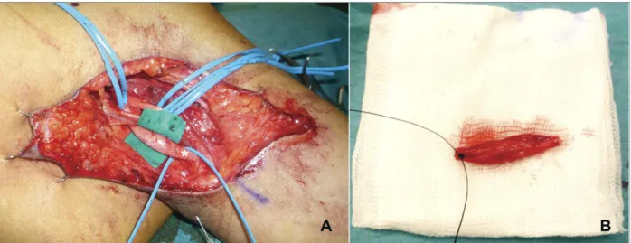
fIGure 3.Intraoperative view of a fusiform neurofibroma located at the origin of the ulnar nerve (marked with an asterisk). A) The tumor was located inside the nerve, without any cleavage plane with the healthy nerve tissue. Intraneural dissection proved to be impossible. B) The segment of the nerve involved by the tumor was excised
work of Desai, 12% of patients had been initially mis-diagnosed as having non-nerve tumors, and surgery with curative intent had been abandoned intraopera -tively in favor of lesion biopsy by referring doctors3.
Most primary BP tumors are benign2,3,9. More than
80% of these tumors are schwannomas or neurofibro-mas2,3,9. Other examples of benign primary BP tumors
are plexiform neurofibromas and ganglioneuromas2,3,9.
Primary malignant BP tumors correspond to less than 10% of all BP tumors, and are usually rapid growing, fixed lesions, associated with rapidly aggravating neu-rological deficits2. They include neurogenic sarcomas
and neurofibrosarcomas2,3,9. Secondary BP tumors are
more commonly Pancoast tumors or metastasis from the lung or breast2,3,9.
In our series, 4 patients had schwannomas and one patient had a neurofibroma. Most authors report a lar -ger proportion of neurofibromas over schwannomas2,5.
However, other series have also found a larger number of schwannomas, as we did3,4,9. For example, Desai in
a recent review of 115 benign brachial plexus tumors identified 70 schwannomas and 45 neurofibromas3.
Schwannomas are so named because they arise from Schwan cells within the endoneurium. As they grow, they progressively stretch the perineurium and epineurium that in turn gradually encapsulate the tu-mor. Hence, clinically, they tend to be tightly adherent to only one or two nerve fascicles sparing the remain-der of the nerve2,3,9. In contrast, the neurofibromas
originate from cells of the epineurium and are
there-fore not encapsulated. As these latter tumors grow, they come to involve most of the nerve fascicles, resulting in frequent neurological deficits after tumor re-moval2,3,9. Neurofibromas can be found to be solitary
or multiple2,3,9. The majority of patients with multiple
neurofibromas have neurofibromatosis2,3,9.
The preferred image test to visualize the tumor and its relations to neighborhood structures is MRI9.
How-ever, this test lacks accuracy in the distinction between primary benign and malignant BP tumors, and even in the distinction between schwannomas and neurofi-bromas9. If a malignancy is suspected, a positron
emis-sion tomography (PET) scan can be useful9. Recently,
there has been great interest in MRI neurography, as it allows visualization of the entire course of nerves and its relations with the tumor9,12-16. CT scan is the best
method to assess bony involvement2,3,9.
Regarding pre-operative electroneuromyography (EMG), most authors argue that it adds little to a com-plete medical history, physical examination and ima-giological examination3,17. However, other authors
ar-gue that EMG is useful in identifying subclinical deficits in involved BP elements and may even detect significant denervation usually associated with BP ma-lignant tumors4,6.
Fine-needle aspiration or core pre-therapeutic biop-sy is generally discouraged because these methods lack accuracy, and can cause iatrogenic neurological deficits and/or vascular injuries. Furthermore, they are known to induce significant fibrosis that will make definitive surgical treatment more difficult and hazardous. Fi-nally, there is the potential risk of tumor seeding along the needle track2,3,9.
After a presumptive diagnosis of BP tumor is made, the main problem is to decide to keep the patient un-der close observation, with regular visits to the clinic, or, on the contrary, to operate18. It is generally
accept-ed that this decision will be made basaccept-ed on the per-ceived risks and benefits of either option, according to patient’s wishes18. Disfigurement associated with
tu-mor growth, pain or progressive neurological deficits are usually considered indications for surgery. The lat-ter two signs are associated with a higher risk of ma-lignancy, particularly if a neurocutaneous syndrome is present2,3,18. Most authors suggest surgery if the lesion
is symptomatic or progressing in size2,3,12. If the tumor
is stationary and not associated with neurological dys-function a conservative approach can be taken2,3,12.
Most authors agree that complete extirpation of a schwannoma is more easily achieved than with neu-fIGure 4.Intraoperative view of the patient presented in
Figure 3. The nerve defect resulting from the nerve segment extirpation was reconstructed with 4 cables of autologous sural nerve graft.
rofibromas, because the latter tend to be more adherent to the core of the nerve, often times with a scant or absent cleavage plane3,18,19. Most authors argue
that in either case it is of utmost importance to preserve the largest number of functioning motor fibers, even if this means leaving small amounts of residual benign tumor or tumor capsule3.
Notwithstanding, it should be noted that surgery is far from being devoid of complications18. Besides
anes-thetic and wound healing problems, which are in-creasingly rare, 10 to 17% of all patients operated due to BP tumors present new neurological deficits of varia -ble severity postoperatively4,18. According to Kehoe et
al., for example, the number of neurological deficits doubled after surgery in a series of 15 patients20.
Other complication that has been described, although seldom encountered, is the intra-operative damage of major vascular structures, like the subclavian artery9.
Overall, surgery benefits have been shown to outweigh risks, and surgery is consensually recommended in se-lected patients2,3,9.
conclusIon
It is of utmost importance that every physician who deals with rheumatological patients is familiarized with BP tumors, in order to correctly diagnose them, through a targeted history and examination, combined with the use of appropriate ancillary tests. After establi -shing the diagnosis, the physician may opt to follow the patient or to refer the patient to a surgeon with ex-perience in this field, in order to minimize operative risks and to increase the odds of complete resection of the tumor3,21,22.
AcknowledGments
Diogo Casal received a grant from The Program for Advanced Me-dical Education, which is sponsored by Fundação Calouste Gul-benkian, Fundação Champalimaud, Ministério da Saúde e Funda-ção para a Ciência e Tecnologia, Portugal.
correspondence to Diogo Casal
Rua Luís Pastor de Macedo, N 32, 5D, 1750-159, Lisbon, Portugal
E-mail: diogo_bogalhao@yahoo.co.uk references
1. Courvoisier L. Die Neurome: Eine Klinische Monographie.: Ba-sel; 1886.
2. Das S, Ganju A, Tiel RL, Kline DG. Tumors of the brachial ple-xus. Neurosurg Focus 2007;22:E26.
3. Desai KI. Primary benign brachial plexus tumors: an experience of 115 operated cases. Neurosurgery 2012;70:220-33; discus-sion 33.
4. Siqueira MG, Martins RS, Teixeira MJ. Management of brachial plexus region tumours and tumour-like conditions: relevant diagnostic and surgical features in a consecutive series of eigh-teen patients. Acta Neurochir (Wien) 2009;151:1089-1098. 5. Kim DH, Murovic JA, Tiel RL, Moes G, Kline DG. A series of
397 peripheral neural sheath tumors: 30-year experience at Louisiana State University Health Sciences Center. J Neurosurg 2005;102:246-255.
6. Huang JH, Zaghloul K, Zager EL. Surgical management of bra-chial plexus region tumors. Surg Neurol 2004;61:372-378. 7. Kim DH, Murovic JA, Tiel RL, Moes G, Kline DG. A series of
146 peripheral non-neural sheath nerve tumors: 30-year ex-perience at Louisiana State University Health Sciences Center. J Neurosurg 2005;102:256-266.
8. Kim DH, Cho YJ, Tiel RL, Kline DG. Outcomes of surgery in 1019 brachial plexus lesions treated at Louisiana State Univer-sity Health Sciences Center. J Neurosurg 2003;98:1005-1016. 9. Binder DK, Smith JS, Barbaro NM. Primary brachial plexus tu-mors: imaging, surgical, and pathological findings in 25 pa-tients. Neurosurg Focus 2004;16:E11.
10. Artico M, Cervoni L, Wierzbicki V, D'Andrea V, Nucci F. Benign neural sheath tumours of major nerves: characteristics in 119 surgical cases. Acta Neurochir (Wien) 1997;139:1108-1016. 11. Das Gupta TK, Brasfield RD, Strong EW, Hajdu SI. Benign
soli-tary Schwannomas (neurilemomas). Cancer 1969;24:355-366. 12. Zhou L, Yousem DM, Chaudhry V. Role of magnetic resonan-ce neurography in brachial plexus lesions. Muscle Nerve 2004;30:305-309.
13. Gupta R, Villablanca PJ, Jones NF. Evaluation of an acute ner-ve compression injury with magnetic resonance neurography. J Hand Surg Am 2001;26:1093-1099.
14. Aagaard BD, Maravilla KR, Kliot M. Magnetic resonance neu-rography: magnetic resonance imaging of peripheral nerves. Neuroimaging Clin N Am 2001;11:viii, 131-146.
15. Chhabra A, Lee PP, Bizzell C, et al. High-resolution 3-Tesla mag-netic resonance neurography of musculocutaneous neuropa thy. J Shoulder Elbow Surg 2012;21:e1-6.
16. Filler A. Magnetic resonance neurography and diffusion tensor imaging: origins, history, and clinical impact of the first 50,000 cases with an assessment of efficacy and utility in a prospecti-ve 5000-patient study group. Neurosurgery 2009;65:A29-43. 17. Angelov L, Davis A, O'Sullivan B, Bell R, Guha A. Neurogenic sarcomas: experience at the University of Toronto. Neurosur-gery 1998;43:56-64; discussion -5.
18. Ball JR, Biggs MT. Operative steps in management of benign nerve sheath tumors. Neurosurg Focus 2007;22:E7. 19. Binder DK, Lu DC, Barbaro NM. Multiple root avulsions from
the brachial plexus. Case illustration. Neurosurg Focus 2005;19:E9.
20. Kehoe NJ, Reid RP, Semple JC. Solitary benign peripheral-ner-ve tumours. Review of 32 years' experience. J Bone Joint Surg Br 1995;77:497-500.
21. Pais D, Casal D, Santos A, Goyri-O'Neill J. A variation in the ori-gin of the median nerve associated with an unusual oriori-gin of the deep brachial artery. Brazilian Journal of Morphological Sciences 2010;27:35-38.
22. Bilsky MH, Vitaz TW, Boland PJ, Bains MS, Rajaraman V, Rusch VW. Surgical treatment of superior sulcus tumors with spinal and brachial plexus involvement. J Neurosurg 2002;97:301-309.