1. Clinic of Rheumatology, Medical University, Sofia 2. Clinical Center of Endocrinology and Gerontology, Medical University, Sofia
3. National Genetic Laboratory, USHATOG “Maichin dom”, Medical University, Sofia
immunostimulatory and antiapoptotic role1. A bidirec-tional association between the pineal gland and the mune system has been suggested based on the im-munomodulating features of melatonin and the pineal regulation by different lymphokines2. Moreover, dif-ferent immune cells and tissues are able to synthesize melatonin3. The hormone might act directly on im-munocompetent cells and it could influence the develop ment of autoimmune diseases as well as their clinical expression1. Circadian rhythm disturbances of the melatonin secretion were already described in pa-tients with rheumatoid arthritis4.
The effects of melatonin on immunocompetent cells and hematopoiesis are accomplished at least in part through its action on the specific melatonin receptors (reviewed in Pandi-Perumal et al. 2008)5. Experimental studies indicated that melatonin receptor type 1B (MTNR1B) could be involved in melatonin-induced enhancement of cellmediated and humoral immune res -ponse6. However, it is not clarified, if the single nu-cleotide polymorphisms of the MTNR1B gene might in-fluence the interactions between the melatonin and melatonin receptor as well as their complex effects on the immune system and autoimmunity. A significant as sociation was found between the MTNR1B rs1562444 polymorphism and the development of rheumatoid fac-tor positive rheumatoid arthritis in Korean patients7, while the role of MNTR1B polymorphisms in systemic lupus erythematosus (SLE) was not clarified.
Therefore, our study aimed to investigate the possi-ble role of MTNR1B gene polymorphisms rs1562444, rs10830962 and rs10830963 for the clinical expres-sion of SLE.
MAterIAls And Methods
subjects
Two hundred and ten Caucasian women were inclu -ded in the study. One hundred and nine patients (mean
Melatonin receptor 1b polymorphisms
in women with systemic lupus erythematosus
Tanev D1, Robeva R2, Andonova S3, Decheva V3, Tomova A2, Kumanov P2, Savov A3, Rashkov R1, Kolarov Z1
ACTA REUMATOL PORT. 2016;41:62-67
AbstrAct
Aim: The pineal hormone melatonin could exert an
important influence on the immune system and autoi mmu -nity. Its effect on the immunocompetent cells might be mediated at least partially through specific melatonin ceptors. However, the role of melatonin - me latonin re-ceptor 1B (MTNR1B) interrelations in human autoim-mune diseases is still unknown. Therefore, the present study aimed to investigate the possible influence of the MTNR1B gene polymorphisms for the development and clinical expression of systemic lupus erythematosus (SLE).
Methods: 109 female SLE patients and 101 healthy
women were genotyped for the MTNR1B rs1562444, rs10830962 and rs10830963 polymorphisms.
Results: No genotype distribution differences were
found between patients and controls. The presence of MTNR1B rs10830963 C/C genotype was related to in-creased prevalence of leucopenia compared to genotypes C/G and G/G after Bonferroni correction for multiple comparisons [36.5% vs. 14.5%, p=0.014]. Moreo ver, the rs10830963 G/G carriers had lower number of lupus criteria in comparison to patients with C/C genotype.
Conclusions: The present data suggested that MTNR1B
polymorphisms could influence the clinical features in lu-pus patients, and especially the susceptibility to leucopenia.
Keywords: Genetic association; Melatonin receptor 1B;
Polymorphisms; Systemic lupus erythematosus; Leu-copenia, Myelosupression.
IntroductIon
age 41.72±11.71 years [20-67]) were recruited from the Department of Rheumatology. They fulfilled the modified 1997 American College Rheumatology (ACR) classification criteria for systemic lupus erythe-matosus8. The Systemic Lupus International Collabo-rating Clinics/ACR (SLICC) index9were determined by one rheumatologist (D.T.). All women underwent a complete general assessment and the presence of the lupus features such as malar rash, discoid rash, pho-tosensitivity, oral ulcer, non-erosive arthritis, serositis, renal disorder, neurological disorder, hematological disorder (including presence of anemia, leucopenia, lymphopenia or thrombocytopenia), immunological disorder (including positive anti-DNA antibodies, posi tive anti-Smith antibodies or positive finding of antiphospholipid antibodies) as well as the presence of antinuclear antibodies were registered. The previous and current medication with corticosteroids and im-munosuppressors such as cyclophosphamide, aza-thioprine, and methotrexate was registered.
One hundred and one age matched controls (mean age 39.36±11.97 years [22-68]) were collected from the medical staff and students. They were all clinically healthy women without connective tissue diseases. The experimental protocol was explained to all participants and written informed consent was obtained. The study was approved by the institutional ethic commission. MelAtonIn receptor 1b polyMorphIsMs All participating women provided peripheral blood samples for DNA. Genotyping was performed by PCR-RFLP analysis. The three different regions rs1562444, rs10830962 and rs10830963 were amplified by PCR in three reactions. Each PCR was performed in a total vo lume of 15 µl containing 2.0 mmol/L MgCl2, 0.5U Pri me Taq DNA polymerase with the appropriate buffer (GenetBio, Korea) and 0.2 pmol/µl of each of the primers: rs10830962: F 5’–TACTAGATATTAGCTGTGTGCTAGT-GACT–3’/ R 5’ TCTGGGCAACTCAGTGAAACC–3’; rs10830963: F 5’–ATGCTAAGAATTCACACCAGCT-3’/ R 5’–CACAGTGCAGACTGTTTTCTAATC–3’; rs1562444: F 5’–GAAAACACTCTTGGTGGTGTCTT–3’/ R 5’-GATGTGGTGGCTATGTGTGTGTGTA-3’.
Thermal cycling was performed with initial denatu -ration 95°C for 7 min, followed by 33 cycles of 95°C for 30 sec/ 60°C for 30 sec/ 72°C for 60 sec (for rs10830962); 95°C for 30 sec/ 54°C for 30 sec/ 72°C for 30 sec (for rs10830963); 95°C for 30 sec/ 60°C for
gation step was 7 minutes at 72°C. PCR products for rs10830962, rs10830963 and rs1562444 were di-gested with restriction endonucleases - HinfI, PvuII and NlaIII (New England BioLabs Inc, USA), respecti -vely. The digested products were analyzed on 2.5% agarose gel stained with ethidium bromide. Since the G to C (rs10830962), C to G (rs10830963) and A to G (rs1562444) transitions create an endonuclease recognition site, the PCR fragment following enzyme digestion reveals two types of alleles. The absence of res triction site for rs10830962 G/C referred to allele G (210 bp) and the presence of restriction site - referred to allele C (with 184 bp and 26 bp fragments). For rs10830963 C/G polymorphism the sizes of detected alleles were 105 bp and 20 bp (allele G) and 125 bp (allele C). The amplification region of 400 bp for rs1562444 A/G was digested with NlaIII restriction endonuclease and two fragments were revealed – 319 bp and 81 bp. The A to G transition creates additio nal NlaIII restriction site. The 2.5% agarose gel electro -phoresis reveals three different patterns of genotypes: G/G (with 319 bp and 81 bp bands), G/A (with 319 bp, 163 bp, 156 bp and 81 bp) and A/A (163 bp, 156 bp and 81 bp). Several randomly selected samples were sequenced and their sequence identities were confir -med. The distribution of all investigated genotypes in healthy females was in agreement with the Hardy--Weinberg equilibrium. The genetic team was not aware of any clinical data concerning SLE patients. stAtIstIcAl AnAlysIs
The results were presented as mean ±SD (median) for continuous variables or as a frequency (%) for dichotomou s variables. Categorical data were analyzed through χ2 test or Fisher’s exact test. After a Kol-mogorov–Smirnov test for normality of the distribution differences between two groups were established with an independent ttest or MannWhitney test. Compari -sons between three groups were calculated through one-way ANOVA with post hoc Bonferroni test (equal varian ces assumed) and Tamhane’s T2 test (unequal variances assumed) or non-parametric Kruskal-Wallis test according to normality of the distribution. All re-sults were considered significant at the 0.05 level. Lo-gistic regression analysis was used where appropriate. The Bonferroni adjustment for multiple testing was ap-plied and the significance of the p value was set at 0.017 (0.05/3 considering the three investigated polymor -phisms). Statistical analysis was conducted through
3.134; 95CI [1.035 – 9.486], p=0.043) in comparison to those with G/G genotype (Table II). The presence of C/C genotype was related to increased prevalence of leucopenia compared to both genotypes containing G allele (G/G and G/C) [38.2% vs. 17.9%, p=0.031]. C/C genotype increased the risk for leucopenia develop-ment (OR 2.837; 95CI [1.117 – 7.205], p=0.028). C/C
results
A total of 100 healthy women and 106 female patients were genotyped for the single nucleotide polymor-phism rs1562444 in the melatonin receptor type 1B gene. No significant differences in the genotype frequencies of patients and controls were observed (Figu -re 1). Considering clinical characteristics of the SLE pa-tients, the rs1562444 polymorphism was found to be related to the development of leucopenia, while no other relations with ACR criteria were established (Table I). Patients with G/G genotype had increased risk for leucopenia development in comparison to A/A carriers (OR 3.771; 95CI [1.135 – 12.533], p=0.030), but the results were not significant after the Bonferroni correction for multiple testing.
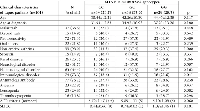
A group of 95 healthy women and 101 female pa-tients was genotyped for the single nucleotide poly-morphism rs10830962 in the melatonin receptor type 1B gene. No significant differences in the genotype distribution of patients and controls were observed (Figu -re 2). Patients with rs10830962 C/C genotype had in-creased risk for immunological disturbances (OR
tAble I. Mtnr1b rs1562444 Genotype dIstrIbutIon In sle pAtIents wIth dIFFerent clInIcAl chArActerIstIcs
MTNR1B rs1562444 genotypes Clinical characteristics of N AA AG GG
lupus patients (n=106) (% of all) n=31 (29.2) n=54 (50.9) n=21(19.9) P
Age 44.19±10.15 41.22±12.17 38.67±12.17 0.235 Age at diagnosis 38.52±9.46 32.67±13.17 32.43±11.00 0.068 Malar rash 40 (37.7) 11 (27.5) 23 (57.5) 6 (15.0) 0.520 Discoid rash 15 (14.2) 6 (40.0) 5 (33.3) 4 (26.7) 0.263 Photosensitivity 76 (71.7) 26 (34.2) 35 (46.1) 15 (19.7) 0.181 Oral ulcers 24 (22.6) 9 (37.5) 9 (37.5) 6 (25.0) 0.339 Non-erosive arthritis 104 (98.1) 30 (28.8) 53 (51.0) 21 (20.2) 1.000 Serositis 18 (17.0) 5 (27.8) 10 (55.6) 3 (16.7) 1.000 Renal disorder 27 (25.5) 7 (25.9) 15 (55.6) 5 (18.5) 0.915 Neurological disorder 35 (33.0) 10 (28.6) 17 (48.6) 8 (22.9) 0.825 Haematological disorder 67 (63.2) 19 (28.4) 32 (47.8) 16 (23.9) 0.382 Immunological disorder 78 (73.6) 21 (26.9) 40 (51.3) 17 (21.8) 0.616 Antinuclear antibody 81 (76.4) 25 (30.9) 41 (50.6) 15 (18.5) 0.753 Anaemia 24 (22.6) 9 (37.5) 12 (50.0) 3 (12.5) 0.481 Leucopenia 26 (24.5) 7 (26.9) 8 (30.8) 11 (42.3) 0.005 Thrombocytopenia 16 (15.1) 7 (43.8) 6 (37.5) 3 (18.8) 0.373
ACR criteria (number) 5.52±1.41 (5) 5.18±1.06 (5) 5.57±1.63 (5) 0.662
SLICC 0.90±1.24 (0) 0.65±0.89 (0) 0.71±1.06 (0) 0.830
Healthy womenSLE patients100%80%60%40%20%0%
MTNR1B rs1562444GGAGAAGGAGAA
Healthy women SLE patients
100% 80% 60% 40% 20% 0% MTNR1B rs1562444 GG AG AA GG AG AA
FIGure 1.MTNR1B rs1562444 genotypes in patients with SLE and healthy controls (p>0.05)
carriers had increased number of ACR criteria com-pared to G allele carriers [5.79±1.47 (5.5) vs. 5.07±1.09 (5), p=0.017]. However, after the Bonferro -ni correction for multiple testing the present results were not statistically significant.
A total of 100 healthy women and 107 SLE patients
were genotyped for MTNR1B rs10830963. No signifi -cant differences in the genotype frequencies of patients and controls were established (Figure 3). The number of ACR criteria was significantly lower in G/G patients (Table III). The presence of C/C genotype was rela ted to increased prevalence of leucopenia compared to genotypes C/G and G/G [36.5% vs. 14.5%, p=0.014]. C/C genotype increased significantly the risk for leu-copenia development (OR 3.383; 95CI [1.324 – –8.645], p=0.011). Similar results were obtained after adjustment for age and immunosuppressive treatment with cyclophosphamide, methotrexate and azathio-prine (OR 3.947; 95CI [1.437 – 10.845], p=0.008). The prevalence of hematological disturbances was in-creased in C/C genotype lupus patients (75.0% vs. 52.7%, p=0.026) compared to G allele carriers, but the results were not significant after Bonferroni correction for multiple testing.
dIscussIon
The present study showed that the MTNR1B
poly-tAble II. Mtnr1b rs10830962 Genotype dIstrIbutIon In sle pAtIents wIth dIFFerent clInIcAl chArActerIstIcs
MTNR1B rs10830962 genotypes
Clinical characteristics N CC GC GG
of lupus patients (n=101) (% of all) n=34 (33.7) n=38 (37.6) n=29 (28.7) p
Age 38.44±12.21 42.26±10.39 44.45±12.38 0.117 Age at diagnosis 31.53±12.63 34.92±10.93 37.21±13.20 0.180 Malar rash 37 (36.6) 10 (27.0) 14 (37.8) 13 (35.1) 0.448 Discoid rash 15 (14.9) 6 (40.0) 4 (26.7) 5 (33.3) 0.642 Photosensitivity 72 (71.3) 22 (30.6) 27 (37.5) 23 (31.9) 0.489 Oral ulcers 22 (21.8) 11 (50.0) 6 (27.3) 5 (22.7) 0.239 Non-erosive arthritis 99 (98.0) 33 (33.3) 37 (37.4) 29 (29.3) 1.000 Serositis 15 (14.9) 7 (46.7) 6 (40.0) 2 (13.3) 0.337 Renal disorder 26 (25.7) 12 (46.2) 7 (26.9) 7 (26.9) 0.266 Neurological disorder 32 (31.7) 13 (40.6) 12 (37.5) 7 (21.9) 0.507 Haematological disorder 65 (64.4) 26 (40.0) 21 (32.3) 18 (27.7) 0.162 Immunological disorder 74 (73.3) 27 (36.5) 31 (41.9) 16 (21.6) 0.041 Antinuclear antibody 77 (76.2) 29 (37.7) 26 (33.8) 22 (28.6) 0.238 Anaemia 23 (22.8) 9 (39.1) 6 (26.1) 8 (34.8) 0.437 Leucopenia 25 (24.8) 13 (52.0) 6 (24.0) 6 (24.0) 0.082 Thrombocytopenia 16 (15.8) 4 (25.0) 9 (56.3) 3 (18.7) 0.298
ACR criteria (number) 5.79±1.47 (5.5) 5.05±1.11 (5) 5.10±1.08 (5) 0.060
SLICC 0.44±0.66 (0) 0.74±0.82 (1) 1.07±1.46 (1) 0.181
Healthy womenSLE patients
100%80%60%40%0%
MTNR1B rs10830962 GGGCCCGGGCCC
70%90%50%30%20%10%
Healthy women SLE patients 100% 80% 60% 40% 0% MTNR1B rs10830962 GG GC CC GG GC CC 70% 90% 50% 30% 20% 10%
FIGure 2.MTNR1B rs10830962 genotypes in patients with SLE and healthy controls (p>0.05)
morphisms rs1562444, rs10830962 and rs10830963 were not related to the development of SLE in women. However, an interesting finding was the significant as-sociation between MTNR1B polymorphisms and the susceptibility for leucopenia development. Patients with rs1562444 GG, rs10830962 CC and rs10830963
CC genotypes were at increased risk for leucocytes number decrease during the lupus flare but after Bon-ferroni correction for multiple comparisons only rs10830963 CC genotype remained as significant risk factor. The leucopenia in lupus could be caused by the presence of autoantibodies, complement cascade dysfunction, bone marrow suppression and splenic poo -ling10. The results suggested that melatonin receptor 1B and its polymorphisms might have an important role for the human myelopoesis or leukocyte survival in vivo. Melatonin administration in animal models pro-tected bone marrow from the damaging effects of cy-totoxic drugs and stimulated bone marrow cells mito-sis11,12. Moreover, recent studies demonstrated that the pineal hormone could reduce apoptosis in human leu-cocytes13,14. Espino et al. studied the underlying mecha -nisms and found that, besides its antioxidant actions, melatonin probably required membrane receptor MTNR1A/MTNR1B interaction in order to counteract the TNF-alpha-stimulated human leukocyte apopto-sis15. According to Lissenko et al. MTNR1B rs10830963 G allele carriers showed higher expression of MTNR1B in pancreas islet cells in comparison to carriers of the
tAble III. Mtnr1b rs10830963 Genotype dIstrIbutIon In sle pAtIents wIth dIFFerent clInIcAl chArActerIstIcs
MTNR1B rs10830963 genotypes
Clinical characteristics N CC CG GG
of lupus patients (n=107) (% of all) n=52 (48.6) n=45 (42.1) n=10 (9.3) p
Age 39.67±11.98 43.56±10.62 44.10±14.07 0.212 Age at diagnosis 32.33±11.96 35.69±11.76 39.40±14.88 0.162 Malar rash 42 (39.3) 23 (54.8) 16 (38.1) 3 (7.1) 0.589 Discoid rash 15 (14.0) 7 (46.7) 5 (33.3) 3 (20.0) 0.268 Photosensitivity 76 (71.0) 34 (44.7) 36 (47.4) 6 (7.9) 0.170 Oral ulcers 24 (22.4) 15 (62.5) 8 (33.3) 1 (4.2) 0.335 Non-erosive arthritis 105 (98.1) 51 (48.6) 44 (41.9) 10 (9.5) 1.000 Serositis 18 (16.8) 12 (66.7) 6 (33.3) 0 (0.0) 0.180 Renal disorder 29 (27.1) 17 (58.6) 9 (31.0) 3 (10.4) 0.362 Neurological disorder 35 (32.7) 18 (51.4) 17 (48.6) 0 (0.0) 0.053 Hematological disorder 68 (63.6) 39 (57.4) 24 (35.3) 5 (7.4) 0.052 Immunological disorder 79 (73.8) 42 (53.2) 31 (39.2) 6 (7.6) 0.237 Antinuclear antibody 82 (76.6) 42 (51.2) 32 (39.0) 8 (9.8) 0.526 Anaemia 24 (22.4) 12 (50.0) 9 (37.5) 3 (12.5) 0.789 Leucopenia 27 (25.2) 19 (70.4) 6 (22.2) 2 (7.4) 0.022 Thrombocytopenia 16 (15.0) 6 (37.5) 9 (56.2) 1 (6.3) 0.522
ACR criteria (number) 5.79±1.40 (6) 5.09±1.16 (5) 4.60±0.70 (4.5) 0.005
SLICC 0.56±0.96 (0) 0.87±1.14 (0) 0.90±0.88 (1) 0.162
Healthy womenSLE patients
100%0%
MTNR1B rs10830963 GGCGCCGGGCCC
80%70%90%60%50%40%30%20%10%
Healthy women SLE patients 100% 0% MTNR1B rs10830963 GG CG CC GG GC CC 80% 70% 90% 60% 50% 40% 30% 20% 10%
FIGure 3.MTNR1B rs10830963 genotypes in patients with SLE and healthy controls (p>0.05)
C allele16. A potential increase of MNTR1B expression in leukocytes amplifying the melatonin signal could protect lupus rs10830963 G carriers from leucopenia. Leukocytes number decrease might be associated not only with the lupus disease, but also with the SLE treatment10. The main limitation of our study was the fact, that almost all patients had conducted long-term treatment with immunosuppressive drugs and corti-costeroids that could had influenced the bone marrow function. However, the association between leucopenia and MTNR1B polymorphism was still present after adjus tment for immunosuppressive treatment.
The MTNR1B rs10830962 and rs10830963 poly-morphisms have been predominantly investigated in the context of metabolic disorders16-18. MTNR1B rs10830962 and rs10830963 G alleles were associa ted with reduced insulin secretion, increased fasting plas-ma glucose concentrations and increased risk for type 2 diabetes in different populations16-18. To the best of our knowledge this is the first study investigating the relationships between systemic lupus erythematosus and MTNR1B polymorphisms. In conclusion, our re-sults showed that melatonin receptor 1B polymor-phisms could affect the clinical expression of SLE in women, and especially the development of leucopenia. Further studies are needed to reveal the significance of MTNR1B receptor polymorphisms for bone marrow function in lupus patients from other ethnic groups and for women with myelosuppression due to diffe -rent causes.
AcknowledGeMent:
This study was financially supported by the Medical University So-fia (Grant Nº 26/2010, Grant Nº 59/2011).
correspondence to
Ralitsa Robeva
Clinical Center of Endocrinology and Gerontology Medical University - Sofia, 2, Zdrave Str., Sofia 1431 Bulgaria
E-mail: [email protected]
reFerences
1. Maestroni GJ, Cardinali DP, Esquifino AI, Pandi-Perumal SR. Does melatonin play a disease-promoting role in rheumatoid arthritis? J Neuroimmunol 2005; 158: 106-111.
2. Maestroni GJ. The immunoneuroendocrine role of melatonin. J Pineal Res 1993; 14: 1-10.
3. Gómez-Corvera A, Cerrillo I, Molinero P, Naranjo MC, Lardo-ne PJ, Sanchez-Hidalgo M, Carrascosa-Salmoral MP, Medrano-Campillo P, Guerrero JM, Rubio A. Evidence of immune system melatonin production by two pineal melatonin deficient mice, C57BL/6 and Swiss strains. J Pineal Res 2009; 47: 15-22.
tered circadian rhythms in rheumatoid arthritis patients play a role in the disease’s symptoms. Autoimmun Rev 2005; 4: 497--502.
5. Pandi-Perumal SR, Trakht I, Srinivasan V, Spence DW, Maes-troni GJ, Zisapel N, Cardinali DP. Physiological effects of mela-tonin: role of melatonin receptors and signal transduction path-ways. Prog Neurobiol 2008; 85: 335-353.
6. Drazen DL, Nelson RJ. Melatonin receptor subtype MT2 (Mel 1b) and not MT1 (Mel 1a) is associated with melatonin-indu-ced enhancement of cell-mediated and humoral immunity. Neu-roendocrinology 2001; 74: 178–184.
7. Ha E, Choe BK, Jung KH, Yoon SH, Park HJ, Park HK, Yim SV, Chung JH, Bae HS, Nam M, Baik HH, Hong SJ. Positive rela-tionship between melatonin receptor type 1B polymorphism and rheumatoid factor in rheumatoid arthritis patients in the Korean population. J Pineal Res 2005; 39: 201-205.
8. Hochberg MC. Updating the American College of Rheumato-logy revised criteria for classification of systemic lupus erythe-matosus. Arthritis Rheum 1997; 40: 1725.
9. Stoll T, Seifert B, Isenberg DA. SLICC/ACR Damage Index is va-lid, and renal and pulmonary organ scores are predictors of se-vere outcome in patients with systemic lupus erythematosus. Br J Rheumatol 1996; 35: 248-254.
10. Cervera R, Espinosa G, D’Cruz D. Systemic lupus erythemato-sus: pathogenesis, clinical manifestations and diagnosis. In: Bi-jlsma JW (ed) EULAR Compendium on Rheumatic Diseases. BMJ Publishing Group and European League Against Rheuma-tism, 2009, 257-268.
11. Anwar MM, Mahfouz HA, Sayed AS. Potential protective effects of melatonin on bone marrow of rats exposed to cytotoxic drugs. Comp Biochem Physiol A Mol Integr Physiol, 1998; 119: 493--501.
12. Ferreira SG, Peliciari-Garcia RA, Takahashi-Hyodo SA, Rodri-gues AC, Amaral FG, Berra CM, Bordin S, Curi R, Cipolla-Neto J. Effects of melatonin on DNA damage induced by cyclophos-phamide in rats. Braz J Med Biol Res 2013; 46: 278-286. 13. Espino J, Bejarano I, Redondo PC, Rosado JA, Barriga C, Reiter
RJ, Pariente JA, Rodríguez AB. Melatonin reduces apoptosis in-duced by calcium signaling in human leukocytes: Evidence for the involvement of mitochondria and Bax activation. J Membr Biol 2010; 233: 105-118.
14. Espino J, Bejarano I, Paredes SD, Barriga C, Rodríguez AB, Pa-riente JA. Protective effect of melatonin against human leukocy-te apoptosis induced by intracellular calcium overload: relation with its antioxidant actions. J Pineal Res 2011; 51: 195-206. 15. Espino J, Rodríguez AB, Pariente JA. The inhibition of
TNF-α-induced leucocyte apoptosis by melatonin involves membrane receptor MT1/MT2 interaction. J Pineal Res 2013; 54: 442-452. 16. Lyssenko V, Nagorny CL, Erdos MR, Wierup N, Jonsson A, Spé-gel P, et al. Common variant in MTNR1B associated with in-creased risk of type 2 diabetes and impaired early insulin se-cretion. Nat Genet 2009; 41: 82-88.
17. Staiger H, Machicao F, Schäfer SA, Kirchhoff K, Kantartzis K, Guthoff M, Silbernagel G, Stefan N, Häring HU, Fritsche A. Po-lymorphisms within the novel type 2 diabetes risk locus MTNR1B determine beta-cell function. PLoS One 2008; 3: e3962.
18. Rönn T, Wen J, Yang Z, Lu B, Du Y, Groop L, Hu R, Ling C. A common variant in MTNR1B, encoding melatonin receptor 1B, is associated with type 2 diabetes and fasting plasma glucose in