UNIVERSIDADE DE LISBOA
FACULDADE DE CIÊNCIAS
DEPARTAMENTO DE QUÍMICA E BIOQUÍMICA
F
UNCTIONALC
HARACTERIZATION OFP
ROTEINSI
NTERACTING ORM
ODULATINGCFTR
ANDEN
AC
T
RAFFIC ANDA
CTIVITYDiana Delgado Faria
DOUTORAMENTO EM BIOQUÍMICA (Especialidade: Genética Molecular)
UNIVERSIDADE DE LISBOA
FACULDADE DE CIÊNCIAS
DEPARTAMENTO DE QUÍMICA E BIOQUÍMICA
F
UNCTIONALC
HARACTERIZATION OFP
ROTEINSI
NTERACTING ORM
ODULATINGCFTR
ANDEN
AC
T
RAFFIC ANDA
CTIVITYDiana Delgado Faria
Tese co-orientada pela Prof. Doutora Margarida D. Amaral (FCUL) e
pelo Prof. Doutor Karl Kunzelmann (Uni. Regensburg), especialmente
elaborada para a obtenção do grau de doutor em Bioquímica,
especialidade de Genética Molecular
Diana Delgado Faria foi bolseira de Doutoramento da Fundação para a Ciência e Tecnologia do Ministério da Ciência, Tecnologia e Ensino Superior SFRH / BD / 43313 / 2008
De acordo com o disposto no artigo 40° do Regulamento de Estudos Pós-Graduados da Universidade de Lisboa, Deliberação n°961/2003, publicada no Diário da República – IIa Série, n° 153 de 5 de Julho de 2003, foram incluídos nesta tese resultados dos artigos abaixo indicados:
Faria D, Dahimène S, Alessio L, Scott-ward T, Schreiber R, Kunzelmann K, Amaral MD. (2011) Effect of Annexin A5 on CFTR: Regulated Traffic or Scaffolding? Mol Membr Biol, 28(1): 14-29.
Faria D, Schreiber R, Kunzelmann K. (2009) CFTR is activated through stimulation of purinergic P2Y2 receptors. Pflügers Arch, 457: 1373-80.
Faria D, Lentze N, Almaça J, Luz S, Alessio L, Tian Y, Martins JP, Cruz P, Schreiber R, Farinha CM, Auerbach D, Amaral MD, Kunzelmann, K. (2012) Differential regulation of biogenesis of ENaC and CFTR by the stress response protein SERP1. Pflügers Arch, 463(6):819-27
Almaça J*, Faria D*, Sousa M, Conrad C, Sirianant L, Reiss M, Clarke LA, Martins, JP, Santos M, Heriché J-K, Huber W, Schreiber R, Pepperkok R, Kunzelmann K, Amaral MD. (2012) High-content siRNA screen reveals CNTFR as a novel ENaC regulator and DAG kinase as a robust therapeutic target for cystic fibrosis. Submitted to Cell (* equal contribution)
No cumprimento do disposto na referida deliberação, a autora esclarece- serem da sua responsabilidade, excepto quando referido em contrário, a execução das experiências que permitiram a elaboração dos resultados apresentados, assim como a interpretação e discussão dos mesmos. Os resultados obtidos por outros
autores (com menção nas respetivas legendas) foram incluídos com autorização dos mesmos para facilitar a compreensão dos trabalhos.
Outros artigos publicados em revistas internacionais contendo resultados obtidos durante o doutoramento:
Martins JR*, Faria D*, Kongsuphol P*, Reisch B, Schreiber R, Kunzelmann K. (2011) Anoctamin 6 is an essential component of the outwardly rectifying chloride channel. Proc Natl Acad Sci USA, 108 (44):18168-72. (* equal contribution)
Tian Y, Schreiber R, Kongsuphol P, Sousa M, Uliyakina I, Palma M, Faria D, Traynor-Kaplan AE, Fragata JI, Amaral MD and Kunzelmann K. (2012) Control of TMEM16A by INO-4995 and other inositolphosphates. British J Pharmacology. Epub ahead of print.
Kunzelmann K, Tian Y, Martins JR, Faria D, Kongsuphol P, Ousingsawat J, Wolf L, Schreiber R. (2012) Airway epithelial cells-Functional links between CFTR and anoctamin dependent Cl- secretion. Int J Biochem Cell Biol, 44(11):1897-1900.
Kunzelmann K, Schreiber R, Kmit A, Jantarajit W, Martins JR, Faria D, Kongsuphol P, Ousingsawat J, Tian Y. (2012) Expression and function of epithelial anoctamins. Exp. Physiol, 97(2):184-92.
Kunzelmann K, Tian Y, Martins JR, Faria D, Kongsuphol P, Ousingsawat J, Thevenod F, Roussa E, Rock J, Schreiber R. (2011) Anoctamins. Pflügers Arch. 462(2):195-208
Acknowlegments/Agradecimentos
A
CKNOWLEDGMENTS/
A
GRADECIMENTOSÀ Professora Doutora Margarida Amaral, por me ter aceitado no seu laboratório e pela confiança em mim depositada. Agradeço por me ter proporcionado as bases necessárias para eu iniciar este doutoramento, e também pela supervisão, e empenho dedicados que foram fundamentais para a realização deste trabalho.
I would like to thank Professor Karl Kunzelmann for accepting me in his lab in Regensburg and for all his dedication to the work I developed there. I am grateful for his guidance and support. The knowledge and experience that I have gain made me also grown more as a person.
Ao ministério da Ciência e Ensino Superior e à Fundação para a Ciência e a Tecnologia, por terem possibilitado a realização deste trabalho, através da concessão da bolsa de doutoramento de que fui recipiente de 2008 a 20012.
I thank all the collegues that contributed with their work to the development of my thesis and that allowed their results to be included here.
A PhD is like a long journey with ups and downs that tests your strength and determination. What made it quite an enjoyable one were the people that surrounded me during this time. I am truly grateful to all my colleagues in the lab that have become friends.
Obrigada a todas as pessoas do laboratório de Lisboa com quem eu convivi no início desta jornada e que se mantiveram sempre presentes durante a minha estadia em Regensburg: à querida Marta por todo o seu apoio e boa disposição e à sempre enérgica Marisa. À Inna e Joana de quem eu tenho saudades pelos tempos passados em Regensburg. Aos bem dispostos Simão e Mário, to Toby for all his initial help, Filipa, Anabela, Ana Carina, André, Luka e Carlos. Obrigada também pela vossa contribuição para este trabalho.
To everyone in and outside the lab in Regensburg with whom I shared so many good moments during my PhD: dear Ji, Brigitte, Julia, Tina, Tini and Fadi thank you for your friendship and for the help and fun I had in the lab. Thank you Rainer, for
all your help, patience and guidance in the lab and for the valuable scientific discussions. Um grande beijinho à Ana Marta e ao Arthur por toda a alegria que trouxeram ao lab. A big Thanks! to my cuban friends René, David and Nesty for being part of my “family” in Regensburg. To Aim and Joana (Raquel) for being my “sisters” in Regensburg, for always being there for me...this wouldn’t have been the same without you!
Aos meus amigos de sempre e à minha querida família, por me apoiarem, por estarem lá para mim quando preciso de desabafar. É sempre bom reencontrar-vos quando vou a Portugal, obrigada por apesar da distância continuarem por perto.
Aos meus pais que eu tanto amo, esta tese é vossa também! Obrigada por toda a força que me transmitem sempre, por todo o vosso incansável apoio, dedicação e confiança!
Table of Contents
T
ABLE OFC
ONTENTS ACKNOWLEDGMENTS /AGRADECIMENTOS ... ix SUMMARY ...xv RESUMO xvii ABBREVIATIONS ...xxi I General Introduction ... 11 Cystic Fibrosis: Overview ... 1
2 CFTR ... 2
2.1 CFTR Structure, Function and Trafficking ... 2
3 CFTR Functions in Epithelia ... 6
3.1 CFTR Activity as a Cl- Channel ... 6
3.2 CFTR as Regulator of Epithelial Ion Transport ... 6
4 Epithelial Na+ Channel (ENaC)... 8
4.1 ENaC: Structure and Function ... 8
4.2 Nedd4-2 and ENaC Ubiquitination ...10
4.3 ENaC Regulatory Pathways ...11
4.3.1 Hormonal Regulation of ENaC ... 11
4.3.2 Regulation of ENaC activity by Proteolytic Cleavage. ... 13
4.3.3 Role of PIP2 and PIP3 in regulating ENaC ... 14
5 Cystic Fibrosis Therapeutic Approaches ...15
5.1 Rescuing CFTR ...15
5.2 The Bypass Approach ...17
II Objectives of the Present Work...21
III Materials and Methods ...25
1 Cell Culture ...25
1.1 Mammalian cell lines and Culture conditions ...25
1.2 Transfections ...26
2 Molecular Biology ...27
2.1 Plasmids and siRNA ...27
2.2 RNA Extraction, Reverse Transcription and Real-Time PCR ...27
Summary
xii
4.1 Immunocytochemistry ...28
4.2 Chemiluminescence ...30
4.3 Western blot and Antibodies ...30
4.4 Co-Immunoprecipitation ...31 4.5 Affinity Chromatography ...32 4.6 2D-Gels ...33 5 Functional Analysis ...33 5.1 Patch Clamp ...33 5.2 Ussing Chamber ...34
5.3 cRNA and Double Electrode Voltage Clamp ...34
5.4 Microscopic FMP Assay ...35
6 Chemicals and statistical analysis ...36
IV Results and Discussion ...37
Part I Effects of Annexin A5 on CFTR: Regulated Traffic or Scaffolding? ...39
1 Abstract ...41
2 Introduction ...41
3 Results and Discussion ...43
3.1 Annexin A5 inhibits CFTR in Xenopus Oocytes ...43
3.2 Annexin A5 Inhibits Membrane Trafficking of CFTR in Xenopus Oocytes ....45
3.3 Regulation of CFTR by Annexin A5 in Mammalian Cells ...48
3.4 Effect of Annexin A5 on F508del -CFTR ...51
3.5 Do Annexin A5 and CFTR Interact in Vivo? ...51
3.6 Effect of Annexin A5 on Endogenous CFTR Expression in Physiologically Relevant Cellular Systems ...56
4 Discussion ...57
4.1 Annexin A5 inhibits CFTR in Oocytes ...57
4.2 Enhancement of CFTR Function by Annexin A5 in Mammalian cells ...58
4.3 Does the Stabilizing Effect of Annexin A5 on CFTR Result from Direct Interaction? ...59
5 Supplementary Data ...62
Part II CFTR is Activated Through Stimulation of Purinergic P2Y2 Receptors ...67
1 Abstract ...69
Table of Contents
3 Results ...70
3.1 Activation of CFTR by stimulation of P2Y2 receptors in Xenopus oocytes ...70
3.2 Activation of CFTR by P2Y2 depends on phospholipase C and an unknown downstream kinase ...74
3.3 P2Y2 dependent activation of CFTR in human airway epithelial cells ...76
4 Discussion ...79
Part III Regulation of ENaC Biogenesis by the Stress Response Protein SERP1 81 1 Abstract ...83
2 Introduction ...83
3 Results ...84
3.1 SERP1 Interacts and Co-localizes with βENaC in Airway Cells ...84
3.2 SERP1 Regulates ENaC ...87
3.3 SERP1 Inhibits Trafficking of ENaC ...91
3.4 Nedd4-2-Independent Inhibition of ENaC ...91
3.5 SERP1 does not Suppress Expression of CFTR ...93
4 Discussion ...95
4.1 SERP1 Inhibits Biogenesis of ENaC ...95
4.2 Hypoxic Inhibition of ENaC ...96
4.3 SERP1 Activates CFTR ...97
5 Supplementary Datta ...97
Part IV High-content siRNA screen reveals CNTFR as a novel ENaC regulator and DAG kinase as a robust therapeutic target for cystic fibrosis ...101
1 Abstract ...103
2 Introduction ...103
3 Results ...106
3.1 DGK as a Novel Regulator of ENaC. ...106
3.2 CNTFR as a Novel Regulator of ENaC ...112
3.3 Validation of DGK as Drug Target for ENaC in Human CF Airways ...114
4 Discussion ...116
V General Discussion, Conclusions and Future Perspectives ...123
Summary
S
UMMARYCystic Fibrosis (CF) is a genetic disease that leads to airways obstruction and progressive impairment of lung function which is the main cause of morbidity and mortality. It is caused by mutations in the gene encoding the Cystic Fibrosis Transmembrane Conductance Regulator (CFTR) protein, which, in CF, has impaired function and/or expression. CFTR is a cAMP-dependent, PKA-regulated Cl- channel expressed at the apical membrane of epithelial cells, where it is required to control ion and fluid homeostasis on epithelial surfaces. Transepithelial ion transport is crucial for hydration and protection against infection of epithelial tissues. CFTR is also a regulator of other ion channels relevant for CF disease such as the amiloride-sensitive epithelial Na+ channel (ENaC) which is inhibited by CFTR. CF disease is
thus also characterised not only by reduced Cl- secretion mediated by CFTR but also
by Na+ hyperabsorption through ENaC. The latter results in dehydration of the airway
surface liquid (ASL), which then impairs mucociliary clearance (MCC). Having a complete knowledge of the regulatory mechanisms underlying CFTR and ENaC activities is of great importance to both understand CF pathophysiology and for the development of new therapeutic approaches.
In the first part of this doctoral work, we aimed at identifying novel CFTR binding proteins and studying their functional effects on CFTR. We thus identified Annexin A5 (AnxA5) as a CFTR-interacting protein (CIP) and showed that it inhibits CFTR-mediated whole-cell membrane conductance in Xenopus oocytes. Our results indicate that AnxA5 acts by a mechanism independent of the PDZ-binding domain at the C-term of CFTR but dependent on protein kinase C (PKC). In contrast, to oocytes data, however, co-expression of AnxA5 in human cell lines augmented CFTR whole-cell currents. Such an effect was also independent of CFTR PDZ-binding domain but dependent on its internalization motif. We conclude that AnxA5 has multiple effects on CFTR, so that the net effect observed is cell system-dependent.
Next, we then studied the regulation of CFTR by P2Y2-receptor stimulation in
both Xenopus oocytes and human bronchial epithelial cells. Activation of CFTR by P2Y2 was shown to be dependent on phospholipase C and an unknown downstream
Summary
xvi
We then focused on the characterization of novel ENaC binding proteins identified in a yeast split-ubiquitin screen, namely on the Stress-associated ER protein 1 (SERP1) and we found that SERP1 overexpression strongly inhibits ENaC activity. SERP1 co-immunoprecipitated and co-localized with β-ENaC in the endoplasmic reticulum, together with the chaperone calnexin. In contrast to the inhibitory effects on ENaC, SERP1 appears to promote CFTR expression and activity.
To achieve a global understanding of molecular regulators of ENaC trafficking and function and identify novel drug targets for CF, we used data from a large-scale siRNA microscopy screen using a live-cell assay for ENaC activity in human airway cells. Among the 739 primary hits identified as putative ENaC activators and validation of 166 pathway classification and bioinformatics led us to select diacylglycerol kinase iota (DGK), as a novel drug target candidate for CF and ciliary neurotrophic factor receptor (CNTFR) to demonstrate the screen potential in identifying novel hits. To get mechanistic insight into the regulation of ENaC activity by DGK, additional work was carried out in Xenopus oocytes, human airway epithelial cells and mouse epithelial tissues. We found that DGK promotes ENaC activity by maintaining PIP2 levels in the inner plasma membrane leaflet. While
CNTFR was demonstrated to regulate ENaC by acting through the mTOR pathway. Taken together, these results give new insights into the fine tuned regulation of CFTR and ENaC, which are both key players in the regulation of ion transport in the airways. Moreover, these results may contribute to a better understanding of the pathophysiological basis of CF disease, which can help to adopt new protein targets for drug development as a therapeutic strategy.
Key words: Cystic Fibrosis, CFTR, ENaC, regulation, ion channels, epithelial cells
Resumo
R
ESUMOA Fibrose Quística (FQ) é a doença autossómica recessiva letal mais comum na população Caucasiana com uma prevalência de 1 em 2500-4000 nascimentos. A principal causa de morbilidade e morte é a doença pulmonar crónica que afeta os doentes. A desidratação das vias respiratórias e a produção de secreções brônquicas extremamente viscosas resultam de uma composição iónica alterada do líquido que reveste as vias respiratórias. Estes fatores originam um ineficiente transporte mucociliar, o que facilita a colonização por bactérias, principalmente Pseudomonas aeruginosa. As recorrentes infeções bacterianas e o processo inflamatório exacerbado que daí resulta conduzem à perda progressiva da função respiratória e à morte prematura. Outros sintomas da FQ incluem insuficiência pancreática, obstrução intestinal (íleo meconial em 5-10% dos recém-nascidos) e infertilidade masculina. Um dos meios de diagnóstico mais utilizado continua a ser a medição da concentração de cloreto (Cl-) no suor (esta está elevada em doentes
com FQ).
Esta doença é causada por mutações no gene localizado no braço longo cromossoma 7 e que codifica a proteína CFTR (do inglês, Cystic Fibrosis Transmembrane Conductance Regulator). Esta é composta por uma única cadeia polipeptídica de 1480 resíduos de aminoácidos e, funcionado como um canal de iões Cl- na membrana apical das células epiteliais, a sua atividade é essencial para a manutenção do correto transporte de sais e água nos epitélios. A ativação deste canal depende da sua fosforilação pela proteína cinase A (PKA – do inglês, protein kinase A) dependente do cAMP. Embora já tenham sido identificadas e descritas mais de 1900 mutações no gene CFTR como provavelmente causadoras de FQ, a maioria (~90%) dos doentes apresenta a mutação F508del pelo menos num alelo. A F508del corresponde à deleção de três nucleótidos que levam à perda do resíduo de fenilalanina da posição 508. Esta mutação impede a proteína CFTR de ser correctamente processada até à membrana apical das células epiteliais, pois devido a um folding incorreto, fica retida intracelularmente a nível do retículo endoplasmático (RE) onde é rapidamente enviada para a via de degradação
Summary
xviii
proteolítica do proteassoma associada ao RE, não chegando assim à membrana plasmática.
A função da CFTR na fisiologia dos tecidos epiteliais estende-se para além do seu papel como canal de Cl-, sendo também reconhecida a sua função como proteína reguladora de outros canais iónicos relevantes na patofisiologia da FQ. Entre estes, destaca-se o canal epitelial de Na+ ENaC (do inglês, Epithelial Na+ chanel), o qual é negativamente regulado pela CFTR. Deste modo, a FQ é caracterizada não só por uma redução da secreção de Cl- mediada pela CFTR, mas também por uma hiperabsorção de sódio (Na+) mediada pela ENaC. A ENaC forma um canal de Na+ funcional após oligomerização das suas três subunidades diferentes: α-, β-, γ-ENaC. Para além da relevância da ENaC no contexto da FQ, algumas mutações nas suas cadeias polipeptídicas estão também relacionadas com diferentes formas herditárias de hipertensão (p.ex., Síndrome de Liddle) ou de hipotensão (p.ex. pseudohipoaldosteronismo), uma vez que a sua expressão no rim funciona como fator limitante de reabsorção de Na+.
Desde a descoberta do gene responsável pela FQ, que a comunidade científica tem concentrado esforços na elucidação dos mecanismos moleculares que estão na base da patofisiologia desta doença. O objectivo central tem sido o desenvolvimento de medicamentos destinados a corrigir o defeito básico da FQ. O composto químico ideal seria não só capaz de corrigir a localização errada da principal forma da CFTR mutada (F508del) permitindo a sua inserção na membrana plasmática, mas também de potenciar a sua função como canal de Cl-. No entanto, os compostos que têm sido testados clinicamente como entre os mais promissores (p.ex., o VX-809, Vertex Pharmaceuticals) têm até à data demonstrado resultados limitados. Assim, não existe ainda cura para a doença, e o aumento da esperança média de vida (aproximadamente 37 anos) verificado ao longo das últimas décadas resulta sobretudo de tratamentos sintomáticos mais eficazes, como p.ex., potentes antibióticos. É por isso premente a descoberta de abordagens terapêuticas alternativas que permitam normalizar o transporte iónico na célula FQ. Em particular, o desenvolvimento de compostos químicos que normalizem os níveis de absorção de Na+ por parte da ENaC, essencial para a correta hidratação da superfície das vias respiratórias. O desenvolvimento destas estratégias terapêuticas requere um
Resumo conhecimento aprofundado das características e mecanismos de regulação dos canais iónicos em questão.
A primeira parte do presente trabalho doutoral foi dedicada à identificação de novas proteínas que interatuam com a CFTR, analisando em particular a interação funcional duma dessas proteínas, a Anexina A5 (AnxA5) com a CFTR. Como resultado, verificámos que em oócitos de Xenopus as correntes mediadas pela CFTR são inibidas pela co-expressão da AnxA5. Este efeito parece dever-se ao aumento da endocitose do canal iónico, num processo dependente da proteína cinase C (PKC, do inglês protein kinase C) e independente do domínio de ligação PDZ localizado no C-terminal da CFTR. No entanto, em contraste com estes resultados, em linhas celulares humanas, a co-expressão da AnxA5 juntamente com a CFTR aumenta os níveis de corrente mediados por esta. Este efeito é dependente das sequências de sinalização PDZ para internalização presentes na proteína CFTR e que são fundamentais na regulação desta proteína nas células de mamífero. Concluímos assim que a AnxA5 pode desempenhar múltiplas funções a nível do tráfego da CFTR e o efeito final depende do sistema celular em estudo.
A segunda parte do trabalho incluído nesta tese centrou-se na análise do mecanismo de regulação da CFTR por parte da estimulação dos receptores purinérgicos P2Y2, quer em oócitos de Xenopus quer em linhas celulares humanas.
Assim, mostrámos que a ativação da CFTR após estimulação dos recetores P2Y2
por ATP não depende do aumento dos níveis intracelulares de cálcio, mas está dependente da fosfolipase C e de outra cinase ainda desconhecida.
A terceira parte deste trabalho doutoral focou-se na validação funcional de novas proteínas identificadas como interatuando com a ENaC através dum “split-ubiquitin screen” feito em levedura. Assim, focámo-nos na análise do efeito da SERP1 (do inglês, Stress-associated ER protein 1) na atividade da ENaC. A SERP1 é uma proteína expressa no RE, cujos níveis de expressão aumentam em resposta ao stress celular e que interage com o chaperone calnexina. Os nossos resultados mostram que a sobre-expressão da SERP1 inibe a ENaC, quer em linhas celulares epiteliais de mamífero quer em oócitos de Xenopus. Neste último sistema celular, verificámos que a expressão membranar da ENaC é reduzida na presença da
Summary
xx
SERP1. A SERP1 co-imunopercipita e co-localiza com a β-ENaC no RE, juntamente com a calnexina. Em contraste com o efeito inibidor ao nível da atividade da ENaC, da SERP1 parece ter o efeito oposto sobre a CFTR aumentando significativamente a sua expressão e atividade. Tendo por base estes resultados, a sobre-expressão da SERP1 poderá trazer benefício na terapia da FQ.
Para finalizar, a última parte deste trabalho focou-se no estudo da regulação da atividade da ENaC pela DGK (do inglês, diacylglycerol kinase iota), tanto em oócitos de Xenopus como em linhas celulares epiteliais de mamífero e tecidos de ratinho. Esta cinase foi uma das principais proteínas identificadas como reguladoras da ENaC num screen de microscopia de alto rendimento. Os nossos resultados indicam que a DGK promove a atividade da ENaC através da manutenção dos níveis de PIP2 na membrana plasmática, cuja presença na membrana é fundamental
na abertura deste canal de sódio. Mostrámos ainda que a inibição da DGK em culturas primárias de pulmão de doentes FQ normaliza a atividade da ENaC para níveis fisiológicos, demonstrando assim o valor da DGK como alvo terapêutico para a FQ. Consequentemente, está em curso a descoberta de novos inibidores (mais potentes) para a DGK. Para alem desta cinase, outra proteína foi seleccionada a partir dos resultados do screen de microscopia de alto rendimento, o receptor CNTFR (do inglês, ciliary neurotrophic factor receptor). Este receptor é um novo regulador da ENaC, investigado aqui pela primeira vez.
Em conclusão, as observações incluídas neste trabalho doutoral permitiram aprofundar o conhecimento existente sobre os mecanismos de regulação da CFTR e da ENaC, ambos os canais com um papel determinante na manutenção do transporte iónico nas vias respiratórias. Além disso, os resultados aqui obtidos contribuem para o esclarecimento da fisiopatologia subjacente à doença FQ, o que pode contribuir para novas abordagens terapêuticas que tenham como alvo para o desenvolvimento de medicamentos, novas proteínas reguladoras destes canais iónicos.
Palavras-chave: Fibrose Quística, CFTR, ENaC, canais iónicos, regulação, epitélio.
Abbreviations
A
BBREVIATIONSAb Antibody
ABC ATP-Binding Cassette
Ado Adenosine
AMPK AMP-dependent protein kinase
AnxA5 annexin A5
Ano Anoctamin ASIC1 acid-sensing ion channel 1 ASL Airway surface liquid
ATP adenosine triphosphate
AVP arginine vasopressin
BHK baby hamster kidney
bp base pair
BSA bovine serum albumin
[Ca2+]i Intracellular calcium concentration
CaCC(s) Ca2+ activated Cl- channel(s)
CAL CFTR-associated protein
CAMKII Calmodulin-dependent protein kinase cAMP cyclic adenosine 3',5'-monophosphate
CAPs Channel-activating proteases
cDNA complementary deoxyribonucleic acid
CF cystic fibrosis
CFTR cystic fibrosis transmembrane conductance regulator [Cl]i Intracellular chloride concentration
COPII coated protein II
cRNA complementary ribonucleic acid
C-term carboxyl terminus
(K)Da (kilo)Dalton DAG diacylglycerol
Abbreviations xxii DAPI 4',6'-diamidino-2-phenylindole dNTP deoxyribonucleotide triphosphate DOX doxycycline DTT dithiothreitol
ENaC epithelial sodium (Na+) channel
ER endoplasmic reticulum
ERAD ER-associated degradation
ERQC Endoplasmic Reticulum Quality Control ERK1/2 extracellular signal-regulated kinase 1/2 FBS fetal bovine serum
Forsk forskolin
G conductance, GFP green fluorescent protein
GILZ1 glucocorticoid-induced leucine zipper protein 1 GPCRs G-protein-coupled receptors
Gte transepithelial (G) conductance
HEPES 4-(2-Hydroxyethyl)piperazine-1-ethanesulfonic acid HEK293 Human embryonic kidney
HRP horseradish peroxidase
HTS High-throughput screening
I current I/F IBMX/Forskolin
IBMX 3-isobutyl-1-methylxanthine
IP3 inositol 1,4,5-trisphosphate
Isc short-circuit current (I)
MCC mucociliary clearance mRNA messenger ribonucleic acid MSD Membrane spanning domain
n nano, number
N-term amino terminus
NBD1/ 2 nucleotide binding domain 1/ 2
down-Abbreviations regulated protein
NHERF Na+/H+ exchange regulatory factor NMDG N-methyl-D-glucamine
ORCC outwardly rectifying Cl- channel
PBS phosphate buffered saline
PCR polymerase chain reaction PDZ PSD-95 – DLG-1 – ZO-1 Pen/Strep penicillin / streptomycin PH pleckstrin homology (domain) Phe, F phenylalanine
PI pancreatic insufficiency
PI3K phosphatidylinositol 3-kinase PIP2 phosphatidylinositol 4,5-bisphosphate
PIP3 phosphatidylinositol 3,4,5-trisphosphate
PKA cAMP-dependent protein kinase A PKC protein kinase C PDK1 PI3K-dependent kinase 1 PLC phospholipase C Po open probability PS pancreatic sufficiency RD regulatory domain RT reverse transcriptase Rte transepithelial resistance
SDS-PAGE sodium dodecyl sulphate polyacrylamide gel electrophoresis SERP1 stress-associated ER protein 1
SGK1 serum and glucocorticoid-induced kinase 1 siRNA small interfering RNA
SNARE(s) Soluble N-ethylmalemide-sensitive factor attachment protein receptor(s)
SOC(s) stored-operated Ca2+ channel(s) TBS Tris buffered saline
Abbreviations
xxiv
Ub Ubiquitin
UTP uridine triphosphate
V voltage
Vm membrane voltage
Vte transepithelial voltage
WB Western blot
General Introduction
I G
ENERALI
NTRODUCTION1 C
YSTICF
IBROSIS:
O
VERVIEWCystic fibrosis (CF) is the most common life-threatening autosomal recessive disease among Caucasians with a frequency of 1:2500-4000 newborns depending on the population [1]. Its most dominant feature is the impairment of lung function and airways obstruction caused by thick and viscous mucus and by subsequent repetitive airway infections, especially by Pseudomonas aeruginosa [1]. The deficient mucociliary clearance (MCC) that evolves from the thickness of the mucus together with recurrent infections promotes a chronic inflammatory state of the airways. All together these events contribute to progressive respiratory disease, culminating in lung failure and death [2]. Other CF symptoms involve mucus obstruction in the gastrointestinal tract and testis. These lead e.g., to pancreatic insufficiency (PI) as a result of obstruction of the pancreatic ducts and subsequent fibrosis and destruction of exocrine function. Moreover, 5-10% of CF newborns have a form of intestinal obstruction named meconium ileus, which has to be surgically treated [1]. In adult patients male infertility is dominant, although female infertility has been also observed [1], [3]. The observation that CF patients have an increase salt content in their sweat gave birth to the sweat test, which is still the most common method of diagnosis [3].
The first CF description dates from 1938, provided by Andersen [4]. Early studies indicated that the balance of salt and water absorption is important in the regulation of the airway surface liquid (ASL) layer, contributing to the mucus composition [5]. Nasal and bronchial epithelia of CF patients were described as having abnormalities that reflected altered ion transport. Later, this included abnormally high basal transepithelial electrical potential difference (PD) and decreased chloride (Cl-) permeability across duct of the sweat gland respiratory epithelial cells [5].
Although the genetic component of the disease was known for some time, the breakthrough in the identification of the CF gene, localized in chromosome 7,
Chapter I
2
happened only in 1989 [6–8]. The protein product of this gene was named cystic fibrosis transmembrane conductance regulator (CFTR) [6] and later shown that to function as a Cl- channel [9, 10], corroborating that CF is caused by a defect affecting the Cl- transport across the epithelial tissues. The most common CF-causing mutation occurring in 90% of CF patients worldwide in at least one allele is an in frame deletion of three nucleotides which leads to a loss of the aminoacid phenylalanine (Phe) at position 508 (F508del-CFTR) [11]. Nevertheless approximately 1,900 mutations, most presumed to be CF-causing, have been reported in the CFTR gene (http://www.genet.sickkids.on.ca/cftr/statisticspage.html).
Since the identification of the CF gene, significant progress has been made in understanding the molecular and cellular pathophysiology of CF in order to develop novel therapeutic strategies. These include gene therapy, pharmacological drugs to rescue the function of CFTR mutant protein; activation of alternative Cl- channels or inhibition of sodium (Na+) absorption to normalize the ASL layer [12].
2 CFTR
2.1 CFTR
S
TRUCTURE,
F
UNCTION ANDT
RAFFICKINGCFTR belongs to the superfamily of ATP-binding cassette (ABC) transporters since it shares the common protein structure characteristic of this group. Typically, ABC transporters utilize the energy of ATP hydrolysis to pump substrates across the plasma membrane against their concentration gradient [13]. Each ABC transporter is relatively specific for a given substrate. Nevertheless, the variety of substrates handled by more than 50 members of this family of transporters is enormous, ranging from amino acids, sugars, peptides, metabolites, chemical compounds, etc [13]. CFTR protein, the only inorganic ion channel in this family, exhibits an adaptation of the ABC transporter structural motif resulting into a tightly regulated anion channel present at the apical surface of many epithelia [14].
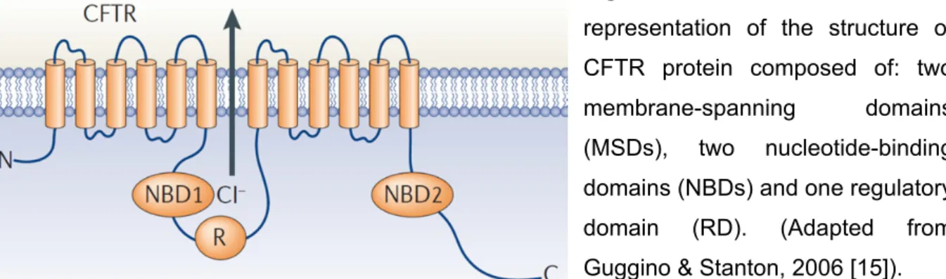
Commonly to most ABC transporters, CFTR is formed by two membrane-spanning domains (MSD 1 and 2) each consisting of six α-helical segments which contributing to the ion pore. In addition, two nucleotide-binding domains (NBD1 and
General Introduction NBD2) which are highly hydrophilic, are situated in the cytoplasmic compartment, were ATP binds to gate the channel. CFTR has a fifth domain, the regulatory domain (RD), which has no equivalent in any other ABC transporter and serving a regulatory function through its multiple phosphorylation sites [15] (Figure I.1)
Figure I. 1: Schematic
representation of the structure of CFTR protein composed of: two membrane-spanning domains (MSDs), two nucleotide-binding domains (NBDs) and one regulatory domain (RD). (Adapted from Guggino & Stanton, 2006 [15]).
The phosphorylation of the RD by the cyclic adenosine 3’,5’-monophosphate (cAMP)-dependent protein kinase A (PKA) is essential for channel activation. It leads to conformational changes of the RD which disrupts its interaction with NBD1 and allows ATP to bind to the NBDs, forming the NBD1-NBD2 dimer. This in turn leads to conformational changes in the MSDs and consequently opening of the channel. The probability of channel opening (Po) is controlled by the extent of RD phosphorylation
at multiple sites, reflecting the balance between PKA and the activity of phosphatases on these sites [11]. The conversion of ATP to ADP then reduces the nucleotide binding affinity towards the NBD binding sites. This facilitates the dissociation of ADP and thereby disrupting the NBD dimer interaction, leading to channel closure [11,16]. While it seems clear that PKA is the main modulator of CFTR Cl- channel gating, there are other kinases involved in CFTR-mediated Cl- transport, namely by modulating CFTR traffic/ cell surface expression, e.g., protein kinase C (PKC), src kinase, AMP-dependent protein kinase (AMPK), casein kinase 2 (CK2) [17–19] and more recently, also SYK - spleen tyrosine kinase [20] and WNK4 – kinase with no lysine 4 [21].
CFTR is composed of a single polypeptidic chain of 1,480 amino acids [6]. Its assembly initiates with synthesis and folding in the endoplasmic reticulum (ER)
Chapter I
4
band B, with a molecular mass of about 150 kDa. Once correctly folded, the immature form migrates through the Golgi where it is processed by glycosyltransferases, thus being converted into its mature form, also known as band C of about 170-180 kDa, which then reaches the cell surface [22]. F508del-CFTR, although still functional as a Cl- channel, is a trafficking mutant as it is mostly retained by the ER quality control (ERQC) due to misfolding. It is then rapidly ubiquitinated and targeted for ER-associated degradation (ERAD) to the proteasome [22].
Several studies suggest that the upregulation of chaperone systems can minimize the protein folding defects that occur in diseases such as CF [23]. Such studies indicate that molecular chaperones can be promising targets in therapeutic approaches aimed at correcting the basic defect of such folding disorders [23]. Chaperones typically recognize and bind to hydrophobic amino acid residues exposed at the surface of unfolded polypeptides thereby preventing unproductive aggregation and promoting proper folding. [22,23]. Furthermore, chaperones are able to distinguish between non-native and native states of newly translated proteins. Once the substrates have reached folded conformations, the affinity to chaperones is lost and the folded proteins are released [26]. However, if the misfolding continues, molecular chaperones are able to crosstalk with the proteolytic systems and thus promote degradation of the substrates [23].
In the case of CFTR, a model has been proposed in which the ERQC of CFTR occurs via a two-step mechanism [27]. Briefly, once biogenesis and insertion in the ER membrane occurs, CFTR nascent domains associate with cytosolic chaperones Hsc70/Hdj2 (or Hsp70/Hdj-1). The Hsc70/Hsp70 association forms the first ERQC checkpoint. If Hsc70 remains associated for too long to its substrate, it recruits other co-chaperones like the C-termin of Hsc70 interacting protein (CHIP) and causes F508del-CFTR to be degraded through the Hsc70-CHIP-UbcH5a proteasomal pathway [28]. The second ERQC checkpoint is the calnexin cycle, where very little F508del arrives. In contrast, wt-CFTR passes the first, Hsp70-mediated checkpoint proceeding in the folding pathway and, through the calnexin cycle, it acquires its native conformation through successive rounds of release-deglucosylation and reglucosylation-rebinding to calnexin until proper folding is achieved [27]. However, in case of prolonged residency in the calnexin cycle, also misfolded CFTR is targeted to
General Introduction proteolytic degradation. Depending on the cell type, only 25-60% of precursor wt-CFTR matures [27,28]. The correctly folded immature form of wt-CFTR is transported to the Golgi via coat protein II (COP II) coated vesicles where a third checkpoint assesses the folding status of proteins en route to the secretory pathway [31].
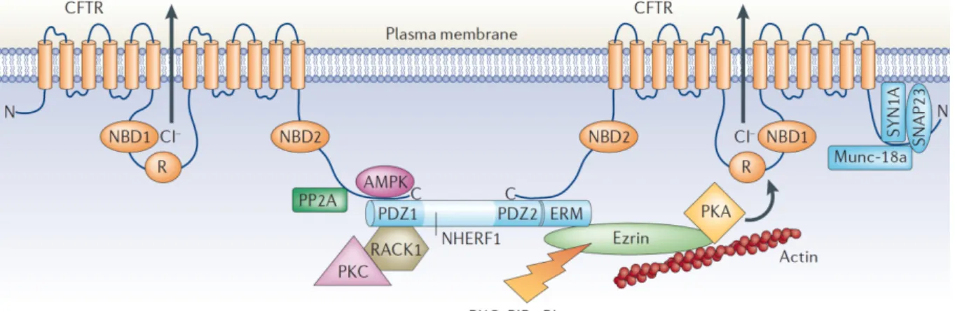
Transport of CFTR protein from the trans-Golgi to the plasma membrane can occur directly to the apical membrane by Golgi-derived vesicles but also indirectly via transcytosis from the basolateral membrane from recycling endosomes [32]. Once at the plasma membrane, the amount of CFTR protein at the surface is tightly regulated by a complex of proteins, which includes PSD-95 – DLG-1 – ZO-1 (PDZ)-domain proteins namely Na+/H+ exchanger regulatory factor isoform-1 (NHERF1) and CFTR-associated protein (CAL) as well as SNAREs (Soluble N-ethylmalemide sensitive factor attachment protein receptors) [15] (Figure I.2). CFTR can then be removed from the cell surface by clathrin-mediated endocytosis through trafficking signal embedded in the primary sequence of CFTR. Furthermore, endocytosed protein can be recycled back to the plasma membrane through recycling endosomes, but damaged protein is targeted for lysosomal degradation [32].
Figure I.2: Proteins that regulate CFTR activity in the plasma membrane: Several proteins interact directly or indirectly with CFTR, including protein phosphatase-2A (PP2A), AMPK, syntaxin-1A (SYN1A), synaptosome-associated protein, 23 kDa (SNAP23) and Munc-18a. These proteins inhibit channel activity and reduce CFTR-mediated Cl– secretion
across the apical plasma membrane in epithelial cells. Other CFTR-interacting proteins that enhance CFTR activity, either directly or indirectly, include NHERF1, receptor for activated C-kinase-1 (RACK1), PKC, PKA and ezrin. ERM (ezrin, radixin, moesin binding domain); PIP2 (4,5-phosphatidylinositol bisphosphate). (Adapted from Guggino & Stanton, 2006 [15])
Chapter I
6
3 CFTR
F
UNCTIONS INE
PITHELIA3.1 CFTR
A
CTIVITY AS AC
L-C
HANNELFully mature CFTR is expressed at the plasma membrane of a variety of secretory epithelial cells such as those in sweat glands, pancreatic ducts and respiratory submucosal glands, where it is required to control ion and fluid homeostasis on epithelial surfaces. As a Cl- channel, CFTR has several
distinguishing characteristics: 1) It has a small single-channel conductance (6–10 pS); 2) its current-voltage (I- V) relationship is linear; 3) it is selective for anions over cations; 4) the anion permeability sequence is Br- > Cl- >I-; 5) It shows time- and voltage -independent gating behaviour; and finally 6) the activity is regulated by cAMP-dependent phosphorylation and by intracellular nucleotides [33].
3.2 CFTR
ASR
EGULATOR OFE
PITHELIALI
ONT
RANSPORTThe role of CFTR in epithelial physiology extends beyond its function as a Cl -channel, namely it is also regarded as a regulator of other ion channels and pathways relevant for CF. Accordingly, this condition is not only characterised by reduced CFTR-mediated Cl- secretion, but also by Na+ hyperabsorption through the amiloride-sensitive epithelial Na+ channel (ENaC). It has been found that CFTR negatively regulates ENaC; hence mutant CFTR eliminates ENaC inhibition, thus leading to increased Na+ permeability. This is widely regarded as the main cause for the dehydration of the ASL, which then impairs MCC [32,33]. Several mechanisms have been proposed and debated to explain how CFTR downregulates ENaC activity. Some studies showed that Cl- currents could account for ENaC inhibition,
regardless of wt-CFTR expression [36]. On the other hand, others suggest that the absence of CFTR from the plasma membrane leads to hyperactivity of ENaC [35,36]. More recently, it was found that wt-CFTR physically interacts with ENaC and such interaction protects ENaC from proteolytic cleavage and stimulated open probability. In contrast, F508del-CFTR failed to protect ENaC from proteolytic cleavage [39].
Moreover, Ca2+-activated Cl- channels (CaCCs) are also functionally related to CFTR since both are main Cl- channels present in the respiratory epithelia. So,
General Introduction CaCCs are among the proteins which could bypass CFTR defective activity in CF epithelia. The latter were finally identified in 2008 [40–42] as TMEM16A, a member of a family comprising 10 paralogs: TMEM16’s (A-H, J, K) also known as the Anoctamin family (Ano 1-10). The roles of most of the Anoctamins are still unknown but they represent alternative, physiologically relevant therapeutic targets for CF.
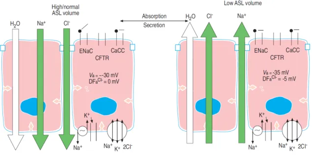
The overall model for ion transport in airway epithelial cells, summarized in Figure I.3, shows how ASL is regulated by Na+ and Cl- transepithelial movement. The transcellular Na+ absorption is mediated by apical ENaC expression together with basolateral Na+/K+-ATPase pump. Apical Cl- channels (CFTR and CaCCs) and the basolateral Na+-K+-2Cl- co-transporter, secrete Cl- when ENaC is blocked and the appropriate secretory driving forces are generated. CFTR activity is regulated by signals in the lumen, e.g. adenosine, which interacts with compartmentalised adenosine receptors A2b, G proteins, adenylate cyclase, and cAMP-dependent
protein kinases [34].
Figure I.3: Regulation of the ASL volume by ion transport. When excess ASL is
present, Na+ absorption mediated via ENaC is dominant (left panel). Cl- is projected to be absorbed passively via the paracellular path due to the fact that there is no electrochemical driving force (DFa Cl-) favoring Cl- exit from the cell. In contrast, both the negative apical
membrane potential (Va) and low intracellular Na+ activity (y20 mM) favour Na+ entry into the
Chapter I
8
membrane potential more negative and generates a driving force for Cl- secretion.
(Reproduced from Boucher, 2004 [34])
Besides this, tidal volume expansion and airflow impart shear stress to airway surfaces, a stimulus that releases nucleotides from many cell types into the extracellular environment. In mammalian airways, extracellular nucleotides interact with P2Y2 purinergic receptors to regulate airway ion transport by activating CaCC,
and by inactivating ENaC via depletion of PIP2 (see 4.3.3). Furthermore, ATP is
metabolised into, ADP, and AMP by ecto-nucleotidases and ecto-apyrases which are typically located on the apical membrane of the superficial epithelia. The product adenosine can then activate CFTR currents upon stimulation of A2b receptors [43]. The stimulation of G-protein-coupled receptors (GPCRs) -like P2Y receptors- by ligands such as ATP leads to the hydrolysis of PIP2 into diacylglycerol (DAG) and
inositol 1,4,5-trisphosphate (IP3). IP3 then promotes Ca2+ release from
ER-intracellular stores which mediates the activation of CaCC’s. Upon store depletion, activation of extracellular Ca2+ influx is assured by store-operated Ca2+ channels (SOCs), which further contribute to CaCC-mediated cellular response.
In addition to ENaC and CaCC, CFTR regulates other ion channels present in epithelial cells such as the outwardly rectifying Cl- channels (ORCCs) or the inward rectifying K+ channels (ROMK = Kir 1.1) in the thick ascending limb of Henle’s loop and in principal cells of the collecting duct. [42,43]. Functional CFTR is required for activation of ORCC’s [46]. It was shown that CF mutations disrupt this regulation, and different CFTR mutations can affect CFTR functions differently. These findings suggest that the severity of pulmonary disease may be more closely associated with CFTR regulatory rather than its Cl- channel function [47].
4 E
PITHELIALN
A+C
HANNEL(EN
AC)
4.1 EN
AC:
S
TRUCTURE ANDF
UNCTIONThe epithelial Na+ channel (ENaC) belongs to the ENaC/Degenerin family of ion channels. ENaC/degenerin channels have a variety of different cellular functions
General Introduction but have some common properties. All members whose functional characteristics have been examined are Na+ selective and amiloride-sensitive to various degrees, and each member is regulated by external ligands and/or is mechanosensitive [46,47]. Compared with its family members, ENaC has a small single-channel conductance of 4-6 pS [50] and is highly Na+ selective (100 Na+ > 1 K+) and amiloride-sensitive (EC50 of 150 nM). In addition, ENaC responds to both external
mechanical and chemical stimuli [51]
Fully functional ENaC is composed of three different subunits alpha-, beta- and gamma-ENaC (α-, β-, and
γ
-ENaC) which have close homology to each other. Two additional subunits delta- and epsilon- (δ and ε) ENaC have also been identified. [50] ENaC subunit stoichiometry has been the subject of a number of studies since the initial cloning of the genes encoding α-, β-, andγ
-ENaC subuunits. Based on the crystal structure of the acid-sensing ion channel 1 (ASIC1), ENaC appears to be a trimeric channel containing one α, one β, and oneγ
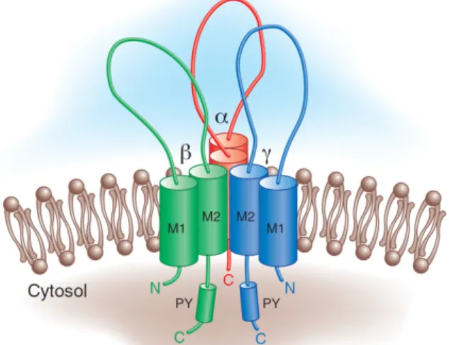
subunit [49]. As a plasma membrane protein, ENaC is co-translationally inserted in the ER membrane and its export requires assembly of the αβγ
subunits into a correctly folded complex [52].Figure I.4: ENaC architecture: Structural
features of the epithelial Na+ channel. ENaC
may exist as a heterotrimer with a single α,β,
γ
subunit. Each subunit has two membrane-spanning domains (M1 and M2) with intracellular N- and C-term. The β- andγ
- subunits each contain a canonical “PY” motif in their COOH-term. (Adapted from Bhalla, 2008 [53]).Each ENaC subunit has two transmembrane domains, each passing once through the plasma membrane, one large N-glycosylated extracellular domain, and relatively short intracellular N- and C-term regions [54] (Figure I.4).
Chapter I
10
ENaC plays diverse roles in maintaining Na+ homeostasis through its functional expression in a variety of tissues. In the apical membranes of epithelial cells in airways, sweat glands, colon, and distal nephron of the kidney, ENaC facilitates transepithelial Na+ transport in conjunction with the Na+-K+-ATPase found in the basolateral membrane. These functions permit the control of extracellular fluid volume and blood pressure, as well as the ASL volume and MCC [51].
Liddle syndrome is caused by gain-of-function ENaC mutations ultimately leading to hypertension. These mutations reside largely in the cytoplasmic C-term tails of the β- and
γ
-subunits of ENaC, resulting in an increased channel surface expression and channel processing by proteases thus enhancing its activity [55]. On the other hand, a partial loss-of-function mutation of ENaC produces pseudohypoaldosteronism type I, characterized by excessive fluid accumulation in the lung mild salt-wasting diuresis and hypotension [50].4.2 N
EDD4-2
ANDEN
AC
U
BIQUITINATIONThe number of functional ENaC channels at the plasma membrane of epithelial cells is primarily determined by the rate of insertion of newly assembled channels. In addition, the rate of retrieval and degradation of channels and the rate of recycling from intracellular pools are also an important factor contributing to ENaC levels at the plasma membrane. Indeed, the mechanism of action of hormones like aldosterone and vasopresin that leads to an increase of the number of functional ENaC channels at the apical membrane, includes the regulation at these levels [50].
Ubiquitin (Ub) conjugation is a prerequisite for ENaC internalization and subsequent degradation or recycling. It requires the specific E3-ubiquitin ligase Nedd4-2 (neural precursor cell expressed developmentally down-regulated protein). Nedd4-2 contains a E6-AP C-term (Hect) domain that is homologous to other ubiquitin ligases, WW domains and a Ca2+/lipid binding domain (CaLB/ C2) [54,55]. Nedd4-2 appears to be the isoform responsible for ubiquitination of ENaC in all mammalian epithelial cells [50]. ENaC subunits have a PPPXYXXL sequence conserved in the C-term of each ENaC subunit. This sequence is similar to two
General Introduction endocytic motifs that mediate ENaC clathrin-mediated endocytosis. This sequence also fits the consensus of a PY motif (PPXY), involved in protein interactions. The ENaC PY motifs function as a binding site for WW domains of Nedd4-2 [52].
4.3 EN
AC
R
EGULATORYP
ATHWAYSENaC activity is regulated by a number of factors that ultimately affect both the number of channels expressed at the cell surface (N) or the channel open probability (Po). N is controlled by hormones and signalling proteins that affect either the
transcription or trafficking of ENaC subunits. Po is influenced by proteolytic
processing and by interactions with cytoplasmic domains with specific membrane acidic phospholipids [49,53].
4.3.1 Hormonal Regulation of ENaC
Given the need for rapid dynamic changes in salt and water reabsorption and secretion, it is not surprising that ENaC is regulated by the action of the volume-regulatory hormones aldosterone and arginine vasopressin (AVP) [52]. ENaC mediates apical entry of Na+ in the aldosterone-sensitive distal nephron and constitutes the rate-limiting step for Na+ reabsorption by the kidney [58]. In states of volume depletion, decreased renal perfusion results in release of renin, activating the renin-angiotensin-aldosterone pathway. Binding of aldosterone to the mineralocorticoid receptor activates transcription of a variety of genes, which increases ENaC expression/current [52]. Some key targets of the mineralocorticoid receptor signalling includes regulatory proteins such as phosphatidylinositol 3-kinase (PI3K) and serum and glucocorticoid-induced kinase 1 (SGK1). By phosphorylating ENaC itself and a variety of ENaC-regulatory proteins, SGK1 alters ENaC expression, trafficking and activity, and stimulates Na+ transport [59] (see Figure I.5 for more details).
Decreased extracellular volume also induces release of AVP from the hypothalamus. In the kidney collecting duct, AVP binds to V2 receptors at the basolateral membrane, increasing Na+ transport through a pathway that includes
Chapter I
12
cAMP and PKA [52]. Nedd4-2 can be phosphorylated by PKA which inhibits its binding to ENaC.
ENaC is also regulated by insulin via the PI3K pathway. It is well established that, acting through the insulin receptor substrate-1, insulin activates PI3K, a heterodimeric enzyme that catalyses the formation of phosphatidylinositol 3,4,5-trisphosphate (PIP3) [60].
Figure I.5: Model of hormonal regulation of ENaC. Aldosterone enters a target cell
and binds MR, which translocates into the nucleus. MR activates the transcriptional machinery, and thus alters expression of aldosterone target genes (in blue arrows). At the apical membrane, ENaC, composed of three subunits (α, β and γ), constitutes the rate-limiting step of apical Na+ entry. Na+ is then extruded into the basolateral space by the
Na+/K+-ATPase pump. In the absence of aldosterone, ENaC proteins interact with Nedd4-2,
an ubiquitin-ligase which targets ENAC to proteosomal degradation. SGK1 is a key aldosterone-regulated target gene that plays a central role in Na+ reabsorption. Upon
aldosterone exposure, PDK1-activated kinase SGK1 phosphorylates Nedd4-2, which in turn dissociates from ENaC, increasing its apical membrane abundance.
PIP3 promotes the activation of protein kinase B (Akt), an important
General Introduction stimulation, the pleckstrin homology (PH) domain of Akt binds to lipid messengers and is phosphorylated [61]. Insulin-induced PI3K signalling cascade leads also to PI3K-dependent kinase 1 (PDK1) activation which phosphorylates SGK1. The immediate natriuretic effect of insulin is attributed to an increase in ENaC Po or an
increase in the number of active ENaC at the apical membrane [62].
A common factor in ENaC hormonal regulation is the activation of SGK1. SGK1 physically interacts with Nedd4-2, and inhibits it by phosphorylation. By inhibiting Nedd4-2-dependent ubiquitination of ENaC an preventing its internalization, SGK1 indirectly enhances apical membrane expression of ENaC [55].
SGK1 has a short half-life of less than 30 min being rapidly targeted to ERAD. Another aldosterone-induced ENaC regulator protein named glucocorticoid-induced leucine zipper protein 1 (GILZ1), protects SGK1 from ERAD [59].
In the absence of aldosterone, when GILZ1 and SGK1 levels are low, Nedd4-2 and Raf-1 associate with and inhibit ENaC [59]. Upon aldosterone stimulation, extracellular signal-regulated kinase 1/2 (ERK1/2) phosphorylates and abrogates ENaC function. At the same time, GILZ1 inhibits the inhibitory Raf-1/MEK1/2/ERK1/2 signalling cascade [59]. GILZ1 is thus an adaptor protein, which interacts and modulates the activity of multiple components of the ENaC regulatory machinery, including Nedd4-2 [55].
4.3.2 Regulation of ENaC activity by Proteolytic Cleavage. Besides the complex regulation of the number of ENaC channels present at cell surface, another crucial mechanism of ENaC regulation is the modulation of channel Po by proteolytic processing.
Channel-activating proteases (CAPs) act by cleaving the extracellular domain of α- and
γ
-ENaC but not of β-subunit, which leads to release of inhibitory peptides. The proteases identified so far can be divided into three subgroups: intracellular proteases, such as furin; cell-attached serine proteases, such as prostasin and matriptase; and soluble serine proteases, such as plasmin and trypsin [63].Chapter I
14
Furin is predominantly found in vesicles of the trans-Golgi network (TGN). It mediates cleavage of α-subunit at two sites and
γ
-subunit at one site during biosynthesis of ENaC. Once at the plasma membrane further proteolytic activation of the channel is accomplished by extracellular membrane-associated serine proteases [63]. The sequential release of inhibitory motifs from α- andγ
-subunits results in channels transitioning from a very low Po (near electrically silent) to an increasinglyhigh Po (constitutively active channel) [53,62].
4.3.3 Role of PIP2 and PIP3 in regulating ENaC
Phosphatidylinositides, such as phosphatidylinositol 4,5-bisphosphate (PIP2)
and phosphatidylinositol 3,4,5-triphosphate (PIP3), directly interact with ENaC
channels to modulate its gating [65].
Channel–phosphoinositide interactions are thought to be of electrostatic nature, whereby the negatively charged head groups of phosphoinositides interact with positive-charged residues within intracellular portions of the channel [65]. PIP2
binding motifs have been identified in the N-term of β- and
γ-
ENaC subunits, but not in the α-subunit [66]. Disruption of these positively charged amino acid residues in the N-term PIP2 binding region leads to strong inhibition of ENaC currents [67]. Onthe other hand, increased PIP2 hydrolysis and depletion from the plasma membrane
in response to purinergic stimulation and activation of endogenous phospholipase C (PLC) inhibits ENaC [68].
The product of PI3K-conversion of PIP2, PIP3, affects ENaC membrane levels
by both a direct mechanism involving physical association of the phosphatidylinositide with the channel and an indirect mechanism involving a signalling cascade that promotes channel retrieval from the plasma membrane. Regulation of ENaC Po by PI3K is a direct consequence of the physical association of
PIP3 with the channel [68]. On the other hand, SGK1 is a downstream effector of
PI3K and is sensitive to both PIP3 levels and phosphorylation by another PI3K
effector, PDK1. As described (see 4.3.1), the activation of SGK1 promotes retention of ENaC in the membrane.
General Introduction The stimulation of luminal purinergic receptors co-localizing with ENaC in luminal membranes transiently activates Ca2+-dependent Cl– secretion and induces long-lasting inhibition of Na+ absorption. This is a relevant issue for the pharmacotherapy of CF patients, where an increased Na+ absorption is found in the airways [67].
5 C
YSTICF
IBROSIST
HERAPEUTICA
PPROACHESSince the discovery of the CF gene, impressive advances in elucidating the molecular basis and cellular pathophysiology of CF have been achieved. Nevertheless, the improvement in mean life expectancy of CF patients has resulted mostly from a better treatment in alleviating symptoms of the disease rather than correcting the basic defect. Thus, life expectancy (mean age at death, in Europe, ~25 yrs) and quality of life are still limited for CF patients [12,67]. In CF, airways disease is the major cause of morbidity and mortality, so most therapeutic efforts have been directed at improving airway function. For the past years, great progress has been achieved in pharmaco-therapy with the aim of normalizing the CF cell physiology, namely ionic transport. This includes rescuing mutant CFTR by promoting its correct folding and traffic to the cell membrane and potentiating its activity as a Cl- channel but also finding agents leading to ENaC inhibition or activation of alternative Cl -channels such as Anoctamin1 (CaCC). These three therapeutic strategies are described below.
5.1 R
ESCUINGCFTR
Although over 1,900 CFTR gene mutations have been described, the emphasis of these therapeutic strategies has been on the F508del mutation, given its high prevalence. As described above (see 2.1), F508del-CFTR fails to fold correctly in the ER and as a result it is retained by the ERQC and rapidly targeted for proteasomal degradation. Very little of this mutant CFTR reaches the cell surface. Nevertheless, rescuing F508del-CFTR to the plasma membrane would be beneficial since it retains some Cl- channel function [69]. High-throughput screens (HTS) of large compound libraries have been carried out to identify small molecules capable of
Chapter I
16
rescuing the traffic of mutant CFTR to the cell membrane. These compounds are known as “correctors”. They can be 1) pharmacological chaperones that specifically rescue a misfolded protein by interacting with and energetically favour its correct folding; 2) chemical chaperones that mimic the effects of molecular chaperones thus lacking target specificity; and 3) compounds that target molecular chaperones by affecting their interaction with the defective protein [69].
Another class of compounds searched by HTS are “potentiators”, i.e., stimulators of pre-activated CFTR Cl- channels when the protein is already at the plasma membrane. Since, when rescued to the cells surface, F508del-CFTR still exhibits reduced Po, an ideal compound to restore F508del-CFTR would combine the
ability of acting both as a corrector and potentiator. Potentiators are also aimed at correcting other CFTR mutations where the protein is correctly localized at the plasma membrane but have impaired channel regulation, notably G551D-CFTR which accounts for 3% of the CF population [12]. At least two experimental drugs, VX-770 (Ivacaftor) and VX-809 (Lmacaftor) and have undergone clinical trials and former already received FDA and EMEA approval in 2012 for patients carrying the rare mutation G551D. Both compounds were produced by a consortium of the Vertex Pharmaceuticals with the US Cystic Fibrosis Foundation (CFF). Using HTS the consortium examined 228.000 compounds. While VX-770 is a potentiator, i.e., restores CCFTR channel gating, and VX-809 is a corrector, i.e., restores CFTR cell surface expression [12].
Another therapeutic strategy contemplates the identification of CFTR-binding proteins modulating CFTR traffic or function as a Cl-channel which may constitute new therapeutic “targets” for CF [15]. Several studies have identified such “drugable targets”, and new compounds, based on the results of these studies, are in clinical trials. However, to gain a complete understanding of CFTR regulation, and to identify more therapeutic targets for CF, we should identify all of the CFTR-interacting proteins and understand how these proteins are dynamically regulated by protein– protein interactions and by post-translational mechanisms [15]. The same approach could be applied to other intervenients of the CF lung disfuction, notably ENaC.
General Introduction
5.2 T
HEB
YPASSA
PPROACHRestoration of normal airway hydration balance by modulation of ion channel activity represents an important therapeutic strategy for CF. Activation of CaCCs which are expressed apically in respiratory epithelia offer an alterntive pathway to restore Cl- secretion in the airways. Ano1 was recently described as a CaCC in the airway epithelium and, as such, represents an attractive target to restore Cl -transport. This concept has been the basis of the CF clinical trials testing P2Y2
agonists such as UTP analogues with increased stability (vs UTP) against degradation by ectonucleotidases. Examples include INS37217 (Denufusol tetrasodium), which, besides increasing Cl- and fluid secretion, increased cilia beat frequency and mucin release. In a 28-day phase 2 clinical trial, Denufusol significantly improved lung function in patients with mild CF lung disease when applied three times daily by nebulizer [12,68]. Nonetheless, Denufosol phase 3 clinical trial (TIGER-2) failed to meet primary endpoint, i.e., a change from baseline in FEV1 (Forced Expiratory Volume in One Second).
Another strategy of potential therapeutic interest consists in the indirec stimulation of CFTR-mediated Cl- secretion by increasing the secretory driving force for Cl-, e.g., by activation of basolateral K+ channels. By causing hyperpolarization of the cell membrane potential, this enhances electrogenic luminal Cl- secretion [69]. This strategy might be particularly useful for CF patients with CFTR mutations not completely abolishing CFTR-mediated Cl- secretion.
An alternative therapeutic strategy for CF is to inhibit ENaC, thereby bringing excessive Na+ absorption to normal levels. This can be achieved by direct blockade of ENaC or inhibition of the channel-activating proteases (CAPs), whose activity regulates ENaC function. Amiloride, which specifically blocks ENaC, has been tested for this purpose. However, the short half-life of this compound on airway surfaces (20–30 min), constitutes a major limitation to its therapeutic efficacy [12,32]. Notwithstanding, complete block of ENaC in the airway epitheia is also undesirable since, as it could lead to pulmonary edema. Instead, we seek compounds reducing
Chapter I
18
ENaC hyperabsorption to its normal levels so as to restore homeostasis of epithelial Na+ and water transport.
Overall, CFTR is more than a Cl- channel; it regulates epithelial ion transport in many organs by interfering with several other ion channels and transporters. Thus the basic defect of CF comprises several events that contribute to a clinically multifaceted disease. Based on the complexity of CF pathophysiology, different therapeutic approaches are being pursued. To this end, however, it is crucial to acquire a deeper and more global knowledge of the proteins interacting and/or modulating CFTR traffic and activity and also more detailed information on the characteristics and regulation of the ion channels involved in CF. The ultimate goal of the current studies, is to identify novel drug targets for CF.
Objectives of the Present Work
II O
BJECTIVES OF THEP
RESENTW
ORKThe present doctoral work aimed at gaining new insight into the regulation of CFTR activity as well as of ENaC which are both critical in the pathophysiology of Cystic Fibrosis. Towards this overall goal, work was carried our focussing on the following specific objectives:
To identify novel CFTR binding proteins and to functionally characterize the effect of one of them (Annexin A5) on CFTR traffic and activity, both in Xenopus oocytes and in mammalian cells.
To analyse the regulation of CFTR by P2Y2-receptor stimulation both in
Xenopus oocytes and in human bronchial epithelial cells.
To functionally validate novel ENaC binding proteins identified in a yeast split-ubiquitin screen, by analysing the effects of one of them (SERP1) on ENaC activity in Xenopus oocytes and in human airway epithelial cells.
To functionally validate novel protein regulators of ENaC activity that were identified in a siRNA HTS, by assessing their effect on ENaC activity in Xenopus oocytes, human airway epithelial cells and mouse epithelial tissues.
Materials and Methods
III M
ATERIALS ANDM
ETHODS1 C
ELLC
ULTURE1.1 M
AMMALIAN CELL LINES ANDC
ULTURE CONDITIONSHuman bronchial epithelial H441 cells were culture in RPMI-1640 media supplemented with 100 units/ml penicillin / 100 μg/ml streptomycin (Pen/Strep), 1% Insulin-Transferrin-Selenium, 200 nM dexamethasone and 10% fetal bovine serum (FBS).
Fluorescently labelled A549 cells stably expressing wt-CFTR or F508del-CFTR after induction with doxycycline (DOX) were created in Professor Amaral’s laboratory. For this, wt-CFTR and F508del-CFTR were fused in the N-term to mCherry, a fluorescent protein obtained from DsRed by changing the chromophore environment [71]. Additionally, a FLAG tag (octapeptide: DYKDDDDK) was inserted by mutagenesis PCR (using Pfu polymerase, annealing temperature at 43ºC and extension at 68ºC, 28 cycles), in the extracellular loop of β-ENaC and in the fourth extracellular loop of CFTR. This construct was inserted by TA-cloning, using a PCR reaction (using Hercules polymerase, annealing temperature of 62ºC and extension at 72ºC, 30 cycles and a final 15min extension at 72ºC with Taq polymerase) and insertion into pCR8 GW TOPO Gateway entry vector (Invitrogen). By LR recombination reactions, this construct was then inserted into a lentiviral destination vector, pLenti4-V5 (Invitrogen) with CMV promoter and pLenti with TetON DOX-sensitive promoter, giving rise to the stable and inducible mCherry-FLAG-βENaC and mCherry-FLAG-wtCFTR and F508del-CFTR cell system, respectively. A549 cells expressing wt-CFTR or F508del-CFTR were grown in Dulbecco's modified Eagle's medium (DMEM). Expression of wt-CFTR or F508del-CFTR was induced with 1µg/ml doxycycline (Sigma-Aldrich, Taufkirchen, Germany) 24h prior to the experiment. These cell lines as well as the A549 parental cell line were supplemented Pen/Strep and 10% FBS. When expression of ENaC was wanted, A549 parental cell line was cultured with a supplement of 1% Insulin-Transferrin-Selenium and 100 nM dexamethasone.