Universidade do Algarve
Cátia Alexandra Ferreira de Sousa
Analyzing Oct4 conserved domains in lower vertebrates
Faculdade de Engenharia e Recursos Naturais
Universidade do Algarve
FERN
Dissertação Engenharia Biologica com Mestrado integrado
2007/2008
Cátia Alexandra Ferreira de Sousa
Analyzing Oct4 conserved domains in lower vertebrates
Supervisor:
Andrew Johnson; David Tannahill; Dr. José BeloAno Lectivo 2007 to 2008
© Cranfield University, 2008. All rights reserved. No part of this publication may be reproduced without the written permission of the copyright holder.
i
Abstract
Oct-4 is a POU-V domain transcription factor which regulates pluripotency in mammals and expressed in ES cells and germ cells, and was believed to be a gene unique to mammals. Recently, it has been demonstrated to be present in the genomes of Xenopus, zebrafish, sturgeon and axolotl. It has been shown that Oct-4 has three transactivation domains, (N), POU and (C), with DNA binding mediated through the POU domain. It was unknown how the activity of these domains has been retained through evolution and how they collectively function to control pluripotency.
This study is new and is the first to analyse the functional conservation of these Oct-4 homologues, and their regulation molecular. Three different assays were developed to study Oct4 functionality: 1) A transactivation assay in which the function of Oct4 protein over-expression on a known Oct4-target sequence was assessed (by luciferase assay using the p6Wtk-luc reporter containing Oct4 binding sites); 2) A heterologous transactivation assay in which the function of specific Oct4 domains by linking them to Gal4-DNA binding domain was specifically assessed (DBD) (by luciferase assay using the pGal4-lux reporter). 3) The subcellular localization of Oct-4 homologues (generating two constructs; either full’ length or POU domain, fused to Green fluorescent protein (GFP).
This study showed the coexistence of DBD conflicts with Oct4 resulting in a decrease on its transactivation capacity when compared to their native state.
Oct-4 function generally conserved among species, with Xenopus Oct91 being the Oct-4 homologue with a transactivation function more similar to mouse Oct-4. Between (C) and (N) transactivation domains linked to DBD, the (C) domain was the one with more activity. The (C) domain is cell-type specific regulated by phosphorylation events, while the (N) domain suffers sumoylation. These two regulatory mechanisms are shared in all Oct-4 homologues. It was also possible to conclude that Oct-4 protein is nuclei transcribed. This project opens many possible studies in Oct-4 regulation.
ii
Acknowledgments
It was a pleasure and an intellectual challenge to work in the Genetics lab of the Nottingham University, it was an experience without precedent.
I would like to thanks to Dr. José Belo by accepting being my supervisor from Algarve University. I would like to thank to my supervisors, particularly Dr. Andrew Johnson for taking me into his laboratory, and Dr. James Dixon for all his time, concern and also for tutoring me and support me during the project. I would also like to thank Dr. Carla Toro and Dr. David Tannahill for their advices and concern. I am grateful to Dr. Niwa for the gift of p6Wtk-luc that was used on the assays.
Also thanks to e to my family and friends that always support me and were there for me, renewing my energy and strength
iii
Table of contents
Abstract ... i
Acknowledgments ... ii
Table of contents ... iii
Abbreviations ... v
List of figures ... vii
List of tables ... viii
Chapter 1: Introduction and literature review ... 1
1.1 Overview ... 1
1.2 Pluripotency ... 1
1.3 Research in lower vertebrates... 4
1.4 Oct-4 belongs to POU transcription factor family ... 7
1.5 Oct-4 domains ... 8
1.6 Oct-4 and pluripotency ... 9
1.7 Oct4 expression ... 10
1.8 Aims and Objectives ... 11
Chapter 2: Material and Methods ... 13
2.1. Construction of expression vectors and reporter plasmids. ... 13
2.1.2. pATG ... 24
2.1.2. pDBD ... 25
2.2.3. pGFP ... 26
2.2. Cell culture and transfection. ... 27
Chapter 3: Results ... 29
3.1. Oct4 domain structure. ... 29
3.2. Oct4 sequence identity as function prediction ... 29
3.3. Amino Acid Composition ... 32
3.3.1 N- Terminal (N) ... 32
3.3.2 POU domain ... 33
3.3.3 C- Terminal (C) ... 33
3.4. Alignments ... 36
iv 3.4.2. POU ... 37 3.4.3. C-terminal (C) ... 39 3.5. Cloning ... 39 3.6. Nuclear localization... 40 3.7. Luciferase Assay ... 42 3.7.1. pATG ... 42 3.7.2. pDBD ... 44 Chapter 4. Discussion ... 50
4.1. Amino Acid Composition ... 50
4.2. SEB (Serine [S]/ Glutamic acid [E]) box ... 52
4.3. Nuclear Localization ... 53 4.4. Luciferase Assay ... 53 4.5. Functional Conservation ... 59 Chapter 5: Conclusions ... 62 5.1. Future work ... 65 References ... 66 APPENDICES ... 72
APPENDIX 1 Ambystoma mexicanum ... 72
APPENDIX 2 Mus musculus ... 73
APPENDIX 3 Acipenser oxyrinchus oxyrinchus ... 74
APPENDIX 4 Danio rerio ... 75
APPENDIX 5 Xenopus laevis ... 76
APPENDIX 6 Xenopus laevis- Oct60 ... 77
APPENDIX 7- Xenopus laevis- Oct25 ... 78
APPENDIX 8. Primers combination ... 79
APPENDIX 9. Amino acid composition of Oct-4 N terminal ... 81
APPENDIX 10. Amino acid composition of Oct-4 POU domain ... 84
APPENDIX 11. Amino acid composition of Oct-4 C-terminal ... 87
APPENDIX 12. pGEM®-T Easy vector (Promega, UK) ... 90
v
Abbreviations
(C) Carboxyl- terminal domain
(N) Amino- terminal domain
µg Microgram (10-6 g)
aa Amino acid
axC Axolotl Oct-4 C domain
axN Axolotl Oct-4 N domain
axOct4 Axolotl Oct-4
Bp Base pairs
BSA Bovine Serum Albumin
cDNA Complementary DNA
CO2 Carbon dioxide
DAPI 4,′6-diamidino-2-phenylindole
DBD DNA Binding Domain
DMEM Dulbecco’s modified eagles medium
DNA Deoxyribonucleic acid
dpc Days poscoitum
ES Embryonic stem
FFT Fast Fourier Transform FGF Fibroblast growth factor
g Gram, unit of mass
GFP Green fluorescent protein
HEK 293T Human Embryonic Kidney 293T hESc Human embryonic stem cells
ICM Inner cell mass
L Liter, unit of volume
LARII Firefly luciferase reagent
LB Lysogeny broth
vi
mC Mouse Oct-4 C domain
MCS Multiple cloning site Ml Milli liter (10-3 l)
mM Milli Molar (10-3 M)
mN Mouse Oct-4 N domain
mOct4 Mouse Oct-4
MW Molecular Weight
NLS Nuclear Localization Site PBS Phosphate buffered saline PGCs Primordial germ cells
SEB Serine [S]- Glutamic acid [E] box
SOB Super Optimal Broth
stC Sturgeon Oct-4 C domain
stN Sturgeon Oct-4 N domain
stOct4 Sturgeon Oct-4
TAD Transcriptional activation domain TFs Transcription factors
Xl25 Xenopus Oct-25
Xl60 Xenopus Oct-60
Xl91 Xenopus Oct-91
zfC Zebrafish pou2 C domain
zfN Zebrafish pou2 N domain
vii
List of figures
Figure 1. 1- Different paths of establish pluripotent embryonic stem (ES) and embryonic
germ (EG) cell lines. ... 2
Figure 1. 2 Model of Oct4, Nanog, Sox2, and FoxD3 interaction during early mouse development (1). ... 9
Figure 1. 4- Oct-4 expression in pre-implantation and early post-implantation during mouse development.. ... 11
Figure 2. 1- Different Oct-4 domain constructs. ... 13
Figure 2. 2- ClustW amino acids alignments ... 14
Figure 2. 3- Enzymes used to create the different Axolotl Oct-4 fragments. ... 16
Figure 2. 4- Enzymes used to create the different Mouse Oct-4 fragments. ... 16
Figure 2. 5- Enzymes used to create the different Sturgeon Oct-4 fragments. ... 17
Figure 2. 6- Enzymes used to create the different Zebrafish Pou2, Xenopus Oct-91, Xenopus Oct-60 and Xenopus Oct-25. ... 18
Figure 2. 7- pATG vector used for cloning. ... 24
Figure 2. 8- pDBD vector used for the cloning. ... 25
Figure 2. 9-Vector pGFP used in cloning. ... 26
Figure 3. 1- Average distance tree using BLOSUM62. ... 31
Figure 3. 2- CD MAFT alignment. ... 35
Figure 3. 3- N-terminal alignment for all species. ... 37
Figure 3. 4- Jalview MAFFT alignments for the different POU domains ... 38
Figure 3. 5- MAFT multiple alignment for the different C-terminal sequences. ... 39
Figure 3. 6- 293 T cells transfected with pGFP Oct-4 full length and POU domain. .... 41
Figure 3. 7- pATG-Oct-4 Full length transactivation in HeLa and HEK 293T cells. .... 43
Figure 3. 8- pDBD- OCT-4 full lenght activation in HeLa and HEK 293 T cells ... 44
Figure 3. 9- pDBD-POU-C Oct-4 transactivation in HeLa and HEK 293T cells ... 45
Figure 3. 10- pDBD- N-POU Oct-4 transactivation in HeLa and HEK 293T cells. ... 46
Figure 3. 11- pDBD-N transactivation in HeLa and HEK 293T cells. ... 47
Figure 3. 12- pDBD-POU transactivation in HeLa and in HEK 293 T cells ... 48
Figure 3. 13- pDBD-C transactivation in HeLa and in HEK 293T cells. ... 49
Figure 4. 1- Sumoylation sites in Oct-4 homologues . ... 58
viii
List of tables
Table 1.1- Different strategies for reprogramming differentiated cells into a pluripotent state . ... 3 Table 2. 1- Domain lengths of Oct4 in the studied samples. ... 15 Table 2. 2-- Axolotl Oct4 enzyme combinations used to generate different fragments. 16 Table 2. 3- Mouse Oct4 enzyme combinations used to generate different fragments ... 17 Table 2. 4- Sturgeon Oct4 enzyme combinations used to generate different fragments. 17 Table 2. 5- Zebrafish Pou2 enzyme combinations used to generate different fragments. ... 18 Table 2. 6- Xenopus Oct-91 enzyme combinations used to generate different fragments. ... 19 Table 2. 7- Xenopus Oct-60 enzyme combinations used to generate different fragments.. ... 19 Table 2. 8- Xenopus Oct-25 enzyme combinations used to generate different fragments. ... 19 Table 2. 9- Enzymes and adaptors used to create different Oct4 fragments for each species. ... 20 Table 3. 1- Sequence identity of Oct4. ... 30 Table 3. 2 Prediction of phosphorylation positions in the C domain, for serine; and tyrosine, according to Netphos 2.0 (56). ... 34 Table 3. 3 Prediction of phosphorylation positions in the C domain, for serine; and tyrosine, according to Netphos 2.0 (56). ... 35
1
Chapter 1: Introduction and literature review
1.1 Overview
Biologists have explored embryonic development, from worms to humans, to understand how different complex organisms can be derived from a single cell, the fertilised egg or zygote [1). It has been demonstrated that many conserved genes and pathways are involved in regulating development. The same genes have been linked to the same cell choices through evolution from simple to complex organism development. We also have a good understanding of the way that the embryo repeatedly uses the same strategies for organogenesis, tissue patterning and cellular specialisation [2]. All species use a common strategy for development, the use of a stem cell to generate and maintain a given tissue or organ. During embryogenesis, cells are initially proliferative and pluripotent; with the ability to differentiate into any cell type; they only gradually become restricted to different cell fates [1, 2]. It is of interest to study if gene functionality is also conserved during evolution, and because the manipulation of these genes and pathways may hold the key to curing all known human disorders
1.2 Pluripotency
Pluripotent cells can give rise to all cells of the embryo or the adult organism [3]. In higher vertebrates Pluripotent cells express Oct-4, Nanog and Sox-2, and these factors are used as pluripotency markers. The potential that pluripotent cells have to develop into specialized cells is being researched for cell and tissue replacement therapies in order to treat disorders such as Parkinson’s disease, rheumatoid arthritis, burns and heart disease [1, 4]. Despite the fact that human embryonic stem cells (hESc) might be used to treat a host of diseases, there are ethical issues regarding the use of human embryos, as well as the problem of tissue rejection followed transplantation [5]. To bypass these issues, there is the possibility of reverting somatic patient cells into pluripotent cells by the constant forced over-expression of pluripotency factors [5]. So
2 far the process of reversion to a pluripotent phenotype is not very well understood however this technology holds much promise for regenerative medicine [6].
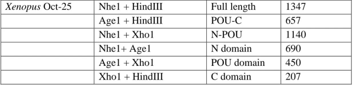
The demonstration that fully differentiated cells can revert to pluripotency cell, has resulted in a longer number of groups interested in the process and the different pathways involved [6]. Pluripotent cells can be found in two main embryonic sources. Mouse and human ES cells are derived directly from the inner cell mass (ICM) of embryos at the pre-implantation stage [1] (Figure 1. 1-. Primordial gem cells (PGCs) produce mature germ cells and generate functional adult gametes. Mouse PGCs can be isolated from the gonadal ridge of the embryo [1, 7], and when cultured with appropriate factors in vitro, can generate embryonic germ (EG) cells [8]. EG cells have many characteristics of ES cells with respect to their differentiation potential [1], and therefore represent an alternative way to study genes that regulate potency and cell differentiation.
Figure 1. 1- Different paths of establish pluripotent embryonic stem (ES) and embryonic germ (EG) cell lines from the inner cell mass (ICM) of mouse blastocysts and from primordial germ cells, respectively [7].
3 There are several methods to re-program the nuclei of differentiated cells to an ES cell-like state (Table 1.1) for therapeutic means [9]. But none of these pathways are fully understood, and they still have many restrictions.
Table 1.1- Different strategies for reprogramming differentiated cells into a pluripotent state [9].
Strategies for reprogramming of differentiated cells Reprogramming
method
Description Restrictions Somatic cell nuclear
transplantation
Introduction of a somatic cell nucleus into an enucleated unfertilised oocyte. For an increasing number of species, a complete organism can thus be formed by the reconstituted oocyte.
Application may be limited by availability of oocytes and the low cloning efficiency. Furthermore, several developmental abnormalities were observed in cloned animals. Ethical and legal obstacles restrict use of this method for human cells. Cell-cell fusion Hybrids of differentiated and
pluripotent cells exhibit characteristics of pluripotency.
The reprogrammed cell hybrids contain an additional set of chromosomes. The nucleus of the pluripotent cell may be required for reprogramming.
Treatment with extracts of pluripotent cells
Permeabilised cells are exposed to cell-free extracts of pluripotent cells. Treated cells re-express pluripotency markers and re-differentiate into multiple lineages.
Limited experience with primary cells. Reprogrammed cells will regain only some of the properties of pluripotent cells.
Stable expression of defined factors
Exogenous expression of Sox2, Oct3/4, Kif4, and c-Myc and subsequent selection for pluripotency markers gives rise to cells with similarity to pluripotent cells. Reprogrammed cells can contribute to tissues of all three germ layers in live chimeric mice
Reactivation of transgenes, in particular of the oncogene c-Myc, leads to considerable side effects in offspring oh chimeric mice.
4 A major breakthrough was made by Takahashi and Yamanaka (2006), who induced pluripotency by retroviral transduction of differentiated cells using four transcription factors: Oct-3/4, Sox-2, c-Myc, and Klf-4 [3, 10]. Later in 2007, Thompson’s team screened genes that were highly expressed in hES cells, and found c-Myc and Klf-4 necessary for reprogramming. They were also able to reprogram somatic cells using Oct-4, Sox-2, Nanog, and Lin-28 using lentiviral transduction [10, 11]. In all cases, the re-programmed cells contribute to live chimeric mice and are transmitted via the germline [3]. Although differentiation of cells involves complex genetic and epigenetic changes [3], it is not known how each transcription factor interacts and contributes to pluripotency.
1.3 Research in lower vertebrates
During evolution, most genes have undergone epigenetic modifications. Pluripotent genes are responsible for regulating the differentiation capacity of a cell, and it is likely that these genes might be found conserved during evolution, with small or no epigenetic modifications. In order to understand how pluripotency is regulated, it is easier to study a basic model organism, where the interference of a complex gene network does not occur.
Mammalian systems have a more complex genetic network than amphibians, and mammalian oocytes are also harder to work and have many legal and ethical problems. For this reason Xenopus oocytes started to be used to do research on reprogramming by using their germ cell factors [5]. It is also easier to do experiments in Xenopus oocytes of than mouse oocytes, as mouse oocytes recover poorly after injection. Both systems have the formation of PGC, and the same genes that are found expressed in inner cell mass (ICM) can also be found in PGC. There are two different ways of generating PCGs.
5 The germline is created by primordial germ cell (PGC) formation, which may either be initiated by cell-autonomous maternal determinants [12] produced during oogenesis (termed germ plasm), this method is called preformation [13]; or by inductive external signals in the absence of germplasm, termed epigenesis [2].
Frogs, such as Xenopus laevis, have germplasm [14], which is seen as dense fibrogranular bodies present in the cytoplasm of oocytes located in the cortex region of the oocyte [13]. During early development there is the inheritance of these maternal determinants [15] leading to the formation of primordial germ cells (PGCs) [13]. By contrast, mammals use Wnt and FGF signalling as key regulators in the inductive methods of PGC formation which takes place later in embryogenesis. The axolotl (Mexican salamander), is actively being used to study PGC development by the Johnson laboratory, due to the fact that mouse and axolotl appear to share the same inductive mechanism. Axolotl germ cells are induced in mesodermal tissues during gastrulation, by a very similar process to that of mouse germline formation [14, 15]. This finding brought a new insight in research, not only do they share the same development process but they also have the advantages of amphibian oocytes.
The two different mechanisms of PGC formation is hypothesised to have lead to the appearance of two major amphibian groups; the anurans (frogs and toads), and the urodeles (salamanders) [14]. When the adult morphology of frogs (anuran) or axolotls (urodele) are compared to other vertebrates it is seen that urodeles have a similar skeletal structure to most vertebrates them frogs [14]. More morphological analyses between those two species also show that the frog’s anatomy has become more specialised, with significant variability among species than axolotls. Frogs also go through many development stages that are not necessary for mammals [14]. Despite the fact that axolotl and frogs diverged from a common ancestor [12], axolotls retained more ancestral features and are less variable than frogs, and therefore is likely that one of the mechanisms of PGC specification in urodeles is more conserved than in frogs [14]. A strong link can therefore be drawn between animals that make PGCs by maternal determinants to allow more rapid speciation and evolution (frogs), and animals that epigenetically specify PGCs which are slower to speciate and evolve (axolotl and mouse).
6 It was assumed that as germplasm determines the formation of PGCs via coding and non-coding maternal RNAs, such as vasa, dazl genes [12, 15] that these would not be present or expressed in animals showing epigenetic PGC formation, but this was contradicted in 2001, when Johnson et al were able to clone the dazl sequence from axolotls.
Oct-4 has been shown to be essential for germ cell development in mammals [12] and is responsible for maintaining the ability of a cell to differentiate into all cell type; in the mammalian germ-line, and it was thought that Oct-4 was an evolutionary gene restricted to mammalian systems. However, in 1992, three homologous Oct-4 (family termed POU-V) sequences were isolated in Xenopus laevis; Oct-25 (Xl25), Oct-91 (Xl91) and Oct-60 (Xl60) [16]. In 2006, a homologous sequence was found in Zebrafish, Pou2 (zfpou2) [17]. Later, the Johnson Group cloned Oct-4 from axolotls (axOct4) [18], and more recently, in sturgeon (stOct4) (unpublished). The presence of Oct-4 in species that specify PGCs by epigenesis and by preformation suggest that Oct-4 not only is an ancient gene, has but also has an important role in both types of PGC formation.
Other genes, such as the well characterised transcription factors Oct-4, Nanog, c-Myc,Sox2 between others, show a significant role in PGC determination, and are also found in lower vertebrates during early development. It is of interest that when vasa, dazl and Oct-4 from axolotl, Xenopus and mouse are aligned the axolotl sequence shows more similarity to mouse than Xenopus sequence [12]. It is likely that in urodeles, like in mammals, RNA binding proteins (vasa and dazl) and the transcription factor Oct-4, might control the formation of PGC and retain the potency for self renewal [15].
Despite species having different modes of embryonic development, they all conserve the same inductive ancestral genes for PGC formation, whether by epigenesis or by preformation. However, it remains to be tested if their function was also conserved during evolution. The fact that axolotl sequences share a higher homology with mice suggest that they may be a good model organism for studying and understanding the biology of mammalian germ cell development, and the regulatory mechanism to produce stem cells. Gathering all the amphibian advantages, new insights might be gained into Oct-4 function through experiments in axolotls instead of Xenopus.
7
1.4 Oct-4 belongs to POU transcription factor family
The POU family of transcription factors share homology to a domain first found in mammalian transcription factors Pit-1, Oct-1 and a nematode regulatory protein, Unc-86 [23]. POU family transcription factors activate expression of genes by binding to an octameric sequence [AGTC(A/T)AAT] found in the regulatory sequences of cell type-specific as well as ubiquitous genes [1]. The POU domain binds to DNA through recognition of the helix-turn-helix region within the POU domain, with the bases in the DNA major groove at the 3’ A/TTTA rich portion of the octamer site [1].
The POU family members share a similar DNA binding domain of approximately 160 residues [19], and contain two structurally independent subdomains, an amino-terminal specific region of 75-amino-acids (POUs) and a carboxy-terminal homeodomain of 60 amino-acids (POUH) [20] connected by a flexible linker of variable length [1]. The POU
domain was highly conserved during evolution – the mouse Oct-4 POU domain shares high similarity with amphibian sequences. It has been shown that the Xenopus Oct-4 homologues can replace the mouse POU domain in ES cells to support their self-renewal; zebrafish pou2 is unable to do this [21]. This suggests that the Oct-4 transactivation function has been preserved in some species during evolution.
POU transcription factors are divided into five classes based on the degree of conservation within these domains and linker region [21].The POU-V family are considered to play an important role during embryogenesis, pattern formation and cellular differentiation [22]. Oct-4 is also known as Oct3, Oct-3/4, POU5F1, OTF3, and NF-A3 [23].
Oct-4 is transported to the nucleus, where it binds the octamer motif and starts to transactivate its target genes [24]. The mechanism by which Oct-4 is transported into the nucleus is still unknown, but a conserved sequence necessary for nuclear transportation has been found [24]. The classical nuclear localization signal (NLS) RKRKR was also found within the POU domain [24).
8
1.5 Oct-4 domains
The POU domain in Oct-4 is surrounded by N- and C- terminal domains that are responsible for the transactivation capacity [1, 15]. Despite both functioning as transactivator domains, the C-terminal (C) shows less activity then the N- terminal (N) when tested in ES cells [16]; however, this has not been tested in other cell types, or by using Oct-4 homologues from other species. The (N), POU and (C) domains by themselves are not sufficient for ES cell self-renewal [16, 21].
The (N) domain is found upstream from the POU domain, and in mouse is 126 aa [21]. Oct-4 like proteins in other species have longer N-domains (e.g. 252 aa for Zebrafish Pou2). Due to the fact that 25% of the domain is proline (13- 60 aa) it is considered a proline-rich transactivation domain [16]. It was shown that the proline-rich region has an important role on Oct-4 transactivation, as when deleted Oct-4 transactivation is reduced [21]. Further studies revealed that deletion of the full N-domain decreases Oct-4 transactivation. Nonetheless, it was still possible to see a significant transcriptional function in ES cells [16]. Mouse (N) domain has a target site for a small ubiquitin-related modifier (SUMO-1) that results in a significant increase of Oct-4 stability and DNA binding [23]. However, if these sumoylation sites are conserved and influence Oct-4 homologues activity, is still not known.
The (C)- domain is found downstream of the POU domain, and in mouse it contains 67 to 95 aa. It has proline, serine and threonine residues [20] and is known as a serine/threonine-rich transactivation domain [16]. The C-domain is cell-type-specific and is mediated by the Oct-4 POU domain [20]. It is believed that (C) might be regulated by phosphorylation events [15, 20].
9
1.6 Oct-4 and pluripotency
It was shown that in the absence of Oct-4, ES cells lose the ability to self-renew (Niwa, 2002). Oct-4 is not the only gene responsible for maintaining pluripotency - other genes act together at different development stages (Figure 1. 2). In mouse, Oct-4 transactivation is also mediated by directly binding Sox2, a Sry/HMG transcription factor [15, 25]. Oct-4/ Sox-2 co-operate to up-regulate genes during pre-implantation development, such as FGF-4 and osteopontin (OPN), and down-regulate human chorionic gonadotropin (HCG) [15].
Nanog, another critical transcription factor that promotes ES self-renewal, was found by Chambers et al. (2003) [26] and Mitsui et a.l (2003) [27]. Nanog is a divergent homeobox factor expressed in vivo during early development [1]. In vitro it is a marker of all pluripotent cell lines (both murine and human) [1, 27]. Nanog and Oct-4 work together in order to maintain a pluripotent phenotype. In mouse, it is known that Nanog and Oct-4 interact directly with each other in a transcriptional complex with chromatin modifiers (James Dixon, personal communication). It is not known whether the ability to bind Nanog is common to all Oct-4 proteins. Nanog regulates Oct-4 and Sox2 levels to preserve self-renewal and to prevent differentiation of the ICM into all three germ layers [28].
Figure 1. 2 Model of Oct4, Nanog, Sox2, and FoxD3 interaction during early mouse development [1].
10 Oct4- has been proven to be a transcription factor necessary to maintain pluripotency in embryonic stem cells (ES cells) along with other transcriptional co-regulators, such as Nanog and Sox-2 [1,29], but once cells start to differentiate, it is switched off. Oct-4 is not restricted to ES cells, but is also found in adult tissues, such as bone marrow, intestinal epithelium, brain, liver and hair follicles [29, 30]. To investigate whether Oct4 is necessary for adult tissue renewal, Jaenish in 2007 deleted Oct-4 in differentiated cell lines, and showed that, despite lacking Oct-4 cells where able to renew. This study shows that Oct-4 does not play a self-renewal role in adult cells [29, 30], and highlights some new ideas and concepts. Despite the fact that Oct-4 has a role in maintaining ES pluripotency, it does not have the same role in adult cells and could mean that pluripotency and self-renewal have different regulatory mechanisms [30].
1.7 Oct4 expression
In mouse Oct-4 is a maternally inherited transcription factor that is expressed at low levels in all blastomeres until the 4-cell stage. Afterwards, the gene undergoes zygotic activation, resulting in high Oct-4 protein levels in the nuclei of all blastomeres until their compaction [1, 15] (Figure 1. 3). After cavitation, Oct-4 is only expressed in the inner cells (ICM) of the blastocyst and it is downregulated in the differentiated trophectodrem (TE). It was thought to be one of the first transcription factors to be regulated in early development [15, 20]. But nowadays it is known that Oct-4 requires the interaction with other transactivation factors such as Sox-2, to maintain ES cells in a pluripotent state [25].
11
Figure 1. 3- Oct-4 expression in pre-implantation and early post-implantation during mouse development. It begins to be present as maternal transcript in the zygote and remains low until the 8-cell stage when the gene is zygotically activated, eventually becoming restricted to the epiblast [15].
A study with different Oct-4 expression levels revealed that when Oct-4 expression is increased by 50%, it leads ES cells to differentiate into extra-embryonic endoderm and mesoderm. However, a 50% decrease result in trophoectoderm differentiation [31]. Nonetheless, subtle Oct-4 changes do not have a drastic effect. Xenopus has a similar expression regulation, when Oct-25 (Xl25), Oct-60 (Xl60) and Oct-91 (Xl91) are over-expressed it suppresses mesoderm formation, while loss of Oct-25 and Oct-60 results in mesoderm differentiation [22]. Oct-4 cell-type specificity and temporal expression suggest unique mechanisms for regulating its expression [32], it is the precise threshold of Oct-4 that determines the three possible cell paths (self-renewal, trophectoderm, or extra-embryonic endoderm and mesoderm) [1].
1.8 Aims and Objectives
Oct-4 passed from a mammalian specific transcription factor to a transcription factor present in at least five embryo vertebrates; mammals; axolotl [12]; sturgeon (not published); in zebrafish and Xenopus [21]. Oct-4 in mammals is responsible for maintaining pluripotency, and essential for the formation of PGCs. This gene is found conserved during evolution, and it is of interest to study if it retained the same function to activate Oct-4 target genes from mammals to lower vertebrates.
12 The aim of the present study was i) to see whether the Oct-4 proteins from different species share the same functional domain; ii) if their activity was conserved during evolution in differentiated and pluripotent cells; iii) to determine how domains influence Oct-4 transactivation in these cells; iv) create different molecular tools for Oct-4 genes to allow further functional analysis and vi) assess subcellular localization.
This study will not only highlight different aspects of Oct-4 conservation and function, but it will also allow determination which of lower vertebrate share the highest functional conservation to mammals and present an alternative model system to study pluripotency pathways.
These aims were addressed by caring out the following studies:
a) Determination of the different domains for all Oct-4 sequences; analyse their length, amino acid composition and conservation;
b) Cloning of the full length and Oct-4 domains into a general utility vector by PCR;
c) Sub-clone these into over-expression GFP and Gal4DBD vectors; d) Assess Oct-4 localization.
e) Analysis of the function of Oct-4 to activate target genes by reporter analysis; f) Analysis of the regulation and conservation of particular Oct-4 domains for their function.
13
Chapter 2: Material and Methods
2.1.
Construction of expression vectors and reporter plasmids.
Oct-4 is divided in three different domains, an amino terminal domain (N-domain) which has a large proportion of proline residues, a carboxyl terminal domain (C-domain), which has a large proportion of proline serine and threonine residues that surrounds the POU domain [16]. Different Oct-4 fragments were constructed for each species, the Oct4 full- length (FL), the N-terminal and POU domain (N-POU), the C terminal and POU domain (POU-C), and also fragments with only the N-domain (N), POUV domain (POU) or C-domain (C), all fragments are described on Figure 2. 1.
Figure 2. 1- Different Oct-4 domain constructs. A) full length Oct-4 domain; B) N-POU domain, that only has the N-terminal and N-POU domain; C) N-POU-C domain, only with the POU domain and terminal; D) POU domain, E) C domain, only the C-terminal, F) N domain, only with the N-terminal.
Oct-4 Sturgeon (stOct4) sequence was supplied by James Dixon. Axolotl (axOct4), mouse (mOct4), Xenopus (XL91, Xl60, Xl25) and Zebrafish (Pou2) Oct-4 sequences were obtained through NCBI database [33], (sequences and accession numbers are given in appendix 1 to 7).
Brikman in “Conserved roles for Oct4 homologues in maintaining multipotency during early vertebrate development” [9], describes the domain lengths for mouse Oct-4,
14
Xenopus Oct-25, Xenopus Oct-91, Xenopus Oct-60 among others. This information was not only used for domain constructs, but also, to predict Pou2, stOct-4 and axOct-4 domains by using ClustW multiple alignments on BioEdit v. 7.0.9 [34]. The resulting alignment is shown on Figure 2. 2.
Figure 2. 2- ClustW amino acids alignments, (the lightened area determines the POU domain, the upstream region correspond to N-terminal domain (N) and the downstream region corresponds to the C-terminal domain (C).)
15 Table 2. 1.
Table 2. 1- Domain lengths of Oct4 in the studied samples.
Specie Domain Length (bp)
Axolotl Oct4 N domain 1-549
POU domain 550- 996 C domain 997- 1197
Mouse Oct4 N domain 1-399
POU domain 400- 846 C domain 847- 1131 Sturgeon Oct4 N domain 1- 705
POU domain 706- 1158 C domain 1159- 1362 Zebrafish N domain 1- 756 POU domain 757- 1209 C domain 1210- 1419 Xenopus 91 N domain 1- 660 POU domain 661- 1143 C domain 1144- 1338 Xenopus 60 N domain 1- 612 POU domain 613- 1076 C domain 1077- 1281 Xenopus 25 N domain 1- 690 POU domain 691- 1140 C domain 1141- 1347
Each fragment was cloned using different restriction enzyme sites surrounding them. Using NEB cutter Ver.2.0 (35), it was possible to identify which enzymes that do not cut in each sequence, and ones that cut within the fragments. The main restriction enzymes chosen where; Nhe, AgeI, XhoI and HindIII and only when necessary replaced by, other restriction enzymes, Sal 1 and and BglII. Restriction enzyme sequences were added to the primer sequence.
16 Axolotl Oct4 has a restriction site at the 82 bp for the Xho1 (35), indicated on Figure 2. 3 with the symbol ( ), so only the fragment N-POU needs to be replaced by Sal1 to avoid double digestion. All enzyme combinations for each domain are indicated on Figure 2. 3 and constructs domain lengths for axOct4 are indicated in the Table 2. 2.
Figure 2. 3- Enzymes used to create the different Axolotl Oct-4 fragments. indicates where the Xho1 cuts the sequence.
Table 2. 2-- Axolotl Oct4 enzyme combinations used to generate different fragments.
Specie Restriction Enzymes Domain Size (bp) Axolotl Oct4 Nhe1 + HindIII Full- Length 1197
Age1 + HindIII POU-C 648
Nhe1 + Sal1 N-POU 996
Nhe1+ Age1 N domain 549
Age1 + Xho1 POU domain 447 Xho1 + HindIII C domain 201
Mouse Oct4 has a restriction enzyme at the 105 bp for HindIII [35], indicated on Figure 2-4 with the symbol ( ), so the only full- length fragment needs a replacement of the HindIII adaptor for BglII. All enzyme combinations and constructs used to make the different fragments are described on the Figure 2. 4, and the respective domain sizes can be found in Table 2. 3.
17
Figure 2. 4- Enzymes used to create the different Mouse Oct-4 fragments. indicates where the HindIII cuts the sequence.
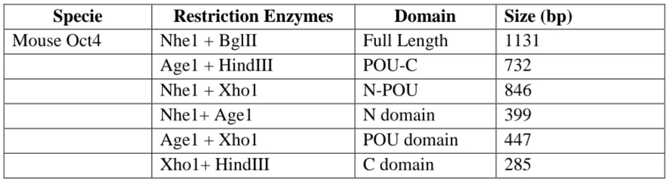
Table 2. 3- Mouse Oct4 enzyme combinations used to generate different fragments Specie Restriction Enzymes Domain Size (bp)
Mouse Oct4 Nhe1 + BglII Full Length 1131
Age1 + HindIII POU-C 732
Nhe1 + Xho1 N-POU 846
Nhe1+ Age1 N domain 399
Age1 + Xho1 POU domain 447 Xho1+ HindIII C domain 285
Sturgeon Oct-4 has a restriction site at the 1024 bp for Xho1 [35], indicated on Figure 2. 5 with the symbol ( ), therefore the fragments N-POU and POU cannot take the Xho1 adapter, being replaced by Sal1. The enzyme combinations used to make the fragments are described in Table 2. 4.
Figure 2. 5- Enzymes used to create the different Sturgeon Oct-4 fragments. indicates where the Xho1 cuts the sequence.
18
Table 2. 4- Sturgeon Oct4 enzyme combinations used to generate different fragments.
Specie Restriction Enzymes Domain Size (bp) Sturgeon Oct4 Nhe1 + HindIII Full Length 1362
Age1 + HindIII POU-C 657
Nhe1 + Sal1 N-POU 1158
Nhe1 + Age1 N domain 705
Age1 + Sal1 POU domain 453 Xho1+ HindIII C domain 204
Zebrafish Pou2 and all Xenopus (Xl25, Xl60, Xl91) cDNA do not have restriction sites for any of the main enzymes (Nhe1, Age1, Xho1 and HindIII) [35] so all the fragments were inserted with the same adapters but generating different size fragments. The restriction enzymes sites are described on Figure 2-6, and all the different combinations and fragment sizes for zfPou2 are in the Table 2. 5, for Xl91 the fragments are described on Table 2. 6, for Xl60 the fragments are on
Table 2. 7 and for Xl25 are in Table 2. 8.
Figure 2. 6- Enzymes used to create the different Zebrafish Pou2, Xenopus Oct-91,
Xenopus Oct-60 and Xenopus Oct-25.
Table 2. 5- Zebrafish Pou2 enzyme combinations used to generate different fragments.
Specie Restriction Enzymes
Domain Size (bp) Zebrafish Nhe1 + HindIII Full Length 1419
Age1 + HindIII POU-C 663
19
Nhe1+ Age1 N domain 756
Age1 + Xho1 POU domain 453 Xho1 + HindIII C domain 210
Table 2. 6- Xenopus Oct-91 enzyme combinations used to generate different fragments.
Specie Restriction Enzymes
Domain Size (bp) Xenopus Oct-91 Nhe1 + HindIII Full length 1338
Age1 + HindIII POU-C 678
Nhe1 + Xho1 N_POU 1143
Nhe1+ Age1 N domain 660
Age1 + Xho1 POU domain 483 Xho1 + HindIII C domain 195
Table 2. 7- Xenopus Oct-60 enzyme combinations used to generate different fragments..
Specie Restriction Enzymes
Domain Size (bp) Xenopus Oct-60 Nhe1 + HindIII Full length 1281
Age1 + HindIII POU-C 669
Nhe1 + Xho1 N-POU 1076
Nhe1+ Age1 N domain 612
Age1 + Xho1 POU domain 464
Xho1 + HindIII C domain 205
Table 2. 8- Xenopus Oct-25 enzyme combinations used to generate different fragments.
20
Xenopus Oct-25 Nhe1 + HindIII Full length 1347 Age1 + HindIII POU-C 657
Nhe1 + Xho1 N-POU 1140
Nhe1+ Age1 N domain 690
Age1 + Xho1 POU domain 450 Xho1 + HindIII C domain 207
Each fragment was amplified with primers corresponding to specific enzyme with the addition of a random nucleotide sequence: CAGT at the 5’ extremity of each primer. The enzymes and their corresponding sequences that were used are described in Table 2.9. The total oligonucleotide sequence with the adaptor sequences are described in Table 2-10.
Table 2. 9- Enzymes and adaptors used to create different Oct4 fragments for each species.
Fragment Enzyme Adaptors A M S Z 25 60 91
FL Nhe1 F+ HindIII R 5’CAGTGCTAGC+ 5’CAGTAAGCTT + - + + + + + FL Nhe1 F+ BglII R 5’CAGTGCTAGC+ 5’ CAGTAGATCT - + - - - - - POU-C Age1 F+ HindIII R 5’CAGTACCGGT+ 5’CAGTAAGCTT + + + + + + + N-POU Nhe1 F+ Xho1 R 5’CAGTGCTAGC +5 CAGTCTCGAG - + - + + + + N-POU Nhe1 F+ Sal1 R 5’CAGTGCTAGC +5’CAGTGTCGAC + - + - - - - POU Age1 F+ Xho1 R 5’CAGTACCGGT+ 5’CAGTCTCGAG + + - + + + + POU Age1 F+ Sal1 R 5’CAGTACCGGT+5’CAGTGTCGAC - - + - - - - CD Xho1 F+ HindIIIR 5’CAGTCTCGAG+ 5’CAGTAAGCTT + + + + + + + ND Nhe1F + Age1 R 5’CAGTGCTAGC+ 5’CAGTACCGGT + + + + + + + Adaptors include the CAGT sequence plus the enzyme sequence. The boldface section corresponds to the enzyme sequence. A- Axolotl, M- Mouse, S- Sturgeon, 25- Xenopus Oct25, 60- Xenopus Oct60, 91-Xenopus Oct91. (+) where the combination was used (-) species without this combination
.
Table 2-10. Oligonucleotides used for creating Oct4 mutants MOUSE OCT4
21 MOCT4-AGE1-R1 5’CAGTACCGGTTTTCATGTCCTGGGACTCCTC MOCT4-AGE1-F1 5’CAGTACCGGTGCCCTGCAGAAGGAGCTAGAAC MOCT4-XHO1-R1 5’CAGTCTCGAGACTTGATCTTTTGCCCTTCTG MOCT4-XHO1-F1 5’CAGTCTCGAGATTGAGTATTCCCAACGAGAA MOCT4-BGLII-R1 5’CAGTAGATCTACCCCAAAGCTCCAGGTTCTC AXOLOTL OCT4 AXOCT4-NHE1-F1 5’CAGTGCTAGCATGGCTGGGCATTTGGGACAG AXOCT4-AGE1-R1 5’CAGTACCGGTCCCTTCCTCGTCTCCGCTGTC AXOCT4-AGE1-F1 5’CAGTACCGGTGGGACGTCGGCGGACCTTGAA AXOCT4-SALI-R1 5’CAGTGTCGACGCTGCGCTTCCCCTTCTGTCG AXOCT4-XHO1-F1 5’CAGTCTCGAGATTTGCCGGGAGGAGTATGAT AXOCT4-HINDIII-R1 5’CAGTAAGCTTGTTGGAGTGCAGGTGCCTTCT STURGEON OCT4 STOCT4-NHE1-F1 5’CAGTGCTAGCATGTCTGATCGGTCTGTCACC STOCT4-AGE1-R1 5’CAGTACCGGTATTCTCCTCTTCTTCCGAGTC STOCT4-AGE1-F1 5’CAGTACCGGTTTGTCCACGGAGGAGCTGGAG STOCT4-SALI-R1 5’CAGTGTCGACGGCCAGACGCTTCCCCTTCTG STOCT4-XHO1-F1 5’CAGTCTCGAGCTGCCCTTTGATGAGGAGGGT STOCT4-HINDIII-R1 5’CAGTAAGCTTGCTGGTCAGGTGTCCCAGCCC ZEBRAFISH OCT2 ZFOCT4-NHE1-F1 5’CAGTGCTAGCATGACGGAGAGAGCGCAGAGCCCA ZFOCT4-AGE1-R1 5’CAGTACCGGTCAGAGTCTCCTCTTCCTCAGA ZFOCT4-AGE1-F1 5’CAGTACCGGTACTACTGAAGATTTGGAGCAG ZFOCT4-XHO1-R1 5’CAGTCTCGAGCAAAGCTAGACGCTTTCCCTT ZFOCT4-XHO1-F1 5’CAGTCTCGAGCCCTTTGATGACGAGTGTGTT ZFOCT4-HINDIII-R1 5’CAGTAAGCTTGCTGGTGAGATGACCCACCAA XENOPUS OCT91
XNOCT91-NHE1-F1 5’CAGTGCTAGC ATGTATAACCAACAGACCTACCCT XLOCT91-AGE1-R1 5’CAGTACCGGT GGCTTCCTCCTCACTGTCACT XLOCT91-AGE1-F1 5’CAGTACCGGTCCTAATTCTGGGGAGATGGAG XLOCT91-XHO1-R1 5’CAGTCTCGAGCTCCCCGCCATTCTCCCTAAT XLOCT91-XHO1-F1 5’CAGTCTCGAGCCTTATGACGCCCCCCAAACC XLOCT91-HINDIII-R1 5’CAGTAAGCTTGTTGCCTTGGTTACCCATGCC XENOPUS OCT60
XLOCT60-NHE1-F1 5’CAGTGCTAG ATGGACCAGCCCATATTGTACAGC XLOCT60-AGE1-R1 5’CAGT ACCGGTTCCATCCTCTTCAGTTCCAG XLOCT60-AGE1-F1 5’CAGT ACCGGTATGACCCTTGAGGAGATGGAA XLOCT60-XHO1-R1 5’CAGT CTCGAG TTGGACATTCTGAACTTGCTC XLOCT60-XHO1-F1 5’CAGT CTCGAGGGGGCATGAGTTTGTGGGTGG XLOCT60-HINDIII-R1 5’CAGT AAGCTTGCCGGTCAGGACCCCCATAGA XENOPUS OCT25
XLOCT25-NHE1-F1 5’CAGTGCTAGCATGTACAGCCAACAGCCCTTCCCA XLOCT25-AGE1-R1 5’CAGTACCGGTGGGAACCTCCTCCTCATTGTC
XLOCT25-AGE1-F1 5’CAGTACCGGTAGCGAATCAGAAATGGAGCAG
22 XLOCT25-XHO1-F1 5’CAGTCTCGAGATGCCCACCGTTGAGGAGAAC XLOCT25-HINDIII-R1 5’CAGTAAGCTTGCCAATGTGGCCCCCCATGGC XLOCT25-FULL LENGTH F1 5’ATGTACAGCCAACAGCCCTTC XLOCT25-FULL LENGTH R1 5’TCAGCCAATGTGGCCCCCCAT
The different fragments were generated by PCR from plasmids containing the cDNA templates of the genes to be amplified. This plasmids were supplied by James Dixon and Jodie Edgson. The amplification was done using the primers described above using REDTaq REadyMix (Sigma- Aldrich, UK) according to the manufacturer’s instructions. The amplification was performed on an Eppendorf Mastercycle epGradient S cycler machine. PCR reactions were heated to 94°C for 5 minutes followed by 45 cycles of denaturation at 94°C for 1 minute, annealing at 56°C for 5 1 minute and 30 seconds, and extension at 72°C for 1 minute and 30 seconds. After amplification the PCR samples were loaded on a (0.5X) TAE Agarose gel (1.2%) and separated at 135v. Their sizes where compared to 1kb and 100bp molecular weight markers (Biolabs, New England). After isolating the correct amplified size segments, they were purified from the gel using Qiaspin column and Gel Extraction Kits (QIAGEN, UK) according to the manufacturer’s instructions.
PCR products were ligated into the pGEM®-T Easy vector (Promega, UK) according to the manufacturer’s instructions. The ligation mix was used to transform DH5-α E. coli competent cells. The culture cells were plated in LB agar medium supplemented with (10 mg/ml) Amplicilin and IPTG to 0.5mM and left to grow overnight at 37ºC. Individual colonies were selected and transfered to liquid LB medium with Amplicilin, overnight at 37ºC.
Plasmid DNA was extracted from cultures using QIAprep Spin Miniprep Kit using a microcentrifuge (QIAGEN, UK). To confirm that the fragments were inserted into the pGEM®-T Easy vector, 5 µ L of the miniprep DNA was digested with EcoRI (10000U/ml) (BioLabs, New England) for 1 hour and 30 minutes at 37ºC with 2µL of Buffer 2 (BioLabs, New England), 2 µ L of 10X BSA, 2µ L o EcoRI (BioLabs, New England) and 9µ L of purified water. EcoRI cuts on either side of pGEM®-T Easy (appendix 12) vector at the 23 and 70 bp after the transcription site, it also cuts the
23 stOct-4 insert at 398 bp, due to an internal EcoRI site. When the fragments are correctly inserted into pGEM®-T the fragment sizes described in the Table 2-11, should be obtained.
Table 2-11- Size of the different fragments constructed of each species. Species Domain Size (bp)
Axolotl Oct4 Full- Length 1197
POU-C 648
N-POU 996
N domain 549
POU domain 447
C domain 201
Mouse Oct4 Full Length 1131
POU-C 732
N-POU 846
N domain 399
POU domain 447
C domain 285
Sturgeon Oct4 Full Length 964 and 398
POU-C 657
N-POU 1158
N domain 398 and 310 POU domain 453
C domain 204
Zebrafish Full Length 1419
POU-C 663
N-POU 1209
N domain 756
POU domain 453
C domain 210
Xenopus 91 Full length 1338
POU-C 678
24
N domain 660
POU domain 483
C domain 195
Xenopus 60 Full length 1281
POU-C 669
N-POU 1076
N domain 612
POU domain 464
C domain 205
Xenopus 25 Full length 1347
POU-C 657 N-POU 1140 N domain 690 POU domain 450 C domain 207
The primary objective is to generate plasmids for the generation of fragments that can be ligated into specific vectors, pATG and pDBD, for transcriptional assays.
2.1.2. pATG
pATG vector is derived from pEGFP-C1 (Clontech) GenBank accession #: U55763. The vector and the alterations can be seen on Figure 2. 7. The Multiple cloning site (MCS) of pEGFP-C1 vector can be found in appendix 10. The MCS of pATG consists of Nhe1, Age1, Xho1, HindIII, BamHI (James Dixon, personal communication)
25 The enzymes in the pATG MCS are compatible with the fragments generated above. In order to clone the fragments into pATG, the vector and the fragments need to be digested with the respective enzymes that are described on Table 2. 2 and on
Table 2. 8. To increase cloning efficiency pATG was also desphosphorylated by alkaline phosphatase, calf intestinal (CIP) (10,000 units/ml) (BioLabs, New England) after a double digestion with the restriction enzymes.
Enzymatic digestion was performed using 300 ng/µL of plasmid, 3 µ L of enzyme (1,5 µl each), 3µ L of BSA (10X) (BioLabs, New England) and 3µ L of the respective buffer to a final volume of 30 µ L. The buffers used for plasmid digestion were chosen using BioLab instructions [36]. Digestion was performed at 37ºC for 1h and 30 minutes. After digestion, the samples were loaded into a (0.5X) TAE 1.2% Agarose Gel and run at 135V. The correct fragments were identified by comparison to 1kb and 100bp molecular weight (MW) makers (BioLabs, NewEngland); extracted and purified using Qiaquick Spin- Gel Extraction Kit (QIAGEN, UK) according to manufacturer’s instructions.
2.1.2. pDBD
pDBD- Gal4 vector was prepared by James Dixon (personal communication). It has the same similar MCS as pATG with the exception for Xho1which was replaced by Sal1 (cohesive ends of Xho1 and Sal1) [36]. The vector can be seen in Figure 2. 8.
26 The vector was digested with the appropriate enzymes using the buffers suggested by BioLab instruction [36]. To create some of the vectors it necessary to do sequential digestion, due to the incompatible enzyme buffers [36]. The digestions were purified purified using Qiaquick Spin columns (QIAGEN, UK). Each digestion was done at 37ºC for 1h and 30 min. After digestion, the samples and 1Kb and 100bp DNA ladder (BioLabs, New England) were loaded into TAE 1.2% Agarose Gel and fragments separated at 135V. The correct fragments were extracted and purified using Qiaquick Spin- Gel Extraction Kit (QIAGEN, UK) according to manufacturer’s instructions. The ligation of fragments to vector was done to a ratio 5:1 in the presence of T4 DNA ligase (2000,000 units/mL) (BioLabs, New England) in appropriate buffer (BioLabs, New England). The reaction was performed at room temperature for 1h and 30 min. After ligation the mix was used to transform DH5α competent cells. The transformations were performed by adding 100µ L of competent cells to the ligation reaction, and leaving on ice for 30 min. Then heatshock was performed at 42ºC for 45 seconds followed by returning to ice for 3 minutes. 500µL of SOB is then added and the cells incubate in a 37ºC shaker for 1 hour and 30 minutes. After transformation, 200µ L of the mixture is spread on an Agar (30µ g/ml) plate and incubated overnight at 37ºC. Two distinct colonies are then picked and grown overnight in a 37ºC shaker, in liquid LB media supplemented with Kanamycin ((30µg/ml). Plasmid DNA is then extracted using Qiagen Plasmid Mini Kits (QUIAGEN, UK) according to manufacturer’s instructions.
2.1.3. pGFP
pGFP vector is identical to pDBD vector but instead of the DNA binding domain is has a green fluorescent protein (GFP) molecule, Figure 2. 9. The ligation and transformation were done in the same way as above described.
27
Figure 2. 9-Vector pGFP used in cloning.
HeLa cells, were transfected with 0. 25µg/well of pGFP using GeneJuice Trasfection Reagent (Novagen, Germany) and incubated at 35ºC (5% CO2). Cells were fixed at
room temperature for 25 min with 2% formaldehyde in phosphate buffered saline (PBS). Nuclei were stained with 4,′6-diamidino-2-phenylindole (DAPI) and GFP expression was followed by fluorescence microscopy.
2.2.
Cell culture and transfection.
HeLa cells and HEK 293T cells were cultured in Dulbecco’s Modified Eagle’s Medium (DMEM; Sigma) supplemented with 10% fetal calf serum, 1X non-essential amino acids, and 100µg/ml antibiotics (penicillin and streptomycin). For reporter assays, approximately 200,000 HeLa and HEK 293T cells were seeded in 24-well cell culture plates and incubated at 37ºC (5% CO2) overnight. Cells should be 50-80% confluent
before transfection. For the reporter assay two different luciferase reporter were used. The reporter plasmid 6Wtk-luc (appendix 13) that was kindly provided by Dr. Niwa (16), and the Gal4-lux reporter. The plasmids were also co-transfected with pGL4.74 (hRluc/TK) (Promega, UK). The transfection was done using GeneJuice Transfection Reagent (Novagen, Germany) according to manufacturer’s instructions. Cells were transfected with 0.25µg/well of the luciferase reporter DNA, and with 0.25µg/well of the test expression vector DNA and 0.05µg/well of the RL-TK DNA. Experiments were done in triplicates for each construct and reporter. The cells were left two days at 37ºC (5% CO2).
The dual-luciferase assays were conducted by James Dixon. Dual luciferase assays were performed using a dual luciferase assay system (Promega, UK) in a Veritas™ Microplate Luminometer (P/N 9100-002) (Promega, UK). Cells were lysed in 200ul
28 passive lysis buffer. For luminescence measurements, 25µl of firefly luciferase reagent (LARII) was added to 25µl of lysate sample, where the firefly luciferase activity is measured. Followed by addition of 25µl of stop and glow (the Renilla luciferase reagent and firefly quenching). The results are expressed as the ratio of firefly to Renilla luciferase activity, (Fluc/Rluc). Experiments were conducted in triplicate.
29
Chapter 3: Results
3.1. Oct4 domain structure.
Oct-4 protein can be divided in three distinct functional domains; a POU domain which mediates DNA-binding function; an amino (N)-terminal (N), which has been shown to act as a ubiquitous transcriptional activation domain (TAD), and a carboxyl (C)-terminal (C) as a cell-type specific TAD (16). The importance of these domains has so far been investigated using mouse Oct-4 (mOct4). So far no work has been carried out to determine the domain function of Oct-4 proteins in other species. Before, determining how the domains influence Oct-4 transactivation, and if that function is shared in similar Oct-4 proteins; it was necessary to localise the domains.
As previously shown in Chapter 2, the domains have similar lengths, with the POU domain being the most conserved among all species. By using standard molecular biology techniques and mammalian over-expression analyses Oct-4 domain function through vertebrate evolution was investigated. To achieve this, vectors were created to express Oct-4 proteins by isolating and cloning different combinations of the N, POU and C domains. These fragments were isolated and recombined to form the constructs shown in Figure 2.1. The different vectors were used in different assays.
3.2. Oct4 sequence identity as function prediction
Apart from directly testing Oct-4 function experimentally, it is possible to predict how Oct-4 proteins might function by studying their amino acid sequence; domain conservation and sequences within them. As many Oct-4 studies have been conducted with mOct-4 and Xenopus Oct-4 (Xl91, Xl60, and Xl25) proteins, I aimed to predict protein behaviour by comparing the amino acid conservation and similarity of these two
30 species with axolotl (axOct-4), sturgeon (stOct-4), and zebrafish (zfPou2); it was also possible to determine the phylogenetic relationship between Oct-4 family members. This was practically examined by reporter analyses, which will be given later.
The amino acid sequences of the Oct4 proteins were obtained using the cDNA sequences in Appendix 1 to 7, and afterwards were compared to previously published sequences in NCBI Genbank, except for stOct-4. The protein sequences where entered into BioEdit [34] to calculate the identity for the full length sequence, and also each domain individually. According to the values described inTable 3.1, axolot Oct4 has highest identity to mouse Oct4, when compared with all the species.
Table 3. 1- Sequence identity of Oct4. Protein
sequence
A/M A/S A/Z A/25 A/60 A/91 M/S M/Z M/25 M/60 M/91
Complete 0.263 0.234 0.224 0.253 0.256 0.249 0.232 0.195 0.222 0.222 0.219 POU 0.469 0.483 0.032 0.273 0.435 0.417 0.423 0.022 0.106 0.376 0.410 (C) 0.298 0.418 0.393 0.350 0.380 0.361 0.277 0.270 0.266 0.242 0.270 (N)l 0.076 0.055 0.063 0.069 0.063 0.068 0.012 0.059 0.056 0.053 0.059 The percentage was measured using BioEdit [34]. A corresponds to Axolotl; M to Mouse; S to Sturgeon; Z to Zebrafish Pou2; 25 to Xenopus Oct25; 60 to Xenopus Oct60; 25 to Xenopus Oct25 Protein sequence S/Z S/25 S/60 S/91 Z/25 Z/60 Z/91 25/60 25/91 60/91 Complete 0.263 0.265 0.246 0.254 0.257 0.236 0.241 0.266 0.297 0.240 POU 0.024 0.052 0.590 0.590 0.029 0.029 0.032 0.058 0.059 0.616 (C) 0.581 0.373 0.411 0.380 0.319 0.360 0.356 0.342 0.443 0.376 (N) 0.051 0.076 0.063 0.089 0.083 0.043 0.059 0.073 0.130 0.100 The percentage was measured using BioEdit (34). A corresponds to Axolotl; M to Mouse; S to Sturgeon; Z to Zebrafish Pou2; 25 to Xenopus Oct25; 60 to Xenopus Oct60; 25 to Xenopus Oct25
31 According to Table 3.1, Oct-4 mouse full length has more similarity with axOct4 (0.263) that with any other species and the lowest identity is with Zebrafish Pou2 (0.195). As expected, the POU domain is the most well conserved domain. Despite mouse and axolotl having a high level of similarity, it is not as high as sturgeon and zebrafish, but these two species came from the same ancestor (12). The identity starts to decrease drastically on the surrounding domains, (C) and (N), but even in those domains mouse and axolotl still share the highest similarity value.
Using the full length amino acid sequence it is possible to draw a phylogenetic tree based upon average distance (Figure 3. 1).
Figure 3. 1- Average distance tree using BLOSUM62 [34].
According to Figure 3.1 there is an evident division of the Xenopus Oct expression to all the others. Within the Xenopus, the Xl60 is closer to Xl25 than to Xl91. It also shows
32 that axOct4 and mOct4 share more similarities between them, that with any other species. StOct4 and zfpou2 also have a higher similarity between them.
3.3. Amino Acid Composition
It is important to determine the amino acid composition, similarity between the different Oct-4 homologues. It is possible that a difference between and within the domains is based on their different amino acid composition.
The amino acid composition of each species and the respective amino acid graphics can be found in Appendix 9 to 11. The amino acid composition was calculated using BioEdit program [34]. The full length values are not shown because it represents the sum of the domains.
3.3.1 N- Terminal (N)- domain
The N-domain was known by being a proline rich domain, due to the fact that 25% of its constitution is proline [16, 20], and that this amino acid plays an important role in mouse Oct-4 transactivation [16].
According to the amino acid composition predicted by BioEdit [34], mouse (N) does not have 25% of proline, but 14.29%, and also glycine is also the amino acid present in higher quantity (17.29%) followed by proline and then glutamic acid. In general, the proline values oscillate from 8.2% (axolotl) to 14.29% (mouse), the species with similar amount of proline to mouse is Xenopus Oct-25 followed by zebrafish.
The amino acid found in higher quantity in axolotl and mouse is glycine; while in sturgeon, Xl91 and Xl60 is serine; in zebrafish Pou2 and in Xl25 is proline. Xenopus Oct-4 homologues (Xl25, Xl60, Xl91) have in common proline and serine constitution; and Xl60 is similar on the amount of glutamic acid when compared to mouse.
33
3.3.2 POU domain
The values and respective graphics for POU amino acid composition are in Appendix 9. According to the results obtained, the amino acid present in higher amounts in axolotl, mouse, sturgeon, and zebrafish is leucine. In Xl91 and Xl25 is lysine while in Xl60 is glutamic acid.
The second amino acid present in higher amount in axolotl is glutamic acid; in mouse is lysine; in sturgeon and zebrafish are lysine and arginine; in Xl91 is glutamine; in Xl60 is lysine and leucine; and in Xl25 is leucine, glutamine and arginine.
3.3.3 C- Terminal (C)-domain
The amounts of amino acid present on the C-terminal can be found in Appendix 11. C-domain is known for having high amounts of proline, serine and threonine [16], but according to Table 3.4, proline is in fact present in significant levels, but not serine and threonine, the amino acids that are present in higher amounts are leucine and proline (13.68% and 12.63%). Proline is in fact present in high levels in stOct4 (16.18%), zbpou2 (20.00%), Xl91 (20%) and Xl60 (16.18%). In this same species, the second amino acid present in higher levels is glycine, fact also shared with Xl25.
The tryptophan is the only amino acid present in mOct4 that is not present on the other species. In Nanog, the tryptophan is associated with the development of extra-embryonic tissue (James Dixon, unpublished data).
a) Phosphorylation prediction
If the C-terminal is correlated with phosphorylation events [40], tyrosine, serine and threonine are known to be involved in phosphorylation reactions. According to the table
34 on appendix 11, Xl60 is the one with more serine, but very low amounts of threonine and tyrosine. Axolotl (C) has big quantities of serine and tyrosine. The amino acid present in higher quantities in mOct4 is serine, but it also has threonine and tyrosine. All the other species also have the three amino acids related to phosphorylation.
For phosphorylation events, the amino acid constitution is not enough, it is necessary a favourable composition of the surrounded amino acids. By using NetPhos 2.0 [38] it is possible to predict the probability of phosphorylation events to happen on a given sequence. The probability is scored from 0.5 to 1, only the values above 0.500 are considered. The results are shown in Table 3.2 and Table 3.3.
Table 3. 2 Prediction of phosphorylation positions in the C domain, for serine; and tyrosine, according to Netphos 2.0 [38].
Serine Tyrosine
Position Sequence Score Position Sequence Score AxOct4 28 HLPTSYIAQ 0.930 6 CREEYDGFQ 0.670 56 SEMYSQTVS 0.735 55 DSEMYSQTV 0.653
Moct4 5 EAPTSPHST 0.964
8 TSPHSTQSL 0.959
45 GDAVSQGKG 0.553
StOct4
Zbpou2 12 EAQYYEQSP 0.928
XLOct25 12 DGEGYDVAQ 0.860
43 APQIYASAG 0.746
XlOct60 26 XAVPSHGSG 0.829
35
Table 3. 3 Prediction of phosphorylation positions in the C domain, for serine; and tyrosine, according to Netphos 2.0 [38].
Threonine
Position Sequence Score
AxOct4 mOct4 34 PCIQTEAPA 0.768 StOct4 Zbpou2 XLOct25 17 DVAQTMGRP 0.544 XlOct60 7 IGLSTPQPS 0.561 XlOct91 7 DAPQTLTPP 0.862
According to Netphos 2.0 [38] stOct4 does not have relative phosphorylation sites, despite having threonine. Mouse C-terminal has four phosphorylation sites that might justify the hypothesis that this domain is regulated by phosphorylation events. Nonetheless, axC also has a high number of phosphorylation amino acids, two in serine and two in tyrosine. The Xenopus domains have less phosphorylation sites, but with a higher probability of occur.
To know if those regions are conserved among the species in study, the sequence given by Netphosh (Table 3.2; Table 3.3) as phosphorylation place, were localized in a MAFT alignment and the phosphorylation sites where highlighted. The result can be seen in Figure 3.2.
Figure 3. 2- C-Domain MAFT alignment with the phosphorylation sites highlightened; in blue is where it might be a phosphorylation in a serine amino acid; in red is where there might be phosphorylation in a threonine amino acid; in green is where phosphorylation might occur in a tyrosine amino acid.
36 Zebrafish Pou2, Xl91 and Xl25 have similar phosphorylation sites(10- 30 aa), and axolotl and mouse have a similar phosphorylation area (30 and 70 aa).
3.4. Alignments
To study the conservation between the different species, it is necessary to make amino acid alignments. There are several alignment programs such as BioEdit [34]; Jalview [37]; Blast; Emboss:: Neddle; Emboss::Water. All of these have different algorithms for sequence alignment.
MAFFT makes sequence alignments using a fast Fourier transform (FFT) approximation [39]. It is able to align more than 50 sequences with a higher accuracy than ClustalW [39], program that can be found in BioEdit. MAFFT version 5, can be found in Jalview v.2.3, that combined with Jalview tools display information about the quality conservation and consensus within the sequences [37]. The consensus display for the alignment is scored from 1 to 9, where 1 is the lower level, and when there is a 100% match, it is scored with a (*). The consensus graphic combines all the sequences into the amino acid that is present in all sequences for the same position. For being more accurate, and for displaying more information in one data, the program used to make all sequence alignments was MAFFT from Jalview v.2.3. The amino acid constitution from each sequence was also calculated by Jalview v2.3 [37].
3.4.1. N-terminal (N)-domain
The proline region of mOct4 is found between the 13-60 amino acid, and when that region is deleted there is a significant decrease in Oct-4 transactivation [16]. It was possible to see that the proline is not present in similar values for all species. However, it might be possible that proline is concentrated in the same area as mouse. The influence of this region on other species, is not known.
37
Figure 3. 3- N-terminal alignment for all species. The green delimitation shows where most of the proline is concentrated. The red delimitation shows a conserved area within the N-terminal.
According to Figure 3.3, the proline region is not conserved within species, but there is a region where most of the proline can be found (160 to 180 aa). It is possible to see a different conserved area, where most of the amino acids are serine and glutamic acid, but this similarity is not shared in mouse sequence, now termed SEB Box.
3.4.2. POU-domain
POU alignment can be seen in Figure 3.4. Duanquin Pei et at. (24) identified a nuclear localization signal (NLS) in mouse Oct-4. The NLS was found in the Pou domain, the sequence responsible for its localization in the nuclei and required for the transactivation of its target genes is RKRKR [24]. By comparing outa multiple
![Figure 1. 1- Different paths of establish pluripotent embryonic stem (ES) and embryonic germ (EG) cell lines from the inner cell mass (ICM) of mouse blastocysts and from primordial germ cells, respectively [7]](https://thumb-eu.123doks.com/thumbv2/123dok_br/18044353.862437/13.892.331.616.615.900/different-establish-pluripotent-embryonic-embryonic-blastocysts-primordial-respectively.webp)
![Table 1.1- Different strategies for reprogramming differentiated cells into a pluripotent state [9]](https://thumb-eu.123doks.com/thumbv2/123dok_br/18044353.862437/14.892.121.770.373.1121/table-different-strategies-reprogramming-differentiated-cells-pluripotent-state.webp)
![Figure 1. 2 Model of Oct4, Nanog, Sox2, and FoxD3 interaction during early mouse development [1]](https://thumb-eu.123doks.com/thumbv2/123dok_br/18044353.862437/20.892.266.629.798.999/figure-model-nanog-foxd-interaction-early-mouse-development.webp)