Submitted 13 September 2017 Accepted 26 January 2018 Published 30 March 2018 Corresponding author Aschwin H. Engelen, [email protected] Academic editor
Mario Alberto Flores-Valdez Additional Information and Declarations can be found on page 20
DOI 10.7717/peerj.4377 Copyright
2018 Aires et al. Distributed under
Creative Commons CC-BY 4.0 OPEN ACCESS
Acidification increases abundances of
Vibrionales
and Planctomycetia associated
to a seaweed-grazer system: potential
consequences for disease and prey
digestion efficiency
Tania Aires1,*, Alexandra Serebryakova1,2,*, Frédérique Viard2,3, Ester A. Serrão1
and Aschwin H. Engelen1
1Center for Marine Sciences (CCMAR), CIMAR, University of Algarve, Campus de Gambelas, Faro, Portugal 2Sorbonne Université, CNRS, Lab Adaptation and Diversity in Marine Environments (UMR 7144 CNRS SU),
Station Biologique de Roscoff, Roscoff, France
3CNRS, UMR 7144, Divco Team, Station Biologique de Roscoff, Roscoff, France
*These authors contributed equally to this work.
ABSTRACT
Ocean acidification significantly affects marine organisms in several ways, with complex
interactions. Seaweeds might benefit from rising CO2through increased photosynthesis
and carbon acquisition, with subsequent higher growth rates. However, changes in
seaweed chemistry due to increased CO2may change the nutritional quality of tissue
for grazers. In addition, organisms live in close association with a diverse microbiota, which can also be influenced by environmental changes, with feedback effects. As gut microbiomes are often linked to diet, changes in seaweed characteristics and associated microbiome can affect the gut microbiome of the grazer, with possible fitness consequences. In this study, we experimentally investigated the effects of acidification on the microbiome of the invasive brown seaweed Sargassum muticum and a native
isopod consumer Synisoma nadejda. Both were exposed to ambient CO2 conditions
(380 ppm, pH 8.16) and an acidification treatment (1,000 ppm, pH 7.86) for three weeks. Microbiome diversity and composition were determined using high-throughput sequencing of the variable regions V5-7 of 16S rRNA. We anticipated that as a result of acidification, the seaweed-associated bacterial community would change, leading to further changes in the gut microbiome of grazers. However, no significant effects
of elevated CO2on the overall bacterial community structure and composition were
revealed in the seaweed. In contrast, significant changes were observed in the bacterial community of the grazer gut. Although the bacterial community of S. muticum as whole did not change, Oceanospirillales and Vibrionales (mainly Pseudoalteromonas) significantly increased their abundance in acidified conditions. The former, which uses organic matter compounds as its main source, may have opportunistically taken advantage of the possible increase of the C/N ratio in the seaweed under acidified conditions. Pseudoalteromonas, commonly associated to diseased seaweeds, suggesting that acidification may facilitate opportunistic/pathogenic bacteria. In the gut of S.
nadejda, the bacterial genus Planctomycetia increased abundance under elevated
acidification. Planctomycetia are slow-acting decomposers of algal polymers that could be providing the isopod with an elevated algal digestion and availability of inorganic compounds to compensate the shifted C/N ratio under acidification in their food. In conclusion, our results indicate that even after only three weeks of acidified conditions, bacterial communities associated to ungrazed seaweed and to an isopod grazer show specific, differential shifts in associated bacterial community. These have potential consequences for seaweed health (as shown in corals) and isopod food digestion. The observed changes in the gut microbiome of the grazer seem to reflect changes in the seaweed chemistry rather than its microbial composition.
SubjectsEcology, Marine Biology, Microbiology, Climate Change Biology, Biological Oceanography
Keywords Invasive seaweeds, Ocean acidification, Grazer microbiomes, Algae microbiomes, Metabarcoding, Sargassum muticum, Synisoma nadejda
BACKGROUND
Ocean acidification significantly affects marine organisms in diverse ways (Fabry et al.,
2008;Kroeker et al., 2013). In the case of species interactions (e.g., predator–prey), the outcome of such effects can be difficult to predict as antagonistic or synergistic effects
may be observed (Asnaghi et al., 2013; Branch et al., 2013;Poore et al., 2013). This is
particularly true for non-calcifying seaweeds, which in contrast to most other organisms
can benefit from rising CO2through increased photosynthesis and carbon acquisition, and
subsequently acquire higher growth rates (Porzio, Buia & Hall-Spencer, 2011;Harley et al.,
2012;Koch et al., 2013;Olischläger et al., 2013). However, changes in plant leaf chemistry in response to elevated carbon supply are expected to result in higher C:N and C:P ratios
and, as such, reduce the nutritional quality of tissue for grazers (Urabe, Togari & Elser,
2003;Van De Waal et al., 2010) and the same is expected for seaweeds. Variations in the palatability of seaweeds may lead to changes in consumption rates by herbivores, which will have to absorb nutrients more efficiently or consume more to compensate for low
concentrations of essential nutrients (Gutow et al., 2014). Thus, ocean acidification (OA)
could have positive effects on seaweed growth rate, but may also induce behavioral changes on the herbivores and increased grazing rates. Therefore, it is important to understand the effects of ocean acidification on prey (bottom-up effects) and, as a top-down effect, on predation.
Interactions among organisms and their associated bacterial communities affect the
holobiont physiology and health (Hollants et al., 2013; Egan et al., 2013), and play an
important role in the functioning of hosts as, in the case of this study, seaweeds (Singh et al.,
2011;Singh & Reddy, 2014). Seaweeds and marine organisms feeding on them live in a close
association with diverse and abundant microbial communities (King et al., 2012;Hollants
et al., 2013;Egan et al., 2013;Dudek et al., 2014). Seaweeds comprise dynamic species-specific bacterial communities (Aires et al., 2015;Aires, Serrão & Engelen, 2016;Vieira et al., 2016). The communities are recognized to have growth-promoting and nutritional effects (Head & Carpenter, 1975;Dimitrieva, Crawford & Yüksel, 2006), and to be involved
in the production of biologically active (Chojnacka et al., 2012) and defensive (Burgess et al., 1999) compounds. At a higher trophic level, symbiotic bacteria inhabiting the guts of
marine herbivores are also known to support important physiological functions (Hacquard
et al., 2015), including the mediation of the digestion of food components by producing
critical digestive enzymes for breaking down complex molecular structures (Mackie et al.,
2004). In addition to digestive functions, grazers depend on seaweed-associated microbiota
for nutrients found in the algal biofilm (i.e., proteins, polysaccharides, lipids, etc.;Tietjen,
2014). As such, diet represents an important factor in shaping microbial diversity in the
intestinal systems of grazers. So, any changes in bacterial composition of the seaweed may result in diet-induced changes in the gut microbiota of grazers that may eventually affect
their metabolism, as well as its fitness and biology (Mattila et al., 2014;Tietjen, 2014).
Because carbon acquisition is expected to be facilitated for seaweeds at elevated CO2levels,
higher nutrient uptake is anticipated to help obtain other nutrients in the right balance with carbon. Part of these nutrients might be obtained through the microbiome and, therefore, the specific bacteria responsible for such acquisitions (e.g., phosphorous, nitrogen and
iron) (Thomas et al., 2008;Burke et al., 2011b) might be positively selected and increase
their abundance.
Because OA is expected to affect the interactions between marine herbivores and
seaweeds through increased consumption of carbon enriched algal tissue (Gutow et al.,
2014), the microbiomes of grazers might help with nutrient acquisition. While better
understanding of the diversity and functions of associated symbiotic bacteria is needed, few studies have addressed the diversity and composition of gut microbiota of marine grazers (but seeHong et al., 2011;Devine, Pelletreau & Rumpho, 2012;Davis et al., 2013;
Dudek et al., 2014).
To predict the responses of aquatic organisms to OA, it is necessary to understand
responses of the host-associated microbiota to increasing CO2and reduced pH. Little is
known about the responses of associated microbiota to changes in pCO2(partial pressure of
carbon dioxide) including microbial metabolic capabilities or the ability to rapidly shift the
host range (Morrow et al., 2015). Also, there is no consensus regarding whether a decrease
in pH causes increase (Kerfahi et al., 2014), decrease (Taylor et al., 2014) or no changes
(Hassenrück et al., 2016) in microbial richness and prevalence of dominant microbial taxa under acidification conditions. Furthermore, the current knowledge of acidification effects on the host-associated microbial communities is mostly based on the results of experiments conducted on corals. These experiments demonstrated that reduced pH initiates shifts in
the coral microbiota towards microorganisms associated with stress and disease (Thurber
et al., 2009;Meron et al., 2011;Webster et al., 2013). Therefore, there is a need for relevant studies on seaweeds, with a particular interest in species interactions as species may not respond similarly to OA and as effects can act synergistically. This response is particularly relevant to be examined in marine introduced seaweeds, because they are expected to benefit from future OA conditions, and thus increase their invasiveness.
In this study, we experimentally investigated the effects of acidification on the microbiomes of an emblematic invasive seaweed, the brown alga Sargassum muticum, and the gut microbiome of a native isopod consumer, Synisoma nadejda. This was done by
following a three-week mesocosms exposure to elevated pCO2followed by 16S amplicon
sequencing in order to compare the bacterial community (hereafter microbiome) responses in these two hosts. Based on bacterial community characterization, our main hypotheses are that in acidified conditions (1) the seaweed-associated microbiome will have a different composition, (2) the grazers’ gut microbiome will mirror the changes in food source (assuming that seaweed nutritional content will change with acidification), when compared with ambient conditions. Considering the existing evidence for corals responses to OA (Thurber et al., 2009;Meron et al., 2011;Webster et al., 2013) and seaweeds responses to
other environmental stresses (e.g., temperature,Case et al., 2011;Mensch et al., 2016) we
expect seaweed microbiome to shift towards a community composed by stress related bacteria and putative pathogens. Also, raw plant consumers’ (e.g., fish, humans, etc.)
gut microbiome and health is directly affected by environmental conditions (Sullam et
al., 2012) or, following the ‘‘you are what you eat’’ premise, indirectly through changes in their food source/quality (Meziti et al., 2010;Berg et al., 2014), we predict that host’s ‘‘normal’’ gut bacterial composition could be affected by OA. An increased abundance of bacteria potentially assisting digestion could provide the isopod with an elevated algal digestion and availability of inorganic compounds to compensate the shifted C/N ratio under acidification in their food. Furthermore, if S. muticum showed a shift under OA conditions, we will investigate the presence of possible bacterial taxa that could assist
the seaweed obtaining nutrients (e.g., nitrogen fixing bacteria) under elevated pCO2
conditions. These predictions, for both seaweed and gut microbiome, are limited to 16S taxonomic assignments and literature description and inherent reservations to this method will be considered.
METHODOLOGY
Experimental set-up
The experiment was performed at Centro de Ciencias do Mar (CCMAR) field station
(Ramalhete) during the spring of 2014. Ambient (380 ppm CO2, and pH 8.16—global levels
of today’s CO2conditions) and elevated pCO2(1,000) ppm CO2, and pH 7.86—the year
2100 predictions by IPCC, A1FI scenario (Houghton et al., 2001) conditions were controlled
by two separate CO2 sensors systems. For acidified conditions, CO2was injected in seawater
deposits that provided seawater for the experimental units. In both systems salinity was 36,
alkalinity 2,550µmol kg−1and seawater temperature 15◦C. Experimental units consisted
of 3 L flowthrough mesocosms receiving each 30 L of seawater per hour. Experimental units were placed in one square meter tanks with 15 cm of the overflown seawater of the experimental units to stabilize temperature conditions in the units. Sargassum muticum and
Syniosoma nadejdawere sampled independently and kept isolated. Thus, the grazers were
‘naive’ and not previously on a diet containing S. muticum. After sampling, seaweeds and isopods were acclimated to ambient conditions in separate tanks for 1 week. A wet weight biomass of 1 g seaweed per experimental unit was used as a preliminary test and showed it did not affect the pH conditions in experimental units at the used volume and flow.
The two factors considered for the experiment were (1) CO2 conditions (two levels:
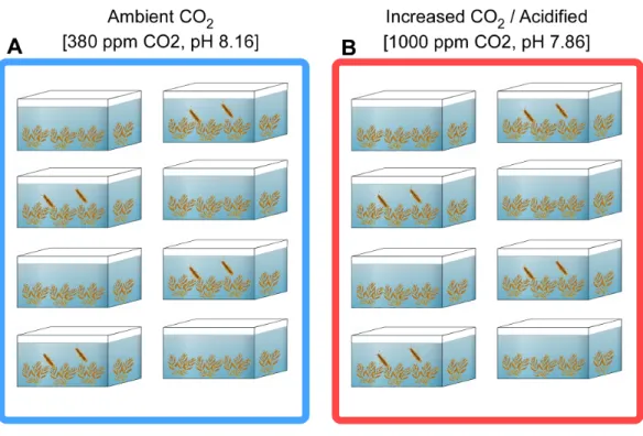
Figure 1 Schematic representation of the mesocosms experiment. (A) Ambient (380 ppm) and (B)
acidified (1,000 ppm) conditions each with four 3 L experimental units only containing 1 g wet weight (WW) S. muticum and four 3 L experimental units containing 1 g WW S. muticum along with the grazer S. nadejdawere placed randomly in each CO2treatment. Each unit represented a replicate from which
sam-ple(s) (seaweed or seaweed and grazer) were taken.
Full-size DOI: 10.7717/peerj.4377/fig-1
without grazers, the grazers that fed on the seaweed). These were combined in a factorial design (all possible combinations of all levels of the two factors). The number of replicates
within treatment combination was four. Therefore, in total, 24 samples (2 CO2× 3 hosts
× 4 replicates) were collected (Fig. 1). The experiment ran for three weeks which was estimated sufficient for microbiomes to reflect the applied conditions considering the high turnover rate of food in the grazer gut and the multiplication rate of bacteria. During this period the units were cleaned, twice a week, to avoid epiphyte overgrowth on the experimental unit walls. In each experimental unit, seawater pH and the calibration of the
automated CO2injection system was manually checked daily to make sure the pH was
stable. At the end of the three weeks, the isopods and seaweeds were flash frozen, transported
to the laboratory in liquid nitrogen and stored there at −80◦C until further processing.
Hight-throughput sequencing of the microbiome
For both seaweeds and isopodes’s guts (also refered to as grazers, hereafter), DNA was
extracted from all the 24 replicates using the Quick–gDNA kit (Zymo ResearchTM, Irvine,
CA, USA) according to the manufacturer protocol for ‘‘Solid Tissue Samples’’ (page 4 of the manual). Before extraction, isopods where dissected by removal of both ends and pulling out the intestinal tract. The total 16S rRNA was amplified using the universal
an initial denaturation at 95◦C for 2 min, 35 cycles of denaturation at 95◦C for 20 s,
annealing at 55 ◦C for 20 s, and extension at 72◦C for 90 s, with a final extension was
at 72 ◦C for 3 min. The 25 µl reaction mixture contained 250µM dNTPs, 0.6µM of
each primer, 1 × 2 PCR buffer mix, 2µl of template DNA (with a final concentration
of about 10 ngµl–1) and 0.3µl of Taq polymerase (Advantage R2; Clontech, Mountain
View, CA, USA). PCR products were cleaned using ExoFastAP enzyme following the
Thermo ScientificTM protocol. Amplified DNA was sent to Molecular Research (MR
DNA), Shallowater, Texas, where a nested–PCR was performed prior to sequencing. The modified 8 bp key–tagged primer 799F along with the reverse primer 1193R, covering the regions V5–V7 from 16S rRNA and amplifying a fragment of ∼400 bp, were used to avoid
chloroplast cross amplification (Bodenhausen, Horton & Bergelson, 2013). PCR conditions
were as follow: 95◦C for 3 min, 10 cycles of 95◦C for 20 s, 50◦C for 30 s, 72◦C for 30
s, and a final elongation of 72◦C for 3 min. All amplifications and sample preparation
procedures were the same for both seaweeds and isopodes. Samples were pooled together in equal proportions based on their molecular weight (calculated based on the size of
the amplicon) and DNA concentrations (using QubitTM; Invitrogen , Carlsbad, CA,R
USA) and purified using calibrated Agencourt AMPureR XP beads. DNA libraries wereR
prepared by following Illumina TruSeq DNA library preparation protocol and paired–end (2 × 250 bp) sequencing performed at MR DNA (http://www.mrdnalab.com; Shallowater, TX, USA) on a MiSeq following the manufacturer’s guidelines.
Detailled protocols for sampling procedures, DNA extraction and PCR amplification
mentioning all the important measures to avoid contamination, can be found in (Aires
et al., 2018).
Sequence analysis and bioinformatics
A total of 3,204,094 partial 16S rRNA gene sequences were obtained from the 24 samples
(i.e., two CO2conditions X four replicates for seaweed in the presence and the absence of
grazer and four grazer gut samples with two CO2conditions). The bacterial community
analyses were performed using Quantitative Insights into Microbial Ecology (QIIME
version 1.8.0) software (Caporaso et al., 2010). Sequences were screened and filtered for a
minimum read length of 350 bp (after reads were paired) and less than two undetermined nucleotides. Selected high-quality sequences were clustered into Operational Taxonomic Units (OTUs) within reads using denovo OTU picking method. Representative sequences for each OTU were selected using the ‘‘most-abundant’’ method and OTU sequence
alignment was carried out using PyNAST (Caporaso et al., 2010) and Greengenes v.13.8
(McDonald et al., 2012). Taxonomic assignments were done using the UCLUST (Edgar,
2010) method with a 97% confidence threshold. To assign each OTU to the closest matching
described taxon, searches were performed against the Greengenes taxonomy database v.13.8
for16S rRNA (McDonald et al., 2012), and sequences were putatively assigned to a described
taxon with a minimum threshold of 0.001 (default value). Eukaryotes (i.e., chloroplasts and mitochondria) matching sequences were excluded from the OTU table in downstream analyses as well as rare OTUs (singletons and doubletons) and unassigned sequences (those sequences that did not match any of those from the Greengenes database, with a minimum threshold of 0.001).
Quality filtering resulted in 2,877,493 high-quality sequences, with an average of 119,896 ± 46,294 reads per sample, which were clustered into 42,730 unique operational taxonomic units (OTUs). The OTU table was rarefied to the minimum number of sequences (66,831). As a result, a total of 41,139 unique OTUs remained. Public access to the data can be done
through:https://doi.org/10.6084/m9.figshare.5346316.v3.
All the statistical and diversity (alpha and beta) analysis were done using the filtered rarefied (to the minimum number of sequences—66,831) OTU table and considered significant at P< 0.05.
Alpha diversity indexes, including Chao I richness (Chao, 1984), observed number
of species (OTUs) and Shannon diversity, were calculated using QIIME software. Bacterial community structure (beta diversity) was assessed by permutational multivariate analyses of variance (PERMANOVA) using Bray–Curtis dissimilarity matrices from square-root transformed data. PERMANOVA tested for differences among samples with different levels of a priori factors: Type of sample: Seaweed vs Grazer gut; for both
CO2 treatments: Ambient vs Acidified; for Seaweed: Grazed vs Non-grazed, and the
interactions among these factors. The homogeneity of multivariate dispersions (based on mean distance to group centroid for all groups within each factor) was tested using a resemblance based permutation test (PERMDISP). To visualize differences and to assess dissimilarity between samples, Canonical Analysis of Principal coordinates (CAP) plots were constructed to test the assignment/clustering of treatments interaction (S.muticum XGrazingxAcidification and Grazer gutxAcidification) as a priori factor. Similarities and dissimilarities in bacterial communities between acidification treatments were explored using, similarity percentage analyses (SIMPER). For those bacterial taxonomic groups that displayed a high contribution (concerning their differential abundances in the treatments being compared) for the differences between the grazer gut and the seaweed and the two
CO2levels, two-way analyses of variance (ANOVA) were performed (with the preliminary
tests for normality and homogeneity of variances being implemented). Species (S. muticum
and isopod) and acidification (CO2ambient and elevated) were tested as factors affecting
the structure/composition bacterial communities. For bacterial OTUs for which significant interaction were detected, a post-hoc t -test was implemented using a Bonferroni correction
and a conservative alpha (considering the comparisons made: CO2effect in S. muticum,
CO2effect on isopod gut), effect of type of tissue (seaweed/gut) in ambient CO2and effect
of type of tissue in elevated CO2(P (T ≤ t ) two tail< 0.0125).
All bacterial community structure statistical analyses were performed using the software
program PRIMER-E + PERMANOVA v.6 (Clarke & Warwick, 1994;Clarke & Gorley,
2006).
RESULTS
Bacterial communities associated with S. muticum and S. nadejda’s gut
Overall, 563 bacterial OTUs from 74 classes (apart from bacteria classified as ‘Other and non-ID’) distributed across 28 phyla (plus ‘Other and non-ID’) were identified. Among them, 551 bacterial OTUs distributed across 27 phyla (plus Other and non-ID) were
0.4 0.2 -0.4 -0.2 0.4 0.2 -0.4 -0.2 0 0 CAP1 C A P 2 Ambient (380ppm pCO2) Acidified (1000ppm pCO2) Grazed S. muticum Non-grazed S. muticum
Synisoma nadejda gut
Grazed S. muticum Non-grazed S. muticum
Synisoma nadejda gut
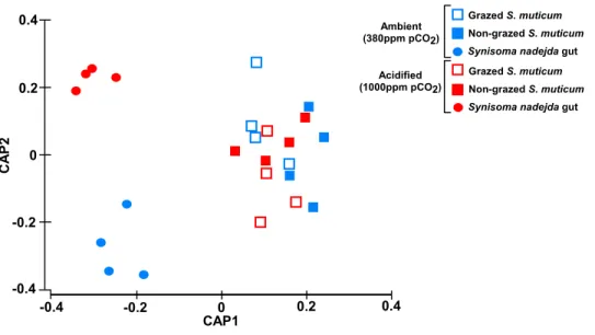
Figure 2 Community structure. Plot of canonical analysis of principal coordinates (CAP) based on
Bray–Curtis distances calculated on square-root transformed bacterial abundances, showing the axes that best discriminate the bacterial assemblages across CO2levels (blue-ambient versus red-acidified), grazing
by S. nadejda on S. muticum (open squares-grazed seaweed vs filled squares-non-grazed seaweed) and the gut of the isopod on a diet of S. muticum (circles).
Full-size DOI: 10.7717/peerj.4377/fig-2
associated with S. muticum (over all samples and treatments), compared to 450 OTUs distributed across 22 phyla (plus Other and non-ID) for gut biome. Note that those sequences labelled as ‘‘others’’ were ambiguous assignments by the classifier and ‘‘no-ID’’ sequences result from a good match with a reference sequence but that reference sequence is poorly defined (not named at a certain taxonomic level and below). Public access to the
data can be done through:https://doi.org/10.6084/m9.figshare.5346316.v3.
Alpha diversity of associated bacterial communities (Table S1) was similar or slightly higher in S. muticum than in the grazer gut for Shannon index (2-way ANOVA, F = 2.507,
P =0.139), OTU richness (2-way ANOVA, F = 3.361, P = 0.092) and Chao 1 (2-way
ANOVA, F = 6.226, P = 0.028), respectively. Acidification did not affect bacterial diversity
at OTU level, for both seaweeds and isopodes gut, as estimated by diversity indexes: Shannon index (2-way ANOVA, F = 0.048, P = 0.831), unique OTU richness (2-way ANOVA, F = 0.178, P = 0.681) and Chao 1 (2-way ANOVA, F = 0.003, P = 0.960).
Despite the overall PERMANOVA results showing that bacterial community composition was significantly different for the different type of samples (grazed and
non-grazed S. muticum and grazer gut, P = 0.001,Fig. 2,Table S2), predation (grazing)
did not significantly change bacterial community structure of S. muticum (P = 0.372,
Table S3A). The main differences among S. muticum (grazed and non-grazed) and the isopode gut (Table S2) were due to a higher abundance of Bacteroidetes (73.7%) associated to S. muticum (contributing 15.11% to the dissimilarity; SIMPER analysis) and to more
Proteobacteria(47.2%) and Planctomycetes (32.4%) in the gut of S. nadejda, contributing
100% 90% 80% 70% 60% 50% 40% 30% 20% 10% 0% Ambient pCO2 No grazing Acidified No grazing Ambient pCO2
Grazing AcidifiedGrazing S. muticum microbiome
Ambient pCO2 Acidified
Verrucomicrobia Proteobacteria Planctomycetes Firmicutes Chloroflexi Bacteroidetes Acidobacteria
S. nadejda gut microbiome
R e la ti ve P h yl a a b u n d a n c e
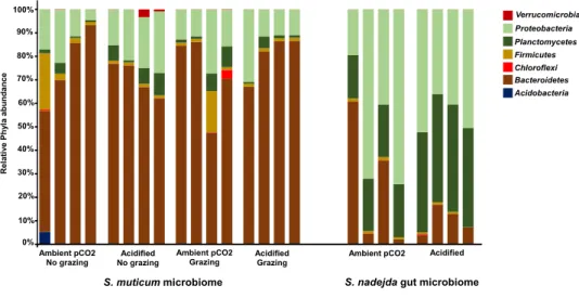
Figure 3 Host and treatment effects on associated bacteria phyla. Relative abundance and distribution
of the bacteria phyla associated to the brown seaweed Sargassum muticum, without (No grazing) and with (grazing) Synisoma nadejda isopods, and the gut of the isopod after three weeks on a Sargassum muticum diet, under ambient (380 ppm) and elevated/acidified (1,000 ppm) CO2conditions.
Full-size DOI: 10.7717/peerj.4377/fig-3
Bacterial phyla-specific to host (refered in PERMANOVA analysis as ‘‘Type of sample’’)
and specific conditions are presented inTable S4.
As mentioned before, grazing did not show significant effects on S. muticum bacterial community structure so, most of further analyses focused on the comparison of grazed
S. muticum vs grazer gut. Grazed S. muticum associated bacterial communities were
dominated by Bacteroidetes (75.4%, Fig. 3; Flavobacteriia 75.2%) and Proteobacteria
(16.2%, Fig. 3; Alphaproteobacteria 12.2%), while the isopod gut communities were
dominated by Proteobacteria (47.2%,Fig. 3; Alphaproteobacteria 43.5%), Planctomycetes
(32.4%, Fig. 3; Planctomycetia 32.1%), and Bacteroidetes (17.7%,Fig. 3; Flavobacteriia
16.8%). Flavobacteriales (73.3%) was the most common bacterial order associated with S.
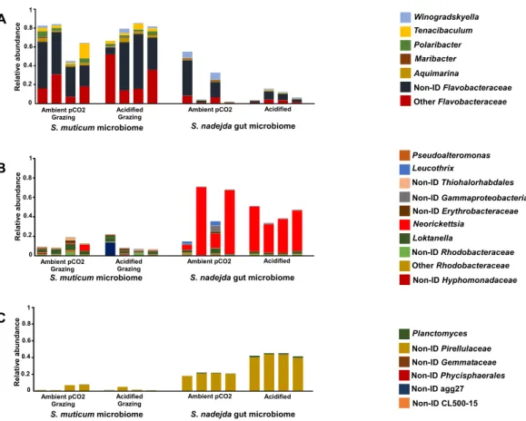
muticum, while Rickettsiales (38.4%), Pirellulales (30.9%) and Flavobacteriales (16.8%) were the most abundant grazer gut-associated orders. The distribution of the bacterial genera belonging to the main phyla found for the two different systems and described above (S.
muticum—Bacteroidetes and S. nadejda gut—Proteobacteria and Planctomycetes) showed a
clear higher relative abundance of genera assigned as Non-ID and ‘‘other’’ Flavobacteraceae (Fig. 4A) for the seaweed microbiome and a isopode gut dominated by Neorickettsialles (Fig. 4B) and non-identified Pirellulaceae (Fig. 4C).
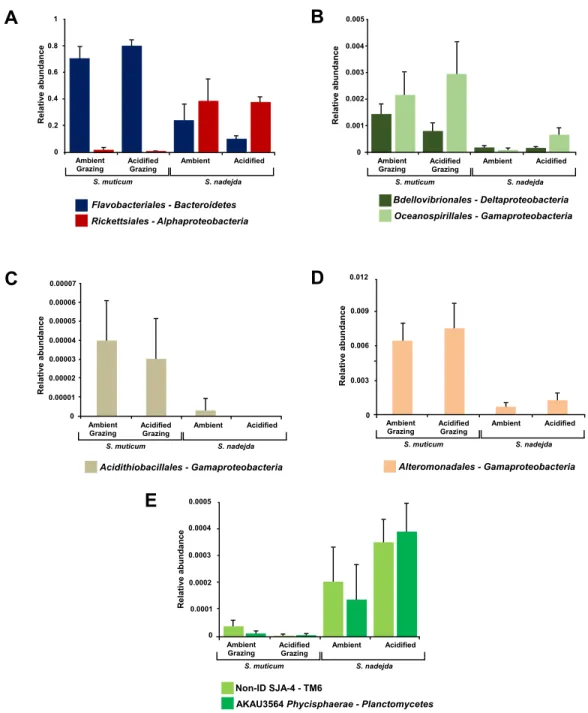
Several bacterial orders were significantly more abundant within grazed S. muticum than in the isopod gut: Flavobacteriales from the phylum Bacteroidetes (2-way ANOVA, F =
49.124, P < 0.001); Bdellovibrionales from the class Deltaproteobacteria (2-way ANOVA,
F =13.221, P = 0.003); as well as Acidithiobacillales (2-way ANOVA, F = 22.589, P =
0.0005), Alteromonadales (2-way ANOVA, F = 15.285, P = 0.002), and Oceanospirillales
(2-way ANOVA, F = 6.407, P = 0.026) from the class Gammaproteobacteria (Fig. 5). In contrast, the isopod gut microbiome had higher abundances of Rickettsiales from the class
1 0.8 0.6 0.4 0.2 0 R e la ti ve a b u n d a n c e 1 0.8 0.6 0.4 0.2 0 1 0.8 0.6 0.4 0.2 0 Ambient pCO2
Grazing AcidifiedGrazing S. muticum microbiome
Ambient pCO2 Acidified S. nadejda gut microbiome Ambient pCO2
Grazing AcidifiedGrazing S. muticum microbiome
Ambient pCO2 Acidified S. nadejda gut microbiome
Ambient pCO2
Grazing AcidifiedGrazing S. muticum microbiome
Ambient pCO2 Acidified S. nadejda gut microbiome
A B C Winogradskyella Tenacibaculum Polaribacter Maribacter Aquimarina Non-ID Flavobacteraceae Other Flavobacteraceae Pseudoalteromonas Leucothrix Non-ID Thiohalorhabdales Non-ID Gammaproteobacteria Non-ID Erythrobacteraceae Neorickettsia Loktanella Non-ID Rhodobacteraceae Other Rhodobacteraceae Non-ID Hyphomonadaceae Planctomyces Non-ID Phycisphaerales Non-ID agg27 Non-ID CL500-15 Non-ID Pirellulaceae Non-ID Gemmataceae R e la ti ve a b u n d a n c e R e la ti ve a b u n d a n c e
Figure 4 Relative abundance of the genera belonging to the main bacterial phyla. (A) Bacteroidetes, (B)
Proteobacteria, and (C) Planctomycetes, associated with the brown seaweed Sargassum muticum grazed by Synisoma nadejdaisopods (left side), and the gut microbiome of the isopod on a Sargassum muticum diet (right side), after three weeks under ambient (380 ppm; −CO2) and elevated/acidified (1,000 ppm; +CO2)
CO2conditions.
Full-size DOI: 10.7717/peerj.4377/fig-4
Alphaproteobacteria(2-way ANOVA, F = 18.574, P = 0.001); the low abundance bacteria
from the phylum TM6 —class SJA-4 (2-way ANOVA, F = 12.491, P = 0.004); and the
low abundance order AKAU3564-Phycisphaerae from the phylum Planctomycetes (2-way ANOVA, F = 7.325, P = 0.019) than S. muticum in the presence of grazer (Fig. 5).
Bacterial diversity and composition under acidification conditions
Acidification did not affect the overall bacterial composition (PERMANOVA, P = 0.093, Table S2) and in particular that associated with S. muticum (P = 0.056, P = 0.584, Table S3B), but significantly affected the bacterial composition associated with the
gut of S. nadejda (PERMANOVA, p = 0.022,Table S3B) (Fig. 2). However, under acidified
conditions (and in the presence of grazers), the number of phyla in S. muticum bacterial community dropped from 26 to 17 (including one unidentified), compared to the ambient conditions (One-way ANOVA, F = 7.228, P = 0.009). In contrast, the number of phyla in the grazer gut-associated bacterial community under acidification treatment increased from 18 to 23 (including one unidentified), compared to the ambient conditions (One-way ANOVA, F = 1.923, P = 0.171).
0 0.2 0.4 0.6 0.8 1 R e la ti ve a b u n d a n c e Ambient Grazing Acidified Grazing Ambient Acidified S. muticum S. nadejda Flavobacteriales - Bacteroidetes Rickettsiales - Alphaproteobacteria 0 0.001 0.002 0.003 0.004 0.005 R e la ti ve a b u n d a n c e Ambient
Grazing AcidifiedGrazing Ambient Acidified
S. muticum S. nadejda Bdellovibrionales - Deltaproteobacteria Oceanospirillales - Gamaproteobacteria 0 0.00003 0.00004 0.00005 0.00006 0.00007 R e la ti ve a b u n d a n c e 0.00002 0.00001 Ambient
Grazing AcidifiedGrazing Ambient Acidified
S. muticum S. nadejda Acidithiobacillales - Gamaproteobacteria 0 0.003 0.006 0.009 0.012 R e la ti ve a b u n d a n c e Ambient
Grazing AcidifiedGrazing Ambient Acidified
S. muticum S. nadejda Alteromonadales - Gamaproteobacteria 0 0.0001 0.0002 0.0003 0.0004 0.0005 R e la ti ve a b u n d a n c e Ambient Grazing Acidified Grazing Ambient Acidified S. muticum S. nadejda Non-ID SJA-4 - TM6
AKAU3564 Phycisphaerae - Planctomycetes
A
B
C
D
E
Figure 5 Mean relative abundances of bacterial classes, and respective orders (A, Flavobacteriales and Ricketsiales; B, Bdellovibrionales and Oceanospirillales; C, Acidithiobacillales; D, Alteromonadales; E, Non-ID SJA-4 and AKAU3564 Phycisphaerae), significantly more abundant in either grazed Sargassum muticumor the gut of Synisoma nadejda. After three weeks under ambient (380 ppm) and
elevated/acid-ified (1,000 ppm) CO2conditions. Alpha = 0.05, error bars show standard error per treatment (n = 4). Full-size DOI: 10.7717/peerj.4377/fig-5
The phyla specific to the microbiomes of S. muticum and gut of S. nadejda, under
ambient vs acidified conditions, are presented inTable S4.
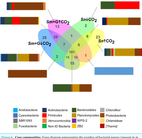
Some bacterial groups were unique to by their hosts under specific conditions (Fig. 6). The seaweed, in the presence of grazer and under the ambient conditions, had the highest
181 15 15 19 7 0 1 13 2 9 23 1 28 29 2 Sm+GiiCO2 Sm+GhhCO2 SniiCO2 SnhhCO2 Acidobacteria Cyanobacteria SBR1093 Fusobacteria Actinobacteria Firmicutes Verrucomicrobia Non-ID Bacteria Bacteroidetes Planctomycetes WPS-2 ZB2 Chloroflexi Proteobacteria Chlamideae [Thermi]
Figure 6 Core communities. Venn diagram representing the number of bacterial genera (present in at
least 75% of samples) shared between the different CO2treatments (ambient CO2−↓ CO2;
elevated/acidi-fied CO2−↑ CO2) and associated to grazed Sargassum muticum (Sm + G), and the gut microbiome of the
isopod Synisoma nadejda on a Sargassum muticum diet. The bar plots show the distribution of Phyla of se-lected intersections.
Full-size DOI: 10.7717/peerj.4377/fig-6
number of unique bacterial OTUs (n = 28, 5%), while the grazer gut in the ambient conditions had the lowest number of unique bacterial OTUs (n = 2) (Fig. 6). S. muticum with the grazer present under the ambient conditions contained many OTUs belonging to Planctomycetes (3 OTUs, 24.8%), Proteobacteria (9 OTUs, 19.8%) and Bacteroidetes (9 OTUs, 19.5%) (Fig. 6). S. muticum with the grazer, but under acidification treatment, contained 13 unique OTUs, most of which belonged to Proteobacteria (7 OTUs, 74.9%) (Fig. 6). Sphingobacterium (Bacteroidetes) and Caldicoprobacter (Firmicutes) were unique to the gut of S. nadejda in the ambient conditions, compared to 23 OTUs unique to the grazer gut in the acidified conditions, a significant part of which belonged to Planctomycetes (2 OTUs, 50.7%) and Proteobacteria (8 OTUs, 15.8%) (Fig. 6).
A core bacterial community (present in at least 75% of samples) was composed of 181 bacterial OTUs (32.1%; including unidentified) (Fig. 6). Within this core bacterial
community, the highest number of OTUs belonged to Bacteroidetes (n = 29; 55.3%),
Proteobacteria(n = 90; 26.7%) and Planctomycetes (n = 7; 13.2%) (Fig. 5). Shared bacterial
communities within S. muticum in the presence of grazer between the ambient and acidified conditions (n = 29; 5.2%) had the highest number of bacterial OTUs belonging to
Proteobacteria(n = 14; 47.2%), Firmicutes (n = 8, 19.9%), and Bacteroidetes (n = 5; 18.5%)
(Fig. 6). Shared bacterial communities in the gut of S. nadejda between the ambient and acidified conditions (n = 9; 1.6%), belonged mostly to Planctomycetes (n = 3; 54.8%) and
Proteobacteria(n = 4, 32.3%) (Fig. 6).
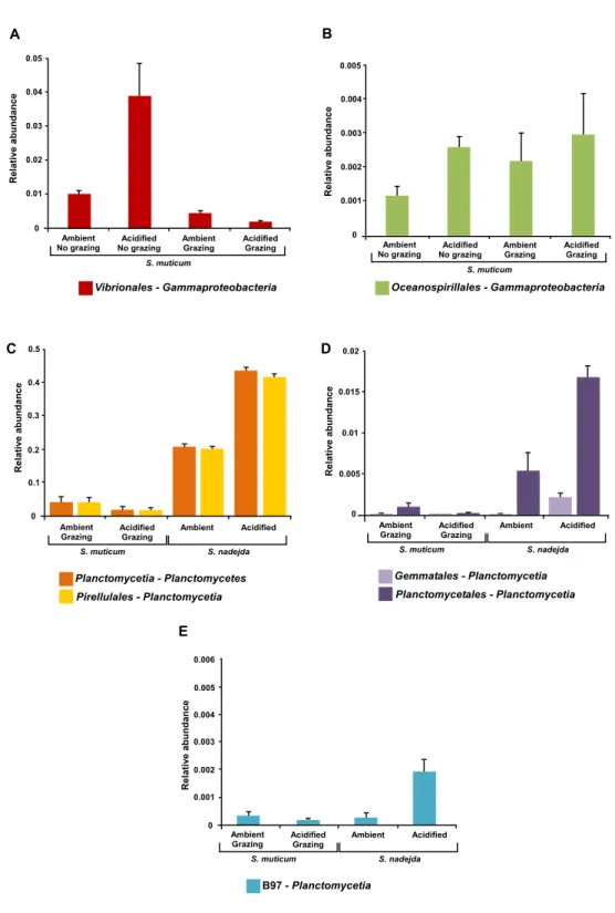
Under elevated CO2treatment, relative abundances of Gammaproteobacteria from the
orders Oceanospirillales and Vibrionales (particularly Pseudoalteromonas), increased on S.
muticumbut only significantly in the absence of grazers (One-way ANOVA, P = 0.037
and P = 0.047, respectively;Figs. 7Aand7B). Under acidification, the abundance of
Planctomycetiaassociated with the grazer gut was significantly higher than at the ambient
CO2levels (Two-tailed t -test, P< 0.001), as was their abundance (at elevated CO2) in the
grazer gut than on S. muticum (Two-tailed t -test, P< 0.001) (Fig. 7C). Deeper analyses
of bacterial abundance within Planctomycetia revealed that all bacterial orders detected within this class responded to acidification treatment (Figs. 7C–7E). A significant effect
was confirmed within the grazer gut under different CO2conditions and between the
seaweed and the grazer gut under increased CO2. At elevated CO2levels, the abundance
of Pirellulales (Two-tailed t -test, P< 0.001,Fig. 7C), Planctomycetales (Two-tailed t -test,
P =0.008,Fig. 7D) and Gemmatales (Two-tailed t -test, P = 0.009,Fig. 7D), were increased
in the isopode gut. Alongside, under the acidification treatment, the abundance of the
bacterial orders Pirellulales (Two-tailed t -test, P< 0.001, Fig. 7C), Planctomycetales
(Two-tailed t -test, P< 0.001,Fig. 7D) and Gemmatales (Two-tailed t -test, P = 0.008,Fig.
7D), was significantly higher in the grazer gut than associated with the seaweed (in the presence of grazers). The class C6-Planctomycetes also exhibited a significant increase in
abundance under elevated CO2 conditions, but only within the grazer gut (Two-tailed
t-test, P = 0.039) (Fig. 6). This was mostly due to the significant increase of bacteria from
the order d113 (two-tailed t test, P = 0.039) (Fig. 6). The abundance of d113 under acidified
conditions was significantly higher in the grazer gut than within S. muticum (two-tailed t test, P = 0.018) (Fig. 6). Overall, under acidification treatment, Planctomycetes increased in abundance (particularly within Planctomycetia) in the grazer gut from 20.9% to 43.8% (1-way ANOVA, F = 306.663, P < 0.001).
DISCUSSION
The results presented in this experimental study demonstrated that acidification affected specific bacterial groups, but hardly influenced the overall microbiome of the invasive brown seaweed S. muticum. In contrast, acidification caused significant changes in the gut microbiome of a native isopod consumer, S. nadejda. Interestingly, acidification increased abundances of Planctomycetia in the gut of S. nadejda and of Oceanospirillales and Vibrionales associated to S. muticum, raising hypotheses about their functional role under these conditions.
0 0.1 0.2 0.3 0.4 0.5 R e la ti ve a b u n d a n c e Ambient Grazing Acidified Grazing Ambient Acidified S. muticum S. nadejda Planctomycetia - Planctomycetes Pirellulales - Planctomycetia 0 0.005 0.01 0.015 0.02 R e la ti ve a b u n d a n c e Ambient Grazing Acidified Grazing Ambient Acidified S. muticum S. nadejda Gemmatales - Planctomycetia Planctomycetales - Planctomycetia 0 0.001 0.002 0.003 0.004 0.005 R e la ti ve a b u n d a n c e 0.006 Ambient
Grazing AcidifiedGrazing Ambient Acidified
S. muticum S. nadejda B97 - Planctomycetia 0 0.01 0.02 0.03 0.04 0.05 R e la ti ve a b u n d a n c e Ambient
No grazing No grazingAcidified Ambient Grazing Acidified Grazing S. muticum Vibrionales - Gammaproteobacteria 0 0.001 0.002 0.003 0.004 0.005 Ambient
No grazing No grazingAcidified Ambient Grazing Acidified Grazing S. muticum Oceanospirillales - Gammaproteobacteria R e la ti ve a b u n d a n c e A B C D E
Figure 7 Mean relative abundances of associated bacterial orders. (A and B) Sargassum muticum under
grazing/non grazing influence after three weeks under ambient (380 ppm) and elevated/acidified (1,000 ppm) CO2conditions and (C–E) grazed Sargassum muticum and the gut of Synisoma nadejda after three
weeks under ambient (380 ppm) and elevated/acidified (1,000 ppm) CO2conditions, that responded to
acidification, but for which a significant interaction between acidification and type of sample (seaweed or grazer gut) was observed. Alpha = 0.05, error bars show standard error per treatment (n = 4).
The guts of isopods are populated by symbiotic bacteria (e.g.,Zimmer & Bartholmé, 2003;Wang, Brune & Zimmer, 2007;Fraune & Zimmer, 2008;Eberl, 2010) that assist in
food utilization by the host (Zimmer, 2002;Zimmer et al., 2002;Zimmer & Bartholmé,
2003; Fraune & Zimmer, 2008). Because the diversity of gut bacterial communities is
shaped by host diet (Tietjen, 2014), changes in seaweed characteristics and its microbiome,
as a result of ocean acidification, were expected to affect the gut microbiome of the grazer with possible fitness consequences. Here, we documented that the most striking change resulting from acidification was a significantly increased abundance of Planctomycetes in the gut of S. nadejda, more specifically due to an increase of Non-ID Pirellulaceae. In this case, as seaweed microbiome was not overall affected by OA, these general changes in the grazer’s gut cannot be directly attributed to changes in its food microbiome. Although the functions of Pirellulaceae are hardly known, its presence was documented in the gastrointestinal tract
of fish (Parris et al., 2016) and as part of the resident microbiomes of marine copepods
but in very low abundance in starved specimens (Moisander, Sexton & Daley, 2015).
Planctomycetesconstituted the second most prevalent phylum (after Proteobacteria) in
the gut of S. nadejda. These bacteria are known to widely colonize aquatic and terrestrial
ecosystems (Lage & Bondoso, 2014) and were, until recently, considered environmental
organisms. However, Planctomycetes have been reported associated to the gut of, not only marine taxa (Singh & Reddy, 2014), but also terrestrial herbivores (Fuerst & Sagulenko,
2011), mammals (Frey et al., 2006) and even humans (Cayrou et al., 2013).
In this study, acidification strongly increased the abundance of Planctomycetes in the bacterial gut communities of S. nadejda compared to ambient conditions. An increase of this Phylum in response to simulated OA has also been shown in sandy sediments (Currie et al., 2017). Although it is clear that in our study this was a consequence of acidification, the lack of seawater samples does not allow us to determine whether the observed Planctomycetes increase was due to acidification of the seawater or a direct response of the grazer’s gut to acidification. This phylum was also abundant (the third most abundant) in the bacterial community associated to S. muticum. These widespread bacteria
have been often found associated to macroalgae (Lage & Bondoso, 2014), among which
Caulerpa taxifolia(Meusnier et al., 2001) and the kelp Laminaria hyperborea (Bengtsson, Sjøtun & Øvreås, 2010). Bacteria from the phylum Planctomycetes are suggested to have potential benefits for their hosts, through their ability to mineralize organic molecules into inorganic compounds that match the nutritional requirements of macroalgae (Lage & Bondoso, 2014). Planctomycetes are also proposed to function as ‘‘slow-acting
decomposers of organic matter’’ (i.e., algal polymer degradation;Bodelier & Dedysh, 2013)
and important contributors to the global nitrogen cycle (i.e., anammox Planctomycetes,
Fuerst & Sagulenko, 2011). So, the presence of these bacteria in the isopod gut might also be related to the ingestion of S. muticum. Its increased abundance (particularly those from the order Pirellulales) could also be a response to the change of the seaweed nutritional
value under acidification, which likely increased its photosynthetic rate driven by high CO2
and consequent C/N ratio shifts (Cornwall, Revill & Hurd, 2015;Briggs, 2017). An increase
in Planctomycetes abundance could provide the isopod with an elevated algal digestion capacity to compensate the highly carbonated food. Again, with the lack of control samples
as starving isopods (empty guts) we can only raise new hypotheses about the direct (through its microbiome) or indirect (through its nutritional value/quality) influence of the food in the grazer’s gut microbiome. Yet, there is no doubt that acidification resulted in a significant overall change in the isopod gut microbiome that is not directly related to bacterial
community shifts of S. muticum, which did not occur for the seaweed at elevated CO2.
Another important change resulting from acidification was a significant increase in Oceanospirillales (Non-ID Oceanospirillaceae) and Vibrionales (Pseudoalteromonas) associated with S. muticum in the absence of grazers. They all belong to
Gammapro-teobacteriawhich have been previously found in association with various seaweed species
(e.g., Patel et al., 2003;Huggett et al., 2006). Members of the class Gammaproteobacteria
are known to produce biologically active metabolites that mediate antifungal (Barbieri
et al., 2001), antifouling (i.e., Alteromonas, Pseudomonas;Maki et al., 1988;Holmstrom, Rittschof & Kjelleberg, 1992;Avelin Mary et al., 1993;Holmström & Kjelleberg, 1999) and
antibacterial activities (Hentschel et al., 2001). There are no studies documenting the
effect of acidification on these bacteria in seaweeds, but environmental samples showed a high increase of Gammaproteobacteria under acidification, in particular from the
order Oceanospirillales (Currie et al., 2017). Bacteria from the order Oceanospirillales are
heterotrophic and capable of degrading complex organic compounds (Garrity et al., 2005;
Goffredi, Johnson & Vrijenhoek, 2007). These bacteria use organic matter as food source and their increase in S. muticum under acidification might be opportunistic and related to the carbon content increase in the seaweed, and consequent seaweed enrichment, due to
elevated CO2levels.
Increased abundance of Vibrionales has often been associated with stressed and diseased
marine invertebrates and they are also known as coral pathogens (Bourne & Munn,
2005;Bourne et al., 2008;Sunagawa et al., 2009;Meron et al., 2011). Vibrionales were also
responsible for a number of infections in humans and animals (Vezzulli et al., 2016),
and identified as potential pathogens of sablefish larvae (Schulze et al., 2006) and bivalve
mollusks (Asplund et al., 2014). Interestingly, it has been shown that under low pH, the
coral-associated pathogen Vibrio sp. increased in abundance (Meron et al., 2011), while the
blue mussel pathogen Vibrio tubiashii became more infectious (Asplund et al., 2014). In this
study, among the Vibrionales that experienced a significant increase were predominantly
Pseudoalteromonas.While certain members of the genus Pseudoalteromonas were reported
to have antibacterial activity in corals, providing it with defense against potential pathogens (Shnit-Orland, Sivan & Kushmaro, 2012), multiple studies identify Pseudoalteromonas as
opportunistic pathogens of marine organisms (Liu et al., 2010;Song et al., 2012;Wang
et al., 2012). Bacteria affiliated to the genus Pseudoalteromonas were found associated to the kelp Laminaria japonica affected by two different diseases (holle-rotten disease
and red spot disease) (Sawabe et al., 1998; Gachon et al., 2010) where its isolation and
posterior reinfection resulted in observable symptoms (Wang et al., 2008). Nevertheless,
particular bacteria that may be otherwise commensal, under stress of the seaweed host,
can become saprophytic (Egan et al., 2013) and this could hypothetically be the case of
the Pseudoalteromonas found in S. muticum under acidification. Ocean acidification could potentially result in shifts from healthy associated bacterial communities within seaweeds
towards a higher prevalence of pathogenic bacteria and/or increased vulnerability to disease. Unfortunately, our taxonomic assignments and our amplicon sequencing approach does not provide more detailed insights into possible bacterial pathogenicity.
Flavobacterialesfrom the Bacteroidetes phylum were among the bacterial groups that
were significantly more abundant in association with S. muticum than in the isopod
gut. Bacteroidetes colonize marine and freshwater environments widely (Thomas et al.,
2011), populating a wide variety of surfaces, including macroalgae (Beleneva & Zhukova,
2006; Staufenberger et al., 2008) and marine sediments (Devine, Pelletreau & Rumpho,
2012). Bacteroidetes were isolated from Caulerpa taxifolia (Meusnier et al., 2001), Ulva
australis and the red alga Delisea pulchra (Longford et al., 2007), suggesting that these
typical marine bacteria are common seaweed associates (Tujula et al., 2010). This phylum
represents some of the most abundant marine bacteria (Glöckner et al., 1999; Simon,
Glöckner & Amann, 1999; Cottrell & Kirchman, 2000) and plays an important role as
degraders of complex organic matter (Church, 2008). Flavobacteriia, the most prevalent
class detected within the seaweed microbiome, are known to produce enzymes for polymer
degradation (Fenchel, 2012). Most Flavobacteriia are able to degrade cellulose, chitin,
proteins, and nucleic acids (Kirchman, 2002;Fenchel, 2012). However, Bacteroidetes are
also related to stress conditions and often found in diseased corals (Barneah et al., 2007),
as in the case of Porites compressa which when exposed to low pH showed an increase of
disease-associated Flavobacteriia (Thurber et al., 2009). Flavobacteriales, the most widely
represented Flavobacteriia order within S. muticum, are mostly associated with degradation
of complex particle biomacromolecules, as well as algal debris (Kirchman, 2002), more
specifically proteins, agars, xylan, fucoidan, cellulose, and chitin (Devine, Pelletreau & Rumpho, 2012). Bacteria from the genera Aquimarina and Tenacibaculum were among the
most frequent OTUs and occur free-living in marine environments (Nedashkovskaya et al.,
2006) and fixed to the surfaces of marine organisms (Suzuki et al., 2001). Bacteria from the
genus Tenacibaculum are thought to induce morphogenesis in algae and possibly enhanced
seaweed growth (Matsuo et al., 2003;Matsuo et al., 2005).
In this study, acidification resulted in a small (non-significant) decrease of seaweed-associated Flavobacteriales and just in the absence of grazer. This contrasts with a study conducted on biofilms from the Great Barrier Reef, which reported that with decreasing pH
there was an increase in the relative abundance of Flavobacteriales (Flavobacteriaceae) (Witt
et al., 2011). However,Witt et al. (2011)focused on natural biofilms on glass slides whereas biofilms from living organisms are the result of environment and host communication and may be differently influenced by any fluctuations that might have occurred in the seawater bacterial communities. Nevertheless, the lack of seawater bacterial community analysis in our study may limit the interpretation of some of these results. The decrease
of these common seaweed associates (Hollants et al., 2013;Egan et al., 2013), although non
significant, may result from the instablity of environmental pH changes. Natural seaweed
bacterial assemblages can be disrupted by environmental pressures (Marzinelli et al., 2015)
and that could contribute to the initial response of S. muticum to acidification.
Seaweed consuming iguanas, which have adapted to use macroalgae as their primary resource, were found to host a large propotion of Bacteroidetes in their gut when compared
to terrestrial related species (Hong et al., 2011). Seaweed polysaccharides, many with sulfated sugars that are absent in terrestrial plants, are easily hydrolized by Bacteroides spp. present in the gastrointestinal tract (Shah & Gharbia, 1993). In particular, these bacteria have been described as contributing to the degradation of brown algal polysaccharides
in the gastrointestinal tract of limpets (particularly Flavobacteriia;Dudek et al., 2014) and
other gastropods that are seaweed consumers (Cardoso et al., 2012). As discussed above,
Bacteroideteswere among the most abundant bacteria associated with S. muticum and,
as shown on copepods (Moisander, Sexton & Daley, 2015), these were the third most
abundant bacterial phylum in the intestinal tract of S. nadejda. Bacteroidetes are within the most abundant phyla in three different copepod species, for both starving and full gut
specimens (Moisander, Sexton & Daley, 2015). These authors also found these bacteria to
be the most abundant in seawater samples which lead to the assumption that they were not ‘‘gut permanent residents’’ but instead colonizers from the seawater. In our case, the lack of seawater samples and starved isopodes (empty gut) limit the interpretation of our results, so its relatively high abundance in S. nadejda gut might be both related with seawater and/or food.
In contrast to Bacteroidetes and Gammaproteobacteria prevailing within S. muticum,
Alphaproteobacteria (Rickettsiales), as well as bacteria from the phylum TM6 (the
class SJA-4) were more abundant in the grazer gut microbiome for both ambient and acidified conditions. Alphaproteobacteria were also reported as one of the most prevalent
Classes in marine copepods (Moisander, Sexton & Daley, 2015). Bacteria from the order
Rickettsiales, which dominated the gut of S. nadejda, are known as pathogenic to humans
and animals (Perlman, Hunter & Zchori-Fein, 2006), and were found in the intestinal tract
of infected isopod Armadillidium vulgare (Dittmer et al., 2016) and several other isopode
species (Wang, Brune & Zimmer, 2007). Neorickettsia is known as a parasite to which the
invertebrates usually serve as vectors (Greiman et al., 2014). This aspect is worth further
investigation once the possibility of these herbivores to function as pathogen vectors may have consequences on human health if they are able to pass them along to some edible algae where they feed on.
The experimentally controlled mesocosm data allowed us to isolate variables to infer their effects, even though they are not able to fully reconstruct the dynamic conditions of nature, as documented for corals that had different bacterial communities in laboratory and
field (Kooperman et al., 2007;Meron et al., 2011). So, the cause of the observed bacterial
community shifts can be easily identified but may not mimick exactly the processes in the field. Another limitation to realistic predictions is that global acidification may be
happening quicker than IPCC models predicted. So, pH and CO2conditions used in this
and several other studies may not be realistic and underestimate the effects of OA that can be more catastrophic than what is expected to be in the next 100 years (the maximum
IPCC prediction) (Thurber et al., 2009;Meron et al., 2011). However, those changes would
happen over a large period of time and not over the course of three weeks, as in our experiment.
The results of this study demonstrate that bacterial communities associated within an isopod—seaweed predator–prey system are dynamic and responsive to changes in
acidification. The observed changes in the associated bacterial communities of the seaweed and the grazer gut might be a type of acclimation that facilitates tolerance and survival,
as suggested by Meron et al. (2011). The capacity of organisms to accommodate and
modify a highly diverse microbiota may influence the fitness and success of the host and
enhance its ability to survive changing environmental conditions (Morrow et al., 2015).
Further research is required to better understand the processes and conditions under which different associated bacteria can increase tolerance of the host organisms to various disturbances.
CONCLUSION
The responses of bacteria associated with S. muticum and the gut of S. nadejda revealed in this study suggest that worst case acidification scenarios may not greatly affect overall bacterial community composition and diversity, but might affect specific bacterial groups.
Contrarily to what was expected, acidification to expected levels seems to have less significant impact on seaweed bacterial ecology than other environmental stressors, such
as increased temperature (Mensch et al., 2016). However, specific groups had significant
abundance shifts in seaweeds under acidification as was the case for Oceanospirillales and Vibrionales (mainly Pseudomonadales). The former might be related to the possible increase of the C/N ratio in the seaweed under acidified conditions and the latter, commonly found associated with diseased seaweeds, could be an indicator that acidification enables an increase of opportunistic/pathogenic bacteria. Unexpectedly, no particular bacteria increased abundance that could assist the seaweed in obtaining nutrients (like nitrogen fixing bacteria) under a higher carbon availability regime.
In contrast with the seaweed host, high CO2 levels globally changed the bacterial
community associated to the isopod gut, particularly the abundance of members of the Class Planctomycetia. However, as no significant changes occurred in the global seaweed microbiome, the overall shift in the grazer gut bacterial community cannot be directly attributed to bacterial changes in the food. Instead, we can hypothesize that the food
‘‘quality’’ changed at elevated CO2 triggering a shift in the isopod gut Planctomycetia
allowing it to better digest the seaweed and compensate the shifted C/N ratio as seaweed becomes less nutritional under acidification, as previously hypothesized. The generality of these findings must be addressed in further studies with other species.
Bacteroidetes (mainly Flavobacteriia), previously isolated and commonly found
associated with other seaweed species (Huggett et al., 2006;Burke et al., 2011a;Lachnit
et al., 2011), were the most dominant phyla, suggesting that they account for core roles in the metabolism of S. muticum. Both in ambient and acidified conditions, the isopod gut bacterial community was dominated by Neorickettsia (Alphaproteobacteria), for which invertebrates are usually the vectors. The pathogenicity of these bacteria has already been
shown in other invertebrate species (Greiman et al., 2014) and the potential of isopod
species as possible vectors for these bacteria has already been suggested (Yuksel, Thompson
& Adams, 2006) and should be further investigated.
Concluding, our results show that after only three weeks of simulated acidification, bacterial communities associated to a seaweed host, when ungrazed, and to an isopod
grazer gut, show shifts in composition. These bacterial community changes were particular and specific in the seaweed and only occurred when ungrazed, but were large and global community shifts in the isopod grazer gut. We hypothesized that these may have potential consequences for seaweed health and isopod food digestion. The observed changes in the gut microbiome of the grazer seem to be a reflection of changes in the seaweed chemistry rather than its microbial composition.
ACKNOWLEDGEMENTS
We would like to thank Dario Nobre and João Reis for conducting the acidification experiment.
ADDITIONAL INFORMATION AND DECLARATIONS
FundingThis study was made possible by the Erasmus Mundus Doctoral Programme MARES on Marine Ecosystem Health & Conservation (MARES_13_08: Acclimation and adaptation of invasive seaweeds) for Alexandra Serebryakova, and funds from FCT (Foundation for Science and Technology, Portugal) fellowships SFRH/BPD/63703/2009 and SFRH/BPD/107878/2015 to Aschwin H. Engelen and SFRH/BPD/116774/2016 to Tania Aires, and the EU SEAS-ERA project INVASIVES (SEAS-ERA/0001/2012) and CCMAR/Multi/04326/2013. The funders had no role in study design, data collection and analysis, decision to publish, or preparation of the manuscript.
Grant Disclosures
The following grant information was disclosed by the authors:
Erasmus Mundus Doctoral Programme MARES on Marine Ecosystem Health & Conservation.
FCT (Foundation for Science and Technology, Portugal): SFRH/BPD/63703/2009, SFRH/BPD/107878/2015, SFRH/BPD/116774/2016.
EU SEAS-ERA project INVASIVES: SEAS-ERA/0001/2012. CCMAR/Multi/04326/2013.
Competing Interests
The authors declare there are no competing interests.
Author Contributions
• Tania Aires performed the experiments, analyzed the data, authored or reviewed drafts of the paper.
• Alexandra Serebryakova performed the experiments, analyzed the data, prepared figures and/or tables, authored or reviewed drafts of the paper.
• Frédérique Viard contributed reagents/materials/analysis tools, prepared figures and/or tables, authored or reviewed drafts of the paper, funding.
• Ester A. Serrão conceived and designed the experiments, contributed reagents/material-s/analysis tools, authored or reviewed drafts of the paper.
• Aschwin H. Engelen conceived and designed the experiments, performed the experiments, analyzed the data, contributed reagents/materials/analysis tools, prepared figures and/or tables, authored or reviewed drafts of the paper, funding.
Data Availability
The following information was supplied regarding data availability:
Engelen, Aschwin (2018): Acidification + Grazing_Effects_Microbiome. figshare. https://doi.org/10.6084/m9.figshare.5346316.v3.
Supplemental Information
Supplemental information for this article can be found online athttp://dx.doi.org/10.7717/
peerj.4377#supplemental-information.
REFERENCES
Aires T, Moalic Y, Serrao EA, Arnaud-Haond S. 2015. Hologenome theory supported by
co-occurrence networks of species-specific bacterial communities in siphonous algae
(Caulerpa). FEMS Microbiology Ecology 91: fiv067DOI 10.1093/femsec/fiv067.
Aires T, Muyzer G, Serrão EA, Engelen AH. 2018. Unraveling seaweeds bacteriomes—
from field site to computer screen. In: Charrier B, Wichard T, Reddy CRK, eds.
Protocols for macroalgae research. Oxfordshire: Taylor & Francis Group, 97–112.
Aires T, Serrão EA, Engelen AH. 2016. Host and environmental specificity in bacterial
communities associated to two highly invasive marine species (Genus Asparagopsis).
Frontiers in Microbiology7:559DOI 10.3389/fmicb.2016.00559.
Asnaghi V, Chiantore M, Mangialajo L, Gazeau F, Francour P, Alliouane S, Gattuso JP. 2013. Cascading effects of ocean acidification in a rocky subtidal community. PLOS
ONE8:e61978DOI 10.1371/journal.pone.0061978.
Asplund ME, Baden SP, Russ S, Ellis RP, Gong N, Hernroth BE. 2014. Ocean
acidifi-cation and host-pathogen interactions: blue mussels, Mytilus edulis, encountering
Vibrio tubiashii. Environmental Microbiology 16:1029–1039
DOI 10.1111/1462-2920.12307.
Avelin Mary S, Vitalina Mary S, Rittschof D, Nagabhushanam R. 1993.
Bacterial-barnacle interaction: potential of using juncellins and antibiotics to alter struc-ture of bacterial communities. Journal of Chemical Ecology 19:2155–2167 DOI 10.1007/BF00979654.
Barbieri E, Paster BJ, Hughes D, Zurek L, Moser DP, Teske A, Sogin ML. 2001.
Phylo-genetic characterization of epibiotic bacteria in the accessory nidamental gland and egg capsules of the squid Loligo pealei (Cephalopoda: Loliginidae). Environmental
Microbiology3:151–167DOI 10.1046/j.1462-2920.2001.00172.x.
Barneah O, Ben-Dov E, Kramarsky-Winter E, Kushmaro A. 2007. Characterization of
black band disease in Red Sea stony corals. Environmental Microbiology 9:1995–2006 DOI 10.1111/j.1462-2920.2007.01315.x.
Beleneva IA, Zhukova NV. 2006. Bacterial communities of some brown and red algae
from Peter the Great Bay, the Sea of Japan. Microbiology 75:348–357 DOI 10.1134/S0026261706030180.
Bengtsson MM, Sjøtun K, Øvreås L. 2010. Seasonal dynamics of bacterial biofilms on the
kelp Laminaria hyperborea. Aquatic Microbial Ecology 60:71–83 DOI 10.3354/ame01409.
Berg G, Grube M, Schloter M, Smalla K. 2014. The plant microbiome and its importance
for plant and human health. Frontiers in Microbiology 5:491DOI 10.1007/10.
Bodelier PLE, Dedysh SN. 2013. Microbiology of wetlands. Frontiers in Microbiology 4:Article 79DOI 10.3389/fmicb.2013.00079.
Bodenhausen N, Horton MW, Bergelson J. 2013. Bacterial communities associated
with the leaves and the roots of Arabidopsis thaliana. PLOS ONE 8:e56329 DOI 10.1371/journal.pone.0056329.
Bourne D, Iida Y, Uthicke S, Smith-Keune C. 2008. Changes in coral-associated
microbial communities during a bleaching event. ISME Journal 2:350–363 DOI 10.1038/ismej.2007.112.
Bourne DG, Munn CB. 2005. Diversity of bacteria associated with the coral Pocillopora
damicornisfrom the great barrier reef. Environmental Microbiology 7:1162–1174
DOI 10.1111/j.1462-2920.2005.00793.x.
Branch TA, DeJoseph BM, Ray LJ, Wagner CA. 2013. Impacts of ocean
acid-ification on marine seafood. Trends in Ecology and Evolution 28:178–186 DOI 10.1016/j.tree.2012.10.001.
Briggs LM. 2017. The effects of ocean warming and acidification on seawed growth and
urchin grazing. Pomona: California State Polytechnic University.
Burgess JG, Jordan EM, Bregu M, Mearns-Spragg A, Boyd KG. 1999. Microbial
antagonism: a neglected avenue of natural products research. Journal of Biotechnology
70:27–32DOI 10.1016/S0168-1656(99)00054-1.
Burke C, Steinberg P, Rusch D, Kjelleberg S, Thomas T. 2011a. Bacterial
commu-nity assembly based on functional genes rather than species. Proceedings of the
National Academy of Sciences of the United States of America108:14288–14293
DOI 10.1073/pnas.1101591108.
Burke C, Thomas T, Lewis M, Steinberg P, Kjelleberg S. 2011b. Composition,
unique-ness and variability of the epiphytic bacterial community of the green alga Ulva
australis. The ISME Journal 5:590–600DOI 10.1038/ismej.2010.164.
Caporaso JG, Kuczynski J, Stombaugh J, Bittinger K, Bushman FD, Costello EK, Fierer N, Peña AG, Goodrich JK, Gordon JI, Huttley GA, Kelley ST, Knights D, Koenig JE, Ley RE, Lozupone CA, Mcdonald D, Muegge BD, Pirrung M, Reeder J, Sevinsky JR, Turnbaugh PJ, Walters WA, Widmann J, Yatsunenko T, Zaneveld J, Knight R. 2010. QIIME allows analysis of high-throughput community sequencing data.
Nature Publishing Group7:335–336 DOI 10.1038/nmeth0510-335.
Cardoso AM, Cavalcante JJV, Vieira RP, Lima JL, Grieco MAB, Clementino MM, Vasconcelos ATR, Garcia ES, De Souza W, Albano RM, Martins OB. 2012. Gut