3D Breast Cancer Models: Multimodal
Data Registration
Cátia Sofia Gonçalves Rodrigues
Integrated Master in Bioengineering Supervisor: Hélder Filipe Pinto de Oliveira, PhD Co-supervisor: Sílvia da Conceição Neto Bessa, MSc
Cátia Sofia Gonçalves Rodrigues
Integrated Master in Bioengineering
O cancro da mama é uma doença amplamente conhecida, principalmente em mulheres. Tem uma mortalidade consideravelmente baixa comparativamente com outras formas de cancro, no entanto é a forma mais comum nas mulheres, trazendo graves consequências a nível físico e psi-cológico. Esta baixa mortalidade deve-se maioritariamente à monitorização desde cedo de mul-heres que se encontrem no grupo de risco para esta forma de cancro, dada a sua idade ou o seu histórico familiar, permitindo uma deteção precoce do cancro assim como um tratamento anteci-pado, que consequentemente será mais eficaz.
A maioria das pacientes necessita de realizar cirurgia mamária para a remoção do tumor, e esta cirurgia pode ter o propósito de remover a totalidade da mama ou apenas a zona onde o tumor se encontra e as suas redondezas. Os resultados desta cirurgia nem sempre correspondem ao que era expectável, e a mama pode de alguma forma ficar deformada após a cirurgia.
A presença de um modelo tridimensional da mama, que é específico à paciente, irá melhorar a comunicação entre a paciente e o clínico, permitindo uma visualização mais clara do modelo da mama e da localização do tumor, antes da cirurgia. Esta visualização permitirá uma melhor compreensão de como será a cirurigia e do que é que a paciente pode esperar desta. Estes mod-elos podem também ser usados para criar modmod-elos biomecânicos ou modmod-elos criados a partir de Machine Learning, que irão ajudar a prever as deformações que irão ocorrer na mama também. No entanto, estes modelos não são fáceis de obter, maioritariamente devido à natureza não rígida e deformável da mama.
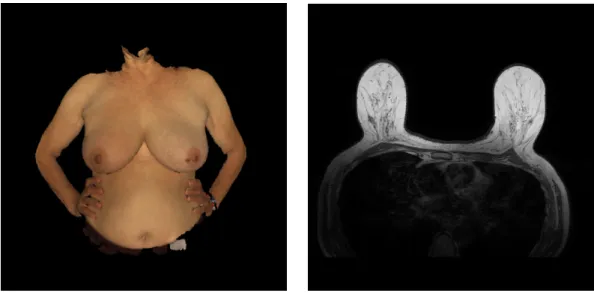
Muitas técnicas de imagem são usadas na atualidade na deteção de cancro da mama, como ultrassons, mamografias e ressonâncias magnéticas, e esta última será usada na aquisição de ima-gens deste trabalho, pois fornece a informação interior da mama dividida por fatias que depois são usadas para construir um volume. Para criar este modelo, imagens de ressonância magnética e de superfície, e a sua correspondência serão feitas para combinar tanto a informação externa como a interna da mama. Os dados de ambas as modalidades não são obtidos com o paciente na mesma posição portanto, primeiramente deve ocorrer uma transformação de pose.
Apesar da mama ter um comportamento não rígido, o registo dos dados obtidos da ressonância magnética e da superfície irão incluir um registo rígido e não rígido. O registo rígido é um passo essencial para a boa performance do registo não rígido, tendo em conta que irá aproximar as duas nuvens de pontos e irá colocá-las no mesmo espaço. O registo rígido incluirá transformações como rotações e translações e a implementação do algoritmo Iterative Closest Point. O registo não rígido será feito pela implementação do algoritmo Free Form Deformation.
Pacientes provenientes de dois projetos diferentes serão usados para formar os conjuntos de dados usados nesta dissertação, que incluirá também um conjunto de dados para validação. Métri-cas como a distância Euclideana e a distância de Hausdorff são usadas para avaliar a precisão das transformações, no entanto essas métricas não consideram que os pontos estão na verdade a representar um objeto tridimensional, não sendo totalmente confiáveis. Para complementar estes
resultados, a visualização das nuvens de pontos e dos passos intermédios do registo é essencial para compreender qual será a melhor metodologia a implementar.
Um conjunto de dados de validação foi também criado com a intenção de validar as defor-mações induzidas na mama. Este conjunto inclui sete pacientes, com pontos de referência marca-dos com cápsulas de óleo de fígado de bacalhau. Os resultamarca-dos mostram que a melhor implemen-tação regista apenas uma mama de cada vez e não o torso completo e usa o paciente na posição vertical, depois da transformação de pose. O registo rígido inclui duas rotações, uma correção de orientação através do plano xy, uma translação através da zona do mamilo e a implementação do Iterative Closest Point. O registo não rígido será realizado usando a Free Form Deformation com uma grelha de pontos de controlo de [6,6,6].
Os resultados obtidos são bastante promissores para uma futura implementação em ambiente clínico, providenciando uma excelente ferramenta para ajudar tanto o paciente como o clínico, para respetivamente compreender e planear melhor as consequências da cirurgia mamária.
Breast cancer is a widely known disease, mostly for its appearance in women. It has a con-siderable low mortality comparing to other forms of cancer, but it is the most common form of cancer in women, bringing meaningful physical and mental consequences for the patients. This low mortality is mainly due to the monitoring of the women who are above a certain age, or have a certain family history, which allows an early detection of the cancer as well as an early treatment, that is consequently more effective.
Most of the patients need to perform breast surgery to remove the tumour, and this surgery can be to remove the entirety of the breast or only the tumour and its surroundings. The outcomes of this surgery do not always match what was previously expected, and the breast can be somehow deformed after the procedure.
The presence of a three-dimensional breast model, that is patient specific, will improve the communication between the patient and the doctor, allowing a clear visualization of the breast and the tumor before the surgery. This visualization will allow a better understanding of how the surgery will be and what can the patient expect from it. These models can also be used to create biomechanical models or models created from Machine Learning, which will help predict the deformations of the breast as well. These models are not easy to be obtained, mostly due to the non-rigid and deformable nature of the breast.
A lot of imaging techniques are nowadays being used in the detection of breast cancer, as ultrasounds, mammograms and Magnetic Resonance Imaging, being the last one the one that is going to be used for the acquisition of breast images since it provides the interior information pf the breast divided by slices that can then form a volume. To create this model, images from Magnetic Resonance Imaging and surface data must be combined, and the matching will be done to combine both interior and exterior information of the breast. The data from both modalities is not acquired with the patient in the same position, so firstly a pose transformation must be performed.
Even though the breast has a non-rigid behaviour, the registration of the data from the Magnetic Resonance Imaging and the surface will include a rigid and a non-rigid registration. The rigid registration is an essential step to the good performance of the non-rigid registration since it will approximate both point clouds and place them in the same coordinate system. Rigid registration will include affine transformations such as rotations and translations and the implementation of a Iterative Closest Point Algorithm. Non-rigid registration is done by performing a Free Form Deformation algorithm.
Patients from two different projects are used to fill the datasets, that will also include a val-idation dataset. Metrics such as the Euclidean Distance and the Hausdorff Distance are used to evaluate the accuracy of the transformations, but these metrics do not consider that the points are actually representing a three-dimensional object so they are not fully reliable. So, to complement these results visualizing the final point clouds and the intermediate steps is essential to understand which is the best methodology.
A validation dataset was also created with the intention of validating the induced deformations in the breast. This dataset includes 7 patients, with reference points marked with codfish oil pills. The results showed that the best implementation registers a single breast at a time, and not the entire torso, and uses the patient in an upright position, after the pose transformation. The rigid registration will include two rotations, a correction of the orientation through the xy plane, a translation through the breast mounds and an Iterative Closest Points algorithm. The non-rigid registration will be performed using the Free Form Deformation algorithm with a [6,6,6] grid of control points.
The results obtained are very promising for a future implementation on a clinical environment providing a great tool to help both the patient and the clinician, to respectively understand and plan better the consequences of a breast cancer surgery.
Em primeiro lugar gostaria de agradecer ao INESC-TEC e ao grupo CTM pela oportunidade dada em realizar esta dissertação no contexto de um grupo tão completo e tão motivado. Um particular agradecimento ao meu orientador, o Professor Hélder Oliveira, por todo o apoio e mo-tivação que me deu ao longo deste ano e por toda a flexibilidade mostrada, as suas ideias foram fundamentais para a boa realização desta dissertação. Um enorme agradecimento à Sílvia, minha co-orientadora, por toda a paciência para resolver as minhas dúvidas, pelo trabalho incansável e por todas as ideias e críticas extraordinárias que complementaram imenso este trabalho. Ainda dentro deste grupo gostaria de agradecer à Sara Oliveira por todo o apoio no tratamento dos dados e por me ter ajudado a descomplicar muita coisa ao longo deste trabalho, ao João Teixeira pela sua disponibilidade e ajuda e ao Henrique Carvalho pelo trabalho fornecido que me deu toda a base para esta dissertação e por todo o trabalho que fez desde o início da minha dissertação que deu mais qualidade aos resultados que aqui apresento. Dentro do projecto do BCCT.plan gostaria de ainda agradecer ao Doutor Pedro Gouveia por todos os dados fornecidos e apoio neste projeto, que foram cruciais para a realização desta dissertação e para os resultados obtidos.
À Adapttech, obrigada por acreditarem nas minhas capacidades e por me acolherem na em-presa que mais parece uma família e que está sempre ansiosa por ver o meu sucesso e felicidade. Um particular agradecimento à Sofia pela ajuda enorme na minha integração como trabalhadora-estudante, pelo suporte dado e por me fazer sentir em casa.
Aos Catos, André, Luis, Hugo e Margarida, sou uma pessoa diferente graças a vocês, toda a vossa paciência comigo e todo o stress que passei convosco me crescer imenso e fizeram-me não ter fizeram-medo das coisas que fizeram-me rodeiam, obrigada for daring fizeram-me to grow e por terem crescido comigo, sou uma secretária orgulhosa. A todo o BEST Porto, obrigada por me proporcionarem as melhores experiências da faculdade, por me darem a oportunidade de conhecer o mundo e por me deixarem fazer parte da direção desta incrível associação. Serei uma alumna saudosa e deixarei parte do meu coração aqui, mas levarei memórias para uma vida.
À Rita, obrigada por toda a tua honestidade e por acreditares mais em mim do que eu própria, deste-me uma enorme motivação ao longo deste tempo todo. À Amélia por todas as conversas na varanda, todos os desabafos e chapadas de realidade, passe o tempo que passar sei que tenho em ti um poço de confiança. À Raquel, tenho o maior orgulho do mundo em ti e ao fim destes 14 anos de amizade, devo-te muitos dos momentos mais felizes e muita da minha recuperação em momentos tristes, obrigada por estares sempre lá, mesmo longe de mim. À Sónia, eu sei que me mandaste dormir durante estes 5 anos da faculdade, mas eu prometo que o vou fazer agora... O meu sucesso académico deve-se imenso ao teu apoio e à inspiração que me dava, obrigada por me chamares à realidade e por saberes sempre dizer o que eu precisava de ouvir. Ao João, por aguentares os meus devaneios e por seres sempre o meu porto seguro, obrigada por seres incansável e por tomares conta de mim (e por me alimentares também), mesmo quando eu não sabia que precisava que o fizessem.
À minha irmã, desculpa por não poder brincar mais contigo, por não ir ao teu primeiro dia na v
escolinha e por ter de te deixar tanto tempo. Prometo que te vou ajudar a fazer os trabalhos de casa e que vou estar sempre lá quando precisares. E acima de tudo, à minha mãe, o meu maior exemplo e a responsável por tudo aquilo que consegui, só espero um dia poder ter tanta força como tu mãe. Isto é tudo por vós.
"We live on an island surrounded by a sea of ignorance. As our island of knowledge grows, so does the shore of our ignorance." John Archibald Wheeler
1 Introduction 1 1.1 Context . . . 1 1.2 Motivation . . . 2 1.3 Goals . . . 2 1.4 Contributions . . . 2 1.5 Structure . . . 3 2 Breast Cancer 5 2.1 Statistics . . . 5 2.2 Breast Imaging . . . 6 2.2.1 Radiological Exams . . . 6 2.2.2 Surface Information . . . 10 2.3 Breast Surgery . . . 13
2.3.1 Types of Breast Cancer Surgery . . . 14
2.3.2 Surgery Planning . . . 15
2.3.3 Psychological Impact of Breast Cancer Surgery . . . 16
2.4 Summary . . . 17
3 Data Registration 19 3.1 Overview . . . 19
3.2 Registration of Radiological Images . . . 22
3.3 Surface Registration . . . 26
3.4 Surface Radiological Matching . . . 26
3.5 Validation Methods . . . 29 3.6 Summary . . . 30 4 Methodology 33 4.1 Datasets . . . 33 4.1.1 PICTURE dataset . . . 33 4.1.2 BCCT.plan dataset . . . 34 4.2 Pre-processing . . . 36 4.3 Registration Strategy . . . 39 4.3.1 Affine Transformations . . . 40 4.3.2 Geometric ICP . . . 40 4.3.3 Deformable Registration . . . 42
4.3.4 Closing the breast . . . 42
4.4 Dataset Validation . . . 43
4.4.1 Evaluation Metrics . . . 45
4.4.2 Target Registration Error . . . 47
4.4.3 Summary . . . 47
5 Results and Discussion 49 5.1 Rigid Registration . . . 50
5.1.1 Affine Transformations . . . 50
5.1.2 Iterative Closest Point Algorithm . . . 51
5.2 Deformable Registration . . . 55
5.2.1 Free Form Deformation . . . 55
5.2.2 Algorithm with complete torso . . . 58
5.2.3 Insertion of the tumor . . . 60
5.2.4 Insertion of the pectoral muscle . . . 61
5.3 Validation Dataset . . . 62 5.4 Prone vs. Upright . . . 66 5.5 Discussion . . . 66 6 Conclusions 71 6.1 Future Work . . . 72 A Additional Information 73 References 87
2.1 Statistics from Globocan 2018 [5]. . . 6
2.2 Categories of percentage mammographic density estimated by radiologists through mammograms A=0. B =< 10%.C =< 25%.D =< 50%.E =< 75%.F =≥ 75% [10]. . . 7
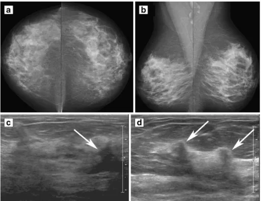
2.3 Selected images of a 54-year-old asymptomatic woman with dense breasts and no previous history of breast cancer. a) Craniocaudal digital mammograms are taken the same day as the ultrasound study. b) Mediolateral oblique digital mammo-grams are taken the same day as the ultrasound study. c) Transverse ultrasound image of the right breast; white arrow shows a 7 mm, grade I, stage 1, invasive ductal carcinoma. d) Ultrasound image of the left breast; two white arrows show 10 mm, grade I, stage 1, invasive carcinoma with lobular carcinoma in situ [12]. . 8
2.4 Block diagram of an MRI scanner [13]. . . 9
2.5 Multifocal carcinoma detected on MRI from a woman with no family history of breast cancer, but history of fibrocystic changes, and negative results after a mam-mography, proving that the accuracy of MRI is superior to the accuracy of the mammography [8]. . . 10
2.6 Three-dimensional acquisition systems for object measurement using non-contact methods [21]. . . 11
2.7 Microsoft Kinect Device. . . 13
2.8 Breast Cancer Surgery. . . 15
3.1 Framework of non-rigid registration. . . 22
3.2 A lattice of control points. The s, t, and u vectors define the local coordinate system 24 3.3 1a) MRimage and 1b) Mammogram. 2) Finite element mesh of the patient’s breast. 3) Definition of the tissue properties and the deformation process. 4) FEM Simulation. 5) Deformed finite element model. 6) Projection of a generated MR image of the deformed breast. . . 25
3.4 Comparison between segmented MRI data, downsampled MRI data, MRI data after biomechanical simulation and surface data [58]. . . 28
3.5 Result of applying the FFD algorithm to the downsampled MRI (after biomechan-ical simulation) PCL of the patient with a [8 8 8] control point grid [58]. . . 29
4.1 Scan and MRI data from the PICTURE project. . . 35
4.2 Scan and MRI data from the BCCT.plan project. . . 36
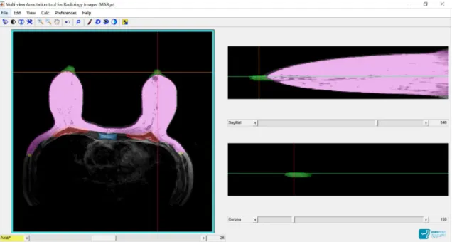
4.3 MARge Software. Pink: breast contour; Green: nipple; Red: pectoral muscle; Yellow: beggining of Latissimus Dorsi muscle; Blue: sternum. Axial view in the left, sagittal view in the top right corner and coronal view in the bottom right corner. 37 4.4 Pipeline of the pre-processing step. . . 38
4.5 Pipeline summary for the registration of MRI PCLs and Surface PCLs. . . 39
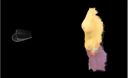
4.6 On the left: original MRI PCL. On the right: surface PCL. . . 40
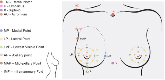
4.7 Reference points for the 3D landmarks, used both for the surface and the MRI. . 43
4.8 MRI and Scan data with breast markers. . . 44
4.9 MRI slice with the cod oil pills showing in pink. Normal pills, intersecting with the skin are surrounded by green. Dislocated pills that show no contact with the skin are surrounded by red. . . 45
4.10 Reference points selected in the surface. . . 45
4.11 Numbered reference points for the MRI. . . 46
4.12 Numbered reference points for the surface. . . 46
5.1 Correction of the orientation. The PCL in red represents the scan, while the PCL in blue represents the MRI after being rotated and centered in (0,0,0) and the yellow the MRI after the correction of orientation using the xy plane. . . 50
5.2 Results after translation. . . 51
5.3 Euclidean distances after the normal ICP. . . 52
5.4 Results after the normal ICP. . . 52
5.5 Euclidean distances after the geometric ICP. . . 53
5.6 Results after the geometric ICP. . . 53
5.7 Patient 6 after the normal ICP. . . 54
5.8 Comparison between the normal and geometric ICP for the patient number 6 using the complete torso. . . 55
5.9 Comparison between the normal and geometric ICP for the patient number 20 using the complete torso. . . 55
5.10 Deformed breasts after the complete registration, using normal ICP and a single breast. . . 56
5.11 Patient 6, after a normal ICP implementation. . . 57
5.12 Surface of patient 18. . . 57
5.13 Euclidean Distance after the FFD using the normal ICP. . . 58
5.14 Breast number 2 of patient 18 after the non-rigid registration using the geometric ICP. . . 58
5.15 Euclidean Distance after the FFD using the geometric ICP. . . 59
5.16 Results after the non-rigid registration for the normal and geometric ICP. . . 59
5.17 Results after the non-rigid registration for the normal and geometric ICP using a grid of [6,6,6] points in the FFD. . . 60
5.18 Complete torso after the non-rigid registration for patient 20 with normal ICP. . . 60
5.19 Complete torso after the non-rigid registration for patient 20 with geometric ICP. 61 5.20 Results after the non-rigid registration for the normal and geometric ICP using the complete torso. . . 61
5.21 Representation of the localization of the tumor of patient 21. . . 62
5.22 Patient 21 with tumor. . . 62
5.23 Representation of the localization of the tumor of patient 21 on a frontal view. . . 63
5.24 Representation of the torso with the pectoral muscle for patient 20. . . 64
5.25 TRE values by breast marker and by patient (Oa, Ob, Oc, Od correspond respec-tively to O5, O6, O7 and O8 in Figure4.7). . . 65
5.26 TRE values for each breast marker, when using a single breast, normal ICP and a [6,6,6] grid for the FFD. . . 66
5.27 Results for the implementation with the patient in a prone position for a single breast, using the normal ICP and a [6,6,6] grid for the FFD. . . 67
5.28 Comparison between the results for patient 20 in a prone and upright position. The PCL in yellow represents the breast in a prone position and the PCL in red represents the breast in an upright position, both after the non-rigid registration. . 68
5.1 Mean TRE value for each breast marker using a single breast, with a normal ICP and a [6,6,6] grid for the FFD. . . 67
A.1 Results after normal ICP with single breast. . . 74
A.2 Results after the geometric ICP with single breast and 100% of the points below the breast mound. . . 75
A.3 Results after the geometric ICP with single breast and 80% of the points below the breast mound. . . 76
A.4 Results after the geometric ICP with single breast and 60% of the points below the breast mound. . . 77
A.5 Results after the geometric ICP with single breast and 40% of the points below the breast mound. . . 78
A.6 Results after non-rigid registration with normal ICP and single breast. . . 79
A.7 Results after the non-rigid registration using geometric ICP with single breast and 100% of the points below the breast mound. . . 80
A.8 Results after the non-rigid registration, using a single breast and a grid of [6,6,6] points for the FFD, after a normal ICP implementation. . . 81
A.9 Results after the non-rigid registration, using a single breast and a grid of [6,6,6] points for the FFD, after a geometric ICP implementation. . . 82
A.10 Results after the non-rigid registration, using the complete torso and normal ICP. 83
A.11 Results after the non-rigid registration, using the complete torso and geometric ICP. 84
3D 3 Dimensional
BCS Breast-Conserving Surgery BCT Breast-Conserving Therapy CC Correlation Coefficient CT Computerized Tomography DoF Degrees of Freedom ECG Electrocardiogram FEM Finite Elements Method FFD Free Form Deformation FoV Field of View
ICP Iterative Closest Points MRI Magnetic Resonance Imaging MSE Mean Squared Error
NaN Not a Number
PCA Principal Component Analysis PCL Point Cloud
PET Positron Emission Tomography QoL Quality of Life
RF Radio Frequency
RMSE Root-Mean-Square Error ROI Regions Of Interest SNR Signal-to-noise ratio
SSD Sum of Squared Differences TRE Target Registration Error
Introduction
1.1
Context
Breast cancer is a widely known disease, being one of the most common forms of cancer in women [1]. Its low mortality, compared to its incidence, is mostly due to all the methods of screening being implemented nowadays [2]. Women start being monitored, around the age of 40, and campaigns are made to raise awareness of how dangerous the disease is, and inform about the ways to monitor the breast’s health, so the diagnosis is made early in order to increase the probability of success of the treatment. This process starts with self breast examination and clinical breast examination, for younger women, and goes on with the execution of a mammography (X-ray of the breast), when the patient reaches a certain age (that depends on the country). Considering some aspects like the clinical history of the patient, the density of the breast or an inconclusive result of the mammography, the patient may be advised to perform an MRI [2] in order to get a more accurate result of the breast analysis.
When it comes to the treatment it includes breast surgery to remove the tumour, if chemother-apy does not prove to be efficient. The surgery may require the removal of the entirety of the breast, which is called a mastectomy, or just a portion of it, which is called a BCS (Breast-Conserving Surgery). Mastectomy was previously almost the standard choice for all the patients going through breast cancer surgery, since the patients felt more safe and less scared of recurrence of the cancer. Now it is easier to localize the tumour in the breast and remove only that part and the surrounding tissues, which makes it unnecessary to remove the whole breast. The planning of this type of surgery and the accomplished result, may not match, which may come from the lack of experience of the surgeon or the location of the tumour in the breast.
Some deformations caused by breast cancer surgery can go against what was expected by the patient, and interfere in their personal and social life. The lack of satisfaction with their looks, that may have come from chemotherapy, will increase after surgery, along with depression, anxiety and feelings of sexual unattractiveness.
Currently, effort has been made in order to include the patient in the decision-making process, making her more aware of the risks and capable of deciding some aspects of the surgery, which in
consequence will improve the acceptance of her own body after the surgery.
The next step should include the presence of 3D models of the patient’s breast, combining the information gathered through radiological exams and data from the surface, to make the changes and deformations that will occur after breast cancer surgery, more visible to both the patient and the surgeon, involving the patient in the decision-making process and making them more comfortable and aware of the process.
1.2
Motivation
The task of creating 3D models of the breast is not easy due to the non-rigid and deformable na-ture of the breast. But nowadays, this task becomes necessary and useful, not only in the decision-making process of the breast surgery, but in other types of clinical applications, like in orthodontics [3] or in preoperative models of the liver [4], for example.
These models can then be used to create biomechanical models or models created from ma-chine learning, which will help predict the deformations of the breast. This process allows a better and more clear visualization of the breast deformations after the surgery to the patient, allow-ing a more informed decision and consequently a more fittallow-ing surgery accordallow-ing to the patient’s expectations.
1.3
Goals
The main goal of this dissertation is to create a 3D model of the breast, by registering images of different modalities, matching both the interior and the surface data of the breast. The 3D model could be created from only radiological modalities, but the combination of both radiological information and surface data, will allow not only a view of the outside part of the breast, but a view of the interior of the breast as well. This will allow not a generic model, but patient specific models. This task can be difficult mostly due to the nature of the breast, since when acquiring images from the interior of the breast, the patient may be standing up or lying down, while in the acqui-sition of surface data, the patient is normally standing up. In both poacqui-sitions, the breast is shaped differently. So, for the matching of both radiological and surface data a pose transformation shall also be done, taking into account all the specifications of the characteristics of the breast.
1.4
Contributions
In this work, the dataset used will include more patients than the previous works, which means the algorithm will become more universal, since the range of breast sizes, shapes and deformities will be much wider. When using a larger dataset, problems that might not have been found previ-ously, might now arise.
To infer the accuracy of the algorithm, a validation dataset will also be used, avoiding the problems inherent to the evaluation metrics normally used in Image Registration, such as the Euclidean Distance.
To approximate the results and the visualization to the reality the breast with the pectoral muscle on the back will be used, as well as the complete torso, instead of only using the frontal contour of the breast. The tumour will also be inserted in the breast, and the its behaviour thorough the algorithm will be analyzed.
The robustness of the algorithm will also be improved, mostly in the rigid registration ap-proach.
The dataset augmentation, the introduction of a validation dataset and the study of different methodologies and their different varieties will make it possible to understand what is the best possible model to register data from different modalities, and which conditions. By understanding what is in fact the best model to register the breast with the information of different modalities, it is possible to create a patient specific 3D model and start planning further advances to create a model that is able to predict the deformations that the breast will go through when performing breast surgery.
1.5
Structure
The following document is divided in 6 chapters.
The second chapter,Breast Cancer, will refer to the basic principles of breast cancer, including the statistics (its incidence and mortality), proving that the stakeholder is numerous, how the treat-ment is performed, including radiological exams and the surgeries performed and how to gather information from the surface of the breast. This chapter frames the problem treated in this work in the current reality.
The third chapter, Data Registration, focuses on the current state of the technology, when it comes to the registration of images, going through all the steps of finding a 3D model of the breast with all the needed information. It includes the registration of radiological images, the registration and the reconstruction of the surface data, and subsequently its matching and validation. All the challenges of these steps are presented as well as some solutions found in the literature.
In the fourth chapter,Methodology, the Methodology used for the acquisition of the datasets and the image registration process is described..
In the fifth chapter, Results and Discussion, the final results are presented and in the sixth chapter, some considerations are made about the results and future needed work is defined.
Breast Cancer
Among females, breast cancer is the most common form of cancer [1]. Its mortality is not the highest compared to other forms of cancer, due to all the screening methods and the awareness of the population that is made. These precautions include screening routines like breast self-examination, to young women, and clinical breast examination or mammography [2] to women over 40/50 years old.
There are two types of breast cancer, depending on where the cancer is formed1: ductal cancer, where cancer starts in the ducts that conduct the milk; and lobular cancer, where cancer starts in the milk-producing glands. Ductal cancer is the most common type of breast cancer.
2.1
Statistics
According to Globocan 2018 [5] (an online database providing estimates of incidence and mortality in 185 countries for 36 types of cancer, and for all cancer sites combined) among all new cancer cases during the year of 2018, 11.6% were breast cancer. That represents 2.1 million newly diagnosed female breast cancer cases worldwide, meaning that 1 in 4 cancer cases among women, will be breast cancer. Its mortality represents 6.6% of all deaths caused by cancer. This difference in the numbers of incidence and mortality, as shown in Figure2.1, proves that the prevention of breast cancer using the multiple screening routines that are available, improves the mortality rates of breast cancer patients.
Yet, among females, breast cancer is the most commonly diagnosed cancer. According to National Cancer Intelligence Network [6], over 80% of women with breast cancer need to get through surgery in order to remove the tumour. In Europe, breast cancer occurs most commonly after the age of 72 [5].
Comparing to the global scenario, in Portugal there are over 6000 new cases per year (in a population of 5 million women in the country), and in terms of mortality, 4 women die every-day because of breast cancer2. Only 5-10% of these cases appear to have hereditary or genetic
1https://www.cancer.org/cancer/breast-cancer/about/what-is-breast-cancer.html
2https://www.ligacontracancro.pt/servicos/detalhe/url/programa-de-rastreio-de-cancro-da-mama/
(a) Number of new cancer cases in 2018. (b) Number of deaths because of cancer in 2018. Figure 2.1: Statistics from Globocan 2018 [5].
influences, that need an earlier treatment comparing to other asymptomatic patients.
2.2
Breast Imaging
Screening for breast cancer has been proven to improve the mortality rates, by making the diagnosis and the control of the disease easier and affordable. Since 1990, breast cancer mortality has decreased by 30% [7] mostly due to the improvement of breast imaging techniques. Screening techniques come after self breast examination or clinical examination, when the results are positive or inconclusive for breast cancer. They are also included in some national plans for breast cancer prevention, for women above a certain age. They can be performed just by routine, in women that require it, because of their age or their family history.
2.2.1 Radiological Exams
The most common way to screen breast cancer is through radiological exams. Radiological exams are not as subjective as self breast examination or clinical examination, since they allow the visualization of the breast tissue and the possible anomalies.
2.2.1.1 Mammography
To check the health status of the breast, women are invited to perform mammography when they reach a certain age. Mammograms are the most common exam when it comes to breast cancer screening [7] and they can detect impalpable tumours.
In Portugal, the recommended age to start performing mammograms is at 40-45 years old, when there are no symptoms in the patient3. After the first exam, it is recommended that the patients repeat it every two years. It is also possible that women start doing mammograms earlier
(but not earlier than 25 years old) if they have a history of breast cancer in their family (first degree family like mothers or sisters) [7].
Although mammograms show great results, women with higher density of breast tissue may have inconclusive results when performing mammography, and some supposed local cancers, can have a greater extension that is not seen in the mammography. The accuracy is also lower in young women and women with mutations that might lead to breast or ovarian cancer [8]. In those cases, other techniques are recommended in order to achieve accurate results.
Mammograms are also used to quantify the density of the breast [9], which is an indicative factor for the probability of developing breast cancer. Women with high mammographic densities have an increased risk of breast cancer, when compared with women whose breasts are composed mostly of fatty or adipose tissue. In Figure2.2, there are 6 mammograms represented with dif-ferent percentages of breast densities. This is an important characteristic when it comes to pose transformation and in the postoperative results of breast cancer surgeries.
Figure 2.2: Categories of percentage mammographic density estimated by radiologists through mammograms A=0. B =< 10%.C =< 25%.D =< 50%.E =< 75%.F =≥ 75% [10].
2.2.1.2 Ultrasounds
Ultrasound exams can be used as an addition to mammograms for women with dense breast tissue [7]. Ultrasounds are widely used in medical imaging because they have no known risk to the patient, since they do not use radiation and for that they can be used in pregnant or young women. Ultrasounds can be performed regardless of the woman’s age, which does not happen in mammograms [11].
Figure 2.3: Selected images of a 54-year-old asymptomatic woman with dense breasts and no previous history of breast cancer. a) Craniocaudal digital mammograms are taken the same day as the ultrasound study. b) Mediolateral oblique digital mammograms are taken the same day as the ultrasound study. c) Transverse ultrasound image of the right breast; white arrow shows a 7 mm, grade I, stage 1, invasive ductal carcinoma. d) Ultrasound image of the left breast; two white arrows show 10 mm, grade I, stage 1, invasive carcinoma with lobular carcinoma in situ [12].
The ultrasound uses a transducer, that couples to the body with an acoustic gel. A pulse-like acoustic wave is produced, propagates through the body, and reflects when it finds reflecting surfaces and small scatterers. The transducer receives these waves and converts them into an electrical signal and amplifies, stores and displays them [13].
In Rotten et al.[11], it is shown that by combining both mammograms and ultrasounds, the per-centage of false negatives is drastically reduced. When used in combination with mammograms, ultrasounds show great results in women with dense breast tissue since the diagnostic yield4 in-creases from 3.6 per 1000, when only using mammography, to 7.2 per 1000, when using both combined. In Figure2.3, this efficiency is proven since the invasive carcinomas are only shown in
4Diagnostic yield is the likelihood that a test or procedure will provide the information needed to establish a
the ultrasound, in a patient with high-density of the breast. The ability to find cancers smaller than 10 mm, is also greater when using both techniques combined [12].
2.2.1.3 Magnetic Resonance Imaging
An MRI scanner is composed by five parts, as represented in Figure2.4:
1. The main magnet: superconducting, with coils of niobium-titanium wire, immersed in liquid helium around 4oK;
2. A set of coils to provide a switchable spatial gradient in the main magnetic field, placed inside the bore of the magnet;
3. Resonators for the transmission and reception of radio-frequency pulses;
4. Electronics for programming the mining of transmission and reception of signals;
5. A console for viewing, manipulating and storing images: it allows the selection of the pro-tocol, setting the gaining to the patient’s electrocardiogram (ECG) and breathing (synchro-nizing the acquisition to the appropriate physiologic process), selecting the orientation of the scan plans to image, reviewing the images obtained and changing variables in the pulse sequence in order to modify the contrast between tissues.
The magnet, the gradient coils and the RF coils must be isolated from electronic noise, so they are placed in a copper-lined room, that acts as a Faraday cage. The patient undergoing this exam needs to be lying on a sliding table inside of a cylinder, under the effect of a magnetic field of 1.5 Tesla [13].
Figure 2.4: Block diagram of an MRI scanner [13].
MRI is performed in women with high risk of breast cancer, women who had breast or ovarian cancer before or neoplasia diagnosed by a biopsy, or in women with dense breast tissue as the only risk factor. It is used as an adjunct to the standard screening routines, like mammography or
clinical examination. It can also be used to select the treatment for the patient [8]. It allows to see not only potential tumours but other kinds of lesions [14].
MRI has a very high sensitivity, but its specificity depends on factors like reader expertise [14], so it should be performed by someone with great knowledge of the necessary MRI techniques. If it is known that the woman has a mutation that will lead to a case of breast cancer, MRI shows greater sensitivity than mammograms, for example, as it is seen in Figure 2.5. But, due to the limited size of the opening, larger breasts might be compressed during the MRI.
(a) MRI (b) Mammography
Figure 2.5: Multifocal carcinoma detected on MRI from a woman with no family history of breast cancer, but history of fibrocystic changes, and negative results after a mammography, proving that the accuracy of MRI is superior to the accuracy of the mammography [8].
Even though the MRI does not use any radiation, it might require the injection of a contrast agent which can be an inconvenient [15].
There are other imaging techniques being used nowadays, but still not so common, as Positron Emission Tomography (PET) and Computerized Tomography (CT). PET is currently being used in the detection of metastatic disease. It uses a radioactive substance that is injected and moves to places in the body where the cells are most active, especially highlighting cancerous tissue. PET is normally not used on the first stage of breast cancer since it does not reliably detect tumours smaller than 5-10 mm [16]. CT is also used to monitor the spreading of the cancer, and uses an iodinated contrast media in order to study the presence of axillary lymph nodes [17]. Both CT and PET can be used together to evaluate the staging of metastatic cancer.
2.2.2 Surface Information
Using 2D images of the breast is not the most intuitive way to show the patient how the surgery will be performed or what part of the breast is going to be removed since they do not allow the visualization of the deepness of the structures.
On the other side, 3D imaging is defined as any technique for recording visual information or creating the illusion of depth in an image [18]. A 3D representation also allows the visualization
of the breast surface from multiple angles. The creation of a 3D model allows not only simple vol-umetric analysis but also, by using more sophisticated software, to perform quantitative measures on the breast and simulate post-operative outcomes [19].
The 3D construction that will allow the obtainment of the exterior information of the breast, can be divided into 3 phases [20] :
1. Data acquisition; 2. Processing; 3. Analysis.
Currently, most of the approaches for acquisition are mainly based on 3D laser scanners [19]. Laser scanning uses the triangulation principle, where a laser beam is projected on the patient’s torso, and the reflected rays are captured by a detector that is sensitive to the orientation of those rays. The breast region of interest (ROI) can be marked on the patient before the scan or can be placed in the 3D image.
More generally, 3D reconstruction and measurement techniques can be divided in: contact and non-contact. Contact methods include coordinate measuring machines and rulers for example, while non-contact methods include photogrammetry and laser scanning. The non-contact methods can be seen in Figure2.6.
Figure 2.6: Three-dimensional acquisition systems for object measurement using non-contact methods [21].
Nowadays, the most used methods for 3D reconstruction are the non-contact ones, using active or passive sensors. The difference between the two type of sensors is mainly due to the time of processing of the 3D coordinates. While active sensors provide the 3D coordinates, necessary
for the generation of the mesh, immediately, passive sensors need processing to derive the 3D coordinates. The collection of these xyz coordinates is called a point cloud.
Considering these two types of sensors, there are four methods that can be distinguished for object and scene modelling [21]:
1. Image-based rendering (IBR): creates novel views of 3D environments, using input images. This requires that the exact position of the cameras is known or that automatic stereomatch-ing is performed.
2. Image-based modelling (IBM): mostly used for geometric surfaces. It uses 2D image surements to recover 3D object information. Passive methods involve acquiring 3D mea-surements from multiple views. It uses projective geometry or perspective camera models. 3. Range-based modelling: captures 3D geometric information of the surface automatically.
Uses active sensors and provides a very accurate result. The sensors use artificial lights or pattern projection.
4. Combination of images and range-based modelling: photogrammetry and laser scanning are combined in order to produce better results, in particular for large and complex architectural objects.
Usually, basic shapes use image methods, while more complex, detailed shapes use range methods.
After the acquisition the object needs to be sampled, being the number of samples directly proportional to the curvature, for example, a square only needs a few number of points, while the breast, needs a much higher number of points [20]. The acquired data also needs to be structured in order to form the polygonal surface of the mesh. To make it more realistic, the surface can be textured with data from images.
Most of the referenced systems can be bulky and not practical to use in a hospital, so there has been more investigation in smaller, more portable and cost-effective equipment. One of them is the Microsoft Kinect Device used for Xbox360, represented in Figure2.7[22]. It uses a pseudo-structured light scanning approach, where the distance to the objects in the field of view is calcu-lated, possible because of its 3D depth camera, which enables the generation of a 3D colourized model [23]. Wheat et al. [23] tested the accuracy and repeatability of this system when imaging the breast. He used two calibrated Kinnects, the minimum number of cameras required to produce a complete point cloud. With the object in the FoV (Field of View) of both of the Kinnects, data from the RGB and depth cameras were obtained sequentially from both devices. Another advan-tage of this system is the acquisition time, which in this case was 2 seconds. After the acquisition, the point clouds were created, one for each Kinnect, using the depth data and the intrinsic param-eters from the depth camera and using colour from the RGB camera, which is projected onto the points of the PCL, originating a coloured model. The software Kinect for Windows is free and allows the obtainment of the depth maps and consequently the point clouds of the object. Digital measurements of Euclidean and surface distances between landmarks showed great results when
compared to manual measurements. But when more complex objects are being studied, some problems might arise. In the case of large ptotic breasts, the inframammary fold might not be captured [24]. A third Kinnect camera in a lower position in an upward view could be used to work around these problems.
Figure 2.7: Microsoft Kinect Device.
2.3
Breast Surgery
The treatment of breast cancer always depends on the stage of the disease, and factors like5: • The size of the tumor in relation to the size of the breast;
• The results of specific pathology tests;
• If the woman has gone through menopause already or not; • The general health of the patient;
• Age;
• Family history.
In general, there are five treatment options, and most treatment plans include a combination of the following6:
• Surgery: involves removing the tumor and nearby margins;
• Radiotherapy: uses high-energy rays to kill cancer cells; may be used to destroy any re-maining mutated cells that remain in the breast or armpit area after surgery, beginning 3-4 weeks after surgery;
• Hormone therapy: the pathologist will perform tests on the breast cancer cells to determine if they have receptors that feed on estrogen or progesterone, stimulating their growth; if the cells have those receptors, hormone therapy will be performed with blockers or inhibitors of those hormones;
5https://www.nationalbreastcancer.org/breast-cancer-clinical-trials 6https://www.nationalbreastcancer.org/breast-cancer-treatment
• Chemotherapy: uses a combination of drugs to either destroy cancer cells or slow down the growth of cancer cells. It can be used to shrink the tumour before surgery. It will be administered in short courses with several weeks in between, to allow the normal cells to recover;
• Targeted therapy: treatments that can attack specific breast cancer cells without harming normal cells, normally used in combination with chemotherapy; they have less severe side effects than standard chemotherapy drugs.
Surgery and radiation are considered local treatments, targeting just the area around the tu-mour, while the others are systemic, targeting the whole body with cancer-fighting agents. The medical team that is following the patient will choose a combination of treatments that are more effective for that specific case.
Surgery is likely to be part of any breast cancer treatment and it may also be considered to find out if the cancer has spread out to the lymph nodes, to restore the shape of the breast after removal or to relieve the symptoms of advanced cancer.7
Breast cancer surgery is performed with the goal of removing not only the tumor but also enough of the margin to be able to test for the spread of cancer. For that, the doctor analyses the results of breast imaging exams that the patient has performed, to decide on what type of surgery will be done and the amount of tissue that needs to be removed. Women with Stage 2 or Stage 3 cancer may receive chemotherapy before the surgery, which is known as preoperative or neoadjuvant chemotherapy8, with the goal of reducing the size of the tumour before the surgery.
2.3.1 Types of Breast Cancer Surgery
When it comes to removing the tumour there are two types of surgery that are currently being performed, as represented in Figure2.89:
1. Breast-Conserving Surgery (BCS): also called lumpectomy or partial mastectomy. 2. Mastectomy.
The main difference between these two types of surgery is the amount of tissue that is removed. In the first one, only the tumour and the surrounding tissue are taken out, but the volume of the breast that is taken depends on the location of the tumour. Lumpectomy is a first treatment option for some women with early-stage breast cancer. On the other side, mastectomy involves removing the entire breast (all of the breast tissue, nipple and skin and sometimes the surroundings as well)10. Very rarely, the muscles of the chest are also removed11.
The surgeon recommends BCS if12:
7https://www.cancer.org/cancer/breast-cancer/treatment/surgery-for-breast-cancer.html 8https://www.nationalbreastcancer.org/breast-cancer-surgery 9https://ww5.komen.org/BreastCancer/Surgery.html 10https://www.cancer.org/cancer/breast-cancer/treatment/surgery-for-breast-cancer.html 11https://www.cancerresearchuk.org/about-cancer/breast-cancer/treatment/surgery/types-surgery 12https://www.cancerresearchuk.org/about-cancer/breast-cancer/treatment/surgery
(a) Lumpectomy (b) Mastectomy Figure 2.8: Breast Cancer Surgery.
• The size of the tumour is small, comparing to the size of the breast; • The tumour is in a suitable position;
• The cancer is only in one zone of the breast. And recommends a mastectomy if:
• There is a large lump (in a small breast); • The tumour is in the middle of the breast; • There is more than one zone where the cancer is; • The patient has had radiotherapy in the past.
2.3.2 Surgery Planning
A breast surgical oncologist will advise the woman on the type of surgery that should be performed, but this decision can be discussed between both parts, in the case that both types of surgeries can be performed. The factors affecting the medical choice of surgery are11:
1. The size of the cancer;
2. The location of the cancer in the breast; 3. The size of the breast;
4. The patient’s wishes.
BCS might sound like the best option since it does not require the entirety of the breast to be removed, but the patient will need radiation after the surgery. So the patient needs to deal with the radiation therapy and its consequences, and there is also the possibility that the breast looks too different or misshapen, particularly if a large portion of the breast is removed, which is the main factor of women opting to perform a mastectomy instead of a lumpectomy.
Doing a mastectomy may give the idea that the possibility of the cancer coming back is lower, but studies show that it does not have an influence13. Women choose to perform a mastectomy due to enhanced fear, because of family history, and give more value to that factor than to statistics, even if the doctor informs that both give the same chance of survival [25].
The choice of the patient goes between having only part of the breast removed, but having to do radiation therapy after, or removing the whole breast, giving the feeling that there is no chance of recurrence, and performing breast reconstruction surgery after.
2.3.3 Psychological Impact of Breast Cancer Surgery
After surgery, the breast may have a completely different aspect and shape, and that difference may cause the patient to feel less confident about their body, less attractive and more abnormal. If this surgery comes after treatment, the consequences of the surgery are added up to the previous consequences of radiation therapy like loss of muscle strength, depression and anxiety. These feelings may affect several aspects off the life of the patient, including her sexual life14. The sexual life of a patient may be affected not only by the lack of confidence, but also because the patient loses her sensations in the affected area. The social and family well-being deteriorate after the surgery, until at least six months after it [26]. Studies also show that women who have undergone mastectomy are more at risk for postoperative sexual dysfunctions, compared to women who have undergone BCS [27].
Studies show that women that have undergone BCS instead of a mastectomy, feel better about their body image after surgery [26]. These studies also show that some of the main concerns of these women are:
• Swollen/Tender arms after surgery; • Worried-risk of cancer in the family; • Worried-effects of stress on illness; • Body image scale.
These concerns are mostly greater in women who have undergone mastectomy, but in a long term, it shows little influence in the quality of life (QoL) of the patient.
If it did not appear during the treatment, patients may suffer from depression and anxiety, due to the lack of confidence, the impact of the surgery on the daily life and relationships and the fear of recurrence [28].
After the surgery, the woman can make a reconstruction of the breast, or can choose to leave the breast the same way or use a prosthesis.
13https://www.cancer.org/cancer/breast-cancer/treatment/surgery-for-breast-cancer.html
14
2.4
Summary
Taking into account all the mentioned aspects in this chapter, it is possible to conclude that breast cancer is a widely known disease, and the way of proceeding with diagnosis and treatment has been carefully studied through the years, to make the patient’s life the most pleasant and nor-mal possible. Different methods of diagnosis are done depending on the background and physics of the patient, and the type of surgery also depends on this. To make this process even more fluent and easy, with positive results, one of the suggestions is involving the patient more in the decision making process, suiting the results to the patient’s expectations.
Data Registration
In the previous chapter, the need to find a 3D model of the breast surface is explained because the stakeholder of this problem is numerous. The satisfaction of the patients after breast cancer surgery is not great and can be improved, using a 3D model of the breast that will be a huge step to improve the communication between the doctor and the patient. To do this, the breast surface needs to be registered, using a multiplicity of techniques, that are then combined. These techniques involve radiological modalities, and surface information, that after registration are matched to form the 3D model of the breast.
3.1
Overview
Registration is a fundamental task in image processing used to match two or more pictures taken, for example, at different times, from different sensors, or from different points of view [29]. To obtain a complete 3D model of the breast, several modalities and the information they provide must be combined. When it comes to breast cancer, data from radiological images and surface data should be combined since they provide information from both the inside of the breast and the outside, giving a complete model that is able to help both the patient and the doctor in the decision-making process. This matching presents some challenges, including:
• Patient change of pose during the acquisition of the radiological images;
• The deformable nature of the breast: its anisotropic and inhomogeneous tissue and its non-rigid behaviour [30];
• Different times of acquisition;
• Different points of view in theacquisition.
The final objective is to align images from two methodologies, using the same coordinate system. In order to perform this, some frameworks are normally used, including finding out the feature space, the transformation that needs to be done, the similarity measure (quality of match-ing) and defining a search strategy. To perform this, some transformations can be done [31]:
• Rigid transformations: that include rotations and translations of rigid objects. It is also used when there are only small changes in the object shape (for example, a sequence of MRI images) or in its intensity. It is widely used, because it uses few parameters, it is not too complex and it can approximate both images without having them changing their spatial relations.
• Affine transformations: that include, not only rotations and translations but scaling and shearing. It maintains the parallelism between lines, but not their lengths and angles. This type of transformation has more degrees of freedom (DoF) than rigid transformations. • Projective transformations: used in tilted images. Straight lines are kept that way, but
paral-lel lines are transformed in order to converge and then vanish. It behaves like a constrained-elastic transformation.
• Curved transformations: also referred as elastic, deformable, or fluid transformation [32], they may map a straight line onto a curve [33]. Gefen et al. [34] proposed a planar-to-curved surface alignment, matching data of 2D images with their corresponding images overlaid on a curved-surface within a volumetric image.
• Non-rigid transformations: these type of transformations are very challenging because they require a high number of DoF, being the computation time also high.
Rigid transformations include methods like PCA (Principal Component Analysis), ICP (Itera-tive Closest Points) and SVD (Singular Value Decomposition) [35].
PCA is a dimension-reduction tool, used to reduce a large set of variables to a small number of variables, mantaining the same information. It projects data on a new orthonormal basis in the direction of the largest variance.
ICP is a method proposed by Besl and McKay [36] where the transformation parameters of two point sets are calculated through the relationship between the corresponding matching points of two point sets to satisfy the given convergence precision, and finally the translation and rotation parameters between the two points are obtained to complete the registration process [37]. ICP uses convergence to the nearest local minimum of a mean square distance metric [35].
The ICP algorithm can be described as follows: Considering the rigid transformation T be-tween the target point set S and the reference point set M, and assuming that the coordinates of the target point set S are {Si|Si∈ R3, i=1,2,...,NS} and the coordinates of the reference point set
Mare {Mi|Mi∈ R3, i=1,2,...,NM}, in the k-th iteration, the coordinates of the corresponding point
corresponding to the coordinates of the point set S are {Mki|Mik∈ R3, i=1,2,...,NM}
The transformation matrix between S and Mk is calculated and the original transform is up-dated until the distance between the data is less than the given threshold τ. In the following points, the ICP algorithm will be described:
1. Calculate the Mki∈ Mk in the reference set M so that
Mik− Sk i
=min;
2. Calculate the rotation matrix Rkand the translation vector Tkso that ∑Ni=1 RkSki − Mk i 2= min; 3. Calculate Sk+1= Sik+1|Sk+1 i = RkSki + Tk, Ski∈ S ; 4. Calculate dk+1=∑Ni=1 Sk+1i − Mk i 2;
5. If dk+1is not less than the given threshold, repeat from (1) until dk+1is under the threshold or the number of iterations is above k (the preset number of iterations).
Non-rigid transformations include transformations like [31]:
• Splines: splines are functions defined piecewise by polynomials. This type of transforma-tion uses control points in the target and source images, and uses splines to define points away from these ones. Some of the most used splines are thin-plate splines and B-splines. Thin plate splines are used globally, meaning that one small change can introduce a greater transformation, which is not the desired situation in complex models. B-splines are used locally, because by changing one control point the transformation is only affected in the neighbourhood of that point. B-splines are very computationally effective;
• Elastic Models: use the source image as a linear, elastic solid image and deform it by us-ing forces derived from an image similarity measure, stoppus-ing when the forces reach an equilibrium. It can not handle large deformations;
• Fluid Registration: works with highly localized deformations. Has a vast number of DoF; • Diffeomorphic Registration: preservse the topology and prevents folding;
• Finite Element Method: widely used in biomechanics. It models the interrelation of different tissue types when applying displacements or forces. It helps to predict deformations and from that, derive or quantify tissue properties.
In breast image registration, the most suitable type of transformation is non-rigid, due to the nature of the breast. Given two images R (the reference image) and F (the floating image), that are defined in the grid Ω and mapping to the grey values r,f= 1,...,n, then the registration can be described by the following mathematical formula [38]:
ˆ
T = argτminS(R, F(τ)) (3.1)
Where τ represents the transformation space and S represents the similarity measure. If there is a perfect alignment between the two images R(x) is equal to F(τ), x ∈ Ω. The goal is to find the transformation that maximizes S, the similarity measure.
The framework of a non-rigid registration process is represented in Figure3.1 [38]. As the floating image is being transformed, there needs to be an interpolation method, changing the floating image space into the coordinates of another image space. After that, the similarity between
Figure 3.1: Framework of non-rigid registration.
the reference image and the floating image is measured, another τ is calculated, in order to improve the value of the similarity measure. This process is repeated until the value of the difference of two consecutive τ is lower than a specified threshold [38].
In order to define the model of deformation, there are two possible approaches: space trans-form models and physical based transtrans-formation models [38]. Space transform models include Free Form Deformation and Markov Radon Field Base Transform, both based on interpolation and ap-proximation theories, that allow any kind of deformation. Physical based models can recover large deformations, but they have a high computational cost.
3.2
Registration of Radiological Images
Breast images acquired at the same or different times, or with different modalities are of-ten combined in order to have a better visualization and diagnostic. This is ofof-ten done with pre and post-contrast images of MRI exams [30], but it might be done with images from different modalities, for example: a mammogram and an MRI. Combining information from more than one modality, takes advantage of the information given by both modalities.
Registration methods can be classified as intra or inter-modality.
In intra-modality, the images that are suffering an alignment belong to the same modality, but are from different sessions or points of view, for example. In the case of different sessions, this task can become challenging because the anatomy of the patient might suffer some alterations due to the treatment of breast cancer, for example.
In the case of intra-modal registration, registration techniques can be divided into two cate-gories: feature-based or intensity based, but both can be combined [30].
1. Feature-based: it requires the identification of landmarks in each of the mammograms. That task becomes difficult due to the nature of the breast and its compressibility (noticeable during the mammograms), being the landmarks non-rigid [39]. These control points can
be pointed out manually or automatically by finding, for example, the nipple in the mam-mograms or other boundaries. There is currently a lot of investigation being done in this area, for example: Vujovic et al. [40] with the objective of making a detailed comparison between mammograms of the same patient acquired at different screening to detect changes that are indicative of cancer, defined a 2 step strategy, being the first one analyzing each im-age independently in order to find potential control points, like the crossing of two elongated structures, and the second one being a correspondence between potential control points (a subset of control points is established using accumulator matrices and signatures, and those are able to find local patterns). A pair of reference points is used to reduce the number of false positive control points. Sivaramakrishna [41] proposed a textural approach, to register mammograms non-rigidly, where they are converted into texture maps where control points are selected. These techniques are only a small sample of all the approaches that have been investigated and more techniques are described by Guo et al. in [39].
2. Intensity-based: these techniques operate on the image pixel values [30]. Richard et al. [42] introduced a matching based on Regions of Interest (ROI), for mammogram registra-tion, combining both feature and intensity based models and using an energy minimization problem with free boundary conditions.
Intra-modality registration in MRI is different from intra-modality registration in mammo-grams, since they provide different types of information. Mammograms show some difficulties in terms of accuracy due to the inherent compression of the breast during the exam. In the case of MRI, the entire 3D internal structure of the breast is provided. Firstly, the pre and post-contrast im-ages need to be aligned or registered. Here the feature based selection has two stim-ages: the selection of control points and their matching [30]. These points, can be registered manually, automatically from edges or breast contour, or randomly. Intensity based selection has been thoroughly studied. In MRI intra-modality registration, non-rigid approaches, like Free Form Deformation (FFD) have been used through many papers.
FFD is a modeling technique that enables the deformation of objects by deforming the space around them [43], and it was first described by Sederberg et al. [44]. Free Form Deformation means that whatever the object is, whatever its description and topology are, deformations are always possible [45]. Those deformations are defined by 3D splines, whose values are determined by the location of the control points. Describing the FFD, conventionally, is done by manipulating the control points. FFD has the advantage of being applicable to any parametric or polygonal model, not being restricted to any class of objects, since the FFD is based on the notion of deform-ing the underlydeform-ing space. The deformation of the control points of an object, starts by assigndeform-ing local coordinates to each of its points within the deformation lattice. Those local coordinates are defined by a parallelepiped-shapped lattice of control points, being their axes the orthogonal vectors s,t and u, as it can be seen in Figure3.2[43]. All object points within the space of the par-allelepiped are assigned local coordinates through a mapping. Any point X with the coordinates (s,t,u) can be defined by [44]:
X = X0+ sS + tT + uU (3.2)
The (s,t,u) coordinates can be found by:
s=T×U(X − X0) T×U · S ,t = S×U(X − X0) S×U · T , u = S× T (X − X0) S× T ·U (3.3)
Taking into consideration that 0<s<1, 0<t<1, 0<u<1. The lattice can be defined as:
Pi jk= X0+ i lS+ j mT+ k mU, (3.4)
being Pijk the grid of control points, that form l+1 planes in the S direction, m+1 planes in
the T direction, and n+1 plates on the U direction.
Figure 3.2: A lattice of control points. The s, t, and u vectors define the local coordinate system When the control points are moved, it is possible to determine the new location of the object points, using a weighted sum of the control points. These weights are functions of the originally assigned local coordinates to the point. So, the positional change of the control points, changes the locations of the object points.
So, the deformed position can be defined as follows [44]:
Xf f d= l
∑
i=0 li(1 − s)l−isi " m∑
j=0 m j(1 − t)m− jtj " n∑
k=0 nk(1 − u)n−kukPi jk ## , (3.5)Where Xf f d is a vector containing the Cartesian coordinates of the displaced point, and Pi jk
is a vector containing the Cartesian coordinates of the control point.
Hsu et al. [43] uses as a deformation function a trivariate spline tensor product, since B-splines are greater in local control properties and desirable for both aesthetic value and for efficient computation with large control point lattices. B-splines also guarantee continuity when any of its
control points are moved. Hsu et al. approach also allows both direct manipulation of the object and manipulation of the control points.
Rueckert et al. [46] found a new approach for the non-rigid registration of contrast-enhanced breast MRI using normalised mutual information. He proposed a technique that describes the global motion of the breast using affine transformation models, while the global transformation is described using spline-based FFD models. Schnabel et al. [47] presented a validation study for non-rigid registration of 3D contrast enhanced magnetic resonance images. In this work, it was used a Finite Elements Method (FEM), where biomechanical, physically plausible deformations are generated in order to simulate a gold standard1deformation vector field.
Inter-modality registration is used in order to combine information from more than one modal-ity, information that is complementary. Since no modality is perfect, the ability to detect breast cancer can be improved when complementing information from more than one modality. The two most interesting and used modalities are definitely mammograms and MRI. Ruiter et al. [48] proposed an automatic approach. To overcome the deformation of the breast during the mammog-raphy it uses a a finite element model. It also imposes a deformation in the MRI images, in order to adopt the same configuration as in the mammography. After that deformation, a 3D projection of the MRI images is done, which enables a 3D visualization of the deformation in the breast caused by the mammography. This method is clarified in Figure 3.3[48].
Figure 3.3: 1a) MRimage and 1b) Mammogram. 2) Finite element mesh of the patient’s breast. 3) Definition of the tissue properties and the deformation process. 4) FEM Simulation. 5) Deformed finite element model. 6) Projection of a generated MR image of the deformed breast.
Kruger et al. [49] presented a method for analyzing 2D/3D intra-individual correspondences between mammography and MRI datasets, using an ICP-based B-spline registration to approxi-mate the breast deformation differences.
Mertzanidou et al. [50] proposed an intensity-based image registration framework, where the biomechanical transformation model parameters and the rigid-body transformation parameters
1Gold standard refers to a benchmark that is available under reasonable conditions. It is not the perfect test, but the

![Figure 2.6: Three-dimensional acquisition systems for object measurement using non-contact methods [21].](https://thumb-eu.123doks.com/thumbv2/123dok_br/18151372.872017/33.892.241.672.674.985/figure-dimensional-acquisition-systems-object-measurement-contact-methods.webp)
![Figure 3.4: Comparison between segmented MRI data, downsampled MRI data, MRI data after biomechanical simulation and surface data [58].](https://thumb-eu.123doks.com/thumbv2/123dok_br/18151372.872017/50.892.161.691.490.998/figure-comparison-segmented-mri-downsampled-biomechanical-simulation-surface.webp)