MULTIFACTORIAL APPROACH TO NON-VIRAL
GENE THERAPY: DEVELOPMENT OF AN EFFICIENT
SYSTEM FOR THE RETINA
Sofia de Amaral Melo Calado
Tese para obtenção do grau de Doutor em Ciências Biomédicas
Trabalho efetuado sob a orientação de: Doutora Gabriela Araújo da Silva
Co-orientação: Doutora Inês Maria Pombinho de Araújo
Multifactorial approach to non-viral gene therapy: development of an efficient system for the retina
Declaração de Autoria de Trabalho
Declaro ser a autora deste trabalho, que é original e inédito. Autores e trabalhos consultados estão devidamente citados no texto e constam da listagem de referências incluída.”
_____________________________________
Copyright – Sofia de Amaral Melo Calado, Universidade do Algarve
A Universidade do Algarve tem o direito, perpétuo e sem limites geográficos, de arquivar e publicitar este trabalho através de exemplares impressos reproduzidos em papel ou de forma digital, ou por qualquer outro meio conhecido ou que venha a ser inventado, de o divulgar através de repositórios científicos e de admitir a sua cópia e distribuição com objetivos educacionais ou de investigação, não comerciais, desde que seja dado crédito ao autor e editor.
A todos os que me tornaram mais forte.
“We don’t know how strong we are until being strong is the only choice!” (Bob Marley)
“I’m not ill…My pancreas is just lazy.” (Anonymous)
Agradecimentos/Acknowledgements
Em primeiro lugar gostaria de agradecer à Doutora Gabriela Silva por me ter aceite no seu laboratório e me ter (des)orientado nestes últimos seis anos. Por confiar em mim e no meu trabalho. Por fazer sempre os possíveis e impossíveis para que nada nos faltasse, nem que para isso tivesse que “chatear” meio mundo e, acima de tudo, ser mais do que uma chefe, ser uma amiga. Muito obrigada por tudo!
I would also like to thank to Doctor Shomi Bhattacharya for accepting me in his lab, allowing me to work with his amazing group.
Não podia deixar de agradecer também à Professora Doutora Leonor Cancela, diretora da Licenciatura e Programa Doutoral em Ciências Biomédicas, por todo o apoio nos últimos nove anos em que frequentei a Universidade do Algarve. Por nos ouvir e apoiar, e pelos sábios conselhos.
À Doutora Inês Araújo e ao Doutor Luís Almeida, membros da minha comissão de acompanhamento, por todos os conselhos e discussões científicas e por introduzirem uma visão diferente ao meu trabalho.
Ao Doutor José Belo e todo o seu grupo, por toda a ajuda e “empréstimos” de material e reagentes que nos fizeram e continuam a fazer.
Gostaria também de agradecer a todos os meus colegas e amigos do lab 2.33, Andreia, Vanessa, Sónia, Susana, Daniela, Liliana e Bitoque por me terem ajudado em tudo o que precisei, não só profissionalmente como também pessoalmente. À Mónica e à Marinella que, apesar de não fazerem parte do lab 2.33, são como se fizessem. Obrigada por todos os cafezinhos, almoços e momentos de parvoíce pegada que fizeram com que a ciência se tornasse ainda mais divertida.
To all my Sevillian friends from CABIMER, Ana Belén, Ana Artero, Rossio, Dani, Andrea, Lourdes, Berta and specially Fran, THANK YOU SO MUCH for receiving me with open arms and integrating me in your desayunos, for sharing with me your gastronomic specialties and for teaching me to be a better scientist. I loved working with you and I hope our paths cross again in a near future.
Aos meus queridos amigos de Faro, principalmente à Teresa, Joanas, Iolanda e Ricardo, por terem feito do Algarve a minha segunda casa, por fazerem com que o meu Erasmus para o Continente fosse bem mais simples, por todas as Semanas Académicas, noitadas, jantaradas e afins.
Às fifinhas mais fifinhas de sempre, Margarida e Inês, por me fazerem ter a certeza de que mudar é bom! Mudar traz coisas boas! Neste caso trouxe-vos à minha vida! Nunca pensei vir a gostar tanto de duas pessoas em tão pouco tempo.
A todos os membros do CEDOC, por me terem recebido de forma tão simpática e sempre com um sorriso no rosto. Fizeram, sem dúvida, com que os meus últimos três meses de trabalho fossem os melhores.
À Anokas, Kisinha, Ninês, Isabel e Patrícia por estes mais de 15 anos de amizade, por estarem sempre comigo. Por me mostrarem que a amizade é imutável mesmo estando a um Oceano Atlântico de distância.
À minha Iris, por ser a “minha pessoa”, minha partner in crime e partner in wine! Por todos os brainstormings, chats e SMS quando ambas estávamos longe de casa. Por
todas as gargalhadas, lágrimas, abraços e discussões… Por ter estado sempre comigo nos maus mas, principalmente, nos bons momentos! Uma vez disseste-me que os amigos que fazemos quando estamos na universidade são aqueles que ficam para a vida…Contigo isto é verdade! Obrigada por fazeres parte da minha vida!
Aos meus pais, por apoiarem todas as minhas decisões e mostrarem que mesmo longe estão sempre perto. Por acreditarem em mim e me mostrarem que tudo é possível, mesmo quando o Mundo à nossa volta está a desabar. Por me darem força e terem sempre algo sábio para me dizer, nem que seja “Eu avisei-te!”. Obrigada por terem feito de mim, aquilo que sou hoje. Obrigada Mãeínha e Paínho.
À minha irmã, Mónica, por partilhar tudo comigo, até mesmo os momentos de doença. Ao meu cunhado, por convencer a famelga toda a vir passar férias ao Algarve para vir ter com a Ti Bi (apesar de eu saber que vocês só vêm por causa do sol…ou pensavas que me enganavas?!).
E como não podia deixar de ser, ao meu sobrinho e padrinho Gongas, por ser o Bé mais chato de todos (para mim vais ser sempre bebé), preencher o meu coração e fazer de mim a sua Ti Bi.
Finalmente gostaria de agradecer ao Rafa, simplesmente por existir. Pela paciência, amor, dedicação, carinho, apoio, força, já tinha dito paciência? PACIÊNCIA, etc, etc, etc. Por partilhar comigo as minhas lágrimas de dor, alegria, frustração e aguentar isto tudo, sempre com um sorriso no rosto. Por me ter feito a primeira injecção de insulina, por cuidar de mim e ajudar-me em todas as picadas, medições de glicémia, nódoas negras, mudanças alimentares e, ainda assim, continuar a gostar de mim e da minha nova bagagem. Sei que para ti também não foi fácil, estes últimos quatro anos trouxeram imensas mudanças, mas juntos conseguimos superar! Obrigada por caminhares de mão dada comigo pela Serra Algarvia…”Because we are Better Together”…Até aos 80! Obrigada!
Este trabalho não teria sido possível sem o apoio de todos os que me acompanharam nestes últimos quatro anos e é para eles que vai o meu grande OBRIGADA!
A produção deste trabalho teve o financiamento da Fundação para a Ciência e Tecnologia, à qual expresso os meus agradecimentos. Referência da bolsa SFRH/BD/76873/2011.
List of Contents
Resumo ... xv
Abstract ... xix
List of abbreviations and acronyms ... xxi
Chapter 1 – General Introduction ... 1
1.1 Gene Therapy ... 3
1.2. The eye as target for gene therapy ... 4
1.3. Retinal diseases ... 8
1.3.1. Retinitis Pigmentosa ... 9
1.3.1.1. Vitamin Therapy ... 10
1.3.1.2. Retinal Implant ... 10
1.3.1.3. Stem cells and retinal transplantation ... 10
1.3.2. Diabetic retinopathy ... 11
1.3.2.1. Laser photocoagulation ... 13
1.3.2.2. Vitrectomy ... 13
1.3.2.3. Anti-VEGF Therapy ... 14
1.4. Barriers to Gene Therapy ... 16
1.4.1. Extracellular barriers ... 16
1.4.2. Extracellular barriers in the eye ... 16
Blood-Aqueous Barrier (BAB) ... 17
Blood-Retinal Barrier (BRB) ... 17
Corneal barrier ... 18
Vitreal barrier ... 18
1.4.3. Intracellular barriers of gene delivery ... 19
1.5. Gene Delivery Systems ... 21
1.5.1. Viral-based Gene Delivery Systems ... 22
1.5.1.1. Adenoviruses ... 23
1.5.1.2. Adeno-Associated Virus... 23
1.5.1.3. Retroviral vectors ... 24
1.5.1.4. Herpes Simplex Virus ... 25
1.5.2. Non-viral Gene Delivery ... 25
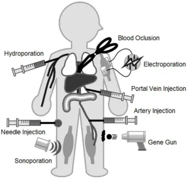
1.5.2.1. Physical methods for gene delivery ... 26
Needle injection ... 26
Gene gun ... 26
Electroporation ... 27
Photoporation ... 28
Magnetofection ... 28
Hydroporation ... 29
1.5.2.2. Chemical methods of gene delivery ... 29
Inorganic particles ... 30
Cationic Lipids ... 30
Cationic polymers ... 31
Peptide-mediated gene transfer ... 32
1.6. Optimization of expression vectors for long-term gene therapy ... 32
1.6.1. Minicircle: a plasmid devoid of bacterial backbone ... 34
1.6.2. MIDGE vectors ... 37
1.6.3. Plasmids Free of Antibiotic Resistance Gene (pFARs) ... 39
1.6.4. Self-replicating episomal plasmids ... 39
1.6.4.1. Self-replicating viral-based vectors ... 40
1.6.4.2. Chromosome-based vectors ... 40
1.6.4.3. Episomal vectors containing S/MARs ... 41
1.7. Specific Aims ... 44
Chapter 2 – Identification of Molecular Targets for Gene Therapy for Retinitis Pigmentosa ... 45
2.1. Abstract ... 49
2.2. Introduction ... 50
2.3. Materials and Methods: ... 52
2.3.1. Antibodies ... 52
2.3.2. Cell Lines ... 52
2.3.3. Primary cilium induction ... 52
2.3.4. Immunofluorescence ... 52
2.3.5. Western blot ... 53
2.3.6. ATR and γ-tubulin immunoprecipitation ... 53
2.3.7. ATR inhibition and cilia length measurement ... 54
2.3.8. Statistical Analysis ... 54
2.4. Results and Discussion ... 55
2.4.1. ATR colocalizes with the centrosome of the ciliated cells ... 55
2.4.2. Inhibition of ATR (by caffeine) affects ciliary length ... 58
2.5. Conclusion ... 63
Chapter 3 – Identification of Molecular Targets for Gene Therapy for Diabetic Retinopathy ... 65
3.1. Abstract ... 69
3.2. Introduction ... 70
3.3.1. Cell lines ... 72
3.3.2. Detection of GLUT1 expression by Western blot ... 72
3.3.3. Cellular localization of GLUT1 by immunocytochemistry ... 73
3.3.4. Glucose consumption assay... 73
3.3.5. PEDF detection in the culture medium of the RPE cells ... 74
3.3.6. Animals ... 74
3.3.7. Statistical analysis ... 75
3.4. Results ... 76
3.4.1. Hypoxia induces overexpression of GLUT1 in RPE cells ... 76
3.4.2. GLUT1 translocation to the cell membrane of RPE cells increases in response to hypoxia ... 77
3.4.3. Glucose consumption is affected by hypoxia ... 78
3.4.4. Secretory function of RPE cells is impaired by high glucose and hypoxia ... 79
3.4.5. GLUT1 and PEDF expression is altered in the RPE of diabetic mice .. 80
3.5. Discussion ... 82
3.6. Conclusion ... Erro! Marcador não definido. Chapter 4 – Expression Systems for Retinal Gene Therapy ... 87
4.1. Abstract ... 91
4.2. Introduction ... 92
4.3. Materials and Methods ... 94
4.3.1. pEPI-1 and pEPito-based vectors ... 94
4.3.2. Transfection Efficiency Assay ... 95
4.3.3. Colony-forming Assay ... 95
4.3.4. Injection of pEPito vectors in C57Bl6 mice and eGFP expression ... 96
4.3.5. Statistical analysis ... 97
4.4. Results ... 98
4.4.2. pEPito plasmids express GFP for three months in mitotically active human RPE cells ... 99
4.4.3. pEPI and pEPito gene transfer to mouse retinas: ... 102
4.5. Discussion ... 105
Chapter 5 – Gene Therapy for Diabetic Retinopathy ... 109
5.1. Abstract ... 113
5.2. Introduction ... 114
5.3. Materials and Methods ... 116
5.3.1. Vector construction ... 116
5.3.2. Evaluation of PEDF expression driven by the pEPito vector in vitro .. 116
5.3.3. Evaluation of PEDF expression driven by the pEPito vector in vivo ... 117
5.3.5. PEDF expression by Western blot analysis ... 118
5.3.6. Statistical analysis ... 119
5.4. Results ... 120
5.4.1. Characterization of PEDF in the retina of Ins2Akita diabetic mice ... 120
5.4.2. pEPito enables long-term expression of PEDF in mitotic RPE cells ... 121
5.4.3. Restoration of PEDF protein levels in the retina of Ins2Akita mice ... 121
5.5. Discussion ... 125
Chapter 6 – General Discussion ... 126
xv Resumo
A terapia génica é uma estratégia terapêutica que se caracteriza pela entrega de material genético a uma célula alvo. A terapia génica tem sido vastamente utilizada no combate a diversas doenças genéticas e adquiridas, tais como as doenças cardiovasculares, cancro e doenças oculares, entre outras.
Devido às suas características únicas, como tamanho reduzido, relativo isolamento da circulação sistémica e fácil acessibilidade a diferentes tipos celulares e tecidos, o olho é considerado o órgão ideal para o desenvolvimento de estratégias de terapia génica. A Retinite Pigmentosa e a Retinopatia Diabética são exemplos de doenças oculares genéticas e adquiridas, respetivamente, que afetam a retina e conduzem a uma perda irreversível de visão. Apesar da sua etiologia ser diferente, ambas as patologias partilham o facto de, atualmente, não existir um tratamento eficaz, o que faz destas bons alvos para terapia génica.
Atualmente, cerca de 80% das estratégias de terapia génica usadas para doenças oculares são baseadas na utilização de vírus como vetores de entrega do material genético. Apesar da sua eficácia, esta abordagem acarreta uma série de desvantagens, principalmente do ponto de vista de segurança a nível imunológico e mutagénico. Assim, os vetores não-virais aparecem como estratégia alternativa aos vetores virais, pela sua fácil utilização, ilimitada capacidade de empacotamento de genes e ausência de resposta imunitária. Porém, a sua aplicação clínica encontra-se limitada, não só pela sua reduzida eficiência de transfeção como pela expressão genética transiente conferida pelos, até agora utilizados, sistemas de expressão.
Neste sentido, nas últimas décadas têm sido desenvolvidos sistemas de expressão baseados em plasmídeos de ADN, que visam uma expressão prolongada, como os minicírculos, os vetores MIDGE, os pFARs e os plasmídeos epissomais com capacidade de replicação. Exemplos de plasmídeos com capacidade de replicação são os pEPI-1 e pEPito. Nestes plasmídeos, a capacidade de auto-replicação é conferida pela presença de S/MARs (Scaffold/Matrix Attachment Regions)
xvi
no corpo do plasmídeo. As S/MARs são sequências de ADN ricas em nucleótidos Adenina (A) e Timina (T) capazes de ancorar a cromatina à matriz nuclear. As S/MARs parecem estar envolvidas na destabilização e abertura da dupla hélice de ADN, sugerindo um envolvimento na replicação e expressão do mesmo, uma vez que a transição de cadeia dupla para cadeia simples é necessária para a replicação e transcrição genética. Além disso, vetores com S/MARs são capazes de prevenir o silenciamento epigenético, protegendo o transgene das sequências regulatórias adjacentes e da heterocromatinização, mantendo o vetor num estado transcricionalmente ativo e conferindo, assim, estabilidade mitótica. As S/MARs têm, também, a capacidade de mediar a associação da partícula episomal aos cromossomas metafásicos, permitindo, assim uma distribuição igualitária dos epissomas para as células filhas, após a mitose.
O objetivo deste trabalho foi desenvolver uma estratégia de terapia génica não-viral para doenças da retina, recorrendo ao uso de sistemas de expressão epissomais com capacidade de auto-replicação (pEPito) e de um sistema de entrega eficaz (eletroporação), de modo a conseguir uma expressão prolongada dos genes terapêuticos. Experimentalmente é possível dividir este trabalho em três secções: i) identificação de genes com potencial terapêutico em patologias da retina, como RP e RD; ii) clonagem dos genes em questão em vectores epissomais, pEPito e iii) administração dos vetores desenvolvidos in vivo, na retina de ratinhos, utilizando eletroporação como método de entrega dos sistemas de expressão.
Foi recentemente descrito em ratinhos que mutações no gene ATR (Ataxia
telangiectasia and Rad3 related) induzem uma acumulação de pigmento na retina,
com consequente degeneração dos fotorrecetores (bastonetes e cones), semelhante à que acontece em pacientes com Retinite Pigmentosa. Nos animais Wild-Type esta proteína está localizada nos cílios dos fotorrecetores. Nos mutantes, a presença da proteína mutada induz um encurtamento dos cílios, originando uma degeneração dos bastonetes e, posteriormente, dos cones. Neste contexto, tentámos investigar qual a
xvii função desta proteína na formação e alongamento dos cílios retinianos. Os nossos estudos in vitro demonstraram que esta proteína está associada ao centrossoma das células ciliadas. Nestas células, o centrossoma é o local onde o cílio se forma e por onde começa a alongar. A inibição de ATR (pela cafeína) originou uma diminuição da expressão proteica e, como consequência, verificou-se uma diminuição no comprimento dos cílios das células tratadas, demonstrando uma relação direta entre a expressão de ATR e a função ciliar. Assim, podemos inferir que mutações no gene
ATR podem ser responsáveis por alguns dos casos de RP que não estão associados
aos genes normalmente implicados na doença.
A Retinopatia Diabética é uma das principais complicações da Diabetes Mellitus. É considerada uma doença das barreiras hematorretinianas, na qual a hiperglicemia e isquémia são as principais responsáveis pelo desequilíbrio entre os fatores pro- e anti-angiogénicos que conduzem à neovascularização e consequente perda de visão. Neste estudo avaliámos os efeitos da hiperglicemia e isquémia na barreira hematorretiniana externa, in vitro, em culturas de células do epitélio pigmentar da retina, sujeitas a glicose elevada e in vivo, no epitélio pigmentar da retina de ratinhos diabéticos Ins2Akita. Os nossos resultados mostraram um aumento do transportador de glicose (GLUT1) nos nossos modelos diabéticos. Este aumento está associado, não só, a um aumento no número de transportadores na membrana das células do epitélio pigmentar da retina, como também a um aumento da sua atividade, aumentando o consumo de glicose. Este aumento no consumo de glicose induz uma diminuição na produção e secreção de factores anti-angiogénicos, como o PEDF (Pigment
Epithelium-Derived Factor) por estas células. Esta diminuição contribui para o
desequilíbrio entre os fatores pro- e anti-angiogénicos, contribuindo assim para o desenvolvimento da neovascularização.
Baseado nos resultados anteriores, decidimos clonar o gene PEDF no vetor de expressão pEPito e, através de injeção subretiniana, administrá-lo nas células do epitélio pigmentar da retina de ratinhos Ins2Akita. Os nossos resultados mostraram que
xviii
os nossos vetores foram capazes de sobre-expressar PEDF até três meses após a injeção, em níveis semelhantes aos dos animais controlo (não diabéticos). Esta sobre-expressão foi associada a uma diminuição de marcadores inflamatórios e angiogénicos, associados à doença. Estes resultados mostram que a sobre-expressão de PEDF pode constituir uma nova estratégia para o tratamento da RD.
Os nossos resultados indicam que esta abordagem baseada em sistemas de expressão com capacidade de auto-replicação e menos susceptíveis ao silenciamento epigenético, como os pEPito, aliada a um método de entrega de ADN eficaz, como a eletroporação, pode ultrapassar as limitações associadas à utilização de vetores virais, conferindo um padrão de expressão génica prolongado e mantendo um elevado perfil de segurança, consistindo assim numa alternativa eficaz para a terapia génica retiniana.
Palavras-chave: Terapia génica; pEPito; S/MARs, Retinite Pigmentosa; Retinopatia Diabética
xix Abstract
Retinitis Pigmentosa (RP) and Diabetic retinopathy (DR) are blinding disorders that
contribute to 25 to 30% of blindness cases in work-age people. The ineffectiveness of current treatments makes them ideal targets for gene therapy. The gene therapy strategy should include the main advantages of viral vectors – long-term expression and high transduction capacity – without presenting its disadvantages – immune response, random integration and tumor formation. This can be achieved by using self-replicating episomal vectors, such as pEPito.
The goal of this project was to develop new strategies using pEPito vectors encoding therapeutic genes for retinal diseases, such as DR and RP.
RP is an inherited degenerative disease, characterized by loss of photoreceptors. Recently, depletion of ATR in the mouse retina was associated to a RP-like phenotype. Our results show that, in ciliated cells, ATR is localized in the centrosome, corresponding to the connecting cilium. The inhibition of ATR induced a decrease in cilia length, showing a direct relationship between ATR and cilia formation and elongation. These findings suggest ATR as a new therapeutic target for RP.
DR is the main complication of diabetes. Several studies have shown an imbalance between pro- and anti-angiogenic factors, due to ischemia and hyperglycemia. In this work we describe an impairment of the oBRB due to an increase GLUT1 expression and function, in diabetic models. As a consequence, we detected a decrease in PEDF production and secretion. We decided to clone PEDF into pEPito backbone and overexpressed it in Ins2Akita diabetic mice, using subretinal injection and electroporation. Our system showed sustained gene expression for up to three months in levels comparable to WT mice. The overexpression of PEDF was associated with a decrease in several inflammatory and angiogenic factors associated with DR.
Our results indicate that the pEPito-based approach, combined with electroporation may overcome some limitations found in viral-mediated gene transfer,
xx
while maintaining a high safety profile and sustained gene expression, thus constituting an alternative for retinal gene delivery.
Keywords: Gene Therapy, pEPito, S/MARs, Retinitis Pigmentosa, Diabetic Retinopathy
xxi List of abbreviations and acronyms
A
AC – Auxotrophy Complementation Ad – Adenovirus
ADA – Adenosine Deaminase
adRP – Autossomal Dominant Retinitis Pigmentosa AGEs – Advanced Glycation End-products
Am – Amacrine cell
AMD – Age-related Macular Degeneration
arRP – Autossomal Recessive Retinitis Pigmentosa
ARVO – Association for Research in Vision and Ophthalmology ATM – Ataxia Telangiectasia Mutated
ATR – Ataxia Telangiectasia and Rad-3 B
BAB – Blood-Aqueous Barrier BC – Bipolar Cell
BRB – Blood-Retinal Barrier BSD – Blasticidin
C
CAR – Coxsackievirus and Adenovirus Receptor CDK2 – Cyclin-Dependent Kinase 2
CEP290 – Centrosomal Protein 290 CHM - Choroideremia
CMV – Cytomegalovirus CsCl – Cesium Cloride D
DAPI – 4’-6-diamidino-2-phenylindole DDR – DNA Damage Response DMSO – Dimethyl Sulfoxide DNA – Deoxyribonucleic Acid DPI – Days Post Injection DR – Diabetic Retinopathy dsDNA – Double-Stranded DNA E
EBNA1 – Epstein Barr Virus Nuclear Antigen 1 EBV – Epstein Barr Virus
ECL – Enhanced Chemiluminescence
eGFP – Enhanced Green Fluorescent Protein ERG – Electroretinogram
F
FBS – Fetal Bovine Serum
FELASA – Federation for Laboratory Animal Science Association G
GAG – Glycosaminoglycans
GAPDH - Glyceraldehyde 3-phosphate dehydrogenase GC – Ganglion Cell
GCL – Ganglion Cell Layer
GFAP – Glial Fibrillary Acidic Protein GLUT1 – Glucose Transporter 1 H
hCMV/EF1 – Human Cytomegalovirus/Elongation Factor alpha 1 HIV – Human Immunodeficiency Virus
HRP – Horseradish Peroxidase HSV – Herpes Simplex Virus
xxii I
Iba1 – Ionized Calcium-Binding Adapter molecule 1 iBRB – Inner Blood-Retinal Barrier
INL – Inner Nuclear Layer
iPS – Induced-Pluripotent Stem Cell
IRES – Internal Ribosomal Entry Sequence IU – International Units
K
kb – Kilo base M
mcDNA – Minicircle DNA
MIDGE – Minimalistic, Immunogenically Defined Gene Expression MP – Miniplasmid
mRNA – Messenger RNA N
NFDM – Non-fat Dry Milk
NLS – Nuclear Localization Signals
NPDR – Non-Proliferative Diabetic Retinopathy NPs – Nanoparticles
O
oBRB – Outer Blood-Retinal Barrier OCT – Optic Coherence Tomography ONL – Outer Nuclear Layer
Ori – Origin of Replication
ORT – Operator-Repressor Titration P
PBS – Phosphate Buffered Saline
PBS-T – Phosphate Buffered Saline-Triton
pDMAEMA - Poly(2-(dimethylamino)ethylmethacrylate pDNA – Plasmid DNA
PDR – Proliferative Diabetic Retinopathy PEDF – Pigment Epithelium-Derived Factor PEG – Polyethylene Glycol
PEI – Polyethylenimine PFA – Paraformaldehyde
pFARs – Plasmids Free of Antibiotic Resistance PKC – Protein Kinase C PP – Parental Plasmid PR – Photoreceptor PSK – Post-Segregational Killing PVDF – Polyvinylidene Fluoride R
RAS – Renin-Agiotensin System
RIPA buffer – Radioimmunoprecipitation assay buffer RNA – Ribonucleic Acid
ROS – Reactive Oxigen Species RP – Retinitis Pigmentosa
RPE – Retinal Pigment Epithelium
RPGR – Retinitis Pigmentosa GTPase Regulator S
S/MARs – Scaffold/Matrix Attachment Regions SCID – Severe Combined Immunodeficiency
SDS-PAGE – Sodium Dodecyl Sulfate-Polyacrylamide Gel Electrophoresis siRNA – Small Interfering RNA
xxiii ssRNA – Single-Stranded RNA
STZ – Streptozotocin
SV40 – Simian Vacuolating 40 Virus T
TLR4 – Toll-Like Receptor 4 tRNA – Transfer RNA V
VEGF – Vascular Endothelium Growth Factor W
3 1.1 Gene Therapy
Gene therapy is a therapeutic strategy that involves intracellular delivery of exogenous genetic material (DNA or RNA) to correct or modify the expression of genes responsible for a certain genetic or acquired disease (Naik et al., 2009, Ramamoorth et
al., 2015). Depending on the disease, this can be achieved by:
Replacement of a mutated gene that causes a disease by a correct copy of the gene (Kaufmann et al., 2013, Ramamoorth et al., 2015);
Inactivation of a malfunctioning mutated gene by using antisense RNA that binds to the messenger RNA (mRNA) blocking it, or by using the siRNA (small interfering) technology, a class of double-stranded RNA molecules that neutralizes the mRNA transcript, resulting in no translation into protein (Kaufmann et al., 2013, Ramamoorth et al., 2015);
Introduction of new genes, a strategy that is being widely used in cancer gene therapy in which the insertion of specific genes induces the host cell to enter apoptosis (Kaufmann et al., 2013, Ramamoorth et al., 2015).
The ideal gene therapy strategy includes four basic prerequisites: i) it should be efficient and nontoxic, ii) the targeted disease should be well characterized in terms of genetic defect and the tissue/ cell types affected, iii) the expression levels of the therapeutic gene must be tightly controlled, and iv) an adequate animal model should be available for pre-clinical studies and proof-of-concept (Hauswirth et al., 2000).
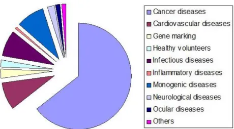
Since the first approved clinical trial in 1990 for the treatment of SCID (Severe Combined Immunodeficiency), using ADA (Adenosine Deaminase) gene (Blaese et al., 1995), several disorders, either genetic or acquired, have been targeted using gene therapy, namely cancer, cardiovascular diseases and ocular diseases (Figure 1.1) (Gascón et al., 2013, Kaufmann et al., 2013, Ramamoorth et al., 2015).
4
Figure 1.1: Targeted diseases in gene therapy clinical trials. Cancer is the most representative disorder in gene therapy applications, but cardiovascular, infectious and monogenic diseases are also noteworthy. Adapted from (Gascón et al., 2013).
The ideal vehicle for gene therapy should penetrate the cell membrane and efficiently deliver genes specifically into target cells, without being toxic or immunogenic. The genes must be directed to the nucleus and integrate the host genome in a non-mutagenic way or be maintained as an episome for a long time period. It is also desirable the therapeutic gene to be delivered independently of the mitotic status of the recipient target cell and the expression level should be constant over time (Chaum et al., 2002, Gascón et al., 2013).
1.2. The eye as target for gene therapy
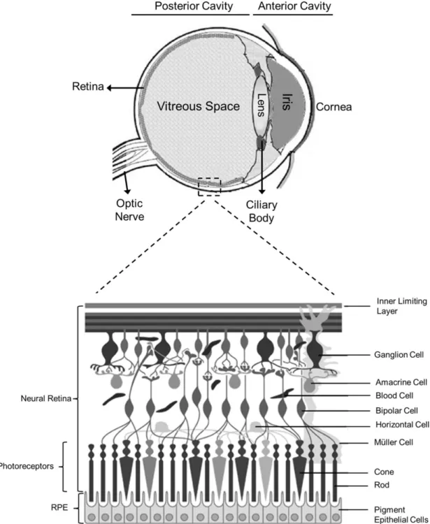
The eye is the organ responsible for the vision. It consists of two distinct anatomic regions: the anterior cavity and the posterior cavity (Figure 1.2) (de la Fuente et al., 2010).
The anterior cavity is the segment between the cornea and the lens, including the conjunctiva. These structures are responsible for focusing the light on the photoreceptor cells of the retina, in the posterior cavity of the eyeball, allowing vision. The cornea is a non-vascularized tissue composed by five to seven layers, conferring
5 high resistance to passive diffusion of ions and molecules. It is also responsible for enduring the intraocular pressure (de la Fuente et al., 2010).
Figure 1.2: Schematic representation of the eye with an enlargement of the retina, showing its main cells types. Based on (Farjo et al., 2010).
The posterior cavity is the space between the lens and the sclera, including the retina and choroid. The posterior cavity contains the vitreous humor, a gel-like substance composed of water, collagen, hyaluronic acid and proteoglycans. The
6
vitreous humor acts as a shock absorber, supports the shape of the eye, and is in contact with the retina, keeping it in place by pressing it into the choroid. The sclera is a hard tissue composed mainly by connective tissue. It is also responsible for maintaining the structure of the eye, by resisting intraocular pressure. The choroid is a vascular layer responsible for providing blood supply to retinal cells (Colthurst et al., 2000).
The retina is the most metabolically active tissue of the human body, with a fast rate of glucose and oxygen consumption. It is the sensory tissue that lines the inner surface of the posterior segment of the eye and is organized in seven major layers composed mainly by three cell types (Naik et al., 2009):
Neural cells (ganglion cells, bipolar neurons and amacrine cells): The ganglion cells (GC) are the output neurons. Its axons constitute the optic nerve and transmit the visual information from the retina to the brain. The bipolar cells (BC) are involved in the synaptic transmission from the photoreceptors to the ganglion cells. Amacrine cells (Am) are interneurons that affect the output from bipolar cells, interacting with ganglion cells (Naik et al., 2009, Hoon et al., 2014).
Photoreceptors (cones and rods): Photoreceptors (PRs) are responsible for the conversion of light energy through changes in the membrane potential that alters neurotransmitter release. There are two classes of PR, rods and cones. Rods are responsible for the low-light vision and are located throughout the peripheral retina. By contrast, cones are located at the central part of the retina (macula) and are responsible for the central and color vision (Colthurst et al., 2000, Naik et al., 2009).
Retinal pigment epithelium (RPE): The RPE is located between the outer segments of the PR and the choroid. It is a monolayer composed by hexagonal cells connected by tight-junctions, containing pigment granules. The apical membrane faces the subretinal space and interacts with the outer segment of
7 the PRs. The basolateral membrane of the RPE is in contact with the Bruch membrane that is in direct contact with blood in fenestrated vessels of the choroid (Strauss, 1995). Figure 1.3 outlines some of the RPE functions.
Figure 1.3: The retinal pigment epithelium (RPE) and its main functions: light absorption, transepithelial transport, reisomeriztion of all-trans-retinal, phagocytosis and secretion of growth factors, such as Vascular Endothelial Growt Factor (VEGF) and neurotrophic factor, like Pigment Epithelium-Derived Factor (PEDF). Adapted from (Strauss, 2005).
RPE is responsible for light absorption, transport of ions, water, and metabolic end products from the subretinal space to blood. RPE takes up nutrients, such as glucose, retinol, and fatty acids, essential for the maintenance of photoreceptors. Another important function of RPE cells is the reisomerization of all-trans-retinal into 11-cis-retinal, a key element of the visual cycle that is further transported back to photoreceptors to be incorporated in the visual cycle pathway. To ensure the excitability of PRs, RPE phagocytes and digests their shed outer segments, where recycling of retinal occurs to be returned and rebuild the outer segments of photoreceptors. Thus, the RPE needs to maintain the structural integrity of the retina by defending it efficiently from free radicals, photo-oxidative exposure and light energy. Furthermore, RPE produces and
8
secretes a wide range of growth and neurotrophic factors to support and maintain the structural basis of photoreceptors and choriocapillaris endothelium. A failure in any of these functions can induce retinal degeneration, loss of visual function, and irreversible blindness (Strauss, 2005, Simo et al., 2010).
The eye is an attractive target for gene therapy. Due to its relative small size, it is only necessary a small amount of a drug to obtain a significant therapeutic effect. The eye also contains different cell types that can be specifically accessed by different delivery methods, enabling a targeted therapy when combined with the vector’s tropism (Bloquel et al., 2006). Moreover, its immune-privileged status due to the presence of the blood-retinal barrier (BRB) limits inflammatory reactions towards the vector (Bloquel
et al., 2006, Farjo et al., 2010). Besides, therapy outcomes can be easily monitored by
non-invasive methods, such as electroretinography (ERG) and optical coherence tomography (OCT), to complement patient input (Petrs-Silva et al., 2014). In particular, retinal disorders are good targets of ocular gene therapy because in most cases the genetic etiology is known and several animal models exist or have been generated to help design new therapeutic strategies. Moreover, PR and RPE cells, which are more often affected by these mutations, can be easily accessed by subretinal injection (Cheung et al., 2010).
1.3. Retinal diseases
Diseases affecting the retina are blinding disorders influenced by genetic and environmental factors, altogether contributing to more than 25% of blindness cases (Naik et al., 2009).
The fact that current treatments for these diseases are ineffective makes them ideal candidates for gene therapy (Naik et al., 2009).
Retinal disorders can be classified as inherited or acquired diseases. Most of inherited retinal diseases are caused by mutations in genes expressed in the PRs and
9 RPE cells. There are more than 200 identified genes responsible for these diseases (http://www.sph.yth.tmc.edu/RetNet/) (McClements et al., 2013). Examples of inherited retinal diseases are Leber Congenital Amaurosis (LCA), Choroideremia (CHM),
Retinitis Pigmentosa (RP), among others. Acquired retinal disorders include, among
others, Age-related Macular Degeneration (AMD) and Diabetic Retinopathy (DR) that results from a combination of aging, environmental, and genetic factors that damage the retina and RPE (Chaum et al., 2002). In the following sections a genetic and an acquired retinal disease will be used as examples of potential therapeutic approaches, mostly focused on gene therapy.
1.3.1. Retinitis Pigmentosa
Retinal degeneration is characterized as a deterioration of the retina, usually caused by a massive cell death. Retinitis Pigmentosa (RP) is one of the most common types of inherited retinal degeneration that can result from defects in more than 60 genes, displaying all three types of Mendelian inheritance: autosomal dominant (30%-40% of cases), autosomal recessive (50%-60%) or X-linked (5%-15%), but it can also occur in combination with other systemic disorders such as Usher syndrome, Refsum disease, Bassen-Kornzweig syndrome, Bardet-Biedl syndrome, and Batten disease. There are still 30%-35% of RP patients whose mutations cannot be identified (Shintani
et al., 2009, Petrs-Silva et al., 2014).
Despite the genetic heterogeneity of this disease, RP patients present several common features, such as pigment accumulation, dysfunction of PRs and/or RPE, massive loss of PRs, abnormal ERG, night blindness and loss of peripheral field vision (Petrs-Silva et al., 2014).
To date there is no cure available for RP, however the effectiveness and safety of several potential treatments are being evaluated (Musarella et al., 2011). A brief description of each of these is presented below.
10
1.3.1.1. Vitamin Therapy
Vitamin A may protect PRs through its trophic and antioxidant properties. Berson
et al. have demonstrated a delay in PRs death after daily ingestion of 15000 IU of
Vitamin A (Berson et al., 1993). More recently, the same group showed that the intake of Vitamin A can retard blindness up to 10 years (Berson, 2007). The administration of Vitamin A in combination with a diet rich in omega-3 fatty acids also show slight improvements (Musarella et al., 2011).
Other studies showed a delay in the progression of the disease with the combination of Vitamin A with the antioxidant lutein (Berson et al., 2010).
1.3.1.2. Retinal Implant
Epiretinal and subretinal implants have been performed in animal models, showing that cortical activity can be induced. This is currently being tested in humans, but the long-term effect of the implants has yet to be evaluated, as well as the effect on the retinal function of the electrodes placed between the neuroretina and the RPE layer (Musarella et al., 2011).
1.3.1.3. Stem cells and retinal transplantation
Some studies showed an improvement in visual acuity and pupillary light response after the implant of fetal retina and PR precursors, respectively. The use of stem cells is advantageous because it is possible to prepare unlimited numbers of progenitor cells. The main disadvantages are the need to predict the ideal number of cells for engraftment and most of time the engrafted cells does not develop synaptic connections (Musarella et al., 2011) and the need of lifelong immunosuppressive therapy due to allograft transplantation (Li et al., 2012). The development of autografts from patient-specific induced pluripotent stem (iPS) cells has shown great potential to solve this problem and has been used as cell therapy in pre-clinical trials (Li et al.,
11 2012), as well as an in vitro model to study the signaling pathways involved in the pathophysiology of the disease (Lukovic et al., 2015).
1.3.2. Diabetic retinopathy
Diabetes mellitus is a group of metabolic diseases characterized by high blood glucose that leads to several complications, including diabetic retinopathy (DR). With the increased survival of individuals with diabetes, DR remains the major cause of vision loss in developed countries, affecting mostly working-age adults, as it develops approximately 20 years after the onset of diabetes (Cheung et al., 2010).
DR is classically regarded as a microvascular complication of diabetes that leads to the neovascularization within the retina (Cheung et al., 2010, Farjo et al., 2010). The pathophysiology underlying diabetic retinopathy is still unknown, but is believed that the chronic exposure to hyperglycemia and other risk factors, such as hypertension, triggers a cascade of biochemical and physiological changes that originate microvascular damage and retinal dysfunction (Cheung et al., 2010). Several biochemical mechanisms have been proposed to be responsible for the pathophysiology of the disease (Figure 1.4) including the accumulation of sorbitol and advanced glycation end-products (AGEs), reactive oxygen species (ROS) production, protein kinase C activation (PKC), inflammation, upregulation of the renin-angiotensin system (RAS) and of vascular endothelial growth factor (VEGF) (Frank, 1995, Antonetti
et al., 2006, Cheung et al., 2010).
Altogether, these biochemical mechanisms contribute to structural and physiological changes, including retinal capillary basement membrane thickening that induces pericyte and endothelial cell death, causing inner BRB (iBRB) breakdown. This loss of retinal capillary function leads to vascular wall leakage, inflammation, and ischemia, contributing to retinal neovascularization, formation of microaneurysms, edema, and hemorrhages leading to irreversible blindness (Frank, 1995, Cheung et
12
ROS
Capillary Dilatation Capillary Leakage Capillary Burst
Microaneurysms Edema Hemorrhages
Retinal Microvascular Sorbitol
pathway AGE’s PKC Inflammation RAS
VEGF Hyperglycemia + Hypertension
Figure 1.4: Pathophysiology of diabetic retinopathy. Hyperglycemia and hypertension initiates a cascade of biochemical events that leads to microvascular damage and ultimately vision loss. Adapted from (Cheung et al., 2010).
Retinal ischemia is an important component of the pathogenesis of retinal neovascularization. Ischemia induces cellular hypoxia that in turn is responsible for a broad-range of signaling pathways to overexpress angiogenic stimulators, such as VEGF, a potent pro-angiogenic factor. In a normal retina, RPE expresses a small amount of vascular endothelial growth factor (VEGF) and high levels of pigment epithelium-derived factor (PEDF), a potent anti-angiogenic molecule. Due to hypoxia during diabetes, the balance between pro- and anti-angiogenic molecules is disrupted, leading to up-regulation of VEGF and down-regulation of PEDF. As a consequence of this imbalance, there is promotion of neovascularization and subsequent vision loss (Farjo et al., 2010).
13 Regarding its progression, DR can be clinically classified as non-proliferative DR (NPDR), characterized by microaneurisms and damage in the retinal capillaries, and proliferative DR (PDR), which is recognizable by the presence of ischemia, neovascularization and hemorrhages, causing irreversible vision loss (Cheung et al., 2010).
At present, there is no effective treatment for DR. Tight glycemic control is the most efficient therapy to slow progression of DR. Blood pressure should also be taken in consideration since hypertension aggravates the disease by increasing blood flow, and causing mechanical damage to the vascular endothelial cells, thus stimulating the production of VEGF (Fong et al., 2004, Cheung et al., 2010).
For advanced stages, more invasive strategies are clinically applied an attempt to block progression of the disease, such as laser photocoagulation, vitrectomy and anti-VEGF therapy.
1.3.2.1. Laser photocoagulation
Laser photocoagulation is the most widely used technique to treat proliferative DR. The aim of photocoagulation is to cause laser burns all-over the retina to help regression and slow progression of retinal neovascularization, by reducing ischemia induced by VEGF production. However, despite its efficiency preventing visual loss, the damaging nature of laser carries several ocular side-effects, including poor light-dark adaptation and decrease in visual acuity. Moreover, if the therapy is not started timely, halting of vision loss cannot be guaranteed (Cheung et al., 2010).
1.3.2.2. Vitrectomy
Vitrectomy is the most effective surgical treatment for complications of advanced retinopathy, such as vitreous hemorrhage and retinal detachment. However, despite its efficiency reducing the risk of retinal neovascularization development, it markedly
14
increases the risk of iris neovascularization and cataract formation (Cheung et al., 2010).
1.3.2.3. Anti-VEGF Therapy
VEGF is an important mediator of angiogenesis and is the main responsible for abnormal neovascularization, leakage and BRB breakdown. Inhibition of VEGF may play an important role the DR prevention. Three VEGF antagonists have been assessed in clinical trials as anti-VEGF therapy: i) Pegaptanib, ii) Ranibizumab and iii) Bevacizumab. These agents are delivered by intravitreal injection, to ensure maximum local efficiency with minimal systemic side effects (Shah, 2008, Cheung et al., 2010).
Pegaptanib (Macugen®) is a RNA aptamer that acts by binding VEGF165 isoform,
the prevalent isoform in the human eye and principal responsible for normal and pathologic neovascularization. The binding of Pegaptanib to VEGF165 leads to its
deactivation and ultimately inhibition of vascular permeability and neovascularization (Praidou et al., 2010).
Ranibizumab (Lucentis®) is a humanized recombinant monoclonal antibody fragment that recognizes all known human VEGFA isoforms (i.e. VEGF121, VEGF145,
VEGF165, VEGF189 and VEGF206) and possesses one antigen binding epitope (Shah,
2008, Praidou et al., 2010).
Similarly to Ranibizumab, Bevacizumab (Avastin®) is also a humanized recombinant monoclonal antibody that binds and blocks the action of all VEGF-A isoforms but displays two antigen binding epitopes (Shah, 2008, Praidou et al., 2010).
Despite its efficacy inhibiting VEGF and reducing neovascularization and its complications, the major disadvantage of these drugs is its short half-life that varies from 3 days for Ranibizumab (Bakri et al., 2007), 10 days for Pagaptanib (Vinores, 2006) and 21 days for Bevacizumab (Praidou et al., 2010), increasing the need of repeated injections and severe side-effects, such as cataract formation, retinal
15 detachment, vitreous hemorrhage, infection and loss of neural retinal cells (Cheung et
al., 2010).
Other strategies using VEGF receptor blockers and siRNA against VEGF mRNA have also being used in order to prevent the interaction of VEGF with its receptor, thus stopping the subsequent signaling pathway, and the production of VEGF protein (Praidou et al., 2010). However, these approaches still need recurrent administrations, halting the importance of an efficient and prolonged strategy to target VEGF.
Regardless of the acquired or genetic nature of the disease, it is clear that conventional therapies are insufficient for retinal pathologies, either because they do not prevent the progression of the disease or because they treat the symptoms, not the cause. In this regard, gene therapy is a very promising approach to treat retinal pathologies, as shown by clinical trials for LCA (Maguire et al., 2009) and CHM (MacLaren et al., 2014). For RP, gene therapy currently represents the most promising strategy. AAVs are the most widely used vector for this purpose, due to tropism for retinal cells, lack of toxicity and absence of immune response, all features that will be later discussed (Petrs-Silva et al., 2014). In the autosomal dominant forms of RP (adRP) both normal and abnormal outer segment proteins are produced in the PRs but the abnormal form of the protein is toxic, causing progressive apoptosis of PRs. Because there are several mutations associated with adRP, targeted gene elimination or repair for each separate mutation is difficult to achieve. An alternative would be to stimulate cell survival, by the use of growth factors, and anti-apoptotic therapies to delay the progression of the disease (Chaum et al., 2002, Petrs-Silva et al., 2014).
In autosomal recessive RP (arRP), the patient has two mutated copies of the gene. In this cases gene replacement of the mutated copies by the correct gene is the most effective strategy (Chaum et al., 2002). X-linked RP is the most severe form of the disease and similarly to arRP, gene replacement is the most viable approach (Chaum
16
et al., 2002, Petrs-Silva et al., 2014). However, for neither two forms exist yet a
proposed gene therapy approach under trial.
For diabetic retinopathy, there are gene therapy strategies being studied (Igarashi
et al., 2003, Zhang et al., 2015) that have yet to reach clinical trials. This is caused by
the fact that the etiology is still not fully elucidated.
For a gene therapy strategy to be successful in treating retinal diseases, it needs to reach the therapeutic target, and for that, it needs to overcome several barriers.
1.4. Barriers to Gene Therapy
Gene delivery is considered the most challenging issue in gene therapy, as the delivery vector needs to overcome a series of extra and intracellular barriers. The next section focuses on these barriers that are common to all administration routes.
1.4.1. Extracellular barriers
Regardless of the route by which the vector is administered, it will interact with a wide-range of different molecules. Vectors administrated intravenously can easily suffer degradation by endo- and exo-nucleases present in the serum. The serum itself is composed by several proteins that can bind to vectors, resulting in aggregation, degradation and removal from the circulation by the reticuloendothelial system, affecting the stability and bioavailability of the vectors. The administration of vectors can also stimulate the immune system resulting in inflammation and complement activation, contributing for the clearance of the vectors (Gottfried et al., 2013).
1.4.2. Extracellular barriers in the eye
Gene delivery through intravenous administration is often associated with reduced bioavailability due to removal from blood circulation. Apart from the blood circulation, retinal gene therapy needs to overcome various barriers present at the anterior and posterior segments of the eye that restrict the entry of naked DNA, or viral and
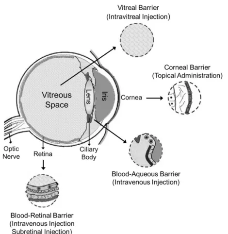
non-17 viral vectors, such as the blood-aqueous barrier, the blood-retinal barrier, the corneal barrier, and the vitreal barrier (Figure 1.5).
Figure 1.5: Scheme of the human eye, showing the main barriers for retinal gene delivery. The blood-aqueous barrier and blood-retinal barrier represent an obstacle to intravenous and subretinal injection. Corneal barrier is a barrier to topic administration and vitreal barrier represents a barrier after intravitreous injection. Adapted from (Naik
et al., 2009).
Blood-Aqueous Barrier (BAB)
The BAB is responsible for the nutrition of the cornea and lens, and its main components are the ciliary body and the iris. BAB regulates nonspecific entry of potential dangerous substances from systemic circulation to the aqueous humor. However, the BAB is less powerful than the blood-retinal barrier (Chen et al., 2008, Tamboli et al., 2011).
Blood-Retinal Barrier (BRB)
18
i) the inner BRB (iBRB) – formed by tight junctions between endothelial cells of the retinal vessels. This barrier allows the transport of small molecules such as glucose, by facilitated diffusion, while preventing widespread diffusion of substances into the retina (Cunha-Vaz, 1979, Farjo et al., 2010, Gottfried et al., 2013);
ii) the outer BRB (oBRB) – formed by intracellular tight junctions in the RPE monolayer. This barrier allows bidirectional flow of essential metabolites, such as glucose, through the Na+/K+-ATPase pump, but restricts the passage of more complex molecules from the choroidal blood supply into the neural retina (Cunha-Vaz, 1979, Farjo et al., 2010, Gottfried et al., 2013).
Corneal barrier
An alternative to intravenous administration would be topical administration. However, this is not an effective way for retinal gene delivery since it comprises penetration of the cornea and diffusion from the vitreous to the retina, against the natural flow of the aqueous humour.
The cornea consists of five to seven layers, in which the outermost one is composed of six to seven layers of stratified epithelium connected by tight junctions, which act as a barrier for vectors entry, limiting the amount of vector that reaches the retina. Moreover, a great portion of the vehicle is returned to the systemic blood circulation through the conjunctival and nasal blood vessels (Naik et al., 2009, Tamboli
et al., 2011)
Vitreal barrier
In order to overcome these barriers, administration can be local. This is achieved by subretinal and intravitreal injections, which have become the most common administration procedures for in vivo retinal gene delivery.
19 Direct injection in the subretinal space, between the retina and choroid, increases contact between the vector and the retinal layers; the contact area is however restricted to the injection site. Additionally, this type of injection can induce lesions in RPE cells and is technically challenging, restricting its clinical application.
Intravitreal injection is more acceptable for clinical applications, because it is a less invasive technique. But it is not as effective as subretinal injection, since the vectors can still be cleared by fluid flow, thus needing repeated intraocular administration to achieve therapeutic levels in the retina, which might lead to lens injury and retinal detachment. Moreover, the passage through the vitreous itself represents a barrier due to its gel-like three-dimensional net of collagen, proteoglycans, glycosaminoglycans (GAG) and serum components that are known to interact with nonviral vectors by immobilizing them in the proteoglycan mesh, binding to negative-charged GAG, causing its aggregation and blocking cellular uptake and/or intracellular trafficking (Naik
et al., 2009).
1.4.3. Intracellular barriers of gene delivery
The vectors that overcome the extracellular barriers and reach the target cells need to be internalized in order to ensure the genetic material is directed to the nucleus. In Figure 1.6 the intracellular barriers are represented: cellular membrane, endocytic trafficking, cytoplasmic transport, nuclear penetration, and gene expression.
The cellular membrane represents the first barrier for the vectors. DNA is an anionic molecule and cannot cross the also negatively-charged cell membrane. The use of positively-charged carriers can minimize this problem by neutralizing the negative charge of DNA, thus promoting cellular uptake by receptor-mediated endocytosis or non-specific endocytosis (Wiethoff et al., 2003, Wong et al., 2007).
Once inside the cell, the endocytic vesicles containing the vectors can be recycled back to the cell surface, converted into acidic vesicles (such as lysosome and phagosome), or delivered to an intracellular organelle (such as Golgi apparatus or
20
endoplasmic reticulum), depending on the internalization path. There are several proposed mechanisms by which the vectors can perform the endosomal escape, including membrane fusion, the proton-sponge effect, and integration of fusogenic peptides (Wong et al., 2007).
Figure 1.6: Cellular barriers to gene transfer: DNA protected by a cationic nanoparticle enters into the cell by endocytosis (1). Inside the cell, the DNA/nanoparticle complex should escape lysosomal degradation (2), cross the cytoplasm (3) and the nuclear envelope (4) and finally access to the cellular transcription machinery (5). Adapted from (Wong et al., 2007).
Vectors based on cationic lipids are able to escape the endosome through fusion with the endosome membrane. In contrast, cationic polymers accomplish endosomal escape through a proton-sponge mechanism. The proton-sponge effect is based on the protonation of the amine groups of the cationic polymers, leading to an accumulation of protons. This accumulation causes influx of chloride ions that induces water entry, resulting ultimately in endosome swelling and rupture (Gottfried et al., 2013).
After the DNA is successfully released, it must be directed to the nucleus. The cytosol represents another significant barrier to gene delivery. It has been postulated
21 that only 1-10% of the transfected plasmid reaches the nucleus (Gottfried et al., 2013). This is due to the presence of nucleases in the cytosolic milieu that can degrade the naked DNA and the mechanical resistance exerted by the cytoskeleton network that impedes the diffusion of the unprotected DNA to the nucleus (Wiethoff et al., 2003, Wong et al., 2007). However, it is believed that several vectors interact with the cytoskeleton proteins to reach the nuclear envelope (Wiethoff et al., 2003, Vaughan et
al., 2006).
There are at least three possible pathways for nucleic acids to enter the nucleus. The simplest way is during mitosis, when there is disruption of the nuclear envelop. If smaller enough, nucleic acids can also cross the nuclear membrane through nuclear pore complexes by facilitated diffusion or it could transverse the nuclear envelope by simple diffusion. DNA transport through nuclear pores can be even facilitated by incorporation of specific sequences recognized by nuclear pore complexes, called nuclear localization signals (NLS) (Wiethoff et al., 2003, Gottfried et al., 2013).
Gene therapy can constitute an efficient therapeutic strategy for the retinal pathologies described previously. In order to develop such a therapy, several points need to be taken into consideration, such as the delivery vector and gene expression system. These will be dealt with in greater detail in the following sections of this chapter.
1.5. Gene Delivery Systems
For overcoming the barriers mentioned in section 1.4 of this chapter and guarantee the success of gene therapy, the gene of interest should be efficiently delivered to the target cells. Since naked DNA is not usually internalized by the cells due to their hydrophilic nature, large size and negative charge (conferred by the phosphate groups), delivery systems have been developed to guarantee efficient gene transfer (Cevher et al., 2012).
22
Gene delivery systems can be categorized in two major groups: viral and non-viral. Non-viral methods can be further divided into physical and chemical delivery methods (Chaum et al., 2002, Cevher et al., 2012).
Each method was developed and optimized to achieve therapeutic gene expression levels based on the nature of the gene to be delivered, target cells and route of administration (Chaum et al., 2002). Both viral and non-viral vectors currently used possess not all characteristics of an ideal vector. Their characteristics, advantages and disadvantages for retinal use will be discussed in this section.
1.5.1. Viral-based Gene Delivery Systems
Viral vectors represent currently more than 50% of the gene therapy vehicles used in clinical trials (Figure 1.7) (Gascón et al., 2013). In the retina, the most widely used viral vectors are Adenovirus (Ad), Adeno-Associated virus (AAV), Retrovirus, Lentivirus, and Herpes Simplex virus (HSV) (Chaum et al., 2002, Cevher et al., 2012).
.
Figure 1.7: Gene delivery systems used in clinical trials. The use of naked/ plasmid DNA is one of the most widely used gene delivery systems, preceded only by Adenoviruses and Retroviruses. Adapted from (Gascón et al., 2013).
The main advantage of viruses as gene vectors is their natural capacity to transduce cells, efficiently delivering their cargo into host cells, originating high levels of
23 gene expression (Atkinson et al., 2010, Cevher et al., 2012). Moreover, some viruses display cellular tropism, such as HSV for neuronal cells (Chaum et al., 2002) and some AAV serotypes for retinal cells (Rabinowitz et al., 2002), allowing for a targeted therapy. Disadvantages associated with viral vectors are their immunogenicity, toxicity, tumorigenicity, limited packaging capacity, and difficulties in large-scale production (Naik et al., 2009, Cevher et al., 2012)
1.5.1.1. Adenoviruses
Adenoviruses are well characterized, non-enveloped, non-integrative, and linear double stranded DNA (dsDNA) viruses that are able to transduce a huge variety of human and non-human cells by binding to specific receptors, such as Coxsackievirus and Adenovirus Receptor (CAR) and Toll-Like Receptor-4 (TLR4) (Chaum et al., 2002, Cevher et al., 2012). In the retina, Adenovirus vectors have been efficiently used to transduce PR (Anglade et al., 1998, Bennett et al., 1998, Von Seggern et al., 2003), GC (Bennett et al., 1994, Cayouette et al., 1997), Müller cells (Di Polo et al., 1998, Fukuhara et al., 1998, Zhou et al., 2014) and RPE cells (da Cruz et al., 1996, da Cruz
et al., 1998, Lam et al., 2014).
These viruses are capable of incorporating DNA fragments up to 36 kb (Cevher et
al., 2012) but it remains as an episomal particle (since it is not integrated into the cell
genome), being eventually silenced or lost, with gene expression decaying in few weeks (Bennett et al., 1994, Jomary et al., 1994). Repeated administrations lead to the most serious problem associated in using Adenovirus vectors: the strong immune and inflammatory response that hampers gene expression (Chaum et al., 2002, Cevher et
al., 2012).
1.5.1.2. Adeno-Associated Virus
Adeno-Associated Virus (AAV) vectors are non-enveloped, single stranded DNA (ssDNA) viruses that, similarly to Adenovirus, are able to infect both mitotic and
post-24
mitotic cells persisting as an episomal particle, without integration into host cell genome. However, the presence of the rep protein in 10% of transduction events enables AAVs to integrate into host genome in a specific locus of chromosome 19, allowing a stable transgene expression and, simultaneously decreasing the possibility of random insertional mutagenesis (Kay et al., 2001). The main disadvantage of using AAV is however its limited packaging capacity of 5 kb (Chaum et al., 2002).
In the retina, AAVs have found widespread use, being used to transduce PR, GC and RPE cells and are currently being used in more than one clinical trial for congenital blindness (Bainbridge et al., 2008, Hauswirth et al., 2008, Maguire et al., 2009, MacLaren et al., 2014).
1.5.1.3. Retroviral vectors
Retroviruses are a family of diploid ssRNA that have the ability to infect and randomly integrate its genome into the host cells’ genome to produce new viral particles, being associated with insertional mutagenesis and tumor formation (Kay et
al., 2001, Chaum et al., 2002, Cevher et al., 2012). The possibility of generating
wild-type (WT) HIV virus during titer production or by viral recombination are also disadvantages of these vectors (Chaum et al., 2002).
The retroviral family is divided into gammaretrovirus and lentivirus, which differ in their capacity to infect only post-mitotic or any cell, respectively.
Retroviral and lentiviral vectors can both pack up to 8 kb of exogenous DNA, but retroviral vectors require cell division to transduce the host cell. Since most retinal cells are post-mitotic, these vectors are not considered the ideal vector for retinal gene therapy, where most of the cells are post-mitotic (Kay et al., 2001, Chaum et al., 2002, Cevher et al., 2012).
Unlike retrovirus, lentiviruses can infect post-mitotic cells. Depending on the proteins present in the viral envelope, it is possible to construct “pseudo-typed” lentivirus to obtain a targeted gene transfer (Chaum et al., 2002, Cevher et al., 2012,
25 Nayerossadat et al., 2012). These pseudo-typed lentiviruses have been used to transduce a broad range of retinal cells (Miyoshi et al., 1997, Bainbridge et al., 2001, Bemelmans et al., 2005).
1.5.1.4. Herpes Simplex Virus
Herpes Simplex Viruses (HSV) are DNA virus with a natural tropism for neuronal cells, representing a great advantage for gene therapy of diseases affecting the nervous system, including the retina (Nayerossadat et al., 2012). Besides neurons, HSV virus is also able to infect other cell types and does not need nuclear envelope disruption to reach the nucleus, where its DNA recircularizes and is maintained as an episome (Chaum et al., 2002).
This viral class is the one with the highest packaging capacity, being able to carry up to 150 kb of transgenic DNA (Nayerossadat et al., 2012).
However, infection with HSV is usually associated with inflammation, cytotoxicity and cell death. For this, it has been mostly used for suicide gene therapy (Chaum et
al., 2002).
1.5.2. Non-viral Gene Delivery
Despite the advantages in terms of transduction efficiency and gene expression, safety issues of viral vectors led the continuous search and development of alternative delivery systems. Non-viral vectors emerge as an alternative to viral vectors, due the lack of specific immune response, no limitation in transgene size, ease of large-scale and low cost of production (Niidome et al., 2002, Nayerossadat et al., 2012). However, the major limitation of using non-viral systems is the low transfection efficiency, precluding transgene expression at therapeutic levels (Cevher et al., 2012, Nayerossadat et al., 2012).
Non-viral gene therapy can be classified as physical and chemical depending on how the transgene is delivered.